and psoriatic arthritis
Section 2. Psoriatic arthritis: Overview and guidelines of care
for treatment with an emphasis on the biologics
Work Group: Alice Gottlieb, MD, PhD,aNeil J. Korman, MD, PhD,b Kenneth B. Gordon, MD,c Steven R. Feldman, MD, PhD,d Mark Lebwohl, MD,eJohn Y. M. Koo, MD,fAbby S. Van Voorhees, MD,gCraig A. Elmets, MD,hCraig L. Leonardi, MD,iKarl R. Beutner, MD, PhD,jReva Bhushan, PhD,kand Alan Menter, MD, Chairl
Boston, Massachusetts; Cleveland, Ohio; Chicago and Schaumburg, Illinois; Winston-Salem, North Carolina; New York, New York; San Francisco and Palo Alto, California; Philadelphia, Pennsylvania;
Birmingham, Alabama; Saint Louis, Missouri; and Dallas, Texas
Psoriasis is a common, chronic, inflammatory, multisystem disease with predominantly skin and joint manifestations affecting approximately 2% of the population. In this second of 5 sections of the guidelines of care for psoriasis, we give an overview of psoriatic arthritis including its cardinal clinical features, pathogenesis, prognosis, classification, assessment tools used to evaluate psoriatic arthritis, and the approach to treatment. Although patients with mild to moderate psoriatic arthritis may be treated with nonsteroidal anti-inflammatory drugs and/or intra-articular steroid injections, the use of disease-modifying antirheumatic drugs, particularly methotrexate, along with the biologic agents, are considered the standard of care in patients with more significant psoriatic arthritis. We will discuss the use of disease-modifying antirheumatic drugs and the biologic therapies in the treatment of patients with moderate to severe psoriatic arthritis. ( J Am Acad Dermatol 2008;58:851-64.)
DISCLAIMER
Adherence to these guidelines will not ensure successful treatment in every situation. Furthermore, these guidelines do not purport to establish a legal standard of care and should not be deemed inclusive
of all proper methods of care or exclusive of other methods of care reasonably directed to obtaining the same results. The ultimate judgment regarding the propriety of any specific therapy must be made by the physician and the patient in light of all the circumstances presented by the individual patient.
SCOPE
This second section will cover the management and treatment of psoriatic arthritis (PsA) with biologics.
Abbreviations used:
AAD: American Academy of Dermatology ACR: American College of Rheumatology AS: ankylosing spondylitis
DAS: Disease Activity Score DIP: distal interphalangeal
DMARD: disease-modifying antirheumatic drug FDA: Food and Drug Administration HAQ: Health Assessment Questionnaire IL: interleukin
NSAID: nonsteroidal anti-inflammatory drug PsA: psoriatic arthritis
PsARC: Psoriatic Arthritis Response Criteria QOL: quality of life
RA: rheumatoid arthritis TNF: tumor necrosis factor
From the Department of Dermatology, Tufts-New England Medical Center, Tufts University School of Medicine, Bostona; Murdough
Family Center For Psoriasis, Department of Dermatology, Uni-versity Hospitals Case Medical Center, Clevelandb; Division of
Dermatology, Evanston Northwestern Healthcare and Depart-ment of Dermatology, Northwestern University, Feinberg School of Medicine, Chicagoc; Department of Dermatology, Wake Forest University School of Medicine, Winston-Salemd; Department of Dermatology, Mount Sinai School of Medicine, New Yorke; Department of Dermatology, University of Califor-niaeSan Franciscof; Department of Dermatology, University of Pennsylvaniag; University of Alabama at Birminghamh; Depart-ment of Dermatology, Saint Louis Universityi; Anacor Pharma-ceuticals Inc, Palo Altoj; American Academy of Dermatology, Schaumburgk; and Baylor University Medical Center, Dallas.l
Funding sources: None.
Conflicts of interest: The authors’ conflict of interest/disclosure statements appear at the end of the article.
Reprint requests: Reva Bhushan, PhD, 930 E Woodfield Rd, Schaumburg, IL 60173. E-mail:rbhushan@aad.org.
0190-9622/$34.00
ª 2008 by the American Academy of Dermatology, Inc. doi:10.1016/j.jaad.2008.02.040
METHOD
A work group of recognized experts was con-vened to determine the audience for the guideline, define the scope of the guideline, and identify clinical questions to structure the primary issues in diagnosis and management (seeTable Iin Section1). Work group members were asked to complete a disclosure of commercial support.
An evidence-based model was used and evidence was obtained using a search of the MEDLINE data-base spanning the years 1990 through 2007. Only English-language publications were reviewed.
The available evidence was evaluated using a unified system called the Strength of Recommen-dation Taxonomy developed by editors of the US family medicine and primary care journals (ie, Amer-ican Family Physician, Family Medicine, Journal of Family Practice, and BMJ USA). This strategy was supported by a decision of the Clinical Guidelines Task Force in 2005 with some minor modifications for a consistent approach to rating the strength of the evidence of scientific studies.1Evidence was graded using a 3-point scale based on the quality of meth-odology as follows:
I. Good-quality patient-oriented evidence. II. Limited-quality patient-oriented evidence. III. Other evidence including consensus guidelines,
opinion, or case studies.
Clinical recommendations were developed on the best available evidence tabled in the guideline. These are ranked as follows:
A. Recommendation based on consistent and good-quality patient-oriented evidence.
B. Recommendation based on inconsistent or limited-quality patient-oriented evidence. C. Recommendation based on consensus, opinion,
or case studies.
This guideline has been developed in accordance
with the American Academy of Dermatology
(AAD)/AAD Association ‘‘Administrative Regulations for Evidence-based Clinical Practice Guidelines,’’ which include the opportunity for review and com-ment by the entire AAD membership and final review and approval by the AAD Board of Directors.
INTRODUCTION
PsA is an inflammatory seronegative spondy-loarthropathy associated with psoriasis. The exact proportion of patients with psoriasis who will de-velop PsA is an area of significant controversy with studies demonstrating a range from as low as 6% to as
high as 42% of patients with psoriasis. The preva-lence of PsA in the general population of the United States has been estimated to be between 0.1% to 0.25%.2In a recent large clinical trial with more than 1000 patients, 84% of patients with PsA had cutane-ous manifestations for an average of 12 years before the onset of PsA.3
Dermatologists are strongly encouraged to ac-tively seek signs and symptoms of PsA at each visit. If PsA is diagnosed, treatment should be initiated to alleviate signs and symptoms of PsA, inhibit struc-tural damage, and maximize quality of life (QOL). Dermatologists uncomfortable or untrained in eval-uating or treating patients with PsA should refer to rheumatologists.
PsA can develop at any time including childhood, but for most it appears between the ages of 30 and 50 years. PsA affects men and women equally. PsA is characterized by stiffness, pain, swelling, and ten-derness of the joints and surrounding ligaments and tendons (dactylitis and enthesitis). The enthesis is the anatomic location where tendon, ligament, or joint capsule fibers insert into the bone. Enthesitis may occur at any such site, although common locations include the insertion sites of the plantar fascia, the Achilles’ tendons, and ligamentous attachments to the ribs, spine, and pelvis. Dactylitis, or ‘‘sausage digit,’’ is a combination of enthesitis of the tendons and ligaments and synovitis involving a whole digit. Symptoms of PsA can range from mild to very severe. The severity of the skin disease and the arthritis usually do not correlate with each other. Nail disease is commonly found in patients with PsA especially those with distal interphalangeal (DIP) joint involvement. PsA may start slowly with mild symptoms, and, on occasion, may be preceded by a joint injury.
The course of PsA is variable and unpredictable ranging from mild and nondestructive to a severe, debilitating, erosive arthropathy. Erosive and de-forming arthritis occurs in 40% to 60% of patients with PsA based on data from rheumatology referral centers and may be progressive as early as within the first year of diagnosis. Studies from the general population indicate that PsA may have a milder course and that it is not associated with excess mortality.4Data on the clinical course of PsA in the dermatology setting are not currently available. Flares and remissions usually characterize the course of PsA. Left untreated, patients with PsA can have persistent inflammation, progressive joint damage, severe physical limitations, disability, and increased mortality.
The spectrum of joint inflammation in PsA is great, ranging from axial to peripheral disease, synovial and adjacent soft tissue inflammation, enthesitis,
osteitis, new bone formation, and severe osteolysis, along with overlapping findings.
Characteristic radiographic features of PsA in-clude joint erosions, joint space narrowing, bony proliferation including periarticular and shaft peri-ostitis, osteolysis including ‘‘pencil in cup’’ defor-mity, acro-osteolysis, ankylosis, spur formation, and spondylitis. The presence of DIP erosive changes may provide both sensitive and specific radiographic findings to support the diagnosis of PsA. In patients with PsA, the hands tend to be involved much more frequently than the feet with a ratio of nearly 2:1. Dactylitis is common among patients with PsA and most often affects the feet in an asymmetric distri-bution. Dactylitis is associated with a greater degree of radiologic damage than occurs in digits not affected by dactylitis (Fig 1).5
PATHOGENESIS
In both the skin and joints there is a prominent lymphocytic infiltrate, localized to the dermal papil-lae in skin and to the sublining layer stroma in the joint and inflammatory enthesis. T lymphocytes, particularly CD41 cells, are the most common in-flammatory cells in the skin and joints, with a CD41/CD81 ratio of 2:1 in the synovial fluid com-partment, matching the ratio found in peripheral
blood. CD81 T cells are more common at the enthesis. PsA synovial tissue is characterized by a T-cell infiltrate with an increase in vascularity and a reduction in macrophages compared with the syno-vial tissue found in rheumatoid arthritis (RA). The synovial lymphocyte population, unlike that of the skin, does not show up-regulation of cutaneous lymphocyte-associated antigen, suggesting that dif-ferent lymphocyte populations migrate to skin and synovial tissues. An important potential role for interleukin (IL)-12/23 in the pathogenesis of PsA is suggested by the finding of increased serum levels of the p40 protein (the shared subunit present on both IL-12 and IL-23) in patients with PsA when compared with healthy control subjects. Furthermore, levels of p40, epidermal growth factor, interferon-a, vascular endothelial growth factor, and macrophage inhibi-tory protein 1-a had the highest discriminant activity when compared with healthy control subjects, and patients with the worst PsA ([4 vs \4 involved joints) had increases in levels of p40 along with IL-2, IL-15, interferon-a, and macrophage inhibitory pro-tein 1-a.6
The observation of an increase in neutrophil infiltration in PsA is consistent with the well-described neutrophil infiltration seen in psoriasis skin and the presence of vascular endothelial growth
Fig 1. Photographs of patients with psoriatic arthritis. A, Dactylitis of third and fourth toes. B, Enthesitis of right Achilles’ tendon. C, Dactylitis of middle finger. D, Radiograph of hands.
factor receptor positive neutrophils in PsA synovium. Angiogenesis is a prominent early event in both psoriasis and Ps. Elongated and tortuous vessels in both the skin and the joint suggest dysregulated angiogenesis resulting in immature vessels.
High levels of tumor necrosis factor (TNF)-a, IL-8, IL-6, IL-1, IL-10, and matrix metalloproteinases are present in the joint fluid of patients with early PsA. Collagenase cleavage of cartilage collagen begins early in the disease and may result from cytokine-driven production of proteases. Treatment with anti-TNF-a therapy is associated with changes in synovial macrophage subsets, reduction in T-cell and neutro-phil numbers, and matrix metalloproteinase-3 expression.
Several findings suggest that osteoclast precursor cells play an important role in the pathogenesis of PsA. Osteoclast precursor cells are increased in the peripheral blood of patients with PsA and during treatment with anti-TNF agents, the frequency of osteoclast precursors decreases significantly within 2 weeks of initiating therapy. A model has been proposed whereby elevated serum levels of TNF lead to an increase in the frequency of circulating osteoclast precursors. Osteoclast precursors then migrate to the joint where they encounter increased expression of receptor activator of nuclear factor kappa B ligand, which favors the differentiation and activation of osteoclasts.7Once formed, osteoclasts are exposed to a variety of activating molecules in the PsA joint, including TNF and IL-1 that trigger osteoclast activation that may, if unchecked, even-tuate in osteolysis.
PROGNOSIS
The prognosis of PsA may range widely from a mild monoarthritic form with a good prognosis to an erosive and destructive polyarticular form with a poor prognosis, comparable in severity with that found in patients with RA. Although it is generally not possible to accurately predict which patients may
progress to disabling PsA, one study of 71 patients suggests that a polyarticular onset ( $ 5 swollen joints) of PsA may predict the appearance of erosive and deforming disease over time, thus necessitating early intervention with effective therapy.8
Much like RA, PsA can lead to chronic joint damage, increased disability, and increased mortal-ity. Social and financial implications are also impor-tant, both in terms of personal loss and the impact of direct (eg, medical care) and indirect (eg, inability to work) costs to the society and patient.
CLASSIFICATION
The classification of PsA is an area of ongoing international discussion. Although the 5 subgroups originally proposed by Moll and Wright9in 1973 are frequently used (Table I), considerable overlap be-tween these groups is now recognized. The recently developed CASPAR (classification criteria for PsA) criteria consist of established inflammatory arthritis, defined by the presence of tender and swollen joints and prolonged morning or immobility-induced stiff-ness, with a total of at least 3 points from the features listed in Table II. These CASPAR criteria have a specificity of 98.7% and sensitivity of 91.4% for diagnosing PsA.10
The diagnosis of PsA is based on clinical judg-ment. Specific patterns of joint inflammation to-gether with the absence of rheumatoid factor and the presence of skin and nail lesions of psoriasis aid clinicians in making the diagnosis of PsA. Although there are no specific serologic tests to confirm the diagnosis of PsA, radiographs can be helpful for diagnosis, to demonstrate the extent and location of Table I. Moll and Wright9criteria for psoriatic
arthritis
dPolyarticular, symmetric arthritis (rheumatoid arthritis-like) dOligoarticular (\5 joints), asymmetric arthritis
dDistal interphalangeal joint predominant dSpondylitis predominant
dArthritis mutilans
To meet the Moll and Wright9 1973 classification criteria for psoriatic arthritis, a patient with psoriasis and inflammatory arthritis who is seronegative for rheumatoid arthritis must present with 1 of the above 5 clinical subtypes. Moll and Wright9specificity is 98% and sensitivity is 91%.
Table II. CASPAR criteria for the diagnosis of psoriatic arthritis (modified10)
The CASPAR (classification criteria for psoriatic arthritis) criteria consist of established inflammatory articular disease*with at least 3 points from the following features:
A. Current psoriasis (assigned a score of 2; all other features are assigned a score of 1)
B. A personal history of psoriasis (unless current psoriasis is present)
C. A family history of psoriasis (unless current psoriasis is present or there is a personal history of psoriasis) D. Current dactylitis or history of dactylitis recorded by a
rheumatologist
E. Juxta-articular new bone formation F. Rheumatoid factor negativity
G. Typical psoriatic nail dystrophy including onycholysis, pitting, and hyperkeratosis
*Prolonged morning or immobility-induced stiffness, and tender and swollen joints suggest an inflammatory joint disease.
joint damage, and to distinguish PsA from either RA or inflammatory osteoarthritis. Radiograph changes may occur early in the course of PsA.
There are two major patterns of arthritis in PsA: peripheral joint disease, which can be polyarticular or pauciarticular, and skeletal or axial disease. Approximately 95% of patients with PsA have in-volvement of the peripheral joints, predominantly the polyarticular form, whereas a minority have the oligoarticular form.11 About 5% of patients have exclusively axial involvement, whereas 20% to 50% of patients have involvement of both the spine and the peripheral joints, with peripheral joint involve-ment being the predominant pattern. Asymptomatic involvement of the spine and sacroiliac joints may occur in patients with PsA. Radiographs, and in some cases, magnetic resonance imaging and/or com-puted tomography imaging, can be helpful in detecting asymptomatic disease.12-15The subclassi-fication of PsA into mild, moderate, and severe is defined inTable III.
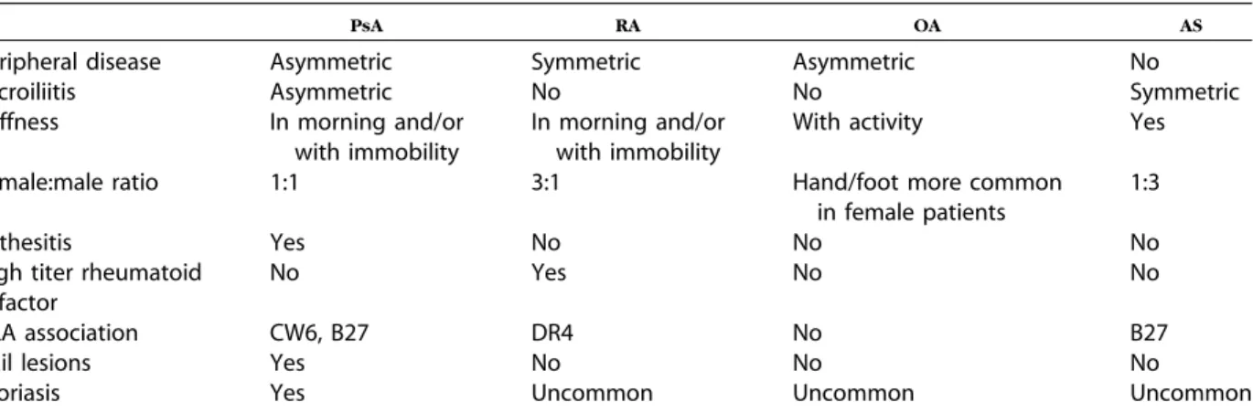
COMPARISON OF PSA WITH RA
As shown inTable IV, the peripheral polyarticular pattern of PsA may share several features with RA. Clinical features are important to differentiate sero-negative (rheumatoid factoresero-negative) RA with co-incidental psoriasis from patients with peripheral PsA. The presence of psoriatic plaques or nail pso-riasis helps to establish a diagnosis of PsA. Patients who display other characteristic signs of RA (ie, rheumatoid nodules, extra-articular involvement, and high titers of rheumatoid factor) should not be given the diagnosis of PsA. Involved joints in PsA are usually less tender and swollen and less symmetric in distribution than in RA. However, 20% of patients with PsA, especially female patients, have a symmet-ric polyarticular inflammatory arthritis resembling RA being differentiated by the presence of cutaneous or nail findings. Dactylitis, enthesitis, and DIP joint involvement common in PsA are uncommon in RA.
COMPARISON OF PSA WITH
OSTEOARTHRITIS
As shown in Table IV, another important differ-ential diagnosis for dermatologists to consider in patients with psoriasis who have joint symptoms is osteoarthritis. In the hands, there may be DIP involvement in both PsA and osteoarthritis but the classic DIP-related Heberden’s nodes in osteoarthri-tis are bone spurs whereas in PsA the DIP involve-ment is joint inflammation. Although morning stiffness and stiffness after prolonged inactivity such as air or automobile travel are common in PsA, stiffness tends to occur with joint activity in
patients with osteoarthritis. Although PsA occurs equally in men and women, osteoarthritis of the hands and feet is more frequent in women. Enthesitis, dactylitis, and sacroiliitis are generally not present in patients with osteoarthritis.
COMPARISON OF AXIAL PSA WITH
ANKYLOSING SPONDYLITIS
As shown inTable IV, patients with PsA who have axial disease (psoriatic spondylitis) may have similar clinical findings to patients with ankylosing spondy-litis (AS). However, patients with PsA are often less symptomatic, have asymmetric disease, and tend to have less severe disease. In addition, the psoriatic plaques or nail changes present in patients with psoriatic spondylitis are absent in patients with AS. Although axial involvement in most patients with PsA is a secondary feature of a predominantly peripheral arthritis, axial PsA may present either as sacroiliitis, often asymmetric and asymptomatic, or spondylitis affecting any level of the spine in a ‘‘skip’’ fashion. When compared with patients with AS, patients with PsA seldom have impaired mobility or progress to ankylosis (total loss of joint space).
IMPORTANT CLINICAL POINTS TO HELP
PRACTITIONERS IN THE DIAGNOSIS OF
PSA
It is important for dermatologists to draw on both history and physical findings in making a diagnosis of an inflammatory arthritis like PsA. Helpful clues Table III. Definition of mild, moderate, and severe psoriatic arthritis
Response to therapy Impact on QOL
Mild NSAIDs Minimal
Moderate Requires DMARDs or TNF blockers
Impacts daily tasks of living and physical/ mental functions; lack of response to NSAIDs
Severe Requires DMARDs plus TNF blockers or other biologic therapies
Cannot perform major daily tasks of living without pain or dysfunction; large impact on physical/mental functions; lack of response to either DMARDs or TNF blockers as monotherapy
DMARDs, Disease-modifying antirheumatic drugs; NSAIDs, nonsteroidal anti-inflammatory drugs; QOL, quality of life; TNF, tumor necrosis factor.
include joint stiffness that lasts more than 30 to 45 minutes in the morning or after long periods of inactivity such as sleep, automobile trips, or air travel. The presence of an inflamed and swollen digit (dactylitis) or of enthesitis (inflammation at sites of tendon insertion such as the Achilles’ tendon or the plantar fascia) makes evaluation of hands and feet, plus large joints, an important consideration in making a diagnosis of PsA. In clinical practice, the physician generally uses subjective qualitative as-sessment of the severity of a patient’s PsA. This assessment combines an objective assessment of the degree of joint involvement, including level of pain, joint tenderness and/or swelling, degree of joint destruction as assessed by radiograph findings, and its effect on the ability of the patient to adequately perform their routine tasks of daily living, along with a subjective assessment of the physical, financial, and emotional impact of the disease on the patient’s QOL.
ASSESSMENT TOOLS USED TO EVALUATE
PSA TREATMENT
AMERICAN COLLEGE OF RHEUMATOLOGY
SCORING
As shown in Table V, The American College of Rheumatology (ACR) scoring system (ACR20/50/70) was originally developed for the clinical evaluation of therapies used to treat RA. The ACR20 scoring criteria are described in Table V. ACR20 response criteria have been used as the primary end point for the majority of clinical trials in PsA, with ACR50/70 response rates as secondary end points. As the pattern of peripheral joint involvement in PsA is different from that of RA, it is important that the DIP joints of the hands and both the proximal interpha-langeal and DIP joints of the feet are counted (ie, a total of 68/66 joints) and assessed for tenderness/sw-elling. A disadvantage of the ACR measurement technique is the time delay required to obtain the results of the C-reactive protein or erythrocyte sed-imentation rate studies.16
DISEASE ACTIVITY SCORE
The Disease Activity Score (DAS), used primarily in Europe, was originally developed as a 44- or 28-joint count used in the assessment of RA. In PsA clinical trials a 78 tender and 76 swollen joint count is frequently used to accommodate the commonly involved DIP and carpal-metacarpal joints.17 The DAS scoring system uses a weighted mathematic formula, derived from clinical trials in RA. Although the ACR rating system only represents change, the DAS system represents current state of disease activ-ity and change. A disadvantage of the DAS rating system is that it requires use of a calculator (square roots are in the formula). Although the ACR20 and DAS scores have been the most widely used Table IV. Comparison of psoriatic arthritis with rheumatoid arthritis, osteoarthritis, and anklyosing spondylitis
PsA RA OA AS
Peripheral disease Asymmetric Symmetric Asymmetric No
Sacroiliitis Asymmetric No No Symmetric
Stiffness In morning and/or with immobility
In morning and/or with immobility
With activity Yes
Female:male ratio 1:1 3:1 Hand/foot more common
in female patients
1:3
Enthesitis Yes No No No
High titer rheumatoid factor
No Yes No No
HLA association CW6, B27 DR4 No B27
Nail lesions Yes No No No
Psoriasis Yes Uncommon Uncommon Uncommon
AS, Anklyosing spondylitis; OA, osteoarthritis; PsA, psoriatic arthritis; RA, rheumatoid arthritis.
Table V. American College of Rheumatology scoring
1. $ 20% reduction in the tender joint count 2. $ 20% reduction in the swollen joint count 3. $ 20% reduction in 3 of 5 additional measures
including:
a. patient assessment of pain
b. patient global assessment of disease activity c. physician global assessment of disease activity d. disability index of the Health Assessment
Questionnaire
e. acute phase reactants, ie, erythrocyte sedimentation rate and C-reactive protein
American College of Rheumatology (ACR)50 and ACR70 analysis include the same criteria as ACR20, with the use of a higher percentage improvement (50% and 70%).
assessment tools in PsA trials, investigation into outcomes measures that do not require blood testing or use of a calculator is ongoing.
PSA RESPONSE CRITERIA
The PsA Response Criteria (PsARC) was specifi-cally developed for PsA and originally used in a large study of sulfasalazine in PsA. The PsARC is defined as improvement in at least two of the following 4 criteria: (1) 20% or more improvement in physician global assessment of disease activity; (2) 20% or more improvement in patient global assessment of disease activity; (3) 30% or more improvement in tender joint count; and (4) 30% or more improve-ment in swollen joint count. A mandatory compo-nent when using the PsARC is improvement in tender or swollen joint count. Furthermore, no worsening in any PsARC component should be observed. Although the PsARC discriminates well between effective treatment and placebo, it does have significant limitations. These include the PsARC’s focus on the peripheral manifestations of PsA, along with its diminished capacity to capture features that are distinctive to PsA such as dactylitis and enthesitis.
RADIOLOGIC FEATURES
Structural damage from PsA can be assessed on conventional radiographs and is an important out-come measure in judging the efficacy of treatment. The most frequently involved joints are those in the hands and wrists, followed by the feet, ankles, knees, and shoulders with DIP involvement, along with an asymmetric pattern being characteristic features of PsA.
The radiographic features of PsA can be grouped into destructive and proliferative changes. Erosions are a typical destructive feature, frequently starting at the margins of joints and then progressing toward the center. Erosions with accompanying increased bone production are typical in PsA and may become extensive enough to give the appearance of a wid-ened joint, rather than a narrowed joint space. Widespread erosive changes may lead to the char-acteristic ‘‘pencil in cup’’ phenomenon produced by a blunt osseous surface on the proximal bone of a joint protruding into an expanded surface of the distal bone of the joint. Marked osteolysis may be observed in severely destroyed joints, such that the whole phalanx may be destroyed. Ankylosis of joints may also be observed.13
SCORING METHODS
Several scoring methods for the assessment of structural damage in peripheral joints in PsA have
been proposed, adapted for use from existing scoring systems for RA. Scoring methods developed for use in AS have been successfully applied to assess spine and sacroiliac joint abnormalities in PsA.18
PSA SCORING BASED ON THE SHARP
METHOD FOR RA
In developing a scoring method for PsA, erosions, joint space narrowing, and radiographic changes should be scored (Table VI).
SHARP-VAN DER HEIJDE MODIFIED
SCORING METHOD FOR PSA
This method, adopted from RA, is a detailed scoring method evaluating erosions, joint space narrowing, subluxation, ankylosis, gross osteolysis, and ‘‘pencil in cup’’ phenomena. In addition to the joints evaluated for RA, the DIPs of the hands are also assessed in PsA.
QUALITY OF LIFE
QOL may be assessed either using specific scales such as the PsAQOL index or more generic instru-ments such as the short form-36, the Health Table VI. Sharp scoring
A. Erosions Hands
Second-fifth DIP joints
All 5 metacarpophalangeal joints The interphalangeal joint of the thumb Wrist bones including first metacarpal base, the
multangulars, the navicular, the lunate, the triquetrum and pisiform, the radius, and the ulna Feet
All 5 metatarsal joints
Interphalangeal joint of the great toe B. Joint space narrowing
Hands
Second-fifth DIP joints
All 5 metacarpophalangeal joints
Wrist bones including fourth, fifth, and sixth carpometacarpal joints, the multangular-navicular, capitate-navicular, capitate-lunate, and radiocarpal joints
Feet
All 5 metatarsal joints C. Radiographic changes
Shaft periostitis
Juxta-articular periostitis Periostitis in the wrist Tuft resorption
Assessment Questionnaire (HAQ), and the Func-tional Assessment of Chronic Illness Therapy. These QOL tools have been tested in PsA and found to be reliable, valid, and responsive to change. The effect of psoriasis and PsA on health-related QOL should be assessed independently.19
TREATMENT
Mild PsAAppropriate treatment of PsA may include phys-ical therapy, patient education, and medication. Because only half of patients with PsA have pro-gressive disease, mild PsA is quite common and often successfully treated with nonsteroidal anti-inflam-matory drugs (NSAIDs).20When only a few joints are involved in patients with PsA, rheumatologists may perform local intra-articular injections of corticoste-roids. Although both NSAIDs and intra-articular injections of corticosteroids can lead to good symp-tomatic relief for patients with mild PsA, neither treatment is capable of inhibiting the development of structural joint damage.
USE OF DISEASE-MODIFYING
ANTIRHEUMATIC DRUGS TO TREAT
MODERATE TO SEVERE PSA
Patients with moderate to severe PsA that is more extensive or aggressive in nature require more potent therapy than NSAIDs or intra-articular corti-costeroid injections, ie, disease-modifying antirheu-matic drugs (DMARDs). Unfortunately, clinical trials evaluating the efficacy of DMARDs in PsA are few, include small patient numbers, and show only mod-erate efficacy with high placebo responses.20 Methotrexate
The data supporting the efficacy of methotrex-ate in PsA, which include two randomized, pla-cebo-controlled trials, are inadequately powered to assess clinical benefit. The first study, performed in 1964, evaluated 21 patients with PsA who were treated with 3 intramuscular injections of metho-trexate at 10-day intervals. This treatment resulted in a decrease in joint tenderness, swelling, and the erythrocyte sedimentation rate.21 In the second study, 37 patients were treated with between 7.5 and 15 mg/wk of methotrexate or placebo. After 12 weeks, the methotrexate group showed supe-rior physician assessment of arthritis activity com-pared with placebo group.22Despite the paucity of evidence demonstrating its clinical benefit, metho-trexate is frequently used as the primary DMARD in PsA, because of its efficacy in treating both skin and joint involvement in patients with psoriatic disease and its low cost.
Other DMARDs
Sulfasalazine showed modest benefit in a study of 221 patients with PsA. After 36 weeks of treatment, 58% of patients treated with sulfasalazine as com-pared with 45% of patients treated with placebo achieved the PsARC.23
Leflunomide, a selective pyrimidine synthesis inhibitor that targets activated T lymphocytes, was studied in a randomized, double-blind, placebo-controlled study of 188 patients with active PsA. After 6 months, 59% of patients treated with lefluno-mide achieved the PsARC, compared with 30% of patients treated with placebo.24 Other DMARDs including antimalarials, cyclosporine, and gold are less frequently used because evidence for their efficacy is even less convincing than for methotrex-ate, sulfasalazine, and leflunomide.20
For patients with moderate to severe PsA that is more extensive or aggressive in nature or that impacts QOL significantly, treatment with metho-trexate, TNF blockers, or both is the standard of care. Although the efficacy of DMARDs appears to be less than for the TNF inhibitors in the limited studies available, prospective, randomized, adequately powered, comparative, head-to-head trials are lack-ing to support or refute this impression.
Numerous medications intended to treat PsA may also have an effect on psoriasis. For example, most dermatologists avoid systemic corticosteroids in the treatment of patients with psoriasis because of the potential risk of pustular and erythrodermic flares when systemic corticosteroids are discontinued. However, rheumatologists often use systemic corti-costeroids in the short- and long-term treatment of PsA, in significantly smaller dosages (5-10 mg/d) than dermatologists traditionally use in chronic der-matoses. Between 10% and 20% of patients entered into the pivotal clinical trials of adalimumab, etaner-cept, and infliximab for PsA were treated with concurrent systemic corticosteroids with minimal observed adverse outcomes.25-27Worsening of skin disease with initiation of NSAID therapy is occa-sionally observed with both nonspecific and cy-clo-oxygenase-2-specific NSAIDs with shunting of arachidonic acid metabolites down the leukotriene pathway postulated as a potential mechanism. By contrast, treatments such as methotrexate and TNF-a antagonists are useful for both the skin and joint manifestations of PsA.
In the last 10 years, there has been growing interest in the pivotal role that TNF, a proinflamma-tory cytokine, plays in inflammation of skin and synovium and this molecule has become a logical target for treatment in PsA with multiple clinical trials demonstrating that TNF-a blockade is effective in the
treatment of PsA. Three TNF-a antagonists, adalimu-mab, etanercept, and inflixiadalimu-mab, are currently Food and Drug Administration (FDA)-approved for the treatment of PsA.
Although TNF-a antagonists are expensive com-pared with DMARDs, there are potential long-term cost savings and long-term benefits. These include reduced need for joint replacement surgery; reduced demands on medical, nursing, and therapy services; reduced needs for concomitant medicines; reduced demands on social services and careers; improved QOL; improved prospect of remaining in the work force; and increased life expectancy.28
GENERAL RECOMMENDATIONS FOR PSA
Dermatologists are strongly encouraged to con-sider the possible concurrent diagnosis of PsA in patients presenting with psoriasis. Although a history and screening examination for PsA should be per-formed at every visit, there are as yet no broadly validated, user-friendly, sensitive, and specific screening tools available specifically for dermatolo-gists to use. The development of one such instru-ment is in progress. The PsA Screening and Evaluation tool was developed to screen patients with psoriasis for signs and symptoms of inflamma-tory arthritis.29 In pilot testing, 69 patients with known psoriasis and PsA before the initiation of systemic therapy were screened using a 15-item questionnaire. Using this self-administered patient tool, a PsA Screening and Evaluation total score of 47 or higher distinguished patients with PsA from patients without PsA (largely patients with osteoar-thritis) with 82% sensitivity and 73% specificity. Larger studies of the PsA Screening and Evaluation tool will be necessary to verify its value as a screen-ing tool for PsA. Dermatologists uncomfortable evaluating or treating patients with PsA should refer patients who they suspect may have PsA to rheumatologists.Upon diagnosis of PsA, patients should be treated and/or referred to a rheumatologist to alleviate signs and symptoms, inhibit structural damage, and im-prove QOL parameters.
Methotrexate, TNF blockade, or the combination of these therapies is considered first-line treatment for patients with moderate to severely active PsA. Although there are no prognostic indicators to iden-tify these patients early, approximately 50% of pa-tients with PsA may develop structural damage.
Not all patients with PsA require treatment with methotrexate or TNF blockade. Patients with mild PsA can be successfully treated with NSAIDs or intra-articular injections of corticosteroids.
Because the clinical trial ACR20 efficacy data at the primary end point with all 3 FDA-approved TNF blockers for the treatment of PsA are roughly equiv-alent, the choice of which TNF agent to use is an individual one with the degree and severity of cutaneous involvement an important consideration. Common safety concerns need to be considered when treating patients who have PsA with TNF inhibitors.
GENERAL RECOMMENDATIONS FOR ALL
PATIENTS WITH PSA WHO WILL BE
TREATED WITH BIOLOGICS
An extensive discussion regarding general recom-mendations for the treatment of patients with psori-asis has been presented in Section 1 of these guidelines devoted to the use of biologics for the treatment of psoriasis. We will not recapitulate this information here, which includes suggestions for laboratory evaluation and issues related to vaccina-tion. The reader is directed to this discussion in Section 1.
TNF INHIBITORS FOR THE TREATMENT
OF PSA
The potential importance of TNF-a in the patho-physiology of PsA is underscored by the observation that there are elevated levels of TNF-a in the syno-vium, joint fluid, and skin of patients with PsA.30
EFFICACY OF THE TNF INHIBITORS
IN PSA
The strength of recommendations for the treat-ment of PsA using biologics that target TNF are shown inTable VII. When TNF inhibitors are used in the treatment of patients with PsA, they are often combined with DMARDs, particularly methotrexate. This combination approach is considered by many to be the standard of care for the treatment of PsA. However, prospective, randomized, adequately powered clinical trials, comparing the combination of methotrexate with TNF blockade with either agent alone (as performed in RA), have not been per-formed in PsA. The results of the use of the 3 TNF Table VII. The strength of recommendations for treatment of psoriatic arthritis using tumor necrosis factor inhibitors Recommendation Strength of recommendation Level of evidence References Adalimumab A I 26, 31 Etanercept A I 17, 27, 32 Infliximab A I 25, 33
inhibitors for the treatment of PsA will be reviewed in alphabetic order.
Adalimumab efficacy
In a phase III study of 313 patients with PsA, adalimumab showed significant benefit for the treat-ment of PsA.26 Adalimumab was administered sub-cutaneously at 40 mg every other week or weekly. In the double-blinded portion of the study at 24 weeks, 57% of patients receiving adalimumab (40 mg every other week) achieved ACR20 response compared with 15% of patients receiving placebo (P \.001). At week 24, the ACR50 and ACR70 response rates were 39% and 23% for patients treated with adalimumab compared with 6% and 1% for patients receiving placebo, respectively (P \ .001). The response rate did not differ between patients taking adalimumab in combination with methotrexate (50% of patients) and those taking adalimumab alone, although the numbers of patients in each cohort were too small to have adequate statistical power. Mean improvement in enthesitis and dactylitis was greater for patients receiving adalimumab than placebo, but this result did not achieve statistical significance. Using a mod-ified Sharp score, radiographic progression of hand and foot joint disease was significantly inhibited by adalimumab. Mean change in the modified Sharp score was e0.2 for patients receiving adalimumab and 1.0 for patients receiving placebo (P \ .001). Mean change in the HAQ was e0.4 for patients taking adalimumab and e0.1 for patients taking placebo (P \.001).
Patients who completed the 24-week double-blind portion of the study were eligible to enter an open-label phase where they received 40 mg of adalimumab every other week for another 24 weeks. The ACR20, ACR50, and ACR70 response rates for patients who had been on adalimumab for 48 weeks were 56%, 44%, and 30%, respectively.31
Improvements in disability as measured by the HAQ were sustained from week 24 to week 48. At week 24 and week 48, the mean changes from baseline in the modified total Sharp score were e0.1 and 0.1, respectively, for patients who received
adalimumab for 48 weeks and 0.9 and 1.0, respec-tively, for patients who received placebo for 24 weeks followed by adalimumab for 24 weeks. There was convincing evidence for both clinical and radiographic efficacy of adalimumab regardless of whether patients were or were not taking meth-otrexate at baseline. Recommendations for adalimu-mab are listed inTable VIII.
Etanercept efficacy
In a phase III study of 205 patients with PsA, etanercept showed significant improvement in signs and symptoms of PsA.27 Etanercept was adminis-tered subcutaneously at 25 mg given twice weekly. Significant improvement in all outcome measures was achieved with etanercept treatment, including the number of tender joints, number of swollen joints, morning stiffness, C-reactive protein levels, and both physician and patient global ratings. In this study, 59% of patients taking etanercept as compared with 15% of patients taking placebo achieved an ACR20 response at 12 weeks (P \.0001).27Response to etanercept was independent of concurrent meth-otrexate use (46% of patients in the study were using concurrent methotrexate at a mean dosage of 16 mg/wk), although the numbers of patients in each cohort were too small to have adequate statistical power, similar to observations made in the adalimu-mab PsA trial. Radiographic disease progression was inhibited in the etanercept group at 12 months; the mean annualized rate of change in the modified total Sharp score was e0.03 U, compared with 11.00 U in the placebo group (P = .0001).27Of the 169 patients who participated in an open-label follow-up use of etanercept between 1 and 2 years, the modified total Sharp score in the 141 patients evaluated showed a change of e0.38 and e0.22 U in the original etanercept and placebo groups, respectively, indi-cating continued inhibition of joint structural dam-age.32Recommendations for etanercept are listed in
Table IX. Table VIII. Recommendations for adalimumab
dIndications: moderate/severe psoriatic arthritis;
moderate/severe psoriasis; adult and juvenile rheumatoid arthritis (as young as 4 y); ankylosing spondylitis; and adult Crohn’s disease
dDosing: 40 mg every other wk subcutaneously dResponse: ACR20 at wk 12 is 58%
dToxicities: seeTable VIIin Psoriasis Guidelines in Section 1
ACR, American College of Rheumatology.
Table IX. Recommendations for etanercept dIndications: moderate/severe psoriatic arthritis;
moderate/severe psoriasis; adult and juvenile
rheumatoid arthritis (as young as 4 y); and ankylosing spondylitis
dDosing for psoriatic arthritis: 25 mg twice wk or 50 mg
once wk given subcutaneously
dResponse: ACR20 at wk 12 is 59%
dToxicities: seeTable VIIIin Psoriasis Guidelines in Section 1 dBaseline and ongoing monitoring: seeTable VIIIin
Psoriasis Guidelines in Section 1
Infliximab efficacy
In a phase III study of 200 patients with PsA, infliximab showed significant benefit for the treat-ment of PsA.25Infliximab was dosed at 5 mg/kg and administered intravenously at weeks 0, 2, and 6, followed by infusions every 8 weeks. Baseline demographic and disease activity characteristics were similar to those of the etanercept PsA phase III trial. At week 14, 58% of patients taking infliximab and 11% of patients taking placebo achieved an ACR20 response (P \ .001). Response to infliximab was independent of concurrent methotrexate use (46% of patients in the study were using concurrent methotrexate at a mean dosage of 16 mg/wk) although the numbers of patients in each cohort were too small to have adequate statistical power, similar to observations made in the adalimumab and etanercept PsA trials. At 6 months, a higher number of patients treated with infliximab achieved ACR20/50/70 responses when compared with those receiving placebo treatment (54%, 41%, 27% vs 16%, 4%, 2%, respectively). The presence of dactylitis decreased in the infliximab group (41%-18%) com-pared with the placebo group (40%-30%) (P = .025). Likewise, the presence of enthesitis, assessed by palpation of the Achilles’ tendon and plantar fascia insertions, decreased in the infliximab group (42%-22%) compared with the placebo group (35%-34%) (P = .016). Using the Sharp-van der Heijde scoring method (for the hands and feet), modified for PsA, patients treated with infliximab showed inhibition of radiographic disease progression at 24 weeks; HAQ scores improved for 59% of patients taking inflix-imab, compared with 19% of patients taking placebo, whereas both the physical and mental components of short form-36 scores improved for patients receiv-ing infliximab. The improvement in PsA by inflix-imab was sustained at 1 year.33 Recommendations for infliximab are shown inTable X.
GENERAL SAFETY RECOMMENDATIONS
FOR PATIENTS WITH PSA WHO WILL BE
TREATED WITH TNF INHIBITORS
The TNF inhibitors have been available for more than 10 years, primarily for the treatment of inflam-matory bowel disease and RA, with more than 1.5 million patients to date having been treated for all indications worldwide. In recent years, the indica-tions for the use of TNF inhibitors have expanded to include PsA and psoriasis. An extensive discussion about the general safety issues of all the TNF inhib-itors, derived in large part from observations made from their use in RA and inflammatory bowel dis-ease, has been presented in part one of these guidelines devoted to the use of biologics for the
treatment of psoriasis. We will, therefore, not reca-pitulate this information here, which also includes safety issues specific for individual TNF inhibitors described in Section 1. It is important to recognize that patients with PsA, as compared with patients with skin involvement only, have a higher likelihood of being treated with the combination of a TNF inhibitor and a DMARD (usually methotrexate). Thus, the general TNF inhibitor safety data detailed at length in the first portion of these guidelines will, of necessity, need to be carefully reviewed to take the combination therapy into consideration.
ALEFACEPT IN PSA
Alefacept, approved for the treatment of adult patients with moderate to severe plaque psoriasis who are candidates for systemic agents or photother-apy, is not FDA approved for PsA, but was studied as a therapy for PsA in a phase II trial in combination with methotrexate, and compared with methotrexate alone. Standard dose alefacept (15 mg) or placebo was administered intramuscularly once weekly for 12 weeks in combination with methotrexate, followed by 12 weeks of observation during which only methotrexate treatment was continued with the pri-mary efficacy end point being the proportion of patients achieving an ACR20 response at week 24.34
In all, 185 patients were randomly assigned to receive alefacept plus methotrexate (n = 123) or placebo plus methotrexate (n = 62) (mean dosage of methotrexate of 14 mg/wk). At week 24, 54% of patients in the alefacept plus methotrexate group achieved an ACR20 response, compared with 23% of patients in the placebo plus methotrexate group (P\ .001).34 The safety profile of patients treated with alefacept plus methotrexate in this trial showed no significant liver abnormalities or other toxicities. Alefacept predictably reduced CD4 T-cell counts; however, no patient discontinued alefacept because of low CD4 T-cell counts, and no association was Table X. Recommendations for infliximab
dIndications: moderate/severe psoriatic arthritis; severe
psoriasis; adult rheumatoid arthritis; ankylosing spondylitis; and Crohn’s disease (pediatric and adult)
dDosing: 5 mg/kg given intravenously at wk 0, 2, and 6,
and then every 6-8 wk; dose and interval of infusions may be adjusted as needed
dResponse: ACR20 at wk 14 is 58%
dToxicities: seeTable IXin Psoriasis Guidelines in Section 1 dBaseline and ongoing monitoring: seeTable IXin
Psoriasis Guidelines in Section 1
apparent between CD4 T-cell counts and the inci-dence of infection. Reductions in CD4 T-cell counts in patients treated with alefacept plus methotrexate were consistent with those reported in the clinical trials of alefacept monotherapy in patients with psoriasis. Results of this phase II trial suggest that alefacept in combination with methotrexate may be an effective and well-tolerated therapeutic option for some patients with PsA. Further studies are war-ranted to confirm these findings.
CONCLUSIONS
PsA is an inflammatory arthropathy associated with psoriasis. Left untreated, a proportion of pa-tients with PsA may develop persistent inflammation with progressive joint damage that can lead to severe physical limitations and disability. Several viable treatment options are available to treat PsA. These include NSAIDs and/or intra-articular injections of corticosteroids for patients with milder, localized PsA, and DMARDs including methotrexate and/or biologics (the TNF inhibitors) for patients with more severe PsA. Because PsA can be a very severe disease with significant functional impairment, early diag-nosis is critical. Early diagdiag-nosis of PsA affords the caregiver the opportunity to improve QOL, improve function, and slow disease progression. Because the large majority of patients (84%) with PsA have psoriasis for approximately 12 years before devel-oping joint symptoms, dermatologists are in an excellent position to make this diagnosis and treat PsA appropriately. Therefore, we strongly encour-age dermatologists to actively seek signs and symp-toms of PsA at every patient visit. If PsA is diagnosed, treatment should be initiated to alleviate signs and symptoms of PsA, inhibit structural damage, and maximize QOL. Dermatologists uncomfortable or untrained in evaluating or treating patients with PsA should refer to rheumatologists.
We thank Bruce Strober MD, PhD, Alexa Kimball, MD, MPH, and the Clinical Research Committee: Karl A. Beutner, MD, PhD, Chair, Michael E. Bigby, MD, Craig A. Elmets, MD, Dirk Michael Elston, MD, Joel M. Gelfand, MD, MSCE, Jacqueline M. Junkins-Hopkins, MD, Pearon G. Lang Jr, MD, Gary D. Monheit, MD, Abrar A. Qureshi, MD, MPH, Ben M. Treen, MD, Stephen B. Webster, MD, Lorraine C. Young, MD, Carol K. Sieck, RN, MSN, and Terri Zylo for reviewing the manuscripts and providing excellent suggestions.
Conflicts of interest: Alice Gottlieb, MD, PhD: Dr Gottlieb served as a speaker for Amgen Inc and Wyeth Pharmaceuticals; has current consulting/advisory board agreements with Amgen, Inc, Centocor, Inc, Wyeth Phar-maceuticals, Celgene Corp, Bristol Myers Squibb Co, Beiersdorf, Inc, Warner Chilcott, Abbott Labs, Roche,
Sankyo, Medarex, Kemia, Celera, TEVA, Actelion, UCB, Novo Nordisk, Almirall, Immune Control, RxClinical, Dermipsor Ltd, Medacorp, DermiPsor, Can-Fite, and Incyte; and has received research/educational grants from Centocor, Amgen, Wyeth, Immune Control, Celgene, Pharmacare, and Incyte. All income has been paid to her employer directly.
Neil J. Korman, MD, PhD: Dr Korman has served on the Advisory Board and was investigator and speaker for Abbott Labs, Genentech, and Astellas, receiving grants and honoraria; served on the Advisory Board and was investigator for Centocor, receiving grants and residen-cy/fellowship program funding; and was investigator and speaker for Amgen, receiving grants and honoraria.
Kenneth B. Gordon, MD: Dr Gordon served on the Advisory Board and was consultant, investigator, and speaker for Abbott Labs, Amgen, and was a consultant and investigator for Centocor, receiving grants and honor-oaria; and was investigator for Genentech, receiving grants. Steven R. Feldman, MD, PhD: Dr Feldman served on the Advisory Board and was investigator and speaker for Galderma, Stiefel, Warner Chilcott, Abbott Labs, and Astellas, receiving grants and honoraria; served on the Advisory Board for Photomedex, receiving stock options; served on the advisory board and was speaker for National Psoriasis Foundation, receiving honoraria; and was an investigator and speaker for Amgen, Centocor, and Genentech, receiving grants and honoraria.
Mark Lebwohl, MD: Dr Lebwohl served on the Advisory Board and was consultant, investigator, and speaker for Abbott Labs, Amgen, Centocor, Galderma, Genentech, and Warner Chilcott, receiving honoraria and grants; served on the Advisory Board and was consultant, investigator, and speaker for Stiefel, receiving honoraria; was consultant and investigator for Astellas, receiving grants and honoraria; was consultant for Biogen, UCB, and Isotechnika, receiving honoraria; was on the Advisory Board and was consultant and investigator for Novartis, receiving grants and hono-raria; and had an ‘‘other’’ relationship with PharmaDerm, receiving grants and honoraria.
John Y. M. Koo, MD: Dr Koo served on the Advisory Board, was speaker, consultant, and investigator for Amgen, Abbott Labs, Astellas, Warner Chilcott, and Galderma, receiving grants and honoraria; was investiga-tor for Genenetech, receiving grants; and was on the Advisory Board and was a consultant and investigator for Teikokio receiving no compensation.
Abby S. Van Voorhees, MD: Dr Van Voorhees served on the Advisory Board, was an investigator and speaker for Amgen and Genentech, receiving grants and hono-raria; was an investigator for Astellas, IDEC, and Roche receiving grants; an Advisory Board and investigator for Bristol Myers Squibb and Warner Chilcott, receiving grants and honoraria; Advisory Board and speaker for Abbott Labs and Centocor, receiving honoraria; served on the Advisory Board for Connetics, receiving honoraria; was a consultant for Incyte and Xtrac and VGX and has received honoraria from Synta for another function. Dr. Van Voorhees’ spouse is an employee with Merck receiving a salary, stock, and stock options.
Craig A. Elmets, MD: Dr Elmets has served on the Advisory Board and was investigator for Amgen and Abbott Labs, receiving grants and honoraria; was consul-tant for Astellas, receiving honoraria; and was an inves-tigator for Genentech and Connetics, receiving grants.
Craig L. Leonardi, MD: Dr Leonardi served on the Advisory Board and was consultant, investigator, and speaker for Abbott Labs, Amgen, Centocor, and Gen-entech receiving honoraria, other financial benefits, and grants for Amgen and Genentech; was speaker for Warner Chillcott, receiving honoraria; was on the Advi-sory Board and was an investigator for Serano, receiv-ing honoraria and other financial benefit; was an investigator for Astellas, Biogen, Bristol Myers, Allergan, Fujisawa, CombinatorRx, and Vitae, receiving other financial benefit.
Karl R. Beutner, MD, PhD: Chair Clinical Research Committee. Dr Beutner was an employee of Anacor, receiving salary, stock, and stock options; and had other relationships and received stocks from Dow Pharmaceu-tical Sciences.
Reva Bhushan, PhD: Dr Bhushan has no relevant conflicts of interest to disclose.
Alan Menter, MD: Chair Psoriasis Work Group. Dr Menter served on the Advisory Board and was a consul-tant, investigator and speaker for Abbott Labs, Amgen, and Centocor, receiving grants and honoraria; served on the Advisory Board and was an investigator and consul-tant for Cephalon and UCB, receiving grants and hono-raria; was a consultant, investigator, and speaker for Warner Chilcott and Wyeth, receiving honoraria; served on the Advisory Board and was an investigator for Galderma and Genentech, receiving grants and honoraria; was a consultant and investigator for Allergan and Astellas, receiving grants and honoraria; was an investi-gator for Collagenex, CombinatoRx, Dow, Ferndale, Leo, Medicis, Photocure, Pierre Fabre, 3M Pharmaceuticals, and XOMA receiving grants; and was an investigator for Connetics, receiving grants and honorarium.
REFERENCES
1. Ebell MH, Siwek J, Weiss BD, Woolf SH, Susman JL, Ewigman B, et al. Simplifying the language of evidence to improve patient care: strength of recommendation taxonomy (SORT); a pa-tient-centered approach to grading evidence in medical literature. J Fam Pract 2004;53:111-20.
2. Gelfand JM, Gladman DD, Mease PJ, Smith N, Margolis DJ, Nijsten T, et al. Epidemiology of psoriatic arthritis in the population of the United States. J Am Acad Dermatol 2005; 53:573.
3. Gottlieb AB, Kircik L, Eisen D, Jackson JM, Boh EE, Strober BE, et al. Use of etanercept for psoriatic arthritis in the dermatol-ogy clinic: the experience diagnosing, understanding care, and treatment with etanercept (EDUCATE) study. J Dermatol Treat 2006;17:343-52.
4. Shbeeb M, Uramoto KM, Gibson LE, O’Fallon WM, Gabriel SE. The epidemiology of psoriatic arthritis in Olmsted County, Minnesota, USA, 1982-1991. J Rheumatol 2000;27:1247-50. 5. Brockbank JE, Stein M, Schentag CT, Gladman DD. Dactylitis in
psoriatic arthritis: a marker for disease severity? Ann Rheum Dis 2005;64:188-90.
6. Szodoray P, Alex P, Chappell-Woodward CM, Madland TM, Knowlton N, Dozmorov I, et al. Circulating cytokines in Norwegian patients with psoriatic arthritis determined by a multiplex cytokine array system. Rheumatology (Oxford) 2007; 46:417-25.
7. Ritchlin CT, Haas-Smith SA, Li P, Hicks DG, Schwarz EM. Mechanisms of TNF-alpha- and RANKL-mediated osteoclasto-genesis and bone resorption in psoriatic arthritis. J Clin Invest 2003;111:821-31.
8. Queiro-Silva R, Torre-Alonso JC, Tinture-Eguren T, Lopez-Lagunas I. A polyarticular onset predicts erosive and deform-ing disease in psoriatic arthritis. Ann Rheum Dis 2003;62:68-70. 9. Moll JM, Wright V. Psoriatic arthritis. Semin Arthritis Rheum
1973;3:55-78.
10. Taylor W, Gladman D, Helliwell P, Marchesoni A, Mease P, Mielants H. Classification criteria for psoriatic arthritis: devel-opment of new criteria from a large international study. Arthritis Rheum 2006;54:2665-73.
11. Helliwell PS, Porter G, Taylor WJ. Polyarticular psoriatic arthritis is more like oligoarticular psoriatic arthritis than rheumatoid arthritis. Ann Rheum Dis 2007;66:113-7.
12. Offidani A, Cellini A, Valeri G, Giovagnoni A. Subclinical joint involvement in psoriasis: magnetic resonance imaging and x-ray findings. Acta Derm Venereol 1998;78:463-5.
13. Ory PA, Gladman DD, Mease PJ. Psoriatic arthritis and imaging. Ann Rheum Dis 2005;64(Suppl):ii55-7.
14. Siannis F, Farewell VT, Cook RJ, Schentag CT, Gladman DD. Clinical and radiological damage in psoriatic arthritis. Ann Rheum Dis 2006;65:478-81.
15. Tan AL, Benjamin M, Toumi H, Grainger AJ, Tanner SF, Emery P, et al. The relationship between the extensor tendon enthesis and the nail in distal interphalangeal joint disease in psoriatic arthritisea high-resolution MRI and histological study. Rheu-matology (Oxford) 2007;46:253-6.
16. Gladman DD, Helliwell P, Mease PJ, Nash P, Ritchlin C, Taylor W. Assessment of patients with psoriatic arthritis: a review of currently available measures. Arthritis Rheum 2004;50:24-35.
17. Mease PJ, Goffe BS, Metz J, VanderStoep A, Finck B, Burge DJ. Etanercept in the treatment of psoriatic arthritis and psoriasis: a randomized trial. Lancet 2000;356:385-90.
18. Mease PJ, Antoni CE, Gladman DD, Taylor WJ. Psoriatic arthritis assessment tools in clinical trials. Ann Rheum Dis 2005; 64(Suppl):ii49-54.
19. Mease PJ, Menter MA. Quality-of-life issues in psoriasis and psoriatic arthritis: outcome measures and therapies from a dermatological perspective. J Am Acad Dermatol 2006;54:685-704.
20. Nash P, Clegg DO. Psoriatic arthritis therapy: NSAIDs and traditional DMARDs. Ann Rheum Dis 2005;64(Suppl):ii74-7. 21. Black RL, O’Brien WM, Vanscott EJ, Auerbach R, Eisen AZ,
Bunim JJ, et al. Methotrexate therapy in psoriatic arthritis; double-blind study on 21 patients. JAMA 1964;189:743-7. 22. Willkens RF, Williams HJ, Ward JR, Egger MJ, Reading JC, Clements
PJ, et al. Randomized, double-blind, placebo-controlled trial of low-dose pulse methotrexate in psoriatic arthritis. Arthritis Rheum 1984;27:376-81.
23. Clegg DO, Reda DJ, Mejias E, Cannon GW, Weisman MH, Taylor T, et al. Comparison of sulfasalazine and placebo in the treatment of psoriatic arthritis: A Department of Veterans Affairs cooperative study. Arthritis Rheum 1996;39:2013-20. 24. Kaltwasser JP, Nash P, Gladman D, Rosen CF, Behrens F,
Jones P, et al. Efficacy and safety of leflunomide in the treatment of psoriatic arthritis and psoriasis: a multinational, double-blind, randomized, placebo-controlled clinical trial. Arthritis Rheum 2004;50:1939-50.
25. Antoni C, Krueger GG, de Vlam K, Birbara C, Beutler A, Guzzo C, et al. Infliximab improves signs and symptoms of psoriatic arthritis: results of the IMPACT 2 trial. Ann Rheum Dis 2005;64: 1150-7.
26. Mease PJ, Gladman DD, Ritchlin CT, Ruderman EM, Steinfeld SD, Choy EH, et al. Adalimumab for the treatment of patients with moderately to severely active psoriatic arthritis: results of a double-blind, randomized, placebo-controlled trial. Arthritis Rheum 2005;52:3279-89.
27. Mease PJ, Kivitz AJ, Burch FX, Siegel EL, Cohen SB, Ory P, et al. Etanercept treatment of psoriatic arthritis: safety, efficacy, and effect on disease progression. Arthritis Rheum 2004;50:2264-72.
28. Kimball AB, Jackson JM, Sobell JM, Boh EE, Grekin S, Pharmd EB, et al. Reductions in healthcare resource utilization in psoriatic arthritis patients receiving etanercept therapy: results from the educate trial. J Drugs Dermatol 2007;6:299-306. 29. Husni ME, Meyer KH, Cohen DS, Mody E, Qureshi AA. The PASE
questionnaire: pilot-testing a psoriatic arthritis screening and evaluation tool. J Am Acad Dermatol 2007;57:581-7.
30. Partsch G, Steiner G, Leeb BF, Dunky A, Broll H, Smolen JS. Highly increased levels of tumor necrosis factor-alpha and other proinflammatory cytokines in psoriatic arthritis synovial fluid. J Rheumatol 1997;24:518-23.
31. Gladman DD, Mease PJ, Ritchlin CT, Choy EH, Sharp JT, Ory PA, et al. Adalimumab for long-term treatment of psoriatic arthri-tis: forty-eight week data from the adalimumab effectiveness in psoriatic arthritis trial. Arthritis Rheum 2007;56:476-88. 32. Mease PJ, Kivitz AJ, Burch FX, Siegel EL, Cohen SB, Ory P, et al.
Continued inhibition of radiographic progression in patients with psoriatic arthritis following 2 years of treatment with etanercept. J Rheumatol 2006;33:712-21.
33. Kavanaugh A, Antoni CE, Gladman D, Wassenberg S, Zhou B, Beutler A, et al. The infliximab multinational psoriatic arthritis controlled trial (IMPACT): results of radiographic analyses after 1 year. Ann Rheum Dis 2006;65:1038-43.
34. Mease PJ, Gladman DD, Keystone EC. Alefacept in combina-tion with methotrexate for the treatment of psoriatic arthritis: results of a randomized, double-blind, placebo-controlled study. Arthritis Rheum 2006;54:1638-45.