Recent
developments
in medical
Recent developments in medical techniques
involving ionising or non-ionising radiation
Recent developments in medical
techniques involving ionising or
non-ionising radiation
Colophon
© RIVM 2011
Parts of this publication may be reproduced, provided acknowledgement is given to the 'National Institute for Public Health and the Environment', along with the title and year of publication.
R Stam
H Bijwaard
Contact:
Harmen Bijwaard
Laboratory for Radiation Research
harmen.bijwaard@rivm.nl
Abstract
Recent developments in medical techniques involving ionising or non-ionising radiation
Based on a literature review, RIVM has made an inventory of the latest medical techniques that use ionising or non-ionising radiation. Both types of radiation are used for diagnosis as well as treatment. An analysis of radiation risks for health care workers and patients was not yet possible, due to the limited data available. The efficacy of the techniques also deserves further investigation.
Ionising radiation
Examples of medical applications that use ionising radiation are X-ray techniques including computed tomography, which are used to generate images of the body's interior. Some of the new applications in this area are very specific, and therefore not widely applicable, or they still have to prove themselves in practice. Three trends are visible in promising developments. The first concerns refinement of existing diagnostic methods to enable better imaging with lower radiation exposure. The second trend concerns the combination of different diagnostic imaging techniques to obtain more information, such as the
combination of PET and MRI scans. Finally, there are treatment techniques new to the Netherlands such as proton therapy, with which tumours can be treated more precisely.
Non-ionising radiation
Medical applications that use non-ionising radiation can be divided in sources of electromagnetic fields, infrared radiation, visible light and ultraviolet radiation. The developments in this area can also be divided into three trends. The first concerns techniques with smaller, faster and cheaper devices that can be used 'at the bedside', such as lasers for anatomical imaging. The second trend
concerns less invasive techniques that are used to monitor vital functions from a distance. One example is using radar to measure heart beat and respiration. The third trend is for so-called miniaturisation, for example in measurement pads that radiographically transmit information about vital functions, or nanoparticles for local heating of tumours.
Rapport in het kort
Recente ontwikkelingen in medische technieken die gebruikmaken van ioniserende of niet-ioniserende straling
Het RIVM heeft op basis van een literatuurreview in kaart gebracht welke nieuwste medische toepassingen worden ontwikkeld die gebruikmaken van ioniserende of niet-ioniserende straling. Beide soorten straling worden voor zowel diagnostiek als behandeling ingezet. Door beperkte gegevens over stralingsrisico’s voor de behandelaar en de patiënt bleek een analyse hiervan voor de meeste van deze toepassingen nog niet mogelijk. Ook de doelmatigheid van de technieken verdient nader onderzoek.
Ioniserende straling
Medische toepassingen die gebruikmaken van ioniserende straling zijn
bijvoorbeeld röntgenfoto’s en CT (Computertomografie), waarmee beelden van de inwendige mens worden gemaakt. Een aantal van de nieuwe toepassingen op dit gebied zijn heel specifiek waardoor ze niet breed toepasbaar zijn, of ze moeten zich in de praktijk nog bewijzen. Bij de veelbelovende ontwikkelingen zijn drie trends waarneembaar: de eerste is verfijning van bestaande
diagnosemethoden, waardoor met minder straling een beter beeld kan worden gemaakt. De tweede trend is de combinatie van diagnostische beeldtechnieken waardoor meer informatie wordt verkregen, zoals bij de combinatie van PET- en MRI-scans. Tenslotte zijn er voor Nederland nieuwe behandeltechnieken, zoals protontherapie waarmee tumoren gerichter kunnen worden behandeld.
Niet-ioniserende straling
Medische toepassingen die gebruikmaken van niet-ioniserende straling kunnen worden verdeeld in bronnen van elektromagnetische velden, infrarood-, optische en uv-straling. Bij de ontwikkelingen op dit gebied zijn eveneens drie trends waarneembaar. De eerste betreft technieken met kleinere, snellere en
goedkopere apparatuur die ‘aan het bed’ kan worden gebruikt, zoals lasers voor anatomische beeldvorming. Bij de tweede trend gaat het om technieken die worden gebruikt om lichaamsfuncties op afstand te volgen, dus zonder dat een ingreep in het lichaam nodig is (minder invasief). Een voorbeeld is radar om de hartslag en ademhaling te meten. De derde trend is de zogeheten
miniaturisering, zoals meetpleisters die radiografisch informatie over
lichaamsfuncties verzenden of nanodeeltjes om tumoren lokaal te verwarmen (hyperthermie).
Contents
Summary—9 1 Introduction—11 1.1 Motivation—11 1.2 Limitations—11 1.3 Aims—11 1.4 Methods—11 1.5 Reading guide—12Techniques involving ionising radiation—13 2 Radiodiagnostics—15
2.1 Conventional X-ray techniques—15 2.1.1 Stereoscopy—15
2.1.2 Tomosynthesis—15
2.1.3 Phase contrast radiography—15 2.1.4 New interventional radiology—16 2.2 CT techniques—16 2.2.1 Virtual colonoscopy—16 2.2.2 CT Angiography—17 2.2.3 Cone beam CT—17 2.2.4 CT optimalisation—18 3 Nuclear medicine—19
3.1 Positron Emission Tomography—19 3.2 Gamma camera imaging—20 3.3 Radiopharmaceuticals—20 3.3.1 Radioimmunotherapy—20
4 Radiotherapy—21
4.1 Particle therapy—21
4.1.1 Boron Neutron Capture Therapy—24 4.2 Recent developments in brachytherapy—24
4.3 Intensity Modulated and Image Guided Radiotherapy—24 4.3.1 Hypofractionation—25 4.4 Stereotactic radiosurgery—25 4.4.1 Gamma Knife—25 4.4.2 Cyberknife—26 4.4.3 Tomotherapy—26 4.4.4 Synchrotron radiation—27
4.5 Intra Operative Radiotherapy—27
Techniques involving non-ionising radiation—29 5 Electromagnetic fields—31 5.1 Introduction—31 5.2 Diagnosis—31 5.2.1 Imaging—31 5.2.2 Monitoring—33 5.2.3 Navigation—34 5.3 Treatment—34
5.3.1 Hyperthermia—34 5.3.2 Ablation—35 5.3.3 Stimulation—35 6 Optical radiation—37 6.1 Introduction—37 6.2 Diagnosis—37 6.2.1 Imaging—37
6.2.2 Monitoring and navigation—40 6.3 Treatment—40
6.3.1 Ablation—40 6.3.2 Stimulation—41 6.3.3 Surgery—42
7 Conclusions and recommendations—43
References—45
List of abbreviations—55
Summary
This report is the outcome of a literature search of new developments and technologies in medical applications that make use of ionising and non-ionising radiation. This was motivated by a request of the Dutch Health Care
Inspectorate to provide an overview of emerging radiation techniques. Although these new techniques may entail risks for patients and staff, the limited data available did not provide enough information for an extensive risk analysis. The report consists of two sections, dealing with applications using ionising and non-ionising radiation, respectively.
Ionising radiation
The section on ionising radiation techniques provides an overview of the recent literature on new developments in radiodiagnostics, nuclear medicine and radiotherapy. These developments comprise many new techniques, indicating that a lot of research is conducted in this field. Naturally, not all new and often experimental techniques currently yield improvements in diagnosis or treatment. The ones that are currently deemed most promising are outlined in the
Conclusions chapter. These comprise phase contrast radiology (though still experimental) and new dose reduction methods for radiodiagnostics; the introduction of PET-MRI, new radiopharmaceuticals (including those for radioimmunotherapy) and perhaps the use of radioprotective drugs
simultaneously with radiation treatment for nuclear medicine; and particle (e.g. proton) therapy, advances in IMRT/IGRT and brachytherapy with pulsed dose rates or using microspheres for radiotherapy.
Non-ionising radiation
The section on non-ionising radiation techniques provides an overview of recent literature on new developments using non-ionising radiation. These techniques can be divided according to frequency (from low to high: electromagnetic fields, infrared radiation, visible light and ultraviolet radiation) and to their general purpose (diagnostic or therapeutic). In diagnostic applications, a wide variety of new imaging modalities using non-ionising radiation enables faster, cheaper and sometimes higher resolution anatomical imaging 'at the bedside'. Vital functions can increasingly be monitored from a distance. Flexible catheters make
minimally invasive imaging and tissue ablation inside the body possible via small incisions in skin or gastrointestinal wall. The ever smaller scale of surgical tools, tissue targeting using radiation-activated nanoparticles and miniaturised
measurement devices may also help to make interventions more precise and potentially less demanding for the patient.
1
Introduction
1.1 Motivation
This study was motivated by a request from the Dutch Health Care Inspectorate. It was noted that new techniques that make use of ionising and non-ionising radiation continue to find their way to medical practice. These techniques may involve risks for patients and practitioners when they are not properly used. Further education and extension of a quality assurance programme could be used to reduce these risks to an acceptable level.
1.2 Limitations
The field of radiation applications in medicine is very broad, so some limitations in what is taken into account are necessary. In identifying new techniques we have therefore limited ourselves to techniques that are new with respect to the application of radiation. This excludes for example new combination therapies in which a new form of chemotherapy is applied together with a conventional form of radiotherapy. Furthermore, our definition of ‘new’ involves techniques for which developments are frequently reported over the last 5 years. These techniques need not be ready for clinical application, but there must be some perspective for them to become clinically applicable in the foreseeable future. Apart from this, we have limited ourselves to reports available in either Dutch or English from a necessarily limited number of sources (see section 1.4 Methods).
Only techniques that have already been applied in humans with some indication of their effectiveness were selected. Techniques that have only been tested in animal experiments or in in vitro systems were excluded. Chapter 5 is limited to applications in which electromagnetic fields are used directly and deliberately for diagnosis or treatment. Applications that rely on the application of electrical current or an electric field to the body via direct contact with electrodes were excluded ('radiofrequency ablation', 'electrosurgery', radiofrequency cosmetic skin treatment, pain management and wound healing). It is possible that the cables or instruments used with such techniques generate secondary
electromagnetic fields which can expose patients or medical personnel. However, this was not the focus of the present report.
In general, it proved impossible to provide an indication of doses involved or risks for staff and patients. This was often not the primary objective of the report and the search results did not provide sufficient information on this topic.
1.3 Aims
The main purpose of this study is to identify new techniques that involve radiation and that are either currently being introduced in the clinical environment or will become available in the clinical environment in the near future.
1.4 Methods
New techniques involving radiation have been identified through an extensive literature study. This study looked at publications of over the last 5 years in two major databases of scientific literature (Scopus and PubMed) and it involved a search in several Dutch newspapers and popular science magazines. Details of
the search method can be found at the beginning of the sections on ionising and non-ionising radiation techniques.
1.5 Reading guide
This report is divided in two sections: the first one deals with new techniques using ionising radiation and the second one describes new techniques that use non-ionising radiation. The section on ionising radiation techniques follows the classical division of radiology in radiodiagnostics, nuclear medicine and radiation therapy. Such a classical division is not available for non-ionising radiation techniques. These techniques were introduced more recently and are used across a wide variety of clinical disciplines. The section on non-ionising radiation techniques is therefore arranged according to increasing frequency and
subdivided in diagnosis and treatment. Since readers may be less familiar with this topic, a general introduction on different types of non-ionising radiation is given. The sections are followed by a final chapter with conclusions and recommendations.
Techniques involving ionising radiation
In order to identify new developments in technology for radiodiagnostics, radiotherapy or nuclear medicine, an extensive literature search was conducted in a way that is comparable to what was done to identify techniques involving non-ionising radiation (see the section on Techniques involving non-ionising radiation).
For searching scientific literature databases the following basic search elements were used:
(ionising OR ionizing OR x-ray OR nuclear OR alpha OR beta OR gamma OR proton OR ion)
(radiation OR irradiation)
(patient OR ((occupational OR worker* OR staff) AND (medical OR hospital OR healthcare)))
(exposure OR dose OR dosimetr*)
(new OR latest OR recent OR future)
(safety OR risk OR hazard OR *protection)
(*therapy OR *therapeutic OR diagnosis OR *diagnostic OR radiologic* OR radiopharma* OR (nuclear medicine))
(intramural OR extramural OR clinical OR community)
These basic search elements were combined and some limits were added so that only titles and abstracts were searched of papers in Dutch or English that
appeared in 2006 or later. Two literature databases were used: PubMed and Scopus. In PubMed the following search was conducted (i.e. the last basic search element was omitted):
(ionising OR ionizing OR x-ray OR nuclear OR alpha OR beta OR gamma OR proton OR ion) AND (radiation OR irradiation) AND (patient OR ((occupational OR worker* OR staff) AND (medical OR hospital OR healthcare))) AND (exposure OR dose OR dosimetr*) AND (new OR latest OR recent OR future) AND (safety OR risk OR hazard OR *protection) AND (*therapy OR *therapeutic OR diagnosis OR *diagnostic OR radiologic* OR radiopharma* OR (nuclear medicine))
[limits: last 5 years, English or Dutch, title or abstract]
This search yielded 71 hits.
In Scopus the following search was conducted:
(ABS((ionising OR ionizing OR x-ray OR nuclear OR alpha OR beta OR gamma OR proton OR ion) AND (radiation OR irradiation) AND (patient OR
((occupational OR worker* OR staff) AND (medical OR hospital OR healthcare))) AND (exposure OR dose OR dosimetr*)) AND SUBJAREA(mult OR agri OR bioc OR immu OR neur OR phar OR mult OR ceng OR CHEM OR comp OR eart OR
ener OR engi OR envi OR mate OR math OR phys OR mult OR arts OR busi OR deci OR econ OR psyc OR soci) AND PUBYEAR AFT 2005) AND ((new OR latest OR RECENT OR future) AND (safety OR risk OR hazard OR *protection) AND (*therapy OR *therapeutic OR diagnosis OR *diagnostic OR radiologic* OR radiopharma* OR (nuclear medicine))) AND (LIMIT-TO(DOCTYPE, "ar") OR LIMIT-TO(DOCTYPE, "re")) AND (LIMIT-TO(LANGUAGE, "English"))
This search differs from the PubMed search in that it only looks at the abstract, a list of subject areas is included, only publications after 2005 are taken into account, and only articles or reviews in English are considered. Nevertheless, this search yielded 484 hits. Some overlap with the PubMed results exists, but all in all well over 500 papers have been identified. A selection of these papers was made, based on their titles. Abstracts of 91 selected papers were studied and based on that the most promising papers were retrieved and studied in full.
Apart from a scientific literature search also some non-scientific media were searched for relevant information. A Google search with search items ‘new’, ‘technology’ and ‘radiology’ was conducted and the first 50 hits were
investigated. Publications from the following organisations were studied: the Center for Devices and Radiological Health, the King’s Centre for the Assessment of Radiology Equipment, Imaging Performance Assessment of CT-scanners, de Raad voor de Volksgezondheid, the National Electrical Manufacturers
Association, de Gezondheidsraad, Zorgonderzoek Nederland - Medische Wetenschappen, the American Association of Physicists in Medicine, the American College of Medical Physics, the American College of Radiology, the European Union, the European Association of Nuclear Medicine, the Health Protection Agency, the International Atomic Energy Agency, the International Agency for Research on Cancer, the International Commission on Radiological Protection, the Institute of Physics and Engineering in Medicine, the National Commission on Radiological Protection, de Nederlandse Vereniging voor Radiologie, de Nederlandse Vereniging voor Klinische Fysica, de Nederlandse Vereniging voor Medische Beeldvorming en Radiotherapie and de Vereniging van ZiekenhuisInstumentatietechnici. This yielded 4 reports for further investigation. Finally, a search was conducted for publications in several Dutch newspapers (de Volkskrant, NRC, NRC next, Trouw) and in several popular science magazines (Scientific American, New Scientist, NWT) with search terms radiology, X-ray, radiotherapy and radiopharmaceutical. This yielded 19 articles in the Dutch newspapers and 6 in the science magazines that merited further study.
The results of these searches have been categorized and described in the following chapters.
2
Radiodiagnostics
2.1 Conventional X-ray techniques
The most prominent development in conventional X-ray techniques is the transfer to digital radiography. The different digital options have been described in an earlier RIVM report (Bijwaard and Brugmans, 2005) and need not be repeated here. Furthermore, the literature search yielded no new developments in digital radiography to be discussed here. Instead, this paragraph focuses on a few other recent technological advances.
2.1.1 Stereoscopy
Stereoscopy is a general technique that uses two images of the same object taken from slightly different angles: one for the left and one for the right eye so that the human brain creates a 3D view. It can also be applied to X-ray images. This could potentially yield 3D images that require much lower exposures than commonly needed for 3D CT views. However, it is currently still in a very experimental setup. Siegbahn et al. (2011) report that it can work well when contrast media are being used, but that the 3D effect is hardly visible without these media. Scientific American already reported in 2007 about a trial using stereoscopic viewing for mammography screening. This yielded slightly better results than conventional viewing (Greenemeier, 2007).
2.1.2 Tomosynthesis
Tomosynthesis is an X-ray technique with strong similarities to CT. It is primarily used in mammography. Instead of taking one or two views from different angles a series of views is taken along an arc spanning some 40 degrees. From these views a 3D image is rendered much like it is commonly done in CT when views are taken along a full arc of 360 degrees. Tomosynthesis usually requires slightly higher exposures than conventional mammography, but gives more 3D information in return. Tingberg (2010) claims that the current state of the art uses low doses and that it is therefore competitive to conventional
mammography. Tomosynthesis is already being used on a small scale in clinical practice. Regarding improvements in mammography a recent article in de Volkskrant reports a new invention that could make the examination less painful: new electronics can calculate the contact surface between breast and mammograph and adjust the exerted force to achieve a predefined overall pressure (de Volkskrant, 2010).
2.1.3 Phase contrast radiography
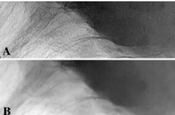
Phase contrast radiography is a collective name for a series of X-ray imaging techniques that exploit the wave properties of X-rays. Conventional X-ray techniques use the absorption of X-rays for imaging, but apart from that X-rays are also refracted and diffracted along their path. The phase shifts that take place when this happens can be detected and used for imaging. This is not a new idea (see e.g. Gao et al., 1998), but it requires much smaller X-ray sources and different detectors to work (Weon et al., 2006). This has only recently led to promising though still very experimental results in dental applications (Sato et al., 2011) and for mammography (Stampanoni et al., 2011). An example is provided in Figure 1.
(Source: http://www.medphys.ucl.ac.uk/research/acadradphys/researchactivities/pci.htm#f13, last visted: 29-11-2011)
Figure 1: Comparison of phase contrast (A) and absorption X-ray (B) images of breast tissue containing a malignant mass (darker in the image).
2.1.4 New interventional radiology
Several new developments in imaging and dose reduction have been reported for interventional techniques. During some interventions both X-rays and MRI are used for imaging. Bracken et al. (2009) report about a shielding system for the X-ray tube to prevent deflection of the electrons in the tube by the strong MRI magnetic field. Schils (2011) advocates the use of an O-arc instead of a C-arc for balloon kyphoplasty. This reduces the fluoroscopy time by 40%. Finally, Schep and van Vugt (2006) write about a GPS-like system for surgery, computer assisted surgery, that helps guiding surgeons without the need for continuous fluoroscopy.
2.2 CT techniques
Many of the recent developments in CT techniques have already been laid down in an RIVM report (Bijwaard and Stoop, 2006). However, for some of these techniques new insights have been obtained since the publication of the report. These are briefly described below.
2.2.1 Virtual colonoscopy
Virtual colonoscopy is a potentially promising technique to detect colon cancer at an early stage (see Figure 2 and Bijwaard and Stoop, 2006). Its main advantage over a conventional colonoscopy is that it is non-invasive. However, it still requires considerable exposures to yield the required image resolution. To remedy this, Wang et al. (2008) have developed software to automatically detect polyps on low-dose, low-resolution images. However, if polyps are detected a conventional colonoscopy is still required to remove them.
(Source: www.terarecon.com, last visited: 29-11-2011)
Figure 2: Example of virtual colonoscopy (arrow pointing to a polyp).
2.2.2 CT Angiography
CT Angiography (CTA) is in itself not a new technique, but due to the continuous development of CT the images yielded by CTA have improved. Two recent papers describe comparisons of CTA with other techniques: Bowman et al. (2009) compare coronary CTA to selective coronary angiography and myocardial perfusion imaging; Anderson and Barnes (2009) compare pulmonary CTA to ventilation perfusion scanning. In the first case the authors note that CTA still has difficulties in determining lumen size in the presence of atherosclerosis, and that artefacts result from movements. However, the authors do present the technique as promising. In the second case the authors find both techniques equally suitable, but note that CTA requires higher exposures.
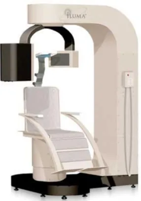
2.2.3 Cone beam CT
Cone beam CT is a slightly adapted form of CT in which a cone-shaped X-ray beam is caught on a flat panel detector. It is mainly used in dental applications for which a single rotation of beam and detector is sufficient to obtain a full 3D image. The recent literature is summarized in Stoop and Van Dillen (2011). Apart from the obvious advantages of the method, Shortt et al. (2008) note that cone beam CT causes stress to the practitioners because rapid decisions are needed with the patient in the treatment position. Additionally, cone beam CT delivers a much larger dose to the patient than conventional radiography. Stoop and Van Dillen (2011) note that there are very few indications for which this method is to be preferred.
(Source: http://www.1stsmiledental.com/ultracone.jpg, last visited: 29-11-2011)
Figure 3: Cone beam CT apparatus for dental applications: notice that X-ray beam and detector can rotate around a seated patient’s head.
2.2.4 CT optimalisation
A trade-off exists between image quality and radiation exposure so that one cannot endlessly reduce exposure and retain enough image quality for diagnostic purposes. However, new techniques continue to be developed that optimise image quality against exposure. A recent example is provided by Sperl et al. (2010), who present an algorithm that modifies the applied tube current and adjusts it to the patient and the scan protocol. In this way an exposure reduction of more than 50% can be achieved.
3
Nuclear medicine
In a recent report on future perspectives of nuclear medicine, Barbet et al. (2008) emphasize mainly two current developments: (1) the use of so-called ‘omics’ (genomics, metabolomics, etc.) techniques to predict tumour and patient responses to different treatments and hence develop personalized medicine, and (2) the promising results of different types of radioimmunotherapy, which uses radiolabelled, specific antibodies that bind to tumour cells. Different antibodies with different radionuclides have been developed for specific tumours. Apart from these main areas of progress, the authors expect improvements in imaging, specifically Positron Emission Tomography (PET), to play a larger role in therapy.
3.1 Positron Emission Tomography
Most of the recent developments and some of the future perspectives of Positron Emission Tomography (PET) are covered by Bijwaard and Stoop (2011). We shall not repeat their findings here. In short, PET is a diagnostic tool to image metabolic function, e.g. sugar uptake by a tumour. It is not a new technique, but its potential was drastically increased by the advent of 18F-FDG. In the last
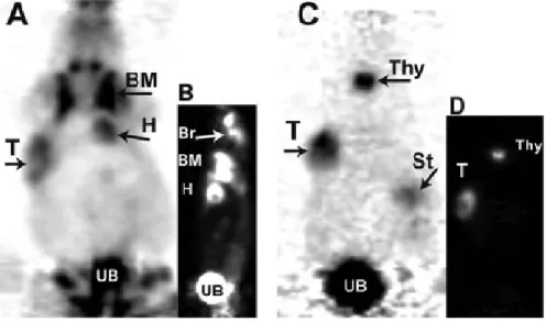
decade the combination of PET with CT has been very successful. Recently, PET has been combined with MRI, which raises new possibilities for application. This topic will be further discussed in paragraph 5.2.1. Barbet et al. (2008) mention a new development in PET: immunopositron emission tomography. The positron emitter is then incorporated in a compound that specifically targets the tumour (see e.g. Figure 4). Its effectiveness has been shown in preclinical studies (van Dongen and Vosjan, 2010).
(Source: Barbet et al. (2008))
Figure 4: Positron emission tomography in mice with (A, B) 18F-FDG and (C, D) 124I-hapten-peptide after a specific antibody. Coronal (A, C) and sagittal sections
(B, D). With 18F-FDG apart from the tumour (T) also the heart (H), bone marrow
(BM), brain (Br) and urinary bladder (UB) show increased uptake. With 124
I-hapten-peptide uptake is concentrated in the tumour, thyroid (Thy), stomach (St) and urinary bladder. The overall picture is much clearer.
3.2 Gamma camera imaging
Many different imaging techniques have been developed that make use of some gamma radiation emitting nuclide that is detected with a gamma camera. Molecular Breast Imaging (MBI) is one such technique that uses radioactive tracers to detect breast lesions. Weinmann et al. (2009) show that by optimising the collimator design doses could be reduced by a factor 1.5-3. This indicates that still some developments are taking place in this technique.
3.3 Radiopharmaceuticals
When radiopharmaceuticals are used for imaging, their radioactivity can lead to unwanted DNA damage. Hosseinimehr (2009) notes that radioprotective drugs can be helpful in reducing the dose. A similar finding was reported in New Scientist (2011): patients who took a mixture of anti-oxidants before having a diagnostic X-ray taken, showed 30-50% less damage to their DNA.
The International Atomic Energy Agency (2009) recently provided an extensive report on the use of Tc-99m for diagnostic imaging. This is in itself not a new technique, but new substances containing Tc-99m continue to be developed for specific diagnostic purposes. This concerns in particular substances containing the Tc-99m-carbonyl, Tc-99m-nitrido and the Tc-99m(III) core complexes. The authors expect these to play an important future role in cardiology, neurology, oncology and inflammation. Especially for neurodegenerative diseases a marker would be most welcome.
Other advances include the use of Ra-223-chloride for the treatment of bone metastases from prostate cancer. In a recent trial it was found that this treatment is much more effective than chemotherapy (Gallagher, 2011).
3.3.1 Radioimmunotherapy
Radioimmunotherapy is a therapy in which antibodies that bind with specific receptors on tumour cells are labelled with a radioactive compound. It has been used to treat different kinds of lymphomas. Monoclonal antibodies are labelled with Y-90 or I-131 and then administered to the patient where they bind specifically to tumour cells. Justice et al. (2006) and Dillman (2006) report promising results for non-Hodgkin’s lymphoma and B-cell lymphoma, respectively. However, the gamma radiation emitted from I-131 is still a drawback, requiring patients to remain separated from their family for several days. Barbet et al. (2008) provide an overview of applications for
radioimmunotherapy. For haematological tumours it is a promising therapy. Especially alpha emitters such as Bi-213 yield good results. For glioblastoma patients who can no longer be treated with external beam radiotherapy, radioimmunotherapy with At-211 is a promising alternative. Finally, for the
4
Radiotherapy
In conventional radiotherapy X-rays or gamma rays are used to irradiate tumour cells in a patient in order to destroy these. Unfortunately, this usually leads to surrounding healthy tissue also being exposed to ionising radiation, which is a well-known carcinogen. This may thus in time lead to secondary tumours induced by the radiation. Many of the current developments in radiotherapy are therefore aimed at optimising the dose distribution so that tumours receive maximum exposure whereas healthy tissues are spared as much as possible.
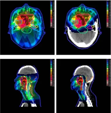
In 2008 the Health Council of the Netherlands produced an outlook for radiotherapy until 2015 (Health Council of the Netherlands, 2008). In this outlook the Council noted as new developments: the combination of PET/CT/MRI with Intensity Modulated Radiotherapy (IMRT) and Image Guided Radiotherapy (IGRT), stereotactic irradiation techniques, and proton therapy. It also noted that IMRT is now widely available in the Netherlands, but IGRT and stereotactic radiotherapy only in a few medical centres. These techniques will be explained and treated below, as well as several others that have been identified in the scientific literature. The use of electromagnetic transponders and laser beams for patient positioning in radiotherapy will be discussed in chapters 5 and 6.
4.1 Particle therapy
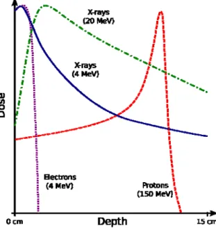
X-rays and gamma rays transfer their energy gradually along their path. Accelerated ions and other particulate matter transfer their energy by collisions with atoms in cells and tissues. In this process the particles are slowed down until they are stopped completely. Just before they come to a stop the energy transfer is maximal, the so-called Bragg peak (see Figure 5). This is in principle advantageous for radiotherapy purposes, because most of the damage to cells is located at the end of the trajectory of the particles, which can be positioned right inside the tumour. State-of-the-art particle treatments make use of the spot scanning technique in which small pencil beams of different energies are used to accurately cover the entire target volume.
(Source: http://en.wikipedia.org/wiki/File:Depth_Dose_Curves.jpg#filelinks, last visited: 29-11-2011)
Figure 5: Energy transfer (dose) of different types of ionising radiation against depth in tissue. Note the Bragg peak for protons at the end of their range.
Current research focuses on two types of particles for therapy: protons and carbon ions. Other types of particles are only marginally investigated. Both types of therapy require a huge investment in infrastructure. They require a particle accelerator to produce particles with very high energies (protons of 70-250 megaelectronvolt). Proton therapy is estimated to be 1.5 to 3 times as expensive as comparable X-ray therapy; Carbon ion therapy is estimated to be 8 times as expensive. Nevertheless, some studies indicate that proton therapy can be cost effective for a select group of breast and prostate cancers (Greco and Wolden, 2007). However, Konski et al. (2007) estimate that proton therapy for prostate cancer costs about twice as much as X-ray therapy and is not cost-effective according to general standards. Worldwide, 37 proton therapy centres are operational (http://en.wikipedia.org/wiki/Proton_therapy, consulted 31 October 2011). In the Netherlands no facility for either of these therapies exists today. Options for a proton therapy centre are currently being
investigated. The Health Council of the Netherlands has estimated that approximately 7,000 patients could benefit from such a facility in the
Netherlands every year (Health Council of the Netherlands, 2009). The Health Insurance Association (College voor Zorgverzekeringen, 2010) has indicated that
Both types of particle therapy are mainly employed for tumours that either require very high doses or a very precise dose delivery. Proton therapy has been applied favourably to treat uveal melanomas, skull base and paraspinal tumours (chondrosarcomas and chordomas), unresectable sarcomas, paediatric cancers (medulloblastomas, retinoblastomas, sarcomas), lung cancer (NSCLC), left-sided breast cancer and prostate cancer (Greco and Wolden, 2007). Recently, good results have also been obtained for mediastinal lymphomas (Li et al., 2011), cervical cancer (Song et al., 2010), glioblastoma multiforme (Mizumoto et al., 2010), Hodgkin’s lymphomas (Chera et al., 2009), corticotroph adenomas (Petit et al., 2008), olfactory neuroblastomas (Nishimura et al., 2007) and pituitary adenomas (Ronson et al., 2006). Protons are scattered by tissue so that only tumours at depths smaller than some 15 cm can be targeted. An example is provided in Figure 6.
(Source: http://en.wikipedia.org/wiki/Proton_therapy, last visited: 29-11-2011)
Figure 6: Irradiation of nasopharyngeal carcinoma by X-rays (left) and protons (right). Note the reduction of the total irradiated volume when using protons.
Carbon ion therapy has successfully been used to treat lung cancer (NSCLC), prostate cancer (Greco and Wolden, 2007) and more recently liver tumours (HCC, Combs et al., 2011), meningiomas (Combs et al., 2010),
adenocarcinomas, melanoma, hepatoma, sarcoma (Okada et al., 2010),
chordomas (Schulz-Ertner et al., 2007a) and chondrosarcomas (Schulz-Ertner et al., 2007b). As the facilities for these therapies are quite rare the numbers of patients treated are still low and the statistics of their supposed superiority over other therapies is still under debate (see e.g. Brada et al., 2009). Carbon ion therapy has the advantage that the clustered damage near its Bragg peak is more difficult to repair than the damage induced by protons (the radiobiological effectiveness (RBE) of carbon ions with respect to X-rays is approximately 3, whereas the proton RBE is 1.1). Similar exposures thus lead to an increased killing of tumour cells. Furthermore, the carbon ions are heavier than protons and are therefore less prone to scattering. They can reach depths in tissue of
some 30 cm and therefore target the entire human body. On the other hand, due to nuclear reactions that produce lighter ions also some tissue beyond the Bragg peak is still exposed.
4.1.1 Boron Neutron Capture Therapy
A somewhat special form of particle therapy is Boron Neutron Capture Therapy (BNCT), which uses neutrons from either an accelerator or nuclear reactor. In order to transfer the neutron energy to tumour cells, boron is used which reacts with the neutrons. The boron is chemically bound to a substance that is
preferentially taken up by tumour cells. Thermal neutrons react with B-10 to produce Li-7 and alpha particles. Both the Li-7 and the alpha particles cause severe DNA damage leading to cell death. BNCT is a very new and still experimental therapy. Recent publications include Kim and Kim (2009), who describe an accelerator based system in which neutrons are produced when accelerated protons hit a lithium target, and Nievaart et al. (2007), who report phase I and II trials for the treatment of glioblastoma multiforme, melanoma and extra-corporeal treatment of liver metastases at the high flux reactor in Petten, the Netherlands. It is yet unclear whether this therapy will have clinical advantages over existing therapies.
4.2 Recent developments in brachytherapy
Brachytherapy generally refers to techniques by which radioactive substances are placed in close contact with a treatment area (often a tumour). Lately, strong progress in brachytherapy for several types of cancer has been achieved using new techniques. Dezarn et al. (2011), Sangro et al. (2009), Cremonesi et al. (2008), and Welsh et al. (2006) provide reviews of brachytherapy for liver cancer using microspheres with Y-90 that are injected in blood vessels that feed the liver and thereby reach the tumour area. Smits et al. (2010) report a phase I clinical trial to investigate the replacement of Y-90 by Ho-166 for liver cancer, because the latter can be imaged better. Blanckenburg (2011) also mentions this experimental method in which holmium is encapsulated in lactic acid
spheres. The spheres are injected in the artery feeding the liver and get stuck in the capillaries where they expose the tumour to the radioactive holmium.
Wilson (2009) demonstrates the use of robotics for the implantation of I-125 seeds for the treatment of prostate cancer. This provides a faster method with lower exposures of medical staff. Polo (2008) reviews the new modality of pulsed dose rate brachytherapy, in which afterloading technology is used to deliver miniature sources controlled by software through cables to the site of interest. This technique can be used for fractionation, as is common in conventional radiotherapy. It is sometimes referred to as high-dose rate brachytherapy. Connell and Hellman (2009) report that it has been applied favourably to treat tongue cancers and uterine cervix tumours. Finally, there
small segments that each deliver radiation of a different intensity (Connell and Hellman, 2009). In IGRT the shape matching can be done real time using diagnostic imaging so that any tumour movement can also be followed.
Recent developments include a real-time tumour tracking system (Tacke et al., 2010) that uses online tumour movement information to calculate new leaf positions for the collimator, and the insertion of markers that delineate the tumour and move with it so that its location is always well defined on images (Nijland, 2011). Because IMRT and IGRT lead to long treatment procedures new systems are being developed that claim to be able to deliver the required dose distribution much faster, like RapidArc (Fogliata et al., 2011). Shortt et al. (2008) note in their report of an IAEA conference on QA that IMRT might have only minimal advantages over 3D conformal radiotherapy, except in special situations. However, due to the complex nature of the technique, stringent quality assurance measures should be taken when introducing the technique.
4.3.1 Hypofractionation
Conventional radiotherapy uses fractionation to optimise survival of healthy tissue against tumour cell killing. Several studies have shown recently that specifically for prostate cancer the radiation fractions could be larger than commonly applied for other tumours (Adkison et al., 2010; Ritter et al., 2009). This is called hypofractionation. Note that apart from the therapeutical benefit, it is also more economical to use fewer fractions and it is also less of a burden for the patient. Similarly, for pancreatic cancer it has been reported that a single low dose of X-rays can stimulate the immune response (Timke et al., 2011).
4.4 Stereotactic radiosurgery
Conceptually the distinction between radiotherapy and radiosurgery is not clear cut. However, radiosurgery usually involves very high, focused, one time exposure (no fractionation). In stereotactic radiosurgery usually many sources of radiation are applied from different angles at the same time.
4.4.1 Gamma Knife
This radiosurgical system is actually a brand name, which is however
synonymous with a certain treatment (see Figure 7). The method was already developed in 1967 and is therefore not new. It is however still further
developed, not only to treat cancer, but also other diseases, mainly affecting the brain (see e.g. Kiran et al., 2007; Sheehan et al., 2007; Chun et al., 2006). For a review see Pollock et al. (2008). Apart from brain diseases, it has also been applied to treat unresectable lung cancers (Connell and Hellman, 2009).
(Source: http://en.wikipedia.org/wiki/Radiosurgery, last visited: 29-11-2011)
Figure 7: Schematic set up of Gamma knife brain surgery: a sort of helmet is attached to the patient’s head and then to multiple radiation sources that deliver a single dose aimed at the brain tumour.
4.4.2 Cyberknife
Cyberknife is a commercially available system that includes a linear accelerator (LINAC) and a robotic arm that is used to very accurately direct the X-ray beam emanating from the LINAC. Its earliest versions date back to 1990. It
incorporates diagnostic X-ray imaging that allows for real time corrections for patient movement. This means that contrary to the Gamma knife no frame needs to be attached to the patient to prevent movement. It may however be necessary to use some form of fiducial marking for tumours that are not well-defined on X-ray images. Wowra et al. (2009) have compared Cyberknife and Gamma knife radiosurgery for brain metastases. They show that Cyberknife uses lower doses and results in longer survival, although it cannot be ruled out that this is in part due to recent innovations in chemotherapy, which is usually also applied.
4.4.4 Synchrotron radiation
Radiation from a synchrotron can also be used for stereotactic irradiation. An advantage of the synchrotron source is that monochromatic X-rays with optimal energy can be used. Edouard et al. (2010) show that 80 kiloelectronvolt is an optimal energy for the stereotactic treatment of brain tumours.
New Scientist (2008) recently reported about a virtual display of radiological images that can be used during surgery. It uses hand gestures to manipulate the images in mid-air and is therefore completely sterile.
4.5 Intra Operative Radiotherapy
Intra Operative Radiotherapy (IORT) is a form of radiotherapy in which during surgery and usually after removal of the tumour the surrounding tissue is irradiated before the surgical wound is closed. The main advantage of the technique is that healthy tissue can be spared. Its main use is currently for breast cancer, both after lumpectomy and after recurrence (when conventional radiotherapy is no longer an option) (Herskind et al., 2008; Kraus-Tiefenbacher et al., 2007; Becker, 2010). However, promising results have also been obtained more generally for soft tissue sarcomas (Tran et al., 2008).
Techniques involving non-ionising radiation
In order to identify new developments in technology involving non-ionising radiation, an extensive literature search was conducted in a way that is
comparable to what was done to identify techniques involving ionising radiation (see the previous section on Techniques involving ionising radiation).
For searching scientific literature databases the following basic search elements were used:
(electromagnetic OR emf OR radiofrequency OR (low frequency) OR microwave) ((optical radiation) OR ultraviolet OR infrared OR (visible light) OR laser)
(patient OR volunteer OR ((worker* OR occupational OR staff) AND (medical OR hospital OR (health care))))
(exposure OR dose OR dosimetr*) (new OR latest OR recent OR future) (safety OR risk)
(therapy OR therapeutic OR diagnosis OR diagnostic)
These basic search elements were combined and some limits were added so that only titles and abstracts were searched of papers in Dutch or English that
appeared in 2006 or later. Two literature databases were used: PubMed and Scopus. In PubMed the following search was conducted:
Electromagnetic fields:
(electromagnetic OR emf OR radiofrequency OR (low frequency) OR microwave) AND (patient OR volunteer OR ((worker* OR occupational OR staff) AND
(medical OR hospital OR (health care)))) AND (new OR latest OR recent OR future) AND (exposure OR dose OR dosimetr* OR safety OR risk OR therapy OR therapeutic OR diagnosis OR diagnostic)
This search yielded 431 hits.
Other non-ionising radiation:
((optical radiation) OR ultraviolet OR infrared OR (visible light) OR laser) AND (patient OR volunteer OR ((worker* OR occupational OR staff) AND (medical OR hospital OR (health care)))) AND (new OR latest OR recent OR future) AND (exposure OR dose OR dosimetr* OR safety OR risk OR therapy OR therapeutic OR diagnosis OR diagnostic)
This search yielded 415 hits.
In Scopus the following search was conducted:
Electromagnetic fields:
(ABS((electromagnetic OR emf OR radiofrequency OR (low frequency)) AND (patient OR volunteer OR ((worker* OR occupational OR staff) AND (medical OR hospital OR (health care)))) AND (new OR latest OR recent OR future) AND (exposure OR dose OR dosimetr* OR safety OR risk OR therapy OR therapeutic OR diagnosis OR diagnostic)) AND SUBJAREA(mult OR agri OR bioc OR immu OR neur OR phar OR mult OR ceng OR CHEM OR comp OR eart OR ener OR engi OR envi OR mate OR math OR phys OR mult OR arts OR busi OR deci OR econ OR psyc OR soci) AND PUBYEAR AFT 2005) AND (LIMIT-TO(DOCTYPE, "ar") OR LIMIT-TO(DOCTYPE, "re")) AND (LIMIT-TO(LANGUAGE, "English"))
Other non-ionising radiation:
(ABS(((optical radiation) OR ultraviolet OR infrared OR (visible light) OR laser) AND (patient OR volunteer OR ((worker* OR occupational OR staff) AND (medical OR hospital OR (health care)))) AND (new OR latest OR recent OR future) AND (exposure OR dose OR dosimetr* OR safety OR risk OR therapy OR therapeutic OR diagnosis OR diagnostic)) AND SUBJAREA(mult OR agri OR bioc OR immu OR neur OR phar OR mult OR ceng OR CHEM OR comp OR eart OR ener OR engi OR envi OR mate OR math OR phys OR mult OR arts OR busi OR deci OR econ OR psyc OR soci) AND PUBYEAR AFT 2005) AND
(LIMIT-TO(DOCTYPE, "ar") OR LIMIT-TO(DOCTYPE, "re")) AND (LIMIT-TO(LANGUAGE, "English"))
This search yielded 570 hits.
This search differs from the PubMed search in that it only looks at the abstract, a list of subject areas is included, only publications after 2005 are taken into account, and only articles or reviews in English are considered. Some overlap with the PubMed results exists, so that the total number of additional papers found in Scopus will be lower than 1322. A selection of these papers was made, based on their titles. Abstracts of selected papers were studied and based on that the most promising papers were retrieved and studied in full (see
References). Occasionally, a specific follow-up search for additional papers was conducted to explain the basics of some of the techniques identified. Additional new technologies involving non-ionising radiation were identified in a survey of medical applications involving EMF in the European Union's 6th framework program (EMF-NET, 2008a; EMF-NET, 2008b).
Apart from a scientific literature search a small number of popular scientific journals and non-scientific media were searched for relevant news items in the years 2006-2011. This included the news sections of the English language journals Science and Nature and the Dutch language journals ‘NWT’, 'Nederlands tijdschrift voor Geneeskunde' and 'Medisch Contact' and the daily newspapers NRC, Trouw and Volkskrant. This yielded 5 articles in the Dutch newspapers and 25 in the science magazines that merited further study. The results of these searches are categorised and described in the following chapters.
5
Electromagnetic fields
5.1 Introduction
Electric fields arise when there is a difference in electric potential between two locations. When the electric field changes in strength and direction, a magnetic field is induced and vice versa. If the variations in the strength of the fields are regular, the speed of this variation is called the frequency. The standard unit of frequency is hertz (the number of variations, or peaks, per second). Because the variable electric and magnetic fields are often closely linked in time, they are jointly called electromagnetic fields (EMF). EMF can also be described as a form of non-ionising radiation with frequencies between 0 hertz and 3×10¹¹ hertz (or
300 gigahertz; Figure 8). The variable for quantifying magnetic fields which is most closely linked to its effects on the body is the magnetic flux density. The standard unit in which the flux density of the magnetic field is measured is tesla.
EMF can have two types of effect on the body. EMF with frequencies between 0 hertz and 107 hertz can result in electrical stimulation of nerve or muscle cells.
If the EMF are sufficiently strong, this can lead to a tingling sensation, pain or heart rhythm disturbances. EMF with frequencies between 105 hertz and
3×10¹¹ hertz can result in heating of body tissues. If the EMF are sufficiently
strong, this can lead to heat stress or burns. EMF are used in two ways in medical practice. On the one hand, patients can be exposed to EMF in order to gain information about their body's anatomy or physiology. New developments in these applications are described under 'Diagnosis'. On the other hand, exposure to EMF can be used to treat diseases or to make other treatment modalities (drugs, chemotherapy, radiotherapy) more effective. New developments in these applications are described under 'Treatment'.
5.2 Diagnosis
5.2.1 Imaging
Techniques using Magnetic Resonance Imaging (MRI) are continually being improved and their applications widened. Three types of magnetic field are generated by MRI devices: a static field and time-varying gradient fields and radiofrequency (RF) fields. There are already two MRI scanners with a static magnetic field flux density of 7 tesla (also called 'ultra-high magnetic field') in the Netherlands, at the University Medical Centers of Leiden and Utrecht. Another 7 tesla scanner is planned for the Spinoza Center in Amsterdam and a 7 tesla scanner and the first 9.4 tesla scanner in the Netherlands for Maastricht. The advantage of ultra-high field MRI is that images of human and animal tissues can be made in even greater detail than before. One example is the detection of microscopic blood clots in the brain (Conijn et al., 2011).
Disadvantages are that movement in a strong static magnetic field can cause nausea and vertigo and that the RF field operates at a higher frequency than in scanners with a weaker static field. This means that the risk of skin burns increases (Eising et al., 2010) and hot spots can occur in the targeted tissues (Kangarlu et al., 2003). However, a modification of the RF pulses makes it possible to measure the absolute temperature in tissues with the MRI scanner and possibly guard against such tissue hot spots (Galiana et al., 2008). This technique can also be used to check the tissue temperature during heat-ablation of tumours (Grissom et al., 2010).
MRI has usually been applied to aid diagnosis and plan subsequent treatments. Recently, it has also started to be applied during surgical procedures in the Netherlands. In brain surgery, the accuracy of traditional neuronavigation techniques is limited by anatomical changes in the brain such as swelling or tumour removal. Such changes can be detected and corrected for by using intraoperative MRI images (Kunneb et al., 2007). The strong static and gradient fields of the MRI scanner can potentially cause vertigo, nausea or tingling of nerves in medical personnel performing tasks near the magnet bore.
MRI-compatible robots now make it possible to perform interventions such as MRI-guided prostate biopsies without the need for medical personnel near the scanner. Although the speed and accuracy of manual and robotic MRI-guided biopsies are similar, the more complicated technical preparations and patient positioning still make robotic biopsies less attractive (Schouten et al., 2011).
Another trend is the combination of MRI with other technologies. MRI can guide the precise focusing of ultrasound for the ablation of tumours, measure the tissue temperature and immediately show the effect (Voogt and Van den Bosch, 2010). A specially shielded MRI system has been developed which allows a particle accelerator to be placed near the scanner. This is normally difficult because magnetic metal in the accelerator is attracted by the static field of the MRI scanner. The MRI-accelerator combination makes more accurate
radiotherapy possible by correcting the anatomical location of the tumour for movements of the body such as breathing (Crijns et al., 2011). It has also become feasible to combine the anatomical detail of MRI with the functional information about tissues of positron emission tomography (PET) in the same imaging session. In contrast, the combination of PET with CT is sequential and therefore less accurate, and it also exposes the patient to more ionising radiation (Bisdas et al., 2010).
In Magnetic Resonance Spectroscopy, small variations in the magnetic
resonance signal due to the local chemical environment can be used to quantify the concentration of certain molecules in the body's tissues. The technique can be combined with anatomical imaging in MRI scanners, requiring an additional 5 to 10 minutes scanning (Mullins, 2006). Recent developments in the technique have proved useful in diagnosing cancer and monitoring the success of cancer treatment. Tumours display high levels of various metabolites and of chemical compounds with choline groups, which can be quantified with spectroscopy. This has led to increased sensitivity and specificity in the diagnosis of breast,
prostate and brain cancer. The technique can also give early indications of the responsivity of these tumours to chemotherapy (Glunde and Bhujwalla, 2011).
5.2.2 Monitoring
EMF with radar frequencies (10 to 24 gigahertz) can be used to monitor vital functions non-invasively. The radar waves are reflected at the surface of the body and at boundaries between different types of tissue inside the body. Rhythmic movements in these surfaces due to heartbeats and breathing can be extracted from these reflections (Mostov et al., 2010). The system can also be used to measure heart rate and respiration in ambulances and in biological or chemical hazard conditions, where traditional methods could cause
contamination of the patient (Suzuki et al., 2009). In a more recent
development, wafer-thin adhesive pads containing electronic circuitry can be attached to the skin with long-lasting adhesion and minimal discomfort to the patient. The microcircuitry in the pads can measure and radiographically send
information about the electrical activity of heart, brain and muscles to a recording device (Kim et al., 2011).
In radiofrequency identification (RFID), a miniaturised transponder ('tag') is fixed to a patient, laboratory animal or piece of equipment which allows it to be tracked and linked to an information database. Transponders can be active (emit an RF signal themselves) or passive (respond to an RF signal from a reading apparatus). The frequencies used are in the megahertz to lower gigahertz range. Passive transponders have to be read in close proximity, while active
transponders can be read at greater distances (HSE Horizon scanning
intelligence group, 2007). RFID technology can be used to identify patients and medical equipment and to track items such as blood supplies and
pharmaceuticals (Anonymous, 2005). A possible drawback is hazardous RF interference with medical equipment (Van der Togt et al., 2008). It is also possible that the strength of EMF generated by RFID devices held close to the body exceeds reference levels for protection of the population, particularly for infants (Fiocchi et al., 2011).
5.2.3 Navigation
Electromagnetic transponders can be applied on or inside the body and used as markers to aid diagnosis or treatment. In dental medicine, sensors that emit radiofrequency signals can be applied to the jaw. These can then be used to track three-dimensional jaw movement and so help identify chewing problems (Santos et al., 2009). In radiotherapy, electromagnetic transponders can also track patient movements, which are then used to adjust the strength and position of radiotherapy beams. This results in improved accuracy of the dose distribution (Keall et al., 2011). As an example, such transponders can be implanted in the region of the prostate. Radiotherapy for prostate cancer is then adjusted for body movements that are detected during the procedure. This gives smaller errors than conventional positioning of the patient with tattoos or lasers before the radiotherapy treatment starts. This has the potential to improve treatment outcome and reduce adverse effects (Quigley et al., 2009).
Magnetic navigation can also be used to guide a catheter in the body from a distance, so that the surgeon is no longer exposed to scattered ionising radiation near the patient. Small magnets inside the tip of a catheter are placed in an external static magnetic field (80 millitesla) with a well-defined direction. Changes in the direction of the external magnetic field change the direction of the catheter tip, which is then moved by a mechanical motor drive (Ernst, 2008). To determine the exact anatomical location of the catheter tip, this navigation system can be combined with weaker magnetic fields of three separate electromagnets (approximately 1 microtesla). A sensor in the catheter tip determines the distance from each of the three magnets by the respective
or chemotherapy, hyperthermia improves survival and tumour suppression in patients with various high-risk tumours. Various mechanisms are thought to be involved: direct cell damage, increased blood flow, increased immune response and inhibition of DNA damage repair (Rao et al., 2010). A recent example of such a combination therapy is the treatment of stomach cancer metastases in the abdominal cavity. A combination of intraperitoneal chemotherapy and local RF heating gives significantly higher survival rates than conventional surgery in a non-randomised study (Mochiki et al., 2007).
A problem with combination therapies involving hyperthermia is that they are not entirely specific to the tumour area and can cause collateral damage to healthy tissues. One novel possibility of making hyperthermia more specific is to target the tumour cells with molecules labelled with metal nanoparticles. This allows controlled, adjustable heating localised to the tumour cells (Cherukuri et al., 2010). When hyperthermia is performed at maximal power levels, patient pain complaints due to heating can be common. It has now become possible to predict the regions of the body where pain may occur from calculations of regions with the highest rate of heat absorption. This makes it possible to adjust the treatment in order to minimise pain (Canters et al., 2011).
5.3.2 Ablation
Strong EMF with microwave frequencies (500 to 2500 megahertz) can be used for heat-ablation of abnormal tissues such as solid tumours or sources of cardiac arrhythmias in heart muscle. Traditionally, the EMF are generated by a group of antennas placed in or around the tumour location. The part of the antenna in direct contact with target tissue (skin, urethra) is actively cooled to prevent damage to healthy tissue (Ryan et al., 2010). Compared to radiofrequency electric heating, microwave heating can achieve larger, more uniform ablation volumes and more uniform heating without charring. However, the ablation volume is still limited by dissipation of heat to surrounding blood vessels (Habash et al., 2007). Minimally invasive techniques, in which the microwave electrodes are inserted through small incisions in the skin, have increased the speed of the procedure and decreased complications and patient discomfort (Bartoletti et al., 2008; Martin et al., 2007). The accuracy and response rates are improved by image guidance of the antenna placement, for example using ultrasound (Yu et al., 2011). Nevertheless, collateral damage is sometimes seen to neighbouring tissue such as bile ducts near hepatic tumours (Inokuchi et al., 2010). Microwaves can also be used to reduce or destroy abnormal tissue that is not cancerous. For example, minimally invasive ablation of bleeding spots in the uterus can be an effective treatment for patients in which hysterectomy is considered too risky (Yeasmin et al., 2009).
5.3.3 Stimulation
In transcranial magnetic stimulation, an electromagnetic field is applied to the head which induces an electric field in a specified brain area. This induced electric field can depolarise nerve cells and change their activity. When the magnetic fields are applied repetitively with pulse frequencies of up to 30 hertz, they can increase or decrease the excitability of the cerebral cortex and thereby modulate brain functions (Wagner et al., 2007a). High frequency repetitive transcranial magnetic stimulation of the prefrontal cortex can reduce symptoms of depression, though not all patients respond (Fitzgerald and Daskalakis, 2011). It can be less effective than electroconvulsive therapy, but also has smaller cognitive side-effects (Hansen et al., 2011). An alternative form of magnetic stimulation was recently developed with lower intensity pulsed EMF. It reduces
symptoms of treatment-resistant depression with few side effects (Martiny et al., 2010). In an earlier assessment, the Health Council of the Netherlands concluded that further cost-effectiveness research is warranted in
treatment-resistant depression, but evidence of usefulness in other disorders was less convincing (Health Council of the Netherlands, 2008).
In the treatment of tinnitus (phantom auditory sensations), repeated sessions of low-frequency magnetic stimulation have longer lasting beneficial effects than single sessions of high-frequency magnetic stimulation. However, the effect size is moderate and variable between individuals. Selection of patients based on disease severity and responsiveness to stimulation may be beneficial (Kleinjung and Langguth, 2009). In normal-hearing patients, the noise associated with repetitive transcranial magnetic stimulation at higher stimulation intensities can induce temporarily hearing problems even with ear protection (Tringali et al., 2011). Treatment in other neurological disorders such as Parkinson's disease (Gottwald and Aminoff, 2011), epilepsy (Bae et al., 2011) and motor injury (Mally and Stone, 2007) has met with mixed results and may need further confirmation and methodological improvements. A recent trend is the combination of transcranial magnetic stimulation with other brain mapping techniques such as MRI, PET, electro- or magnetoencephalography. This helps provide a more precise localisation of stimulation effects (Najib et al., 2011).
RF EMF can be used to transfer energy to regions of the body, generating moderate amounts of heating that do not destroy tissue. The terminology for such techniques is variable and confusing. For example: shortwave diathermy (13 to 27 megahertz) can refer to direct application of electrical fields or current via electrodes in the body or on the skin (Malik and Benzon, 2007), but also to the application of radiofrequency EMF via antennas that do not touch the body (Leitgeb et al., 2010). The main purpose of the latter application is pain relief. Claims have also been made for non-thermal effects such as wound healing, but it is not clear what the mechanism for such effects would be (Guo et al., 2011). It has recently become clear that the heat absorption rate of EMF used in diathermy routinely exceeds the basic restrictions for protection of the population. This makes it important that hard evidence is available that the claimed benefits outweigh the risk (Leitgeb et al., 2010). When 'millimeter wave' EMF (30 to 300 gigahertz) are applied for pain relief, the energy is deposited closer to the surface of the body than at diathermy frequencies. There are some indications for beneficial effects on joint and muscle pain (Giombini et al., 2011), but these are not always confirmed by rigourous randomised-controlled clinical trials (Usichenko et al., 2008).
6
Optical radiation
6.1 Introduction
Optical radiation is non-ionising radiation with frequencies between 3×10¹¹ hertz
and 3×10¹⁶ hertz (or wavelengths between 1 millimeter and 10 nanometer).
Visible light is radiation with frequencies between about 4×10¹² and
8×10¹² hertz (wavelengths between 740 and 380 nanometer) which can be seen
by the human eye. Exposure above recommended safety limits can damage the retina. Infrared radiation is radiation with a frequency lower than that of visible light, which can be felt as heat. Overexposure can lead to heat stress or burns. Infrared radiation is divided into three bands of descending frequencies: infrared-A (near-infrared), infrared-B and infrared-C.
Ultraviolet (UV) radiation is radiation with a frequency higher than that of visible light. The frequency range of ultraviolet radiation can be divided into three bands of increasing frequencies: UV-A, UV-B and UV-C. UV radiation cannot be immediately detected by the human senses, but exposure above recommended safety limits can lead to delayed effects such as damage to the cornea and lens in the eye, burns and skin cancer. Laser radiation is radiation with high power in a small surface area. Laser radiation is also monochromatic (of one wavelength) and coherent (the peaks of subsequent waves are in the same phase over long distances). Its frequency can vary from infrared to UV (Figure 9). Laser radiation that is sufficiently strong can cause skin burns, damage to the retina (infrared and visible) and cornea (UV).
6.2 Diagnosis
6.2.1 Imaging
Infrared radiation, visible light and UV radiation are increasingly used to
generate images of intact, living tissue in patients. Advantages are that they can be safer, faster, cheaper and sometimes show smaller tissue details than more traditional imaging methods such as CT, MRI and ultrasound (Chamberland et al., 2010). The longer wavelength of near-infrared radiation has the advantage that it penetrates the body more deeply than visible light and UV radiation. Dyes sensitive to infrared radiation can penetrate the skin and help clarify the
microscopic shape and arrangement cells in skin diseases and breast cancer (Jayachandran et al., 2007; Ra et al., 2011). Spectroscopic readings from the absorbed infrared radiation, for example from the surface of the brain, can give information about tissue oxygenation in neurological diseases (Tobias, 2006).
In diffuse optical tomography, near-infrared lasers are used to illuminate brain, breast or joint tissue. The scattering and absorption are detected and used to build up a three-dimensional picture with millimeter resolution. It can also give information about chemical composition, blood flow or neuronal activity. The technology is less cumbersome than traditional imaging techniques such as MRI and can be applied at the bedside. One example is detection of the clouding of synovial fluid in the knee joint which can indicate early-stage inflammation (Chamberland et al., 2010). Other recent applications are the detection of breast cancer (Flexman et al., 2011) and functional damage to the cerebral cortex in premature babies (White et al., 2011).