RIVM Report 2014-0070
I. de Waard-Schalkx et al.
Recent developments in medical techniques
involving ionising or non-ionising radiation
Update 2014
RIVM Report 2014-0070 I. de Waard-Schalkx et al.
Page 2 of 49
Colophon
© RIVM 2014
Parts of this publication may be reproduced, provided acknowledgement is given to: National Institute for Public Health and the Environment, along with the title and year of publication.
I. de Waard-Schalkx (auteur), RIVM R. Stam (auteur), RIVM
M. van der Schaaf (auteur), RIVM H. Bijwaard (auteur), RIVM Contact:
Harmen Bijwaard
This investigation has been performed by order and for the account of the Health Care Inspectorate, within the framework of project
V/080138.
This is a publication of:
National Institute for Public Health and the Environment
P.O. Box 1 | 3720 BA Bilthoven The Netherlands
Synopsis
The National Institute for Public Health and the Environment (RIVM) has investigated new medical techniques using radiation that are being developed, are at the experimental stage and are now in use. The techniques investigated are those that employ ionising radiation as well as those that employ non-ionising radiation. Both types of radiation are being used for diagnosis and treatment. These new techniques generally yield more information than existing techniques, which leads to more accurate diagnosis and more effective treatment. In addition, it is generally possible to produce better images with less radiation and therefore a lower patient dose.
This report is an update of research carried out in 2011. It has been compiled by order of the Health Inspectorate. In addition to a literature search, experts have been interviewed to find out which techniques have already been implemented in Dutch clinical practice.
Examples of medical techniques that use ionising radiation are X-ray radiography and computed tomography (CT), both used to image the interior of the human body. New developments involve the use of the wave characteristics of X-rays to extract more information from the data. In addition, techniques that reduce the radiation dose are increasingly being used. These encompass tube current modulation, a technique that adapts the radiation dose to the size of the body part that is being irradiated.
The latest development in radiation therapy is imaging with MRI during irradiation. This combination leads to a more accurate irradiation of tumours that are not in a fixed position in the body. Proton therapy, which is also used to treat tumours more precisely, is also being developed and will be available in the Netherlands soon.
Examples of techniques that employ non-ionising radiation are
electromagnetic (MRI) and optical techniques. Apart from improvements in MRI, compact equipment is being developed to image the skin and blood vessels using weak electromagnetic fields in situations where traditional equipment is unsuitable. Another development is the increasing use of wireless techniques to guide cameras and manage (drug) implants. For optical imaging, infrared radiation and internal techniques are being used more widely.
Publiekssamenvatting
Het RIVM heeft geïnventariseerd welke nieuwste medische technieken met straling in opkomst zijn, welke daarvan nog experimenteel zijn en welke al in de praktijk worden gebruikt. Het gaat om technieken die gebruikmaken van ioniserende en niet-ioniserende straling. Beide soorten worden zowel voor diagnostiek als behandelingen ingezet. Met de nieuwe technieken kan meer informatie worden ingewonnen, zodat een verfijndere diagnose en een effectievere behandeling kunnen worden geboden. Daarnaast kunnen ze met minder straling hetzelfde of een beter ‘beeld’ maken zodat de patiënt aan minder straling blootstaat. De rapportage is een update van een inventarisatie uit 2011 en is in opdracht van de Inspectie voor de Gezondheidszorg (IGZ) uitgevoerd. Hiervoor is een literatuurstudie uitgevoerd en is op basis van ervaringen van deskundigen in kaart gebracht welke technieken in Nederland inmiddels hun weg naar de praktijk hebben gevonden.
Voorbeelden van medische technieken die gebruikmaken van
ioniserende straling zijn röntgenfotografie en CT (computertomografie), waarmee beelden van de inwendige mens worden gemaakt. Een nieuwe ontwikkeling hierin betreft het gebruik van de golfeigenschappen van de röntgenstraling om meer informatie uit de röntgendata te halen.
Daarnaast zijn technieken om de hoeveelheid straling te verminderen sterk in opkomst. Het gaat hierbij vooral om zogeheten
buisstroommodulatie, waarbij de straling aangepast wordt aan de omvang van het te bestralen lichaamsdeel.
De nieuwste ontwikkeling op het gebied van radiotherapie is
beeldvorming tijdens de bestraling (met MRI). Door deze combinatie kunnen tumoren die niet op een vaste plek zitten gerichter worden bestraald. Protonentherapie, waarmee tumoren preciezer kunnen worden behandeld, is al een tijd in opkomst en zal binnenkort in Nederland beschikbaar zijn.
Voorbeelden van technieken die met niet-ioniserende straling werken zijn elektromagnetische velden (bijvoorbeeld MRI) en optische
technieken. Behalve verbeteringen van de MRI is compacte apparatuur in opkomst die gebruikmaakt van zwakke elektromagnetische velden om een beeld te krijgen van huid en bloedvaten. Een andere ontwikkeling is het toenemende gebruik van draadloze technieken om camera’s en (medicijn)implantaten aan te sturen. Voor optische beeldvorming worden steeds vaker infrarode straling en inwendige technieken gebruikt.
Contents
Summary — 9 1 Introduction — 11 1.1 Motivation — 11 1.2 Limitations — 11 1.3 Aims — 11 1.4 Methods — 11 1.5 Reading guide — 12Part I: Techniques involving ionising radiation — 13 2 Introduction — 15 3 Radiodiagnostics — 17 3.1 Conventional radiography — 17 3.2 CT — 19 4 Nuclear medicine — 21 5 Radiation therapy — 23
Part II: Techniques involving non-ionising radiation — 25 6 Electromagnetic fields — 27
6.1 Introduction — 27
6.2 Diagnosis — 27
6.2.1 Imaging — 27
6.2.2 Monitoring and navigation — 28
6.3 Treatment — 29 6.3.1 Hyperthermia — 29 6.3.2 Ablation — 29 6.3.3 Surgery — 30 6.3.4 Pharmacotherapy — 30 7 Optical radiation — 31 7.1 Introduction — 31 7.2 Diagnosis — 31 7.2.1 Imaging — 31 7.2.2 Monitoring — 34 7.3 Treatment — 34 7.3.1 Ablation — 34 7.3.2 Stimulation — 35
8 Conclusions and recommendations — 37 References — 39
List of abbreviations — 45
Page 8 of 49
Summary
This report constitutes an update of Stam and Bijwaard (2011), who conducted an extensive literature search to identify new medical technology using ionising or non-ionising radiation. In this report their literature search is updated to 2014 and supplemented by interviews with experts in the field. The main purpose of the interviews was to determine which techniques have found their way into clinical practice. The report consists of two parts: the first covers new techniques using ionising radiation and the second new techniques using non-ionising radiation. The main findings are outlined below.
Ionising radiation
The main developments in imaging are the increased use of the wave properties of X-rays in phase contrast and spectral imaging and new dose-reduction techniques. Spectral imaging has been introduced in Dutch clinical practice, and dose-reduction techniques such as tube current modulation and iterative reconstruction have become common in hospitals.
In radiation therapy the main developments consist of better imaging, treatment planning and targeting. Real-time X-ray imaging during therapy has become more common and MR imaging (MRI) during therapy is becoming available, although it has not yet entered Dutch clinical practice. Software for automatic treatment planning has been tested in clinical practice and will soon be implemented in Dutch clinics. Finally, the introduction of proton therapy in the Netherlands is under way.
Non-ionising radiation
Apart from the improvements in MRI-based techniques, two other trends are apparent in medical techniques that use electromagnetic fields. The first is the increasing use of devices that are more compact and use weaker electromagnetic fields than those of MRI. The main example is the use of microwave tomography (MWT) in the assessment of
superficial structures such as skin, breast tissue and cerebral blood vessels. The second trend is the increasing use of wireless technology to transfer images from a device to a computer or to transfer energy or instructions to a device. The main examples that have found their way into clinical practice are wireless capsule endoscopy and wirelessly controlled drug implants.
In new techniques using optical radiation two main trends are apparent. First, terahertz imaging has been applied to superficial structures such as skin and breast tissue. Unfortunately, its resolution is still inferior to that of existing imaging techniques. Second, a variety of new optical imaging techniques have been introduced for in vivo analysis of tissue sections. These are often combined with endoscopes and enable real-time histopathology.
For most new techniques that use non-ionising radiation, too little information was found to perform a risk analysis for patients or medical personnel. Further research into dosimetry would therefore be useful.
Page 10 of 49
The efficacy of some of the newer techniques has yet to be measured. More research on the cost–benefit ratio of the new techniques reviewed here is also recommended.
1
Introduction
1.1 Motivation
This study was motivated by a request from the Dutch Health Care Inspectorate. Its main purpose is to identify new techniques that involve radiation and that are either currently being introduced in the clinical environment or will become available in clinics in the near future. This study is an update of a study published in 2011 (Stam and Bijwaard, 2011).
1.2 Limitations
As in the 2011 report, the analysis was limited to techniques that are new with respect to the application of radiation. This excludes for example new combination therapies in which a new form of
chemotherapy is applied in combination with a conventional form of radiotherapy. Furthermore, our definition of ‘new’ applies to techniques in which new developments have been reported over the last five years. Apart from this, we have limited ourselves to reports available in either Dutch or English from a necessarily limited number of sources (see Section 1.4 Methods). Only techniques that have already been applied to humans with some indication of their effectiveness were selected.
Techniques that have been tested only on animals or in in vitro systems were excluded. Chapter 5 is limited to techniques in which
electromagnetic fields are used directly and deliberately for diagnosis, treatment or information transfer. Techniques that rely on the
application of an electrical current or an electrical field to the body via direct contact with electrodes (e.g. radiofrequency ablation,
electrosurgery, radiofrequency cosmetic skin treatment, pain
management and wound healing) were excluded. It is possible that the cables or instruments used with such techniques generate secondary electromagnetic fields to which patients or medical personnel are exposed. However, this was not the focus of the present report. In general, it proved impossible to provide an indication of the doses or field strengths involved or the risks to staff and patients. This was often not the primary objective of the report and the search results did not provide sufficient information on this topic. Where adverse effects on patients were found, these are noted.
1.3 Aims
The first aim was to update the analysis of the literature in a 2011 RIVM report on new medical techniques that make use of ionising and non-ionising radiation (Stam and Bijwaard, 2011). These techniques may involve risks for patients and practitioners when they are not properly used. The second aim was to get some idea of the extent of the use of these new technologies in Dutch hospitals by interviewing relevant experts.
1.4 Methods
New techniques involving radiation were identified through an extensive literature study. This study considered publications from the last five
Page 12 of 49
years listed in two major databases of scientific literature (Scopus and PubMed). It also involved a search of the relevant issues of several English-language and Dutch newspapers and popular science magazines and other sources. Details of the search method can be found in
Appendixes 1 and 2.
In addition, interviews were conducted with experts in the field to get an indication of the degree and methods of implementation of the new techniques in Dutch clinics and hospitals. These experts were approached through the authors’ network of contacts and via the relevant professional organisations.
1.5 Reading guide
This report is divided into two sections: the first deals with new techniques using ionising radiation and the second describes new techniques that use non-ionising radiation. The section on ionising radiation techniques follows the classical division of radiology, namely: radiodiagnostics, nuclear medicine and radiation therapy. Such a classical division is not available for non-ionising radiation techniques, which were introduced more recently and are used across a wide variety of clinical disciplines. The section on non-ionising radiation techniques is therefore arranged according to the frequency of use of the techniques, and is subdivided into diagnostic techniques and therapeutic techniques. Since readers may be less familiar with this topic, a general introduction on the different types of non-ionising radiation is given. The two main sections are followed by a final chapter with conclusions and
2
Introduction
Electromagnetic radiation covers a wide spectrum from long radio waves via microwaves, infrared light, visible light and UV to X-rays and gamma rays. The different physical properties of these waves allow them to be used in different diagnostic and therapeutic medical techniques. For this report the spectrum has been divided into ionising and non-ionising radiation. Ionising radiation is radiation containing enough energy to ionise matter. This means that ionising radiation can damage the DNA in human cells and in the long run cause cancer.
Because the energy at which ionisation takes place depends on the substance being irradiated, there is no clear divide between ionising and non-ionising radiation. For example, hydrogen can be ionised by UV radiation, which is generally considered as non-ionising radiation. For this report we draw the line between UV radiation (non-ionising) and X-rays (ionising).
Apart from electromagnetic radiation consisting of photons, other highly energetic particles can induce ionisation. The most common are alpha particles (He2+) and beta particles (e-). These particles often result from the radioactive decay of radionuclides.
The first part of this report, dealing with ionising radiation, is divided into three sections: the diagnostic uses of (mainly) X-rays
(radiodiagnostics); the therapeutic uses of X-rays and ionising particles (radiation therapy); and diagnostic and therapeutic uses of radionuclides (nuclear medicine).
3
Radiodiagnostics
3.1 Conventional radiography
In the field of conventional radiography, the technical innovations most widely discussed in the new literature (i.e. since 2011) relate to
techniques for radiation protection. Carpeggiani (2014) writes about diagnostic imaging in cardiology. Cardiologists prescribe and perform a large part of the total number of imaging examinations and therefore contribute significantly to the total population dose. Unfortunately, many cardiologists are unaware of the level of radiation their patients are exposed to during radiological examinations. Carpeggiani mentions the 3-A strategy, as proposed by the International Atomic Energy Agency: audit, appropriateness and awareness. Cousins et al. (2013) also mention the increase in the use of radiation imaging examinations in cardiology. Complex coronary interventions can lead to high skin radiation doses, which can cause skin damage. Cousins et al. provide guidance to justification procedures and recommendations for optimising radiation protection in the various imaging procedures.
Another aspect of radiation protection is the identification of the patients most susceptible to the adverse effects of high radiation doses (Ciaurriz-Munuze et al., 2012). These include patients with Crohn’s disease, who have multiple examinations during their lifetime. Ciaurriz-Munuze et al. state that an effective dose of 50 mSv could be the point above which there is an increased risk of developing cancer. The group of patients receiving a dose of more than 50 mSv should therefore be identified and monitored, and their radiation exposure reduced as much as possible. Radiation protection in paediatrics is mentioned in Alzen et al. (2011). The authors’ conclusions are that there are many ways to lower the exposure of children and that proper attention to clinical indications is very important (see also Bijwaard and Valk, 2015). Special X-ray protocols based on age and the use of alternative imaging without radiation are also mentioned. In the end the expertise of the examiner can make a big difference.
Outside the field of radiation protection there is experimental work on
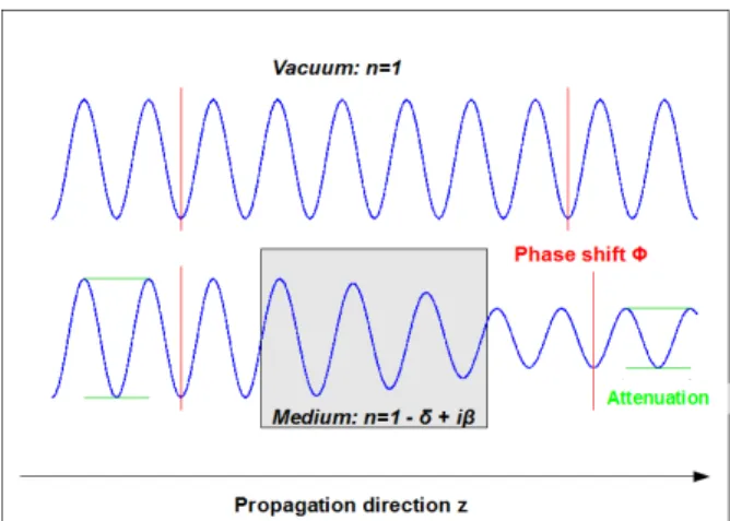
X-ray phase-contrast imaging (XPCi, see Figure 1). Whereas
conventional radiography only employs the attenuation of waves by tissue, XPCi also uses the phase shifts induced by the refraction and diffraction of X-rays in tissue.
Page 18 of 49
Figure 1: Illustration of how a change in transport medium leads to both attenuation (wave amplitude decrease, see green lines) and a phase shift (compare red lines).
Source: http://en.wikipedia.org/wiki/Phase-contrast_X-ray_imaging.
Figure 2 shows what can be achieved with XPCi (Bech et al., 2013). This technique is still in an experimental phase: it has been applied to human tissue, but it has not yet been introduced into the clinic.
Figure 2: Comparison of three X-ray images of a mouse: (a) conventional X-ray image, (b) difference between conventional X-ray image and refraction X-ray image, and (c) the image based on diffraction of X-rays. The black arrows point towards the trachea (well imaged in image b) and the lungs (well imaged in image c).
Source: http://www.nature.com/srep/2013/131113/srep03209/full/srep03209.html.
In the interviews, we asked experts about techniques identified as promising in the previous report (Stam & Bijwaard, 2011), such as
breast tomosynthesis and stereoscopy (3D imaging using just two
images taken from different angles). According to the experts
interviewed, the latter technique has not yet been introduced in Dutch clinical practice. The former technique, tomosynthesis, has been introduced in a few Dutch hospitals, but has as yet been used on very few patients. However, according to the FDA (2014), the technique is
superior to standard mammography in detecting breast cancer. Apart from the above, there have been few changes in conventional radiology since the last report.
3.2 CT
Radiation protection is also an important topic in CT. Brix et al. (2013) refer to radiation protection in clinical SPECT/CT. Clinical studies have demonstrated that the use of a combination of SPECT and CT has an added value as compared to single SPECT. The diagnostic CT in these exams contributes considerably or dominates the patient exposure to radiation. Brix et al. conclude that SPECT/CT studies should be optimised. The exposure due to CT must be reduced to the level of state-of-the-art CT practice and should never exceed diagnostic reference levels.
The potential for cardiac scanning using CT has increased with the recent development of faster and more accurate scanners. Sun et al. (2012) reported on the current state of cardiac scanning with CT in Australia. They concluded that CT is a rapid, accurate and non-invasive method of imaging the cardiac region.
An important development is the measurement of waveform
characteristics in CT: currently the attenuation of X-rays is commonly used to derive 3D images. However, it is possible to derive additional information from the phase shift of X-rays in CT. The method is
especially well suited for generating more contrast in low-density tissue (Lang et al., 2014). However, this method is still experimental and, according to the experts interviewed, has not yet been introduced in clinical practice.
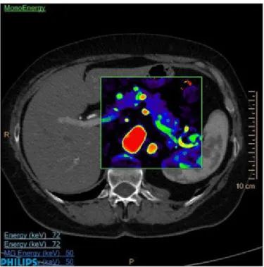
It is also possible to use different wavelengths or imaging to literally add colour to X-rays or CT(see Figure 3. According to the experts
interviewed, dual-energy imaging has been introduced in Dutch
hospitals. It is currently being used to diagnose kidney stones, gout and lung embolism, to limit metal-related artefacts on images, and for CT angiography. However, its use is still somewhat experimental and not routine practice. State-of the-art spectral CT uses eight energy levels (Barnes, 2014). This opens up new possibilities for functional imaging with CT. In December 2013 the first spectral CT scanner was introduced, and in November 2014 it came on the market. It is likely to be
introduced in Dutch hospitals soon. Its main advantages include a dose reduction in scans using contrast agent (because a pre-contrast scan is no longer required) and a reduction in the amount of contrast agent that is needed.
Page 20 of 49
Figure 3: Image taken by a spectral CT scanner.
Source:
http://www.medgadget.com/2013/12/philips-introduces-iqon-worlds-first-spectral-detector-ct.html.
An important development related to spectral CT, but also with implications for conventional CT, is the introduction of hybrid photon
counting detectors. In December 2014 the first detectors became
commercially available (see http://medical.dectris.com/). When used in CT scanners, these detectors will allow for higher-resolution or lower-dose imaging. At the same time, the individual energies of photons may be determined, enabling spectral CT imaging.
In the interviews, we asked about techniques identified as promising in the previous report (Stam and Bijwaard, 2011), such as CT colonoscopy,
CT coronary angiography and CT optimisation. Colonoscopy is currently
replacing conventional imaging of the colon, either virtual (with CT) or by internal examination. Similarly, coronary angiography with CT is replacing conventional screening in suspected cases. In cases of abnormality, this is followed by an interventional angiography (e.g. for placing a stent). To optimise CT scanning, iterative reconstruction techniques have been introduced and are being widely used. The same applies to tube current modulation. It is these dose-reduction techniques (leading to reductions of several tens of percent) that constitute the most significant development in clinical practice since the last report (see also Stam, 2013).
4
Nuclear medicine
In general, there have been developments in the use of radiolabeled
nanoceria, the nanoparticle equivalent of ceriumoxide, an antioxidant.
As mentioned by Bakht et al. (2013), the use of nanoparticles could reduce the radiation risk to the patient. Nanoceria can protect cells from reactive chemical species due to their antioxidant and free radical scavenger properties. The use of nanoceria can therefore reduce the cancer risk from ionising radiation used for diagnostic imaging. This technique is particularly useful for patients who need frequent imaging and children, who are more susceptible to radiation.
Fahey et al. (2012) describe the minimisation and communication of radiation risk in paediatric nuclear medicine. They state that paediatric nuclear medicine is well established. Approaches are suggested for effective communication to patients’ relatives.
There have been no significant changes in the equipment and materials used in nuclear medicine since the last report, though there have been some developments in the use of toolkits for the evaluation of the radiation dose (Ikuta, 2012).
In the interviews, we asked about techniques identified as promising in the previous report (Stam & Bijwaard, 2011), such as (1) the
combination of PET and MRI, (2) options for dose reduction and
(3) radioimmunotherapy. With regard to (1): the first PET-MRI scanner was recently introduced in the VUmc in Amsterdam. As PET-MR imaging is currently not reimbursed by health insurance companies, this scanner is mainly used for research purposes. This is likely to change in the near future and more PET-MRI scanners will follow. However, some experts expect that this will be of benefit to only a few patients. With regard to (2): a dose-reduction technique that is currently being employed is to block the thyroid with sodium- or potassium-perchlorate when using I-123 or I-131. This prevents the thyroid from absorbing large quantities of these radionuclides. With regard to (3): radioimmunotherapy using Bi-213 and At-211 is not used in the Netherlands. Lu-177 is occasionally employed in some hospitals.
5
Radiation therapy
In radiation therapy there has been a development in image-guided
radiation therapy (IGRT). By real-time monitoring of the anatomic
region it is possible to be more precise in targeting the tumour (area) and to avoid irradiating healthy tissue. The imaging can consist of ultrasound, 2D X-ray devices or 3D CT (Sterzing et al., 2011). IGRT is also available in clinical practice, where cone beam CT is often used for 3D and even 4D imaging. The precise imaging of the tumour at every treatment allows for a daily update of the treatment plan, the so-called
plan of the day. This is currently being introduced in clinical practice. At
the same time, treatment planning is changing: software is being introduced that can automatically generate treatment plans. Until recently, these automatic plans were used as a first guess that needed to be optimised by radiation therapy experts, but the first patients were recently treated using only automatic planning (ITN, 2015).
As mentioned in the 2011 RIVM report there is some development in
gamma knife stereotactic radiosurgery. Harris et al. (2012) mention this
technique for the treatment of brain metastases resulting from small-cell lung cancer. Gamma knife can be used when whole-brain irradiation fails as therapy. Kuremsky et al. (2013) describe the development of the use of Gamma Knife Radiosurgery in different types of lung cancer. The successful use of Gamma Knife radiosurgery in adenomas is also mentioned by Sheehan et al. (2013).
A new development is noted in the Dutch article ‘Genezen met geluid’ (Steenhorst, 2014). It is an experimental trial at the University Medical Centre Utrecht, in which a tumour area was heated with ultrasound to 42˚C before the application of a specially designed chemotherapy. The suspected tumour area received a high dose of chemotherapy that was restricted to that area; normal tissue was spared. The application of focused ultrasound to generate local cavitation effects, resulting in the heating of tissue, is also currently being investigated by Philips for application to breast cancer treatment. One of the interviewees noted that the combination of radiotherapy and ultrasound techniques
(high-intensity focussed ultrasound, HIFU) is also being investigated. A
prototype of a combination device is operational and ready for a clinical trials. These trials should clarify the kinds of treatment for which this technique would be most effective. It is expected that good results will be obtained for pancreas, kidney and rectum/bladder abnormalities. The development of particle therapy has not changed much since the previous report. Hashimoto et al. (2012) describe the use of proton
therapy in patients with cancer and the resulting interference with
implantable cardioverter defibrillators (ICDs). Developments are under way to introduce proton therapy to the Netherlands. Currently four consortia aim for introducing proton therapy in the Netherlands:
HollandPTC (a collaboration between LUMC, Erasmus MC and TU Delft), GPTC (UMC Groningen), APTC (a collaboration between AMC, VUmc and NKI) and ZON-PTC (a collaboration between Maastro Clinic, Maastricht UMC and several other hospitals in the south of the Netherlands). The
Page 24 of 49
Ministry of Health, Welfare and Sports has granted permission to the consortia to build proton therapy centres, but negotiations with health insurance companies over the reimbursement of such treatments are still being conducted. The consortia hope to treat the first patients in 2017. According to the experts that were interviewed, radiation therapy with other particles, such as carbon ions or neutrons, is not currently used in Dutch clinical practice.
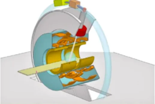
An important development is the combination of MRI and a linear accelerator (linac), MRI-linac. This allows simultaneous MR imaging and irradiation (see Figure 4), which will enable more precise irradiation of tumours in soft tissue without side effects resulting from damage to surrounding tissue (i.e. a reduction of the irradiation of these tissues). This technique is currently being developed at UMC Utrecht. It is still experimental but plans for a commercial version have already been developed. According to one expert, the experiments indicate that the technique might eventually replace surgery in specific cases.
Figure 4: Schematic representation of the MRI-linac, with the MRI in blue and the linac in grey. The linac moves in a ring around the MRI and irradiates through the MRI scanner.
Source: https://tussenhemelenaarde.wordpress.com/tag/mri-scan/.
It should be noted that the combination of MRI and radiation therapy is not entirely new. A system that combines Co-60 irradiation with a low-field MRI is already commercially available (Freeman, 2014). In 2014 the first patients were treated with this system in the United States. The combination of MRI and Co-60 allows, for example, the real-time
tracking and irradiation of lung tumours while the patient is breathing. One of these systems will be installed at VUmc in Amsterdam in the near future (VUmc, 2015).
6
Electromagnetic fields
6.1 Introduction
Electric fields arise when there is a difference in electric potential between two locations. When the electric field changes in strength and direction, a magnetic field is induced and vice versa. If the variations in the strength of the fields are regular, the speed of this variation is called the frequency. The standard unit of frequency is the hertz (the number of variations, or peaks, per second). Because the variable electric and magnetic fields are often closely linked in time, they are jointly called electromagnetic fields (EMF). EMF can also be described as a form of non-ionising radiation with frequencies between 0 hertz and 3×10¹¹ hertz (or 300 gigahertz). The variable for quantifying magnetic fields that is most closely linked to their effects on the body is the magnetic flux density. The standard unit of measurement of the flux density of magnetic fields is the tesla.
EMF can have two types of effect on the body. EMF with frequencies between 0 hertz and 107 hertz can result in the electrical stimulation of nerve or muscle cells. If the EMF are sufficiently strong, this can lead to a tingling sensation, pain or heart rhythm disturbances. EMF with
frequencies between 105 hertz and 3×10¹¹ hertz can result in the heating of body tissue. If the EMF are sufficiently strong, this can lead to heat stress or burns.
EMF are used in two ways in medical practice. On the one hand, patients can be exposed to EMF in order for information to be gained about their body’s anatomy or physiology. New developments in these applications and in data or energy transfer for diagnostic devices are described in Section 6.2 Diagnosis. On the other hand, EMF can be used to treat diseases or to make other treatment modalities (drugs, chemotherapy, radiotherapy) more effective. New developments in these applications and in treatment-related data or energy transfer are described in Section 6.3 Treatment.
6.2 Diagnosis
6.2.1 Imaging
Improvements in, and new applications of, magnetic resonance imaging (MRI) continue to be made. The use of MRI during surgery is limited by the size and accessibility of the scanner and by restrictions on metal surgical equipment because of the static magnetic field. More compact MRI systems combining a relatively weak static field (0.2 tesla) with high image quality are becoming available (Morita at al., 2014). Such systems are easier to use in a standard operating theatre. MRI scanners with static magnetic field flux densities of 3 tesla and higher have the disadvantage that the radiofrequency EMF are less homogeneous than at lower flux densities. This can result in image artefacts and local peaks in energy absorption and temperature that may pose risks for patients. The homogeneity of the radiofrequency field and the image quality can be improved by dual-source transmission, where two independent
Page 28 of 49
radiofrequency transmission channels are used to calibrate the pulses (Jia et al., 2013).
Another recent development is the use of hyperpolarised noble gases for imaging of the lungs with MRI. Lung imaging is generally difficult because of the relatively low proton density of the lungs and the presence of air– tissue borders and movements, which can cause artefacts. Hyperpolarised noble gases such as 3helium and 129xenon can be used in combination with MRI to determine ventilation defects, airspace size and connectivity, and gas exchange. Hyperpolarised noble gas MRI, without ionising radiation, could be a viable alternative to X-ray and CT examinations of the lungs (Walkup and Woods, 2014). Similarly, the detection of injected paramagnetic nanoparticles in lymph nodes with MRI could be an
alternative to X-ray lymphangiography or PET-CT (Niemirowicz et al., 2012). This technique has been used in the Netherlands to diagnose lymph node metastases in patients with prostate cancer (Broersen, 2014).
Volumetric electromagnetic phase-shift spectroscopy is a new technique
for the ambulant measurement of brain conductivity. It uses a transmitter coil that emits radiofrequency magnetic fields with
frequencies between 1 and 100 megahertz through the patient’s head. The current that is induced in the receiver coil depends on the fluid content and conductivity of the brain. The technique can be used to detect abnormalities such as oedema and haematoma. Since the apparatus is relatively small and cheap, it can be used in geographical areas without access to MRI while avoiding the use of ionising radiation (Gonzalez et al., 2013).
Microwave tomography or microwave radar imaging (frequency range:
1 to 15 gigahertz) can be used for the imaging of more superficial areas of the body using smaller, less restrictive and less expensive equipment than that for MRI. It is based on the principle that radar waves are reflected differently by malignant tissue and healthy tissue, because they have different dielectric properties. Microwave imaging has greater specificity than MRI in detecting breast cancer. However, the sensitivity of microwave imaging is lower than that of MRI due to its lower image resolution. One way of overcoming this drawback is to combine MRI and microwave imaging in one diagnostic system with superior accuracy (Meaney et al., 2013). Other possibilities, which are still being
developed, are to combine microwave imaging with a thermoacoustic method (Kirshin et al., 2013) or to inject magnetic nanoparticles as a contrast agent (Scapaticci et al., 2014). Dosimetric analysis shows that microwave radar imaging of the breast normally leads to energy
depositions below the safety limits for the general public (de Santis et al., 2012). It should therefore be safe for patients. Microwave imaging can also be used to detect strokes, using existing MRI images (Irishina et al., 2013) or CT images (Persson et al., 2014) of the brain for orientation.
6.2.2 Monitoring and navigation
Advances continue to be made in the wireless transfer of medical data from body sensors. In wireless capsule endoscopy, images of the walls of the oesophagus, stomach and intestines are made by a miniature
camera inside a swallowed capsule. These images can help to detect bleeding, inflammation and tumours of the gut. Advantages are the absence of stressful catheterisation and the accessibility of remote parts of the small intestine. If necessary, the capsule can be steered by fitting it with a permanent magnet, which is moved by applying the static magnetic field of an external magnet. The image data can be sent to a computer via a radiofrequency EMF (Hale et al., 2014). Conversely, the energy needed to operate the camera and transmitter can be
transferred to the capsule via a second radiofrequency EMF (wireless
power transmission). This removes the need for a battery and makes
smaller capsules possible. The EMF exposure remains below occupational exposure limits (Basar et al., 2014). Wireless power transmission is also becoming available for medical implants such as pacemakers. The absence of a battery should help to further reduce the size of such devices and make it easier to implant them inside the target organ (Ho et al., 2014).
Radar (pulsed EMF with frequencies in the microwave range) continues
to find new applications in monitoring vital functions and screening for disease. A system with two radar sources can be used to monitor breathing in patients with sleep apnoea without disturbances by other body movements (Kagawa et al., 2013). Radar detection of the breathing rate can also be used in combination with non-invasive measurement of the blood flow (laser Doppler) and facial temperature (thermography) to distinguish influenza patients from controls. This combined screening gives a higher predictive value than thermography alone (Matsui et al., 2010). A special type of handheld device, a
micropower impulse radar, sends out short radar pulses over a wide frequency range. It can be used to screen trauma patients quickly for pneumothorax with a sensitivity of 100% (van der Wilden et al., 2012). The frequencies of these radar applications lie in the range of 6 to 24 gigahertz.
6.3 Treatment
6.3.1 Hyperthermia
A hyperthermia technique based on high-intensity MRI in combination with the injection of metal nanoparticles in order to locally heat tissue was discussed in Stam and Bijwaard (2011). It is currently being developed in one hospital in the Netherlands. An integrative software tool for the adaptive application of hyperthermia was recently tested on patients with head and neck tumours at the Erasmus Medical Centre in Rotterdam. The ‘VEDO’ system visualises the rate of radiofrequency energy absorption on a CT scan of the patient’s body and prevents it from exceeding safety thresholds in vulnerable tissues. It also re-adjusts the hyperthermia settings when the patient complains of pain or when the measured tissue temperature exceeds a certain limit (Rijnen et al., 2013).
6.3.2 Ablation
In bronchial thermoplasty, radiofrequency EMF are applied to the bronchial walls or airways via a transmitter in a flexible catheter. This results in a local temperature increase to 65˚C for ten seconds. Its main application is to reduce the symptoms of severe and persistent asthma.
Page 30 of 49
The presumed mechanism is a reduction in the number and a change in the function of bronchial smooth muscle cells, resulting in a widening of the airways (Cayetano et al., 2012). A recent systematic review of randomised controlled trials found that bronchial thermoplasty results in a modestly increased quality of life but no significant difference in lung function in terms of objective parameters (Torrego et al., 2014).
6.3.3 Surgery
In gastro-oesophageal reflux disease, the lower oesophageal sphincter muscle is defective. Acid stomach contents can enter the oesophagus and cause pain and inflammation. Conventional surgical treatment (fundoplication) can have severe side effects. As an alternative, a ring of magnetic beads on a wire can be fitted around the sphincter muscle between the oesophagus and the stomach. The static magnetic fields of the permanent neodymium magnets in the beads produce a precise and permanent force. This force prevents excessive reflux, but the ring can expand to allow the passage of food. The device can reduce symptoms and increase quality of life, but there is a small risk of persistent swallowing problems (Ganz et al., 2013).
6.3.4 Pharmacotherapy
Certain drugs, including polypeptide hormones, cannot be administered orally because they are broken down in the digestive tract. They would normally require daily subcutaneous injections, which can be painful and have poor patient compliance. The first drug delivery microchip tested on humans has a series of reservoirs to administer daily pulses of parathyroid hormone fragment for the treatment of osteoporosis. The device can be remotely programmed using radiofrequency EMF with frequencies from 402 to 405 megahertz. It can also transmit information about its functioning back to a computer (Farra et al., 2012). A version for the administration of contraceptive hormones is being developed. The risk of interference by other devices is reduced by encryption and by the fact that communication between the implant and the
transmitter/receiver occurs close to the skin (BBC News, 2014).
Systems for drug delivery based on wirelessly activated microchips are commercially available but not yet routinely applied in hospitals in the Netherlands.
7
Optical radiation
7.1 Introduction
Optical radiation is non-ionising radiation with frequencies between 3×10¹¹ hertz and 3×10¹⁶ hertz (or wavelengths between 1 millimetre and 10 nanometre). Visible light is radiation with frequencies between about 4×10¹⁴ and 8×10¹⁴ hertz (wavelengths between 740 and
380 nanometre). Intensive exposure can damage the retina. Infrared radiation is radiation with a frequency lower than that of visible light, which can be felt as heat. Overexposure can lead to heat stress or burns. Infrared radiation is divided into three bands of descending frequencies: infrared-A (near-infrared), infrared-B and infrared-C. Ultraviolet (UV) radiation is radiation with a frequency higher than that of visible light. The frequency range of ultraviolet radiation can be divided into three bands of increasing frequency: UV-A, UV-B and UV-C. UV radiation cannot be directly detected by the human senses, but intensive exposure can lead to delayed effects such as damage to the cornea and the lens in the eye, burns and skin cancer.
Laser radiation is radiation with high power in a small surface area. Laser radiation is also monochromatic (of one wavelength) and coherent (the peaks of successive waves are in the same phase over long
distances). Its frequency can vary from infrared to UV. Strong laser radiation can cause skin burns and damage to the retina (infrared and visible lasers) and cornea (UV lasers).
Optical radiation is used in two ways in medical practice. On the one hand, patients can be exposed to optical radiation in order for
information to be gained about their body’s anatomy or physiology. New developments in such applications are described in Section 7.2
Diagnosis. On the other hand, optical radiation can be used to treat diseases or to make other treatment modalities (drugs, chemotherapy, radiotherapy) more effective. New developments in these applications are described in Section 7.3 Treatment.
7.2 Diagnosis
7.2.1 Imaging
Terahertz imaging uses optical radiation with frequencies in the
infrared-C band (3×10¹¹ to 1013 hertz). At these frequencies, the energy is
deposited mainly in the superficial layers of the body (Armstrong, 2012). This limits its application in medical diagnosis to superficial tissues such as skin, breast and intestinal mucosa. Terahertz imaging works by comparing the radiation emitted by the device with the radiation refracted by the body. The absorption and refraction of terahertz radiation changes with the water and fat content of the exposed tissue. Although it has proved possible to distinguish cancerous from normal tissue, the resolution and penetration depth of terahertz radiation is still inferior to that of competing techniques such as ultrasound, optical coherence tomography and confocal microscopy (Yu et al., 2012). Labelling cancer cells with nanoparticles may improve the sensitivity of
Page 32 of 49
terahertz techniques and could also be used to focus hyperthermia treatment (Stylianou and Talias, 2013).
Capsule endomicroscopy is a technique intermediate between classical
catheter endoscopy and the wireless capsule endoscopy described in Chapter 5. It uses a pill-sized scanner that can be swallowed but is still connected to an infrared laser source via a thin bundle of optic fibres. The scanner uses an imaging technique called spectrally encoded
confocal microscopy: near-infrared light is diffracted and the separate
wavelengths are focused and beamed through the gut wall. The
reflected light is collected, filtered and analysed. Since the scanner can rotate within the capsule, it can generate a three-dimensional
microscopic image of an entire section of the gut. Such images have been used to study white blood cells in the inflamed oesophagus wall (Tabatabaei et al., 2013).
A similar technique called optical frequency domain imaging (or volumetric laser endomicroscopy) has been used to image the
microscopic structure of the upper gastrointestinal tract. This technique is based on optical coherence tomography and compares the laser light scattered by living tissue with a reference beam that has not passed the tissue (Gora et al., 2013). It has a greater scanning speed and greater sensitivity than ‘classical’ optical coherence tomography. Further research is needed to determine whether it is sufficiently sensitive, specific and cost-effective to replace or supplement traditional random biopsy and pathology (Carignan and Yagi, 2012). Optical frequency domain imaging has also been used to generate high-resolution images and measurements of the diameter of coronary arteries (Okamura et al., 2011).
Two other endoscopy-based techniques have been developed in order to identify oesophageal and intestinal cancer more directly. The first,
angle-resolved low coherence interferometry, measures the angular
distribution of scattered near-infrared light as a function of tissue depth. It has a greater penetration depth than confocal microscopy and can be used to identify abnormal cells on the basis of the size of their nuclei. It has shown high sensitivity and specificity in patients with Barrett’s Oesophagus or colon cancer. It is potentially faster and less variable than traditional biopsies with histopathology (Carignan and Yagi, 2012; Zhu et al., 2012). The second technique, hypoxia imaging endoscopy, uses a visible light laser to compare the absorption spectrum of a tumour with that of healthy tissue in the colon. It is based on the fact that the oxygen saturation of haemoglobin is relatively low in the tumour, leading to different peaks in the absorption spectrum (Kaneko et al., 2014).
Our previous report (Stam and Bijwaard, 2011) discussed the use of near-infrared spectroscopy in the chemical analysis of superficial structures such as the brain. It has now become possible to perform near-infrared spectroscopy of blood vessels with a flexible catheter. Near-infrared laser light with wavelengths between 2,500 and 800 nanometre (frequencies between about 1×10¹⁴ and 4×10¹⁴ hertz) is applied to the vessel wall and the reflected light is analysed by a
plaques in patients at risk of coronary disease. It can also be used to evaluate the effect of therapies such as treatment with statins
(Jaguszewski et al., 2013).
Fluorescence imaging can help surgeons to visualise anatomical
structures that are otherwise difficult to distinguish from the
surrounding tissue. First, the target tissue is labelled with a fluorescent marker molecule; the operating area is then illuminated at two rapidly alternating wavelengths (for example visible light and a near-infrared laser) and the reflected visible light and fluorescent light are collected by separate cameras and integrated into one digital image. Thus, for the surgeon the fluorescence image is superimposed continuously and in real time on the normal image of the tissue and instruments (Orosco et al., 2013). Although the technique has been available for at least 15 years, it has been applied in humans only in the past five years, and only in clinical trials. One of these involved the fluorescent labelling of ovarian cancer deposits in the abdominal cavity (van Dam et al., 2011). In another trial, it was used to label sentinel lymph nodes in breast cancer patients. In this case, fluorescence imaging may come to replace traditional labelling with a radioactive tracer. One disadvantage is its lesser penetration depth (Verbeek et al., 2014).
Fluorescence angiography is a new technique that can be used to assess
blood flow and the viability of tissues before surgery. A fluorescent dye is injected into the bloodstream and binds to plasma proteins. The tissue, usually skin, is illuminated with a near-infrared laser and the emitted light is measured. Viable, well perfused tissue will show greater fluorescence than poorly perfused tissue. The technique has been used to assess foot perfusion in diabetes and the viability of skin flaps in plastic surgery. It could help to reduce the use of X-ray fluoroscopy or CT angiography in some circumstances, but it is still relatively expensive (Braun et al., 2013; Yeoh et al., 2013). Fluorescence angiography is also applied in the field of ophthalmology (Murakami et al., 2013). The
following information was gathered from the expert interviews: only in some academic hospitals in the Netherlands a further development of this technique is pursued, which allows for imaging deeper inside the body (veins, lymph, central node procedure). In the latter case, this technique is sometimes used as an alternative to imaging with
radiopharmaca. Indocyanine green can in principle be labelled to tracers to mark tumour cells. The technique operates at UV or (blue) optical wavelengths at intensities that do not involve exposure risks, which means that no specific safety measures are necessary. Risk to patients is limited to a possible allergic reaction to the indocyanine green.
Optical coherence tomography (OCT) uses scattered light from pulsed
lasers to capture micrometre-resolution three-dimensional images from within biological tissue. The use of relatively long-wavelength light allows it to penetrate the scattering medium (for more details see Stam and Bijwaard, 2011). OCT-based techniques are widely used in the Netherlands for imaging purposes in ophthalmology and cardiology. The technique is still being improved, and new applications are being
explored. Pilot projects are currently in place in the Netherlands to investigate the feasibility of using catheter-based OCT in the bladder and lungs. According to the experts interviewed, this laser-based
Page 34 of 49
technique, operating at infrared wavelengths and moderate intensities, does not cause safety issues for patients and operators, meaning that no specific safety measures are necessary. OCT-based techniques have now mostly replaced near-infrared spectroscopy for blood vessel
imaging. However, near-infrared spectroscopy is now commonly used in neonatology.
Laser-speckle imaging, discussed in Stam and Bijwaard (2011), is based
on the detection of interference patterns (speckles) due to the reflection of light by tissue. Moving blood cells, for example, cause a fluctuating interference pattern. This technique is used to image blood perfusion (mainly in skin and eyes), and is applied in some hospitals. According to the experts who were interviewed, no specific safety measures are necessary because irradiance levels are low and wavelengths correspond to infrared or near-infrared radiation.
Endoscopic multispectral imaging involves the application of a number of
narrow bands of (optical) wavelengths. According to the experts, this technique is currently being investigated, and is expected to be used in hospitals in the near future. At present, the application of this technique to the monitoring of respiration and heart pulse is being investigated in one hospital. In hospitals, fluorescent endoscopy is commonly used in the bladder, in combination with the dye 5-aminolaevulinic acid.
7.2.2 Monitoring
Our previous report (Stam and Bijwaard, 2011) discussed laser speckle imaging as a new digital technique for quantifying blood flow in tissue near the body surface. Now, a technique called near-infrared diffuse
correlation spectroscopy can be used to measure blood flow with a
greater penetration depth (several centimetres). This makes it possible to measure blood flow in the brain, in muscle and in tumours such as those arising in breast or prostate cancer and to evaluate the effects of treatment. This technique directly quantifies the motion of red blood cells by measuring fluctuations in their scattering of near-infrared photons (wavelengths 650 to 950 nanometre) (Yu, 2012). Near-infrared diffuse correlation spectroscopy equipment is already commercially available, and is used in some hospitals in the Netherlands. The technique can also be used to discriminate between blood vessels and surrounding tissues near the skin surface. An example of such an
instrument is the ‘VascuLuminator’, which is a clinical device designed to visualise blood vessels to increase the success of venipuncture and reduce complications during blood redrawing or the intravenous administration of medication. It operates at a wavelength of 800 nanometre. Another example of a commercially available instrument using this technique is the ‘AccuVein’. Although these devices improve the visibility of blood vessels, they do not necessarily improve
cannulation (de Graaff et al., 2013).
7.3 Treatment
7.3.1 Ablation
Our previous report (Stam and Bijwaard, 2011) discussed photodynamic therapy, where a light-sensitive chemical is given to the patient and the diseased area is illuminated with light of the appropriate wavelength. Interaction between the light-activated dye and local oxygen generates
reactive oxygen species, which cause cell death. Conventional, hospital-based photodynamic therapy can be time-consuming and painful for patients. A portable light-emitting diode has been developed that can be worn as a skin plaster. It delivers visible light with lower irradiance but over a longer period than that used in conventional photodynamic therapy and appears to be less painful. The therapeutic effectiveness of conventional and ambulant photodynamic therapy for non-melanoma skin cancer is similar (Ibbotson and Ferguson, 2012).
From the interviews of experts it can be concluded that optical laser-based skin therapy is applied widely in the Netherlands, e.g. in hair removal and wrinkle treatment (see also Bijwaard and Stoop, 2009). Dosimetry, especially for the eye, should be given appropriate attention. Low-energy pulsed lasers are routinely used for ablation in
ophthalmology. In some cases, pulsed lasers are also used for the treatment of renal lithiasis and cystoliths. Femtosecond high-intensity pulsed lasers are sometimes used in ocular surgery. This technique is expected to be used on a more routine basis in the future.
7.3.2 Stimulation
Since the publication of our previous report (Stam and Bijwaard, 2011), the possible complications resulting from the use of lasers in skin
therapy have been reviewed in more detail (AlNomair et al., 2012). In the case of fractional skin resurfacing with infrared lasers, which is used to treat scars, wrinkles and photoaging, inflammation and
hyperpigmentation can occur, especially in patients with darker skin. Hypertrophic scarring can also occur in more sensitive skin areas such as the neck. In both cases, lower intensities and longer treatment intervals should be used. However, the risk of such complications is lower with fractional laser treatment than with traditional laser skin resurfacing techniques. In general, the potential for complications is reduced if the operator of the equipment generating optical radiation understands not only its settings, but also its technical properties such as wavelength, energy source, power and possible mechanical errors.
8
Conclusions and recommendations
Since the previous report (Stam and Bijwaard, 2011), a number of new techniques has been identified that have found their way into clinical practice.
Ionising radiation
The main developments in imaging are the use of the wave properties of X-rays in phase contrast and spectral imaging and new developments in dose-reduction techniques. Phase contrast imaging is still experimental, but spectral imaging has been introduced in Dutch clinical practice. Dose-reduction techniques such as tube current modulation and iterative reconstruction have become common in hospitals.
In radiation therapy the main developments consist of better imaging, planning and targeting. Real-time imaging for IGRT has become common and MR imaging during therapy is commercially available, although it is not yet used in Dutch clinical practice. Software for automatic treatment planning has been improved and the first
automatically planned treatments have been undertaken. Finally, both MR-guided irradiation and the introduction of proton therapy in the Netherlands will enable more precise irradiation of tumours.
Non-ionising radiation
Apart from the continuing improvement and expansion of MRI-based techniques, two other trends are apparent in medical techniques that use EMF. First, devices that are more compact and use weaker EMF than those of MRI continue to find their way into the clinic. The main example is the use microwave tomography on superficial structures such as skin, breast tissue and cerebral blood vessels. It has good specificity for breast cancer but its sensitivity needs to be improved before it can be used more widely. The second trend is the increasing use of wireless technology to transfer images from a device to a computer or to transfer energy or instructions to a device. The main examples that have found their way into clinical practice are wireless capsule endoscopy and wirelessly controlled drug implants.
Most techniques using optical radiation that have been developed in the past five years were found in the diagnostic domain and two main trends are visible. First, terahertz imaging is starting to be applied in humans to study superficial structures such as skin and breast tissue.
Unfortunately, its resolution is still inferior to that of existing imaging techniques such as ultrasound. Second, a variety of new optical imaging techniques have been introduced for in vivo analysis of tissue sections. These are often combined with endoscopes and enable real-time histopathology of the gastrointestinal tract and blood vessels.
For all these new techniques, the main recommendations of our previous report (Stam and Bijwaard, 2011) still hold. Relatively little information was found about the strength of the non-ionising radiation used and the risks its use may entail for patients or medical personnel. Further
Page 38 of 49
of the newer techniques is yet to be measured in randomised controlled trials. More research on the cost–benefit ratio of many of the new techniques reviewed here is also recommended.
References
AlNomair N, Nazarian R, Marmur E (2012) Complications in lasers, lights, and radiofrequency devices. Facial Plast Surg 28(3):340–346. Alzen G, Benz-Bohm G (2011)Radiation protection in pediatric radiology.
Dtsch Arztebl Int. 108(24):407-14.
Armstrong CM (2012) The truth about Terahertz. IEEE Spectrum. http://spectrum.ieee.org/aerospace/military/the-truth-about-terahertz (last visited on 7 August 2014).
Bakht MK, Hosseini V, Honarpisheh H (2013) Radiolabeled nanoceria probes may reduce oxidative damages and risk of cancer: a hypothesis for radioisotope-based imaging procedure. Med Hypotheses 81(6): 1164–1168.
Barnes E (2014), New Zealand project eyes eight-energy CT, Medical Physics Web,
http://medicalphysicsweb.org/cws/article/research/58731. Basar MR, Ahmad MY, Cho J, Ibrahim F (2014) Application of wireless
power transmission systems in wireless capsule endoscopy: an overview. Sensors (Basel) 14(6): 10929–10951.
BBC News (2014) ‘Remote control’ contraceptive chip available ‘by 2018’. http://www.bbc.com/news/technology-28193720 (last visited on 7 August 2014).
Bech M, Tapfer A, Velroyen A et al. (2013) In-vivo dark-field and phase-contrast X-ray imaging. Sci Rep 3: 3209.
Bijwaard H, Stoop P (2009) Cosmetische toepassingen van lasers buiten de reguliere gezondheidszorg, aanbevelingen voor veilig en
verantwoord gebruik (in Dutch), RIVM report 300080003. National Institute for Public Health and the Environment, Bilthoven.
Bijwaard H, Valk D (2015) Radiologisch onderzoek bij kinderen- Inventarisatie van de Nederlandse praktijk met de focus op
dosisreducerende maatregelen (in Dutch), RIVM report 2014-0071. National Institute for Public Health and the Environment,
Bilthoven.
Braun JD, Trinidad-Hernandez M, Perry D, Armstrong DG, Mills JL Sr (2013) Early quantitative evaluation of indocyanine green angiography in patients with critical limb ischemia. J Vasc Surg 57(5): 1213–1218.
Brix G, Nekolla EA, Borowski M (2013) Radiation risk and protection of patients in clinical SPECT/CT. Eur J Nucl Med Mol Imaging 2013 Sep 20.
Broersen S (2014) Nano-MRI moet patiënten meer duidelijkheid brengen (in Dutch). Medisch Contact 8: 375.
Carignan CS, Yagi Y (2012) Optical endomicroscopy and the road to real-time, in vivo pathology: present and future. Diagn Pathol 7: 98. Carpeggiani C, (2014) Over diagnostic imaging in cardiology (in Italian).
Recenti Prog Med 105(3): 93–99.
Cayetano KS, Chan AL, Albertson TE, Yoneda KY (2012) Bronchial thermoplasty: a new treatment paradigm for severe persistent asthma. Clin Rev Allergy Immunol 43(1–2): 184–193.
Ciáurriz-Munuce A, Fraile-González M, León-Brito H, Vicuña-Arregui M, Miquélez S, Uriz-Otano J, Jiménez-López C (2012) Ionizing
Page 40 of 49
radiation in patients with Crohn´s disease. Estimation and associated factors. Rev Esp Enferm Dig. 104(9):452-7. Cousins C, Miller DL, Bernardi G, Rehani MM, Schofield P, Vañó E,
Einstein AJ, Geiger B, Heintz P, Padovani R, Sim KH (2013) International Commission on Radiological Protection. ICRP
PUBLICATION 120: Radiological protection in cardiology. Ann ICRP. 42(1):1-125.
de Graaff JC, Cuper NJ, Mungra RA, Vlaardingerbroek K, Numan SC, Kalkman CJ (2013) Near-infrared light to aid peripheral
intravenous cannulation in children: a cluster randomised clinical trial of three devices. Anaesthesia 68: 835–845.
De Santis V, Sill JM, Bourqui J, Fear EC (2012) Safety assessment of ultra-wideband antennas for microwave breast imaging.
Bioelectromagnetics 33(3): 215–225.
Fahey FH, Treves ST, Adelstein SJ (2012) Minimizing and
communicating radiation risk in pediatric nuclear medicine. J Nucl Med Technol 40(1): 13–24.
Farra R, Sheppard NF Jr, McCabe L, Neer RM, Anderson JM, Santini JT Jr, Cima MJ, Langer R (2012) First-in-human testing of a wirelessly controlled drug delivery microchip. Sci Transl Med 4(122):
122ra21.
FDA (2014) Clinical studies have shown that 3D breast tomosynthesis can increase the cancer detection rate. Consumer Health
Information:
http://www.fda.gov/downloads/ForConsumers/ConsumerUpdates/ UCM416664.pdf.
Freeman T (2014) First MR-guided radiation treatment. Med Phys Web: http://medicalphysicsweb.org/cws/article/research/56179.
Ganz RA, Peters JH, Horgan S, Bemelman WA, Dunst CM, Edmundowicz SA, Lipham JC, Luketich JD, Melvin WS, Oelschlager BK, Schlack-Haerer SC, Smith CD, Smith CC, Dunn D, Taiganides PA (2013) Esophageal sphincter device for gastroesophageal reflux disease. N Engl J Med 368(8): 719–727.
Gonzalez CA, Valencia JA, Mora A, Gonzalez F, Velasco B, Porras MA, Salgado J, Polo SM, Hevia-Montiel N, Cordero S, Rubinsky B (2013) Volumetric electromagnetic phase-shift spectroscopy of brain edema and hematoma. PLoS One 8(5): e63223.
Gora MJ, Sauk JS, Carruth RW, Gallagher KA, Suter MJ, Nishioka NS, Kava LE, Rosenberg M, Bouma BE, Tearney GJ (2013) Tethered capsule endomicroscopy enables less invasive imaging of gastrointestinal tract microstructure. Nat Med 19(2): 238–240. Hale MF, Sidhu R, McAlindon ME (2014) Capsule endoscopy: current
practice and future directions. World J Gastroenterol 20(24): 7752–7759.
Harris S, Chan MD, Lovato JF (2012) Gamma knife stereotactic radiosurgery as salvage therapy after failure of whole-brian radiotherapy in patients with small-cell lung cancer. Int J Radit Oncol Biol Phys 2012 83(1): e53–59.
Hashimoto T, Isobe T, Hashii, H (2012) Influence of secondary neutrons induced by proton radiotherapy for cancer patients with
implantable cardioverter defribillators. Radiat Oncol. 28(7): 10. Ho JS, Yeh AJ, Neofytou E, Kim S, Tanabe Y, Patlolla B, Beygui RE, Poon
AS (2014) Wireless power transfer to deep-tissue microimplants. Proc Natl Acad Sci USA 111(22): 7974–7979.
Ibbotson SH, Ferguson J (2012) Ambulatory photodynamic therapy using low-irradiance inorganic light-emitting diodes for the treatment of non-melanoma skin cancer: an open study. Photodermatol Photoimmunol Photomed 28(5): 235–239.
Ikuta I, Sodickson A, Wasser EJ, Warden GI, Gerbaudo VH, Khorasani R (2012) Exposing exposure: enhancing patient safety through automated data mining of nuclear medicine reports for quality assurance and organ dose monitoring. Radiology 264(2):406-13. Irishina N, Torrente A (2013) Brain Stroke Detection by Microwaves
Using Prior Informationfrom Clinical Databases. Abstract and Applied Analysis 2013: 1-8.
ITN (2015) Leading UK cancer center performs first radiotherapy treatment with Varian’s RapidPlan,
http://www.itnonline.om/article/leading-uk-cancer-center-
performs-first-radiotherapy-treatment-varians-rapidplan?eid=276868945 &bid=996224.
Jaguszewski M, Klingenberg R, Landmesser U (2013) Intracoronary near-infrared spectroscopy (NIRS) imaging for detection of lipid content of coronary plaques: current experience and future perspective. Curr Cardiovasc Imaging Rep 6: 426–430.
Jia H, Wang C, Wang G, Qu L, Chen W, Chan Q, Zhao B (2013) Impact of 3.0 T cardiac MR imaging using dual-source parallel
radiofrequency transmission with patient-adaptive B1 shimming. PLoS One 8: e66946.
Kagawa M, Ueki K, Kurita A, Tojima H, Matsui T (2013) Non-contact screening system with two microwave radars in the diagnosis of sleep apnea-hypopnea syndrome. Stud Health Technol Inform 192: 263–267.
Kaneko K, Yamaguchi H, Saito T, Yano T, Oono Y, Ikematsu H, Nomura S, Sato A, Kojima M, Esumi H, Ochiai A (2014) Hypoxia imaging endoscopy equipped with laser light source from preclinical live animal study to first-in-human subject research. PLoS One 9(6): e99055.
Kirshin E, Oreshkin B, Zhu GK, Popović M, Coates M (2013) Microwave radar and microwave-induced thermoacoustics: dual-modality approach for breast cancer detection. IEEE Trans Biomed Eng 60(2): 354–360.
Kuremsky JG, Urbanic JJ, Petty WJ, Lovato JF, Bourland JD, Tatter SB, Ellis TL, McMullen KP, Shaw EG, Chan MD (2013) Tumor histology predicts patterns of failure and survival in patients with brain metastases from lung cancer treated with gamma knife radiosurgery. Neurosurgery 73(4):641-7
Lang S, Zanette I, Dominietto M et al. (2014) Experimental comparison of grating- and propagation-based hard X-ray phase tomography of soft tissue. J Appl Phys 116: 154903.
Matsui T, Hakozaki Y, Suzuki S, Usui T, Kato T, Hasegawa K, Sugiyama Y, Sugamata M, Abe S (2010) A novel screening method for influenza patients using a newly developed non-contact screening system. J Infect 60(4): 271–277.
Meaney PM, Golnabi AH, Epstein NR, Geimer SD, Fanning MW, Weaver JB, Paulsen KD (2013) Integration of microwave tomography with magnetic resonance for improved breast imaging. Med Phys 40(10): 103101.