Food, novel foods, and allergenicity H. van Loveren
This investigation has been performed by order and for the account of the Inspectorate for Health and veterinary Public Health, within the framework of project V/640400, A survey of food allergy in the population.
Preface
This report reviews mechanisms of gut immune responses and food allergy, symptoms and manifestations of food allergy, incidences of food allergy in the population, characteristics of food allergens, and discusses approaches of assessment of allergenic potential of foods derived from genetically engeneered foods, including testing in which human sera or testing in humans is applied, or alternatively testing in laboratory animals. The conclusion drawn in this review indicates that it may eventually be possible to use the elements from chemical risk assessment in food allergy risk assessment. However, in order to reach this goal, further information needs to be available especially with respect to characteristics of food allergens, critical windows of exposure, and the validity of laboratory animal models.
Contents
Samenvatting 5 Summary 7
1. INTRODUCTION 9
2. MECHANISMS OF GUT IMMUNE RESPONSES 11
3. SYMPTOMS AND MANIFESTATIONS OF FOOD ALLERGY 15 3.1 Generalized and local symptoms 15
3.2 Gastrointestinal symptoms 16 3.3 Symptoms in the skin 17 3.4 Respiratory symptoms 18
4. INCIDENCE OF FOOD ALLERGY 19 5. FOOD ALLERGENS 21
6. PROPERTIES OF FOOD ALLERGENS 25 6.1 Size and structure 25
6.2 Glycosylation 25
6.3 Solubility and resistance to heat 26
6.4 Sensitivity to enzymatic and acid degradation 26
7. ASSESSMENT OF ALLERGENIC POTENTIAL OF FOODS DERIVED FROM GENETICALLY ENGENEERED FOODS 31
8. PREDICTION OF FOOD ALLERGENICITY USING LABORATORY ANIMALS 39 9. RISK ASSESSMENT 41
10. CONCLUDING REMARKS 43 References 45
Samenvatting
Het immuunsysteem in het maag-darm kanaal vormt een gespecialiseerd compartiment en is bijzonder effectief in weerstand tegen pathogenen, terwijl immuunresponsen tegen voedselcomponenten doorgaans niet tot uiting komen. Dat laatste fenomeen is gebaseerd op een mechanisme van actieve suppressie van dergelijke responsen, en wordt aangeduid met de term orale tolerantie. Soms kan voedsel in sommige individuen toch tot allergische responsen leiden. Belangrijke allergene voeding zijn schaaldieren (garnalen, kreeft, krab), eieren, vis, melk, pinda, soya, noten, en tarwe. De meeste allergenen in die voeding zijn eiwitten. Voedselallergie tegen componenten met een laag moleculair gewicht, zoals kleurstoffen in de voeding, komen niet voor. Voedselallergie kan gepaard gaan met een groot aantal symptomen, zoals gastro-intestinale problemen, problemen, in de huid, en in de ademhalingsorganen. Na opname van voedselallergenen door gesensibiliseerde individuen kunnen levensbedreigende situaties optreden. De prevalentie van voedselallergie is ongeveer 2% van de bevolking. Ook niet-immunologische reacties op voedsel kunnen voorkomen. De mechanismen van dergelijke voedsel-intolerantie zijn verschillend, en kan gebaseerd zijn op irriterende activiteit, farmacologische activiteit, verstoring van enzymactiviteit, of zijn psychosomatisch van oorsprong. Soms gelijken de symptomen van voedsel-intolerantie op echte allergie, maar de responsen zijn meestal mild en nooit levensbedreigend. De aandacht die het probleem van voedselallergie in de algemene bevolking krijgt heeft geleid tot een overschatting van het probleem, waarbij wel wordt aangenomen dat in bijna ieder huishouden wel een persoon met voedselallergie voorkomt.
Omdat allergische reacties gepaard kunnen gaan met levensbedreigende situaties, en omdat de vervaardiging van genetisch gemodificeerde gewassen gepaard kan gaan met de introductie van nieuwe antigene determinanten in die gewassen, bestaat bezorgdheid dat dergelijke nieuwe voedingsgewassen een nadelig effect op gezondheid zouden kunnen hebben.
Het identificeren van potentiële allergeniciteit van genetisch gemodificeerde voeding houdt onder andere in de evaluatie van de bron van het gen dat in het nieuwe voedsel is geïntroduceerd, de beoordeling van eventuele homologieën van de geïntroduceerde genproducten met bestaande allergenen, stabiliteit onder omstandigheden waarbij digestie door maagsappen wordt gesimuleerd, solid phase immunoassay gebruikmakend van positieve humane sera, huid skin prik testen in patiënten met bewezen voedselallergie, en dubbelblinde placebo-gecontroleerde voedsel challenge in patiënten met bewezen voedselallergie. Het zal duidelijk zijn dat genetisch gemodificeerde voeding, waarbij genen zijn geïntroduceerd afkomstig uit niet allergene bronnen, die met name coderen voor eiwitten die resistent zijn degradatie door maagsappen, een groot probleem opleveren voor de beoordeling van het betreffende product. Er wordt geïnvesteerd in het ontwikkelen van diermodellen die gebruikt kunnen worden om allergeniciteit van eiwitten te voorspellen. Daarbij wordt orale of parenterale blootstelling aan de eiwitten toegepast, waarna IgE antilichaamresponsen en andere klassen van responsen worden gemeten, om allergeniciteit en immunogeniciteit te kunnen onderscheiden. De resultaten zijn bemoedigend. Echter, de extrapolatie van dergelijke gegevens naar de mens is bijzonder moeilijk, omdat doorgaans slechts een minderheid van individuen in de bevolking allergisch op een bepaald product reageert. De modellen zijn tot dusver ook nog niet gevalideerd.
In het algemeen is het proces van risico-evaluatie gebaseerd op de identificatie van potentiële schadelijke activiteit, het vaststellen van dosis-respons relaties, en het vaststellen van actuele blootstellingniveaus. Tot dusver zijn dosis-respons relaties nauwelijks vastgesteld. Voor allergie geldt, dat niet alleen sprake is van dosis-respons relaties ten aanzien van
sensibilisatie, maar dat bovendien in eenmaal gesensibiliseerde individuen sprake is van een andere dosis-respons relatie, waarbij veel kleinere hoeveelheden van het allergeen dan bij niet gesensibliseerden al tot een effect kunnen leiden.
Om een aantal redenen wordt adequate risico-evaluatie momenteel belemmerd. In de bevolking is sprake van grote interindividuele verschillen. Gedeeltelijk kunnen deze worden toegeschreven aan genetische verschillen. Het mechanisme van orale tolerantie wordt nog nauwelijks begrepen. Tenslotte moet worden opgemerkt dat met name het zich ontwikkelende immuunsysteem beïnvloed kan worden door immunologisch actieve factoren. Het tijdstip waarop een eerste contact met voedselallergenen plaats vindt kan een grote impact hebben op het al of niet ontstaan van voedselallergie of orale tolerantie. Factoren in de voeding die niet zelf allergeen zijn, kunnen een invloed hebben op de het ontwikkelen van orale tolerantie voor voedselallergenen, zoals bijvoorbeeld omdat die bestanddelen immunologisch actief zijn of de toegang van allergenen tot de mucosa bevorderen.
Summary
The gut immune system forms a specialized compartment of the immune system and is very effective in protection from pathogens, while immune responses to food components do normally not are become evident, The latter phenomenon is based on a mechanism of active suppression of such responses, to which the term oral tolerance has been attributed. However, certain foods lead to allergic responses in certain individuals. Main allergenic foods are Crustacea (shrimp, lobster, crab), egg, fish, milk, peanuts, soybeans, tree nuts, and wheat, and allergens are always proteins. Food allergies to low molecular weight compounds, such as food colours do not occur. A wide array of symptoms can result from food allergy, including gastrointestinal problems, skin problems, and respiratory problems. Severe and life threatening situations can occur after ingestion of allergenic foods in sensitized individuals. The actual prevalence seems to be about 2% of the population. Non-immunologically based reactions to food also occur. The mechanisms of such food-intolerance reations are divers, and include irritant activity, pharmacological activity, disturbance of enzyme activity, or are of psychosomatic nature. Sometimes expression of food intolerance resembles that of true allergies, but the responses are mostly mild and never life treathening. Public awareness in the general population concerning food allergies has led to an overestimation of the problem, suggesting that in almost every household there will be at least one person with allergies to food.
Based on the potentially life treatening consequences of allerig reactions to food, and the possibility to introduce new antigenic determinants when introducion new genes into crops, there is reason for concern that the in genetically engineered food may provide a health risk. Hazard identification concerning the allergenicity of newly engineered foods comprises evaluation of the source of the newly introduced gene, evaluation of sequence similarities of the new gene products with known allergens, sensitivity to digestion by gastric simmulated fluid, solid phase immunoassay, skin prick testing in patients with proven food allergies, and double-blind placebo-controlled food challenge in patients with food allergy. It is obvious that genetically engeneered foods that contain genes from non allergenic sources, but that lead to new proteins (especially those that are relatively insensitive to gastric simmulated fluid degradation) pose the biggest problem for evaluation of potential allergenicity. At present, efforts are undertaken to develop animal models that can be used for prediction of allergenicity of proteins. Either oral or parenteral exposure to proteins, follwed by analysis of IgE and other antibody subclasses have been used and show promise. However, extrapolation of such animal data to humans will be extremely difficult, especially as only a minority of individuals may react with an allergic reactions to food. Hence, as yet tere are no validated animal models to predict the allergenicity of genetically engineered proteins.
In general, the process of risk evaluation is based on hazard identification, assessment of dose-response relationships, and assessment of the expected exposure. Sofar, dose-response relationships have hardly been established. It should be noted that for allergy, dose-response relationships must relate to the sensitization phase, and in addition to the elicitation of already sensitized individuals after exposure. Amounts of allergens required to elicit responses in already sensitized individuals are usually much lower than required for sensitization.
Adequate risk evaluation is at present especially hindered by the great interindividual differences. For a part, they may be attributed to genetic differences. The process of oral tolerance is hardly understood, and it is therefore relatively unknown how genetic influence oral tolerance. Finally, the developing immune system is especially sensitive to immunomodulatory influences. The time at which food allergens are first encounterd may
have an impact on the development allergy or tolerance. Also factors that are in the food but are not allergenic themselves may influence the development of oral tolerance to food allergens, i.e. those moieties that influnece immune responses (immunotoxicants) or may influence entry of proteins into the mucosa.
1.
INTRODUCTION
This report review major aspects of allergic responses to food.
Generally, components in food that are responsible for immunologically mediated allergic reactions are proteins, whereas non-immunological adverse reactions are caused by lower molecular weight agents. Most allergic responses to protein in foods are IgE mediated, but cellular mediated allergic responses also occur. True allergic responses can be life threatening, but as their expression often resembles non-immunologic adverse responses to food, food intolerance and food allergy are often mistaken. In contrast to food allergy, food intolerance reactions are never life threatening, and although they may pose serious problems, they often have a psychosomatic background. This report focussus on true food allergy. Because of deficient knowledge on mechanisms of food allergy and associated symptoms, that are partly overlapping, it has been very difficult to develop assays that would predict the adverse capacity of components in the food.
In the section 2 immune mechanisms, with an emphasis on mechanisms in the gut are presented. In the following section 3, adverse reactions as observed clinically are discussed, while in section 4 prevalence of food allergies are mentioned. In the following sections 5 and 6 existing food allergens are listed, as well as properties that such food allergens have. Sections 7 and 8 deal with hazard identification of genetically engeneered foods using human immune sera or human volunteers, or using animal models respectively. Section 9 provides some perspective on risk evalluation of allergenic foods, and section 10 provides some recommendations for further resaerch.
2.
MECHANISMS OF GUT IMMUNE RESPONSES
The mechanisms of adverse reactions to food are excellently reviewed by Brandtzaeg (1997). For the purpose of this report, a brief summary suffices:
The aim of the immune system is to provide defense to the host against invading pathogens. Infectious agents are degraded by non-specific cells such as macrophages and natural killer cells. Processed antigen determinants are then presented to lymphocytes inducing specific immune responses. These specific immune responses complete the action started by the non-specific effector cells. In addition, non-specific cells carry memory, so that after a secondary encounter with the pathogen the infection is terminated much more efficiently. Specific immune responses are either cellular immune responses or humoral responses. Cellular responses are mediated by T lymphocytes that provide help to specific cytotoxic lymphocytes, or T lymphocytes that attract and stimulate macrophages. Humoral responses are also often mediated by T cells that provide help to the synthesis of specific antibodies.
T and B lymphocytes recognize and combine with different regions of antigenic protein molecules. Antigens are three-dimensional structures and there are usually many peptide regions of the native molecule to which antibodies can bind. Sometimes these are concentrated in particular ‘immunodominant’ regions on the outside of the molecule. Digestion of a food protein in the gut may also reveal new antigenic determinants which have been hidden deep within the native molecule. The key point to note is that antibody and B cells recognize antigens, in solution or on cell surfaces, in their native conformation. On the other hand, it has been known for many years that T cells recognize both native and denatured antigen, indicating that it is primary sequences of peptides which are important for the induction of cellular immunity and for other T cell functions. The T cell receptor sees only a small fragment of the original antigen, after the latter has been internalized by an antigen-presenting cell, partially degraded by proteolytic enzymes, then carried back to be presented at the cell surface in physical association with a MHC (major histocompatibility complex) molecule. The optimum size of peptide for antigen presentation is 8-24 amino acids, a size which fits well into the groove of an MHC molecule. Although different MHC molecules can interact with the same antigen, they will present it to T cells in slightly different ways. Thus, an individual’s genetic make-up as well as environmental factors will determine which polypeptide sequences from a single protein his or her T cells can recognize, which types of T cells are preferentially stimulated, and ultimately, whether the immune response generated is appropriate or harmful –IgE and cell-mediated immune responses to foods being examples of the harmful type of response.
Both cellular and humoral immune responses are operational in the intestinal tract. IgA is especially important, as this immunoglobulin is attached to the mucus in the lumen of the gut, and thus immobilizes pathogens, preventing them from invading the tissues. Those pathogens that do succeed in penetrating the mucosa will then be dealt with by other mechanisms, such as for instance specific IgG and complement.
The gut-associated lymphoid tissue (GALT) is the specialised immune system of the gut, and represents the largest mass of lymphoid tissue in the entire human body. The immune response occurs in different physiologic compartments of this tissue: aggregated in follicles and Peyer’s patches, and distributed within the mucosa, the intestinal epithelium, and secretory sites (Isolauri et al., 2001). After birth, T lymphocytes migrate from the thymus to the thymus-dependent areas of the Peyer’s patches and the epithelium. Exposure to microorganisms in the normal environment is necessary to develop the B cell population, but
as colonisation of the intestine begins immediately from the maternal birth canal and from the surrounding environment, this is rapidly established.
The intestinal epithelium overlying the Peyer’s patches is specialised to allow the transport of antigens into the lymphoid tissue. Epithelial cells termed ‘M’ (microfold) cells, due to the numerous microfolds on their luminal surface carry out this particular function. M cells are able to absorb and transport antigens and, possibly, process and present them to subepithelial lymphoid cells. The M cells overlay lymphoid follicles, and they lack mucus and glycocalyx thereby facilitating contact between molecules and particles and the M cells. Microbial entry into epithelial cells results in the production and release of proinflammatory cytokines. Therefore in response to a bacterial invasion an acute mucosal inflammatory response is initiated. This protects the host from these pathogens within a very short time period. Adherence to and uptake of microorganisms by M cells also result in secretion of protective polymeric IgA antibodies to prevent the uptake of antigens, thus limiting the intensity and duration of the disease. These mechanisms all help to keep infection with pathogens in the gut local and as minimal as possible (Lu and Walker, 2001).
Immunoglobulin A (IgA) is mainly secreted in the lamina propria. In contrast with IgA in serum, secretory IgA is present in a different form. This form is resistant to intraluminal proteolysis, and does not activate complement or inflammatory responses. This makes it ideal for protecting mucosal surfaces. There are differences between the upper and lower parts of the human GALT in the isotype distribution of immunoglobulin-producing cells. IgA-1 immunocytes predominate in the small intestine, whereas IgA-2 producing cells are most frequent in the colon, the latter being more resistant to bacterial proteases (Isolauri et al., 2001; Doe, 1989). The secretory IgA antibodies in the gut are part of the common mucosal immune system. That is the reason why these antibodies can also be effective at other mucosal surfaces. The specific antibody-secreting lymphocytes appear in peripheral blood 2-4 days after antigen exposure, reach a maximum concentration after 6-8 days, and persist in the blood for 2-3 weeks (Isolauri et al., 2001).
Interepithelial T lymphocytes mainly exhibit a suppressor and cytotoxic phenotype, whereas the lamina propria cells exhibit a helper and inducer T cell phenotype. B cells are predominantly found in the lamina propria. T cells in the lamina propria have the ability of increased lymphokine and cytokine production. They even seem to have the potential to produce both T helper 1 as well as T helper 2 cell specific cytokines.
Immune responses are associated with damage to the pathogen, but in this process also some damage is done beyond the pathogen. The flip side of specific immune responses is allergy, i.e. immune responses to harmless agents. In this respect IgE is often a key player. The physiological role is in resistance to parasites, but IgE is often involved in allergic processes. Mast cells in the wall of the gut may acquire IgE from plasma cells in the immediate vicinity, or they may have been sensitized as they migrate prior to lodging in the tissues. The very small quantity of antigen needed for degranulation of IgE-sensitized mast cells probably reaches the tissues via the bloodstream, after entry into the body through the mucous membrane of the mouth, stomach or proximal intestine. Antigen which induces local immune-complex reactions or T cell mediated immunity probably reaches the gut lamina propria through the overlying surface epithelium. As a result of secretion of cytokines and inflammatory mediators, such as histamine, leukotrienes, prostaglandins, etc. These mediators induce capillaries and epithelium of the affected tissue become more permeable to the access of plasma, leukocytes and, in the gut, enteric antigens, increase vascular permeability, can attract eosinophils, and can enhance or suppress immune responses.. Thus, soon after the antigen-specific hypersensitivity response starts, there may develop a variety of other hypersensitivity and inflammatory reactions. Many food antigens have been implicated in
food allergy. Food allergens are always proteins, but there are no unique structural features of the allergens which induce the expression of IgE reactions in the gut. On the other hand coeliac disease is thought to be predominantly mediated by T cells, is provoked by certain cereal proteins. It is obvious that such effects to harmless agents such as peptides, i.e. as can be the case in food allergy, are undesired. Inadvertent cellular immune responses are likewise undesired.
While it is clear that the gastrointestinal tract contains effective mechanisms to eliminate pathogenic antigens, elimination of nutritive antigens is not desired. This has led to the concept of ‘oral tolerance’ (Isolauri et al., 2001; Doe, 1989). This mechanism of oral tolerance is active, by which specific IgE and cellular immune responses are suppressed, while defense such as mediated by IgA is maintained (Srobel and Mowat, 1998). The mechanism is not yet fully understood. It is thought that anergy of antigen-specific T cells plays an important role. This anergy may be induced by class I restricted CD4+ T-cells and by the cytokines they produce. These cytokines may have an additional immunomodulationg, in this case suppressive effect. Dose and frequency of exposure may influence the tolerance and its mechanism to a fed antigen. Feeding high doses of an antigen results in clonal deletion or anergy, whereas feeding low doses results in active suppression subsequent to the induction of regulatory T cells, sometimes called Th3 cells, in Peyer’s patches. The regulatory T lymphocytes function through the production of suppressive cytokines, including IL-4, IL-10, and especially transforming growth factor b. Clonal deletion or anergy is preceded by the local production of IL-12, IFN g (with consequent suppression of IL-4 and transforming growth factor b generation), and involves the apoptosis of Th1 cells. It is therefore suggested that one of the major mechanisms by which the gut-associated lymphoid tissue maintains homeostasis is via local cytokine regulation, particularly transforming growth factor b-associated low-dose tolerance (Isolauri et al., 2001).
This balance of oral tolerance and mucosal immunity is influenced by many factors. These factors include among others genetics, atopy, microbial exposure, nutrition, breast-feeding, age. These factors or combinations thereof, may disturb the balance, leaving an individual with allergic reactions to agents in his diet. It is at present not possible to predict whether an individual, while ingesting materials that have been shown to produce adverse effects, will in fact develop such adverse responses.
Historically, mucosal permeability has been thought to be a major factor in food allergy. Many food proteins cross the gut mucosal membranes, prompting immune responses even in normal individuals, which had already been recognised by Korenblat et al. (1968). However, differences in mucosal absorption of allergens have not been well studied in food-allergic individuals, compared with normal individuals; the relationship of the phenomenon of gut permeability to food allergy has not been resolved either. This is a particularly important issue in infants prior to mucosal gut barrier maturation when large molecules more easily pass though the gut epithelium and induce allergic sensitization.
Besides allergic responses to components in the food, such components may also have non-immunologic effects. These include direct toxic effects, irritant effects through the epithelium of the gut, or simulation of mast cell mediator release in a non-immunologic fashion, the latter called pseudo-allergy. There is an intricate innervation of mast cells in the intestines, which may play a role in neurological mediated adverse reactions to food. Finally, components in food may disturb enzymatic activity.
Generally, components in food that are responsible for immunologically mediated allergic reactions are proteins, whereas non-immunological adverse reactions are caused by lower molecular weight agents. With this array of mechanisms, and associated symptoms, that are partly overlapping, it has been very difficult to develop assays that would predict the adverse capacity of components in the food.
3.
SYMPTOMS AND MANIFESTATIONS OF FOOD
ALLERGY
A broad spectrum of symptoms and signs in children may be caused by food allergy, and generalized reactions, such as anaphylactic shock may also arise (Table 1).
Table 1.
Symptoms of food allergy
Generalized symptoms Anaphylaxis Gastrointestinal Nausea Vomiting Colic Diarrhea Abdominal cramps Bloating Respiratory Rhinitis Sneezing Asthma Recurrent cough Wheezing Laryngeal edema Dermatological Angiooedema Urticaria Eczema Pruritis
Erythema (skin inflammation)
3.1
Generalized and local symptoms
Anaphylaxis is a generalized and potentially life-threatening clinical syndrome resulting from the release of biologically active mediators from mast cells and basophils. Food allergies are, besides drugs and insect stings, the single most common cause of anaphylaxis seen in hospital emergency departments, accounting for about one third to a quarter of cases seen (Wutrich, 2000; Pumphrey, 2000).
Local reactions are primarily in the gastrointestinal tract, but food allergy may also cause local symptoms in the skin and respiratory tract. The pattern of symptoms may vary with age and depends on the selection of patients. In unselected, prospectively-followed birth cohorts, about 50-70% of patients with cow’s milk allergy show cutaneous symptoms, 50-60% gastrointestinal symptoms, and about 20-30% respiratory symptoms (Host, 1994). Among young children with cow’s milk allergy/intolerance, the majority have two or more symptoms with symptoms in two or more organs, as reviewed by Host (1994).
Symptoms of the so called oral allergy syndrome are localized in the pharynx and consist of swelling, itching and burning of the oral mucosa, sometimes accompanied by swelling of the lips. The reaction is due to contact with a food for which the patient is sensitized. The mechanism is based upon histamine release after activation of musosal mast cells by binding of food allergens to allergen specific IgE on these cells (Amlot et al., 1987). This type of reaction may precede more severe allergic reactions like angioedema, urticaria and even anaphylactic shock. Oral allergy syndrome occurs in patients with the para-birch, artemisia pollen-cellery and latex-fruit syndrome allergic to pollen. Allergy to fresh fruits and vegetables is a frequent cause of oral allergy syndrome with local symptoms within 15 min. of food contact (Pastorello et al., 1995).
3.2
Gastrointestinal symptoms
Gastrointestinal symptoms are chronic or acute vomiting, diarrhea, and colic. Colic appears to be a common symptom (Hill et al., 1986), but nearly always in combination with other symptoms (Goldman et al., 1963; Hill et al., 1986). In some studies, a rather high frequency of food allergy and diarrhea appears. Gastrointestinal manifestations of food allergy are reviewed by Ferguson (1997). Acute local reactions are IgE-mediated. Within seconds of oral contact with or swallowing of a provoking substance there may be facial angio-oedema, diarrhea or vomiting.
Food sensitive enteropathy is usually found in children with an atopic background, but there is good evidence for T cell mediated hypersensitivity as well as IgG and IgE, in the pathogenesis. Small intestinal mucosal damage with malabsorption is best documented for cows’ milk protein intolerance, but can also occur with soy, chicken, rice, fish and egg intolerance. Classically the infant has diarrhea and failure to thrive, with vomiting. There is malabsorption, and jejunal biopsy is abnormal, but not usually so severely affected as in coeliac disease. A cows’ milk-free diet (or other elimination diet) induces rapid remission. Once clinical recovery is complete, it may be appropriate to confirm the diagnosis by an in-hospital provocation test. Most of these children will be clinically tolerant of cows’ milk by the age of one year.
Food sensitive colitis is characterized by bloody diarrhea but differs from ulcerative colitis in many clinical and pathological features. Typically, an infant presents before the age of one year with loose stools containing mucus and blood. An elimination diet and clinical monitoring with a rectal biopsy, confirm the diagnosis. The pathology differs from classical ulcerative colitis. In food-sensitive colitis there are many eosinophils in the lamina propria. Severe clinical colitis may be induced by a food challenge and so this should not be done as a routine. As is the case of food sensitive enteropathy, most children can tolerate the offending food by the age of two.
Coeliac disease is a permanent condition of gluten intolerance, defined on the basis of the pathology of the small bowel mucosa, which recovers when the patient is treated with a gluten free diet, and reintroduction of gluten into the diet will produce a histological relapse in the jejunal mucosa. Application of these strict diagnostic criteria has led to the realization that there is a wide clinical spectrum within coeliac disease, and has reinforced the need for
biopsy confirmation in every patient. For reasons that are as yet unknown the clinical effects are remarkably heterogeneous. A coeliac child is often a grumpy, rickety, anemic, growth retarded toddler with diarrhea, a pot belly and muscle wasting numbers A strict gluten free diet for life is the cornerstone of management.
Protein-losing enteropathy, when not accompanied by other signs of GI disease has in the past been attributed to food allergy. Certainly, in animal experiments, considerable increase in gut permeability to marker proteins occurs in association with anaphylaxis. However in clinical practice, a patch of lymphangiectasia or jejunal Crohn’s disease should be considered.
Eosinophilic gastroenteritis encompasses inflammatory lesions, discrete or diffuse, and the disease may affect any part of the gut. The relationship to foods probably varies from one ease to another, and the presence or absence of atopy as an independent variable should be considered.
Abnormal immunity to wheat antigens produces several other diseases, such as coeliac disease (a permanent gluten-sensitive enteropathy), the associated condition of dermatitis herpetiformis, transient gluten-sensitive enteropathy in infancy, wheat-sensitive diarrhea without enteropathy, and baker’s asthma. The latter two conditions are caused by reactions to non-gluten wheat proteins.
3.3
Symptoms in the skin
The skin is an important target organ in non-toxic adverse reactions to food. Dermatological symptoms are atopic dermatitis, urticaria, angiooedema, and contact dermatitis. In selected materials, food allergy has been demonstrated in approximately one third of children with atopic dermatitis (Burks et al., 1988; Host, 1988). In recent Danish studies on intolerance to food additives, the majority of positive reactions were a worsening of atopic eczema and urticaria in atopic children (Fuglsang et al., 1993).
There is, however, no skin disease which is specific for an adverse reaction to food. Dermatitis herpetiformis is probably the only exception. The majority of skin eruptions associated with adverse reactions to food can also be caused by other factors. This often makes the food related diagnosis difficult and unclear.
Acute urticaria is an IgE-mediated adverse reaction to food that may be expressed as acute urticaria, occurring within 30 min. after the intake of the food. Fish, shellfish, nuts and peanuts are on the top ten of food items inducing urticaria. The prevalence of urticaria due to an IgE-mediated reaction to food is not known. There is probably an underregistration since many patients recognize the cause-effect relation, are able to avoid the suspected food and do not seek medical intervention.
In chronic urticaria food is seldom involved as a causal agent.
The most likely candidate for food-induced eczema is the young child with recalcitrant atopic dermatitis. Adults rarely have this problem. About one third of the children with atopic dermatitis visiting an allergy clinic have atopic dermatitis as a manifestation of an adverse reaction to food (Burks et al., 1988). This means that their eczema substantially improves when the suspected food is avoided. Cow’s milk is by far the most frequently involved food item. Soy milk, peanut and eggs are rarely involved in the induction of eczematous skin lesions.
The type of skin reaction occurring after double blind placebo controlled oral food challenges in children with atopic dermatitis and cow’s milk allergy are: (1) an immediate pruritic erythematous rash persisting for 30/120 min.; (2) urticaria; and (3) eczema occurring only after repeated oral challenge (Sampson and McCaskill, 1985).
The immediate reaction seems to be due to the activation of mast cells (basophils?) and the release of histamine. The mechanism behind the food induced eczematous skin reactions is
not completely understood. Atopic dermatitis is a T cell mediated skin disease. This suggests a role for cow’s milk specific T cells in the induction of the skin lesions. Children with atopic dermatitis and cow’s milk allergy have cow’s milk specific T cells in the circulation. Recently it has been reported that in children with eczema as a clinical manifestation of cow’s milk allergy cow specific T cells from peripheral blood are CLA (cutaneous lymphocyte associated antigen) positive, whereas in children with gastrointestinal symptoms of cow’s milk allergy cow’s milk specific T cells are CLA negative (Abernathy-Carver et al., 1995). CLA is considered to be a T cell homing factor for the skin. This suggests that after activation of this type of T cell homing to the skin occurs. Sofar, it is not known how and where the CLA expression is induced and where T cell activation after cow’s milk intake occurs.
The role of cow’s milk specific IgE molecules is not well defined. It has been speculated that they are involved in IgE-mediated antigen presentation to T cells. On the other hand some of the children with eczema as a manifestation of cow’s milk allergy do not have cow’s milk specific serum IgE molecules or positive skin prick tests with this allergen (Isolauri and Turjanmaa, 1996).
Cow’s milk related atopic dermatitis in adults is rare. However, there are some patients with atopic dermatitis and adult onset cow’s milk allergy (De Maat-Bleeker and Bruijnzeel-Koomen, 1996). The cow’s milk related symptoms in these patients start with an exacerbation of their eczema and the occurrence of oral allergy symptoms, urticaria and even systemic anaphylaxis.
Food allergy due to plant derived food is frequently seen in patients with atopic dermatitis with pollen sensitization. The symptoms however are localized in the pharynx (oral allergy syndrome) and sometimes may involve the skin as urticaria. Induction of eczematous responses is very rare. Allergy to plant derived food in the absence of pollen sensitization is uncommon in patients with atopic dermatitis.
3.4
Respiratory symptoms
Respiratory symptoms are recurrent wheezing, asthma, stridor, cough, and rhinoconjunctivitis. These symptoms are rarely the only clinical manifestation of a food allergy. In children with asthma, food allergy has been demonstrated in about 4-6% (Bousquet, 1995). In the UK register of fatal anaphylactic reactions (Pumphrey, 2000), all food-induced fatalities have been accompanied by respiratory problems with respiratory arrest. Atopic individuals with bronchial asthma and prior allergic reactions to the same food are at a particularly high risk.
4.
INCIDENCE OF FOOD ALLERGY
Bock (1992) has, on the basis of severe food reactions in Colorado, estimated that in the U.S. a minimum of 950 fatal or life threatening reactions to food may occur each year. Fatal anaphylactic reactions have regularly been registered in the UK since 1992 (Pumphrey, 2000; Pumphrey and Roberts, 1999). The register holds details of 37 fatalities that occurred from 1992 to 1998 that were induced by food. All fatal reactions judged as food-induced have been accompanied by respiratory problems and, in 86%, even by respiratory arrest. In 14% (5 cases) a ‘combined’ mode (combination of shock and respiratory arrest) was assumed. In none was shock alone recorded as the cause of death.
Beside these fatal cases, life-threatening reactions to food occur more frequently than commonly assumed. In the UK, anaphylactic reactions of 172 patients, aged 5 months to 69 years, were documented in 1994 (Pumphrey and Stanworth, 1996). Food was the most frequent cause of anaphylaxis, accounting for 60% of the cases. Eriksson et al. (1996) studied the cases of patients treated for anaphylaxis at the country hospital of Halland, Sweden, over a period of 13 years. Foods were the most important eliciting factor of anaphylaxis (45%). In 1991-92, a French multicenter survey (Moneret-Vautrin and Kanny, 1995) reported on 794 anaphylactic shock reactions; food-induced anaphylaxis represented only10.2% of the cases, which is somewhat lower than in the studies mentioned earlier. The contribution of food to anaphylatic reactions was also somewhat lower in the study of Rohrer et al. (1998). They analyzed clinical data, causative agents and follow-up in 118 subjects (68 females and 50 males, mean age 41) with severe anaphylaxis who were referred to the Allergy Unit in Bern from May 1994 to October 1996. Food was identified as the cause in 22 individuals; 17 were females.
Much less is known about the incidence of non-life threatening food allergy in the general population. Research suggest that the public’s belief in food allergy is much higher than prevalence data support. Two studies done by the Good Housekeeping Institute addressed this issue. In 1984, 55 of 200 mothers (27.5%) with young children reported that one or more family members was allergic to food (Good Housekeeping Institute Consumer Research Department, 1984). In a 1989 study, 51 of 300 mothers (17%) reported problems with food additives in their household food (Good Housekeeping Institute Consumer Research Department, 1989). It should be noted that food additives have not not been unequivocally reported to induce food allergy. Nearly three quarters of these mothers made changes in their family’s eating habits, mostly without any professional diagnosis or recommendation, demonstrating that the mere perception of food allergy is sufficient to influence eating behavior. In a study performed in 1989 and 1992 in the US, 5,000 households were polled. Overall 16.2%, 16.6%, and 13.9%, respectively, of households reported at least one individual in the home with a food allergy. Each household with perceived food allergy reported an average of 1.17 allergic individuals. Twice as many female subjects reported food allergies in all the years of the study. The distribution among males was skewed toward younger individuals; this age predilection was not apparent in female subjects (Altman and Chiaramone, 1997).
The most reliable technique for confirming the diagnosis of food allergy is the double-blind placebo-controlled food challenge (DBPCFC, Jewitt et al., 1990). Studies employing the DBPCFC suggest that food allergy is relatively uncommon, affecting fewer than 2% of adults (Sampson and Metcalfe, 1991). The prevalence of food allergy in children has been estimated between 0.1 and 8%; most claims of food allergy could not be confirmed by DBPCFC
(Kayosaari, 1982; Bock, 1987). Even in a highly selected group of patients with a positive history of allergy, food allergy can only be confirmed in 40% of the time (Kayosaari, 1982). When a random sample of 1483 Dutch adults was polled, 12,4 claimed to have food allergy (Jansen et al., 1994). Food allergy was confirmed by DBPCFC only in 12 of 73 subjects who completed the protocol. This indicated a probable 2.4% prevalence of true food allergy in the Dutch adult population, assuming that allergy was equal in participants, non-participants, and dropouts.
In the U.K., a food allergy questionnaire was sent to 15,000 households, representing an estimated 20,000 individuals. The overall response was 47%. Of the subjects who responded initially, 19.9% reported food allergy. Of 93 subjects undergoing DBPCFC, 18 (19.4%) had positive test responses to the suspected foods. A briefer survey elicited responses from those households that did not initially respond to the original food allergy survey. The response rate to the second survey was 17.4%, with 6.4% reporting food allergy, none of which were challenged. Assuming that this second group would also have 19.4% positive challenge rate, the investigators estimated 1.4-1.7% prevalence of food allergy in the British population (Young et al., 1994).
In both European studies, woman had a higher prevalence of self-reported food allergy, and, in the British study, confirmed food allergy as well.
Although confirmed food allergy is relatively uncommon, perceived food allergy is a widespread and persistent international problem with significant consequences. Belief in food allergy can lead to potentially deleterious dietary manipulation, or may delay a more appropriate diagnosis and treatment (Robertson et al., 1988; Labib et al., 1989). It has been well documented that unnecessary restrictive diets based on the unsupported belief in food allergy can lead to malnutrition and even death (Bierman et al., 1978; Lloyd-Still, 1979; David et al., 1984; Davidovits et al., 1993).
There may be various explanations for the great disparity between confirmed and perceived food allergy. Widespread reliance on history and skin tests and RAST results as the sole indicators of food allergy has overestimated prevalence (Sampson, 1990; Burks and Sampson, 1992). Social and cultural habits and attitudes may also be a factor (Johnson al., 1993). For instance, food allergy may be more acceptable to the patient than a more serious medical or psychiatric diagnosis. Practitioners of ‘alternative medicine’ are consulted by 34% of Americans (Eisenberg et al., 1993), receive extensive media attention, and may reinforce unproven beliefs in food allergy, and offer ‘cures’ (Jarvis, 1993).
The perceived severity of the food allergy problem is a concern that should be addressed by adequate education of the general public by health authorities. In addition, true food allergy is also a reason for concern. Potentially life-threatening situations may occur after ingesting several foods. There is an increasing variety of foods from all parts of the world used for consumption, and the risk of gaining food allergy to such ingredients may be rising. The complexity of the food production, and the use of a wider variety of ingredients for production of foods may pose a risk. Finally, the production of novel food, that includes the introduction of novel potentially allergenic moieties in these foods may increase the risk.
5.
FOOD ALLERGENS
Virtually all food allergens are proteins, although only a small percentage of proteins are allergens (Taylor, 1987). Any food that contains protein has the potential to cause allergic reactions in some individuals. However, a few foods or food groups are known to cause allergies on a more frequent basis than other foods. At a 1995 consultation on food allergies sponsored by the Food and Agricultural Organization (FAO), a group of international experts confirmed that certain foods (Table 2) constitute the most common allergenic foods (Food and Agricultural Organization, 1995). These foods are responsible for over 90% of serious allergic reactions to foods. Allergies to certain fruits and vegetables are also rather common, but the allergens tend to be labile to processing and cooking, and the symptoms are mild and confined primarily to the oropharyngeal area (Parker et al., 1990).
Table 2
Common allergenic foods and food groups
Crustacea (shrimp, lobster, crab) Egg Fish Milk Peanuts Soybeans Tree nuts Wheat
(Note: adapted from Hefle et al., 1996).
The prevalence of allergic sensitivities to specific foods obviously varies from one country to another on the frequency with which the foods are consumed and the typical age at its introduction into the diet. For example, peanuts are a much more frequent cause of allergies in the U.S. than in most other countries. Americans eat peanuts more often, and introduce peanut butter into the diet of children at an early age. The Japanese experience more soybean and rice allergies than some other countries because of the frequencies of the two foods in the Japanese diets (Food and Agricultural Organization, 1995). For similar reasons, Scandinavians have a relatively high incidence of codfish allergy (Aas, 1966).
Besides these most common food allergies, there is an extensive list of foods that have the capacity to produce allergies at a much less common rate (Table 3). Only some of the foods listed in this table have been documented to cause severe, life-threatening allergic reactions.
The absence of a particular food on this list may not mean that it is non-allergenic, but rather that its allergenicity has not been documented. Conversely, the presence of a specific food on the list merely indicates that it has been listed in one or more reports as a cause of food allergy and does not indicate the prevalence or potential as an allergenic food.
Table 3
Less common allergenic foods and food groups
Abalone Acacia gum Allspice Amaranth Amaranth dye Anise Anatto Apple Aspergillus niger Avocado Balsam of Peru Banana Barley
Beans (garbanzo, green, kidney, lima, pinto, sprouts (taugeh) Beef (cooked and less cooked)
Beer Broccoli Buckwheat Cabbage Caraway Cardamom Carrot Cassia Cassia oil Cauliflower Celery
Celery root (raw, cooked) Celery salt Chamomile (tea) Cherry Chicken Chocolate Cocoa
Cinnamate (methyl, benzyl) Cinnamic acetate
Cinnamic acid Cinnamic aldehyde Cinnamon
Cinnamon oil Cinnamyl alcohol Citral Clams Clove Coconut Coffee (instant) Coriander Cottonseed
Cucumber (raw, pickled) Cumin Curry Cuttlefish Dill Ethanol Fennel Flax seed Garlic Gelatin Ginger Grapes Grapefruit juice Guava
Honey (forest, rape, dandelion, sunflower) Royal jelly Hops Karaya gum Kiwi Lemon Lettuce Lime Limpet Lentils Lupine Mace Maize Maize syrup Maize dextrimaltose Maize invert sugar Maize GF sugar
Maize isomerized dextrose Maize-d-psicose Malt Maple syrup Mango Melon Millet seed
Mushrooms (ramaria flave, shiitake) Mycoprotein (quorn)
Mussel
Mustard (black, white, seed) Nutmeg
Oats
Orange (mandarin, orange juice) Oysters Papain Paprika Parsley Pea Peach Peanut oil Pear
Pepper (cayenne, red, white) Pineapple Plum Pomegranate Poppy seed Pork Potato Psyllium Rape Rice Rye Sesame seed Single cell protein Soybean oil Spinach Squash
Squid (raw, boiled) Strawberry Sugar beet Sunflower seed Sunflower oil Swiss chard Tangerine Tangerine seeds Tomato Tragacanth gum Turkey Turnip Vanillin Vitamin A Vitamin E Wine Yeast Saccharomyces cerevisae Zucchini
6.
PROPERTIES OF FOOD ALLERGENS
Properties of food allergens were reviewed by Taylor and Lehrer (1996). Some foods that are common components in our diets are not very allergenic. For example, beef and pork are important foods in diets in the Western world. These diets have a high protein content. Yet, they are not common causes of food allergy, compared with fish and shellfish, which are major allergenic foods. This is remarkable as both beef and pork contain the muscle protein tropomyosin. This molecule is identified as the major allergen in shrimp (Shanti et al., 1993). Structural differences that contribute to or diminish the allergenicity of molecules must underlie these differences.
6.1
Size and structure
Because all allergens must be able to bridge IgE molecules on the surface of mast cells to cause degranulation, they usually, they contain at least two IgE antibody-reactive sites (B-cell epitopes) to trigger mediator release. Most known food allergens have a molecular weight between 10 and 70 kDa (Taylor et al., 1987). However, some allergens such as the peanut allergens Ara h 1 (mol wt 63.5 kDa) and Aea h 2 (mol wt 17 kDa) (Burks et al., 1991; 1992) exist in native form as large protein polymers that are 200 to 300 kDa in size. It is not clear if these large molecules act as allergens or are disassociated during the digestive process.
When the biochemical structure of various allergenic proteins is compared, there does not appear to be any consistent pattern that is representative of allergens in general, or food allergens specifically. Comparisons of primary amino acid sequences of allergenic proteins have not revealed unique or typical patterns. There also does not appear to be any particular pattern to the protein tertiary structure, such as an alpha helix, beta strand, or loops (King, 1994).
6.2
Glycosylation
Glycosylation seems an important determinant for allergenicity. Many protein allergens are glycosylated. Surprisingly, any relevance of protein glycosylation to the induction of an allergic response to proteins has yet to be demonstrated. Oligosaccharides are naturally added to many proteins during or shortly after their synthesis. Attachment of oligosaccharides most commonly occurs to asparagine or serine/threonine amino acid residues (Marshall, 1972). They thus form an integral part of the resultant glycoproteins and influence their physical properties. These include altered stability, solubility, hydrophobicity, and electrical charge. Any number of these characteristics can influence the stability and uptake of a protein, and hence its antigenic and allergenic potential. Glycosylated epitopes generally make very good B-cell epitopes (Petersen et al., 1998; Weber et al., 1987), with a significant proportion of the IgE directed against a glycoprotein binding to glycosylated epitopes (Batanero et al., 1994: Singh et al., 1999; Tretter et al., 1993). As for B epitopes, the identity of T epitopes may be altered through glycosylation, which may render them either more or less antigenic (Mouritsen et al., 1994). Glycosylated proteins and peptides can show several hundred-fold greater rates of uptake by cells than their non-glycosylated counterparts (Agnes et al., 1998, Jansen et al., 1991; Kindberg et al., 1990). This holds true for the uptake of proteins by blood-derived dendritic cells, considered to be a model of natural APCs (Sallusto et al., 1995). The receptor-mediated uptake of proteins by APCs has been shown to produce a quantitative increase in the antigenicity of proteins (Tan et al., 1997) and peptides (Agnes et al., 1998) by several orders of magnitude. However, it is unclear from these studies in mice whether there
are any alterations in the quality of the immune response. Together, these findings support the hypothesis that APCs process glycoproteins particularly effectively, and thereby mediate an enhanced immune response. Whether glycosylation can bias the response raised against a protein towards a dominant Th2 phenotype in vivo is as yet unknown.
6.3
Solubility and resistance to heat
Most food allergens are soluble in water and or saline solutions, thus belonging to the classes known as albumins (water soluble) and globulins (salt soluble) (Bargman et al., 1992). However, many food proteins fall into these two solubility categories, so these features are not particularly distinguishing.
Many allergenic food proteins are resistant to heat. Cow’s milk proteins have been the most extensively studied in this regard. Heat treatment can reduce the antigenicity of whey proteins, but has virtually no effect on the antigenicity of casein (Lee, 1992). Most other food allergens are also resistant or relatively insensitive to heat. This holds true for the codfish allergen (Elsayed and Aas, 1971); the allergens in shrimp (Hoffman et al., 1981; Lehrer et al., 1990; Nagpal et al., 1989), rice glutelin (Shibasaki et al., 1979), the 7S and 11S globulin fractions of soybean (Burks et al., 1991), and the major peanut allergens, Ara It 1 and Ara It 2 (Burks et al., 1992), the albumin fraction of peas (Malley et al., 1983), the allergens in cottonseed (Yunginger, 1991), ovalbumin and ovomucoid, two of the major allergens from egg whites (Hoffman, 1983). In contrast, some food allergens are quite sensitive to heat denaturation. The application of heat promotes protein denaturation and the loss of IgE-binding epitopes. The allergens in fresh fruits and many vegetables are good examples (Hannuksela and Latti, 1977).
6.4
Sensitivity to enzymatic and acid degradation
Enzymatic or acidic cleavage of polypeptide chains may destroy epitopes. Yet, most food allergens are resistant to proteolysis or hydrolysis (Taylor, 1995). In fact, a few studies have even suggested that digestion might enhance the allergenicity of food proteins. Haddad et at al. (1979) showed that cow’s milk allergens are actually more pronounced after proteolytic digestion. Complete hydrolysis does result in a substantial loss of allergenicity (Gjesing et al., 1986; Taylor, 1985). The hydrolysis of whey proteins with trypsin resulted in a partial hydrolysate that had no sensitizing capacity in guinea pigs (Jost et al., 1987; Pahud et al., 1985). A commercial infant formula made with this hydrolysate was determined to contain less than 20% free amino acids, detectable peptides with chain lengths of up to 10 to 15 amino acids, and a small amount (about 1%) of proteins with mol wt up to 3000 indicating the presence of proteins of 27 amino acids in length (Schwartz et al., 1980). These proteins are apparently reactive with cow’s milk specific IgE based on the occurrence of adverse reactions to this formula among milk-sensitized infants (Businco et al., 1989; Ellis et al., 1991) and the demonstration of significant IgE binding to these partial whey hydrolysates using the sera of certain cow’s milk-allergic subjects (Plebani et al., 1990).
Other food allergens are also resistant to proteolysis and digestion. This applies to the codfish allergen, Gad c 1 (Apold and Elsayed, 1980; Elsayed and Apold, 1977; 1980). However, extensive hydrolysis of a codfish allergen extract with trypsin, pepsin, subtilisin, and pronase resulted in the destruction of its IgE-binding ability. Elastase hydrolysis and simulated digestive proteolysis were only partially effective (Aas and Elsayed, 1969). Similarly, the IgE-binding abilities of ovomucoid and ovalbumin, the major allergens in egg white, were also unaffected by proteolysis and resulted in the isolation of a peptide fragment with IgE-binding activity (Elsayed et al., 1986; Honma et al., 1994; Kurisaki et al., 1981).
The IgE-binding capability of a soybean extract was reduced tenfold when subject to pepsin hydrolysis followed by hydrolysis with trypsin, chymotrypsin, and a mixture of intestinal peptidases. This same procedure led to a 100- fold reduction of the IgE-binding capability of a peanut extract (Burks et al., 1992). The skin test reactivity of wheat extracts was destroyed by treatment with trypsin, but not with pepsin (Kushimoto and Aoki, 1985). The major cottonseed allergens are unaffected by pepsin hydrolysis (Spies et al., 1953.). The rice allergen could be largely inactivated by hydrolysis with actinase, while papain caused a decrease in the IgE-binding activity of the rice allergen. Pepsin, trypsin, chymotrypsin, and pancreatin had no effect on the activity of this allergen (Cordle et al., 1991).
Some food allergens are more sensitive to proteolysis and digestion. Fresh fruits commonly cause oral allergy syndrome (OAS) in affected individuals (Amlot et al., 1987; Ortolani et al., 1988). Apparently, such allergens in fresh fruit are easily digested in the gastrointestinal tract and do not cause systemic effects. Most food allergens, perhaps especially those that do cause systemic effects, are resistant to digestion, proteolysis, and other forms of hydrolysis. Although the assessment of the resistance to hydrolysis of proteins could offer valuable information regarding the potential allergenicity of specific proteins, a rigorous protocol for such experiments has not been established. Ideally, this protocol would mimic digestive proteolysis and include tests on the isolated protein and the protein in the appropriate food matrix. The experience with the evaluation of the immunogenicity of partial whey hydrolysates in animal models dictates that extreme caution be used in the evaluation of results obtained from such studies.
Food allergens are usually quite stable to moderate acid treatments (Taylor, 1992). The treatment of food allergens with acid concentrations simulating stomach acid conditions typically has little effect, except for allergens in fresh fruit. A demonstration of a lack of immunogenic stability under simulated stomach acid conditions could be an additional criterion to apply in the assessment of the potential allergenicity of a given protein.
Assessment of the stability of proteins in a simulated gastric fluid (SGF) was recommended by Metcalfe et al. (1996). This recommendation was based upon the observation that there exists a strong correlation between the ability of a protein to resist digestion in SGF and its potential to induce allergic sensitization (Astwood et al., 1996). Many proteins that are confirmed human allergens are stable in SGF, sometimes for 60 mm or more, while common plant proteins considered not to have allergenic potential are digested very rapidly, often within 15 sec (Astwood et al., 1996). The correlation is considered to be a reflection of the requirement that for sensitization to develop the protein must survive in the stomach for sufficient time to reach the gastrointestinal mucosa and initiate an immune response (Astwood et al., 1996; Metcalfe et al., 1996). Sensitivity to gastric simulated fluid is listed in table 4.
Table 4
Stability of allergen protein in gastric simulated fluid
Protein
Egg white allerergens:
Ovalbumin (Gal d 2) Ovomucoid (Gal d 1) Conalbumin (Gal d 3) Milk allergens: b-Lactoglobulin Casein
Bovine serunm albumin a-Lactalbumin
Soybean allergens:
b-Conglycin (b-subunit) Kunitz trypsin inhibitor Soy lectin b-Conglycin (a-subunit) Glycinin Gly m Bd 30K Peanut allergens: Ara h II Peanut lectin Mustard allergens: Sin a I Bra j IE
Common plant allergens:
Rubisco LSU (spinach leaf) Rubisco SSU (spinach leaf) Lipoxygenase (soybean seed) PEP carboxylase (corn kernel) Acid phsphatase (potato tuber Sucrose synthetase (wheat kernel) b-Amylase (barney kernel)
Introduced proteins: B.t.t. insecticidal protein B.t.k. HD-73 insecticidal protein Stability in minutes 60 8 0 60 2 0.5 0.5 60 60 15 2 0.5 0 60 8 60 60 0 0 0 0 0 0 0 0 0.5 0.5
B.t.k. HD-1 insecticidal protein CP4 EPSP synthase Glyphosate oxidoreductase ACC deaminase b-D-glucuronidase Neomycin phosphotransferase II Phosphinothricin acetyltransferase 0 0 0 0 0 0
While survival in the gastrointestinal tract undoubtedly plays an important role in the acquisition of food allergy, it is unlikely that this is the sole explanation for the association between stability in SGF and sensitization potential. It was reported by Astwood et al. (1996) that ovalbumin (OVA) and bovine serum albumin (BSA) display differential stability in SGF. Thus, whereas OVA remained intact for at least 60 mm in SGF, whole BSA was stable for only .5 min, although fragments were detectable for up to 15 min (Astwood et al., 1996). In subsequent investigations performed by Hilton et al. (1997) and Dearman et al. (2000), the immunological properties of ovalbumin (OVA) and bovine serum albumin (BSA) when administered to mice by parenteral routes of exposure were compared. The former protein is an acknowledged food allergen (Metcalfe, 1985), and has been shown also to cause respiratory allergy (Bernstein et al., 1987). By comparison, BSA is regarded as having a lesser potential to cause sensitization, although there is some evidence for respiratory and gastrointestinal (GI) allergy to this protein (Fiocchi et al., 1995; Joliat and Weber, 1991). It was found that following either intraperitoneal (ip) or intranasal (in) exposure of mice to these proteins, under conditions where they provoked total IgG antibody responses of comparable vigor, OVA stimulated much higher levels of specific IgE antibody than did BSA. Moreover, on the basis of the subclass distribution of IgG antibody responses, OVA was found to provoke a more selective type 2 immune response, consistent with a greater potential to induce IgE antibody production (Dearman et al., 2000; Hilton et al., 1997).
7.
ASSESSMENT OF ALLERGENIC POTENTIAL OF
FOODS DERIVED FROM GENETICALLY ENGENEERED
FOODS
Over 60 different plant species, including the most economically important crops, have been genetically engineered, and the list is still growing (Fisk and Dandekar, 1993). Traits being introduced in these crops include disease protection, insect protection, virus resistance, herbicide tolerance, delayed ripening, modified starch, modified oils, male sterility, and many others (Goy and Duesing, 1995). In some cases, the desired trait will be introduced by introducing a gene that turns off another gene. Therefore, there will be no new proteins introduced. Most traits, newly introduced into crops by genetic engineering, result from the expression of one or a few new proteins. It is the expression of these new proteins that cause concern of the allergenic potential of the newly constructed crops. This concern is underscored by a number of incidents, such as the incident of genetically engeneered maize produced by Aventis. The new insecticidal protein in the maize, Cry9C, was resistant to temperature and digestion, and has therefore the possibility to induce human food allergies. Starlink, the genetically engeneered maize, was approved by EPA for use in animal feed. However, it turned up in human food (i.e. in taco's), and this has led to the recall of over 300 food brands, and Starlink is now withdrawn from the market (Netting, 2000).
Assessment of potential allergenicity of genetically engineered foods should start with evaluating acid sequence similarities to known allergens (Metcalfe et al., 1996). The source from which the gene is derived is the critical parameter in the assessment of potential allergenicity. Both common and less common allergenic foods and food groups contain both major and minor allergens. A major allergen is defined as one to which more than 50% of individuals sensitive to that substance react by skin testing. For instance, all individuals with peanut allergy react to one or both major allergens in peanuts.
Allergen sources include certain plant- and animal-derived foods. Non-food allergens, such as pollen, fungal spores, insect venom, feces and animal dander, and urine should also be considered. Individuals may experience adverse reactions if they have become sensitized through the oral, epidermal, or respiratory route, and subsequently consume that protein after it has been introduced into a food by genetic engineering.
The amino acid sequences of allergenic epitopes are known for relatively few allergens, especially food allergens. Moreover, the tertiary structure is not well predicted from the aminoacid sequence, whereas this structure will have an impact on the immunogenicity However, the approach is reasonable in the absence of comprehensive epitope data for allergens in that no attempt is made to identify matches with known epitopes per se; rather, the emphasis is on identifying a potential match.
Matches of the allergen protein sequences from genetically engineered plants can be searched in information on amino acid sequences of allergens present in the public domain genetic databases such as GenBank, EMBL, PIR, and SwissProt, using computerprorams such as FASTA (Pearson and Lipman, 1988).
Tables 5 and 6 list reported plant and animal food allergens respectively and table 7 lists non-food allergens from which aminoacid sequences are known.
Table 5
Plant food allergens from which sequences are known and can be retrieved from public domain databases
Species Arachis hypogea Bertholletia excelsa Brassica juncea Garcia papaya Glycine max Hordeum vulgare Malus domestica Oryza sativa Phaseolus vulgaris Sinapsis alba Triticum aestivum Triticum durum Triticum turgidium Common name Peanut Barzil nut Leaf mustard Papaya Soybean Barley Apple Rice Kidney bean White mustard Allergen Ara h 1 Peanut lectin Ber e 1 Bra j IE-L Bra j IE-S Papain Glycin b-conglycin soy lectin
kunitz trypsin inhibitor
Hor v 1 Mal d 1 RAP RAG1 RAG2 RAG5 RAG14 RAG17 PR-1 PR-2 Sin a 1.1 Sin a 1.2 WGA WGA 16K allergen (Note: adapted from Metcalfe et al., 1996).
Table 6
Animal food allergens from which sequences are known and can be retrieved from public domain databases
Species Bos taurus Gadus callarius Gallus domesticus Metapenaeus ensis Common name Cow Cod fish Chicken Shrimp Allergen BSA b-lactoglobulin a-lactalbumin casein Gad c 1 Gad d 1 Gad d 2 Gad d 3 Gad d 4 Vitellogenin II Apovitellenin I Met e 1
(Note: adapted from Metcalfe et al., 1996).
Table 7
Non-food allergens from which sequences are known and can be retrieved from public domain databases
Species PLANTS Agrostis alba Alnus glutinosa Ambrosia artemisifolia Ambrosia trifida Ambrosia psilotachya Common name Brent grass Alder tree Ragweed (short) Ragweed (tall) Weed Allergen Agr a 1 Aln g 1 Amb a 1.1 Amb a 1.2 Amb a 1.3 Amb a 1.4 Amb a 2 Amb a 3 Amb a 5 Amb t 5 Amb p 5 (A2) Amb p 5 (A3) Amb p 5 (B1) Amb p 5 (B2)
Anthoxanthum odoratum Artemisia vulgaris Betula verrucosa Caprinus betulus Castanea sativa Corylus avellana Cryptomeria japonica Cynodon dactylon Dactyls glomerulata Festuca elator Glycine max Holocus lanatus Hordeum vulgare Lolium perenne Lycopersicon esculatum Olea europea Parietaria judaica Parietaria officinalis Phleum partense Poa pratensis
Sweet vernal grass Mugwort Birch tree Hornbeam tree European chestnut Hazel tree Japanese cedar Bermuda grass Orchard grass Reed fescue Soybean Medow velvet Barley Ryegrass Tomato Olive tree Parietaria Parietaris Timothee grass Kentucky blue-grass Amb p 5 (B3) Ant o 1 Art v 2 Bet v 1 Bet v 1N Bet v 2 Bet v 3 Car b 1 Cas s 1 Cor a 1-5 Cor a 1-6 Cor a 1-11 Cor a 1-16 Cry j 1-A Cry j 1-B Cry j 2 Cyn d 1 Dac g 2 Dac g 3 Fes e 1-A Fes e 2-B Gly m cim Hol j 1 Hor v 9 Lol p 1 Lol p 1b Lol p 2-A Lol p 2 Lol p 3 Lol p 4 Lol p 9 Lol p 30K Lol p 34K Lol p 50K LAT52 Ole e 1 Par j 1 Par o 1 Phi p 1 Phi p 2 Phi p 5a Phi p 5b Phi p 6 Phi p 32K Phi p 138K Poa p 1 Poa p 9 (KBG31) Poa p 9 (KBG41)
Quercus alba Secale cereale Triticum aestivum Zea mays FUNGI SPORES Alternaria aternata Aspergillus fumigatus Cladosporium herberum MITES Euroglyphus maynei Dermatophagoides farinae Dermatophagoides microceras Dermatophagoides pteronyssinus Lepidoglyphus destructor INSECT VENOMS Apsis mellifera Dolichovespula arenaria Dolichovespula maculata Myrmecia pillosula Polestes annularis Polestes exclamans Polestes fascatus Solenopsis invicta Solenoptis richteri Oak tree Cultivated rye Bread wheat Maize House mite House mite House mite House mite Feces mite Honey bee Yellow hornet Whiteface hornet Bulldog ant Wasp Paper wasp Paper wasp Red fire ant
Black fire ant
Poa p 9 (KBG60) Que a 1 Sec c 30K Tri a 2.1 Tri a 2.2 Tri a 2.3 Zea m 1 Clone c13 Alt a 2 Alt a 6 Alt a 7 Asp f 1A Alt a 2 Alt a 3 Alt a 4 Alt a 5 Eur m 1 Der f 1 Der f 2.1 Der f 2.2 Der f 2.3 Der m 1 Der p 1 Der p 2 Der p 3 Der p 5 Der p 7 Lep d 1 Api m 1 Api m 3 Dol a 5 Dol m 1.02 Dol m 2 Dol m 5 Myr p 1 Pol a 5 Pol e 5 Pol f 5 Sol i 2 Sol i 3 Sol i 4 Sol r 2
Vespa crabro Vespula flavopilosa Vespula germanica Vespula maculiformis Vespula pensylvanica Vespula squamosa Vespula vidua Vespula vulgaris PARASITIC NEMATODES Loa loa SEGMENTED WORMS Ascaris lumbricoides Ascaris suum MAMMALS Felis domesticus Mus masculus Rattus norvegicus European hornet Yellow jacket German yellowjack Eastern yellowjack Western yellowjack Southern yellowjack Yellow jacket Yellow jacket Filarial worm Common roundworm Earth worm Cat saliva Mouse urine Rat urine Sol r 3 Ves c 5.0001 Ves c 5.0002 Ves f 5 Ves g 5 Ves m 1 Ves m 5 Ves p 5 Ves s 5 Ves vi 5 Ves v 5 LL20 Asc l 1 Asc s 1 Fel d 1.1 Fel d 1.2 Fel d 1.3 Mus m 1 Rat n 1
(Note: adapted from Metcalfe et al., 1996).
Foods that contain a gene from allergenic sources should be subjected to immunologic analysis to evaluate allergenic potential. To this end, in vitro and in vivo approaches can be used, in an hierarchical order. First, a panel of at least 14 sera from proven food allergic individuals with different specificites should be used in a RAST, ELISA, or immunoblotting assay (Sampson and Albergo, 1984; Yunginger and Adolphson, 1992; Burks et al., 1986). This number is sufficient to identify allergenic potential at a confidence level of > 99% (Metcalfe et al., 1996). If the in vitro test results are negative, the in vivo skin prick test should be employed (Sampson and Albergo, 1984) as a further screen for allergenicity. If no positive response is observed in either the in vitro or skin prick tests, a final test could consist of performing the DBPCFC (Jewett et al., 1990) under controlled clinical conditions with patients sensitive to the food in question.
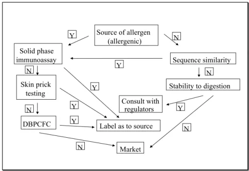
Metcalfe et al. (1996) have proposed a strategy for predictive testing of allergenicity of genetically engineered foods (see Fig. 1). Based on the source of the gene, two pathways may be followed. If the gene that is intoduced in the food is derived from allergenic foods, or if sequence similarities with known allergens are detected, solid phase immunoassay must be performed. Solid phase immunoassay needs to be performed for any product where the gene stems from an allergenic source. In case of negative findings, skin prick testing of at least 14 individuals with different types of proven food allergy, and, in case of negative results,