11
MAILING LIST
1-5 Hoofdinspectie Gezondheidsbescherming 6 Directeur-Generaal van de Volksgezondheid
7 Plv. Directeur-Generaal van de Volksgezondheid, tevens^Hoofddirecteur Financiering en Planning.
8 Mr S van Hoogstraten, directeur Voedings- en Veiligheidsaangelegenheden Produkten (WP)
9 Dr F. Schuring, Hoofdinspecteur Gezondheidsbescherming 10 WJ. de Koe (HIGB)
11 Dr Ir G. Kleter (HIGB)
12 Mevr. Ir. M.A.N. de Schutter (WP) 13 Ir. J. Besling, (IGB, Rotterdam)
14 Depot Nederlandse Publikaties en Nederlandse Bibliografie 15 Directie Rijksinstituut voor Volksgezondheid en Milieuhygiëne 16 Hoofd Bureau Voorlichting en Public Relations
17 Dr Ir G. de Mik, directeur sector VI 18 Dr H. Krasselt, directeur sector UI
19 Dr R.W. Stephany, hoofd Laboratorium voor Analytisch Residu Onderzoek 20 Dr J.G. Vos, hoofd Laboratorium voor Pathologie
21 Dr W.H. Könemann, hoofd Laboratorium voor Farmacologie en Toxicologie i.o. 22 Dr Ir H.J.G.M. Derks, hoofd Unit Biotransformatie Farmaco- en Toxicokinetiek 23 Dr J.E.T. Moen, wnd. hoofd Adviescentrum Toxicologie
24 Drs J.W. Dorpema, hoofd Laboratorium voor Geneesmiddelen en Medische Hulpmiddelen
25 Dr J.G. Loeber, hoofd Unit Teratologie, Endocrinologie en Perinatale screening 26 Dr J.W. van der Laan
27 P.K. Beekhof/Y. Wallbrink-de Dreu/Ing. C.J. Dobbe 28 Dr. A.B.J.T. Boink, Dr. M. Oortgiesen
29 Dr. J. Wemer/Prof.Dr. DJ. de Wüdt 30 Dr. Ir. E.H.J.M. Jansen
31 Ir. P.J.A. Rombout/Dr. L. van Bree 32-40 Auteurs
43 Projecten- en Rapportenregistratie 44 Bibliotheek
m LIST OF CONTENTS MAILING LIST ii SUMMARY iv SAMENVATTING v 1 INTRODUCTION 1 2 MATERIALS AND METHODS 1
2.1 Test substance 1 2.2 Animals and diet 2 2.3 Experimental design 2
2.4 Methods 2 2.5 Statistical methods 4
3 RESULTS 4 4 DISCUSSION 6 5. COMPARISON OF THE SUBACUTE TOXICITY OF ERGOMETRINE
MALEATE AND ERGOTAMINE TARTRATE IN THE RAT 8
6 REFERENCES 9 7 FIGURES 11 8 TABLES 12
IV
SUMMARY
Data on the toxicity of ergot alkaloids are too limited to make a proper risk evaluation. Therefore, the present study was carried out. In a 4-week toxicity experiment six groups of Sprague Dawley (SD) rats (6 animals/sex/group) were fed a diet containing concentrations of 0, 2, 10, 50 and 250 mg ergometrine maleate/kg diet. Two control groups were included: one control group (group 0) received the test diet (SSP-tox) ad libitum, and one control group (group 1) was pair-fed with the highest dose group (group 5). In the fourth week blood was sampled for haematological and biochemical analysis, and urine was collected during a 24-hours period. After 4 weeks blood was sampled for biochemical and endocrinological analysis. The animals were sacrificed and necropsied. Organ weights were determined and tissues were fixed for a complete histopathological examination.
Dietary concentrations of 2, 10, 50 and 250 mg/kg ergometrine maleate resulted in a mean daily intake of approximately 0.2, 1, 5 and 25 mg/kg body weight ergometrine maleate. The homogeneity and the stability of ergometrine maleate mixed through the diet was good during the experiment. No treatment related clinical signs were observed during the experiment. Plasma glucose levels were significantly decreased in females at 50 mg/kg and 250 mg/kg. In males also a tendency to decreased glucose levels was observed, but no significance was reached by multiple group-comparison. In male rats, T4 levels were decreased at 250 mg/kg, and FT4 at both 50 and 250 mg/kg. In female rats there was a tendency to decreased T4 levels at 250 mg/kg, but this effect was not statistically significant. Prolactine levels were reduced at 50 and 250 mg/kg in males and females. No treatment related effects on haematological parameters were seen. The absolute and relative (only females) heart weight were increased at 250 mg/kg. The relative brain weight of the pair-fed control group was increased as compared to the control group. The absolute liver weight was increased in females at 250 mg/kg and the relative liver weight was increased at 50 and 250 rag/kg in both sexes. Both absolute and relative weight of the ovaries were increased at 250 mg/kg, and the absolute kidney weight was increased in males at 250 mg/kg. Macroscopic examination revealed a slight dose-related increase in the incidence of enlarged mediastinal lymph nodes in males and, to some extent, of enlarged parathymal lymph nodes in males. Histopathological examination revealed pale and occasionally swollen hepatocytes in animals treated with 250 mg/kg. These changes were interpreted as increased glycogen storage. Renal mineralization and nephrosis was seen in untreated and treated rats. The increased relative weight of the heart and ovaries was not accompanied by histopathological changes. The no-effect level was 10 mg/kg.
In our Institute, sub-acute and sub-chronic studies with ergotamine tartrate have been performed before (Speijers, 1992 and 1993). The subacute toxicity of ergotamine tartrate and ergometrine maleate (both in the SD rat) are compared. Decreased thyroxine levels, increased relative weights of the heart, liver and ovaries were seen with both compounds. Nephrotoxicity and necrosis of the tail tips was seen only for ergotamine tartrate at a high dose level. Indications for disturbance of the carbohydrate metabolism with increased glycogen storage in the liver was seen with ergometrine only,
A sub-acute study in Sprague Dawley rats with a third ergot alkaloid, ergocryptine, is planned. Then the lexicological profile of the model compounds in three different categories of ergot alkaloids will be compared. A risk evaluation with regard to human exposure will be made.
SAMENVATTING
Er zijn onvoldoende toxicologische gegevens over ergot alkaloïden om een risicoschatting te maken. Daarom werd het volgende experiment uitgevoerd. Sprague Dawley (SD) ratten (6 dieren/geslacht/groep) werden gedurende 4 weken blootgesteld aan ergometrine maleaat, in doseringen van 0, 2, 10, 50 of 250 mg/kg voer. Er waren twee controle groepen: één groep kreeg het SSP-tox voer ad libitum, en één groep (groep 1) werd 'pair-fed' gevoerd met de hoogste doseringsgroep. In week 4 zijn bloed monsters verzameld voor hemato-logische en klinisch-chemische bepalingen, en werd gedurende 24 uur urine verzameld. In week 5 werden de dieren opgeofferd waarbij bloed is verzameld voor biochemische en endocrinologische bepalingen. Een aantal organen zijn gewogen, gefixeerd en er is een uitgebreid histopathologisch onderzoek verricht.
Concentraties van 2, 10, 50 en 250 mg ergometrine maleaat/kg voer resulteerde in een gemiddelde opname van circa 0,2, 1, 5 en 25 mg/kg lichaamsgewicht De homogeniteit en stabiliteit van ergometrine maleaat in het voer was goed. Er werden geen klinische symptomen gerelateerd aan de behandeling waargenomen bij de dieren. Plasma glucose spiegels waren significant gedaald in vrouwtjes behandeld met 50 en 250 mg/kg. Bij mannetjes werd eveneens een tendens tot verlaging van glucose spiegels waargenomen, maar dit effect was niet significant in de multiple-comparison test. In mannelijke ratten waren T4 spiegels verlaagd in de hoogste doseringsgroep (250 mg/kg), en FT4 spiegels bij 50 en 250 mg/kg. Bij vrouwtjes was er een (niet significante) tendens tot verlaging van T4 spiegels. Serum prolactine was verlaagd bij 50 en 250 mg/kg (mannetjes en vrouwtjes).Er werden geen behandelings gerelateerde effecten gezien op hematologische parameters. Het absolute en relatieve (alleen vrouwtjes) gewicht van het hart was verhoogd bij 250 mg/kg. Het relatieve hersenen gewicht van de pair-fed controle groep was hoger dan dat van de controle groep. Het absolute gewicht van de lever was verhoogd in vrouwtjes (250 mg/kg), en het relatieve gewicht van de lever was verhoogd bij 50 en 250 mg/kg in beide sexes. Het gewicht van de ovaria (absoluut en relatief) was verhoogd bij 250 mg/kg, en het absolute gewicht van de nieren was verhoogd bij 250 mg/kg (mannetjes). Macroscopisch werd in mannetjes een lichte dosis-gerelateerde vergroting van de mediastinale lymfeklieren gezien, en een vergroting van de parathymale lymfeklieren. Histopathologisch werden bij mannetjes behandeld met een dosis van 250 mg/kg bleke hepatocyten gezien die in sommige gevallen gezwollen waren. Dit fenomeen werd geïnterpreteerd als een toegenomen glycogeen opslag in de lever. Mineralisatie en nefrose in de nier werd in alle groepen (inclusief controle) gezien. Histopathologisch werden geen veranderingen in het hart en de ovaria gezien. De no-effect level is 10 mg/kg.
Binnen het RIVM zijn reeds sub-acute en sub-chronische studies met ergotamine tartraat uitgevoerd (Speijers, 1992 en 1993). De subacute toxiciteit van ergotamine tartraat en ergometrine maleaat in de SD rat worden vergeleken. Effecten die met beide stoffen gezien zijn: verlaging van thyroxine spiegels, verhoogde relatieve gewichten van het hart, lever en ovaria. Nefrotoxiciteit en necrosis van de staartpunt werden uitsluitend met ergotamine tartaat gezien. Aanwijzingen voor een verstoring van het koolhydraat metabolisme werden uitsluitend met ergometrine maleaat verkregen.
Er wordt nog een experiment met een derde ergot alkaloid (ergocryptine) uitgevoerd. Dan kan de toxicitiet van de modelstoffen uit drie verschillende categorieën ergot alkaloïden vergeleken worden en een risico evaluatie m.b.t humane blootstelling worden gemaakt
1 1 INTRODUCTION
The fungus Claviceps purpurea grows parasitically on some grasses and several cereal crops such as rye, wheat and barley. The resulting sclerotia or "ergot bodies", which can hibernate, may contain a mixture of different alkaloids that have powerful pharmacological properties (Young, 1979; Schoch et al, 1985). These alkaloids consist of lysergic acid derivates amoung which ergotamine and ergocristine are major representatives (Schoch et a l , 1985; Speijers, 1989). Small amounts of ergot alkaloids (0.01-2,36 mg ergot alkaloids/kg) are still frequently reported in flour from rye, wheat and barley (Besling and van der Haar, 1984). Although toxic effects of ergot alkaloids at very high consumption levels have a long history as the disease ergotism (Bove, 1970; Guggisberg, 1964; Opitz,
1984), little is known about a long-term intake of small amounts of ergot alkaloids. The pharmacological actions of the ergot alkaloids are varied and complex: peripheral vasoconstriction, depresssion of vasomotor centers, peripheral adrenergic blockade, secretion of prolactin, stimulation of uterine smooth muscle etc (Rail, 1990). Ergot alkaloids have various therapeutic indications. Ergometrine is used to prevent blood loss during partus or abortion and ergotamine is prescribed as treatment of migraine. According to Schoch et al (1985), ergot alkaloids can be divided in two major classes:
1) lysergin derivates, 2) Clavin alkaloids. Among the lysergin derivates are major representatives and pharmacological active compounds. Lysergin derivates can be subclassified in: a) lysergin amide derivates (e.g. ergin and ergometrine), b) peptids, with sub-classes 'ergotamins' (e.g. ergotamine, ergosine, ergosecaline) 'ergotoxins' (ergocryptin, ergocomin, ergocristin), and 'ergoxins' (ergostin and ergostinin). In the Laboratory of Toxicology, sub-acute and sub-chronic toxicity experiments with ergotamine have been performed (Speijers, 1992 and 1993). For the current experiment, ergometrine (also named ergonovine) is chosen from the class of 'lysergin amide derivates', because limited data on the toxicologic properties of ergometrine are available (Griffith et al., 1978). Data are too limited to make a proper toxicological evaluation. In the future, an experiment with a compound from the subclass 'ergotoxins' will be performed. The goal is to compare the toxicological profile of compounds from the three classes of ergot alkaloids, and to examine whether group evaluation based on a complete file of one ergot alkaloid (ergotamine) can be justified. In this light, the study plan of the current experiment was very similar to the study plan of the sub-acute toxicity study with ergotamine. The choise of dose levels was based on the (limited) acute toxicity data of ergometrine and the acute toxicity and subacute toxicity data of ergotamine tartrate. The study was conducted according to GLP principles.
2 MATERIALS AND METHODS 2.1 Test substance
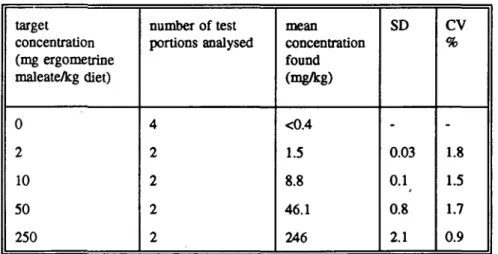
Ergometrine-maleate was obtained from Bufa B.V., Uitgeest, The Netherlands (charge nr 914309). The purity of the compound was indicated to be 98.0-101.0%. The test substance was stored in the original container at +2 to +8 °C in the dark. Ergometrine maleate was added to semi-purified SSP-tox standard diet in a premix. The premixes were diluted to obtain concentrations of 2, 10, 50 or 250 mg/kg diet ergometrine maleate. Homogeneity and stability of ergometrine maleate in the diet was analysed by HPLC, The limit of detection was 0.4 mg/kg.
2.2 Animals and diet
SPF Sprague Dawley rats [HsdVOla/Sprague Dawley (CD)] weighing approximately 60-80 g were obtained from Harlan UK. Upon arrival, the animals were weighed and randomized on basis of their body weight, using a randomized block model. Following allocation, the animals were identified by tattooing the last two digits of their number in their ears. The animals were housed individually in stainless wire cages in room D6B024.
Air pressure, the relative humidity and the temperature were controlled and recorded daily according to the Standard Operating Procedures. The semi-purified diet SSP-tox standard was obtained from Hope Farms, Woerden, the Netherlands. The standard diet and the test diets prepared were stored at a temperature of +2 to +8 °C. The test diets containing ergometrine maleate were stored in the dark avoiding exposure to daylight Diet analyses were carried out on the test compound.
2.3 Experimental design
Six groups of 6 rats/group/sex received a diet containing 0, 2, 10, 50 or 250 mg/kg ergometrine maleate/kg diet for 4 weeks. Two control groups were included: one control group received ad libitum the SSP-tox diet, and the other control group was pair-fed with the high dose group. Drinking water was provided ad libitum. Daily observation of animal appearance was performed, with special attention to the tail tips. Body weight gain was recorded weekly, and food intake was measured three times a week. The remnant of food was removed. At the end of the third week the animals were placed in metabolism cages to collect urine during 24 hours in calibrated plastic tubes. In the metabolism cages the animals were deprived from food, but drinking water was provided ad libitum. Directly after this 24 hour of urine collection 1.5 ml blood was sampled by means of puncture of the venous orbital plexus under ether anaesthesia to prepare EDTA-blood for hematolo-gical examination, and to prepare serum for biochemical analysis. After 4 weeks the rats were sacrificed and necropsied. Blood was collected for endocrinological and biochemical analysis. Organ weights were determined and tissues were fixed for a complete histologi-cal examination.
2.4 Methods
Ergometrine in the diet was determined by HPLC. The method of analysis for ergometrine maleate in SSP-tox standard diet is described in SOP ARO/362. Homogeneity of the test diets was confirmed, and limited recovery experiments were performed. Stability of ergometrine maleate in the diet was determined after 5 weeks of storage in the dark at a temperature of +2 to +8 °C.
In blood the following parameters were determined by the Sysmex E4000 haematological analyser: haemoglobin concentration (Hb), packed cell volume (PCV), red blood cell concentrations (RBC), white blood cell concentration (WBC) and platelet concentration. Mean corpuscular volume (MCV), mean corpuscular haemoglobin (MCH) and mean corpuscular haemoglobin concentration (MCHC) were calculated. In the urine and serum gained after 3 weeks creatinine was determined with an Unimate CREA kit of Roche, and urea was determined with an urea UV UNI-kit (Roche). From urine volume and creatinine concentrations in serum and urine the creatinine clearance was calculated. The same applies to urea clearance. In serum triglycerids were determined with a GPO-PAP testkit of Boehringer (nr 701904), Total cholesterol was determined according to the CHOD-PAP method of Boehringer, high densitiy lipoprotein (HDL) was determined after precipitation of apolipoprotein B containing lipoproteins by the heparin-manganese method (Janssen et
a l , 1988), and y-glutamyl transpeptidase (GGT) with the Ultimate test combination of Roche, Sodium and potassium levels in serum and urine were determined by flame photospectrometry. The volume of the collected urine was noted and the osmolality was determined with a Osmomat 030 osmometer (Gonotec). The urinary protein concentration was determined according to the method of Bradford (1976). The urine was also analysed semi-quantitatively by means of a strip test with N-Multistix SG (Bayer Diagnostics), to indicate pH, protein, glucose, specific gravity, ketone, bilirubin, occult blood, nitrite and urobilinogen.
After 4 weeks blood was sampled for endocrinological and biochemical analysis. Glucose was measured in sodium fluoride plasma with an enzymatic colorimetric testkit of Roche. In serum, alanine transferase (ALAT) and aspartate transferase (ASAT) were determined with Unimate kits of Roche, creatinine kinase (CK) was determined according to the CK-NAC MA-kit of Roche, and alkalische phosphatase (AP) according to IFCC with the ALP Ultimate 3 kit of Roche. Bilirubin was determined with a billirubin testkit of Roche.
Thyroid stimulating hormone (TSH) was determined by means of a radioimmunoassay based on the following reagents: anti-TSH-S5, iodination preparation TSH-I6 and calibration preparation TSH-RP2. These reagents were kindly supplied by the NIDDK (Baltimore, USA). Aldosteron, corticosteron, thyroxine (T4) and free thyroxine (FT4) were determined with RIA kits from Diagnostic Products Corporation (Los Angeles, USA). For the determination of insuline the RIA kit of Pharmacia was used. The plasma renine activity was measured by means of a radioimmunological determination of angiotensin I, according to Elvers et al. (1986). Serum prolactin levels were determined in serum samples that were stored at -20 °C, 8 months after finishing the experiment. Prolactin was determined by means of a radioimmunoassay (Smit et al, 1987) based on the following reagents: anti-S8, iodination preparation 16 and calibration preparation PRL-RP3. These reagents were kindly supplied by the NIDDK (Baltimore, USA).
At completion of the treatment period after 4 weeks all surviving rats were killed free from stress, on day 29 to 33, at a random order. The animals were anaesthetized by a C02/02 mixture, followed by exsanguination from the abdominal aorta, and were then examined grossly for pathological changes. The heart, liver, kidneys, spleen, brain, adrenals, pituitary, thyroid, gonads, uterus, mesenteric lymph node and thymus were weighed. These organs, together with the lungs, aorta, tail, stomach, small intestines (3 levels), caecum, colon, rectum, nervus ischiadicus, musculus quadriceps, spinal cord, pancreas, urinary bladder, prostate, oesophagus, trachea, sternum with bone marrow, caudal lymph nodes and organs showing gross changes were preserved in a neutral aqueous phosphate-buffered 4% solution of formaldehyde. Swiss rolls were prepared from the samples of the stomach and the intestines. All specimens of the control group and the high dose group were processed histologically, embedded in paraffin wax, cut at 5 um and stained with haematoxylin and eosin. All specimen processed histologically were examined microscopically for histopathological changes. Histopathological data "were recorded and processed using the PATHOS pathology data acquisition system.
Histopathology was extended to the liver and kidneys of the other dose groups, because of possible treatment-related changes in these organs, m addition the livers of some control and high dose animals were stained according to the Periodic Acid Shiff (PAS) method, in order to evaluate the glycogen content To confirm the presence of glycogen, diastase digestion was applied to parallel sections prior to PAS staining. The livers and kidneys
were examined microscopically in a "blind and random" fashion, without knowledge of treatment
2.5 Statistical methods
Results were analysed by one factor (group as factor) analysis of variance (ANOVA) using the STATA program, release 2.05. Homogeneity of variance was tested with Barlett's test. Variables with hetereogenous variance were analysed by ANOVA after a single logarithmic (In) transformation. For each parameter first a two-way ANOVA was performed with the variables sex and group, and the interaction between sex and group. In case a sex difference was obtained (P < 0.05), a oneway ANOVA was performed with group as variable, for both sexes apart In case no sex difference was observed, both sexes were analysed together, in a oneway ANOVA. Bonferroni's multiple-comparison test was performed. Semi-quantative parameters in urinalysis were tested non-parametrically with the Kruskal-Wallis Test Histopathological changes were evaluated by Fisher's exact probability test (two-sided). In this report results that are significantly different, or results that are considered important are expressed only. Individual data are expressed in the appendix to report 388802.05 (03) 9305 d.d. September 22, 1993, which is available on request.
3 RESULTS
The homogeneity, and the stability of ergometrine maleate mixed through SSP-tox diet during the experiment were good (Table la and lb).
No treatment related clinical signs were observed during the experiment Tail tips were not affected. One animal (male, 10 mg/kg) died during the experiment probably due to ether anaesthesia. The animal did not show major gross- or microscopic abnormalities that reasonably could have caused the death.
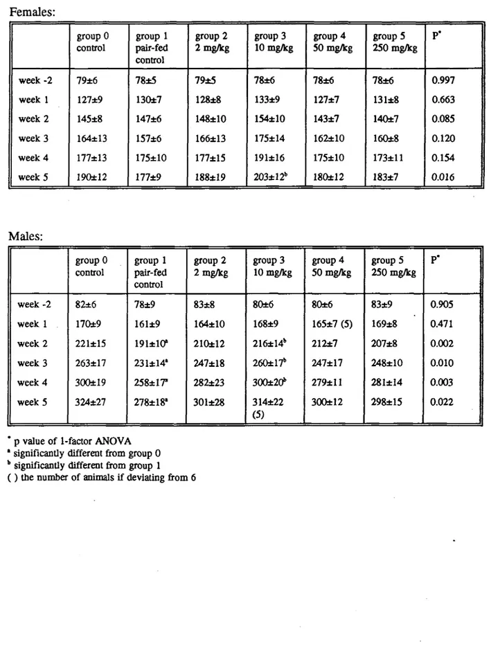
In female rats body weight was not clearly influenced by ergometrine maleate treatment, except for a higher weight of group 3 (10 mg/kg) as compared to the pair-fed control group in week 5. In male rats, the body weight of the pair-fed control group was lower than that of control rats, from week 2 onwards. In the first weeks of treatment the body weight gain was lower in the high dose female rats, as compared to all other groups. In pair-fed control male rats (group 1), the body weight gain was lower as in control animals (group 0) during the experimental period. Results are shown in Tables 2 and 3.
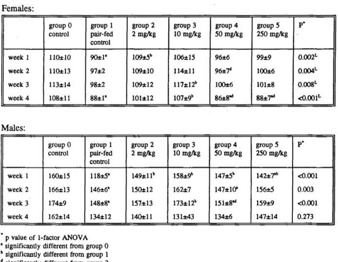
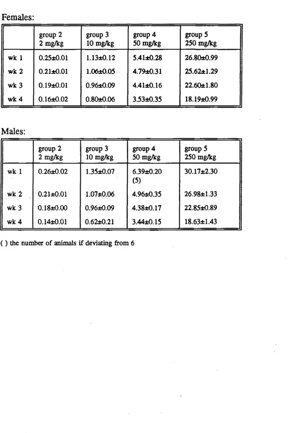
Food intake of female rats was not influenced by ergometrine maleate, except in week 4 when the high dose animals consumed less food than the control animals. Male rats treated with the highest dose consumed less food than the control animals in week 1 only (table 4). Dietary concentrations of 2, 10, 50 and 250 mg/kg ergometrine maleate resulted in a mean daily intake of ca 0.2, 1, 5 and 25 mg/kg body weight ergometrine maleate (Table 5).
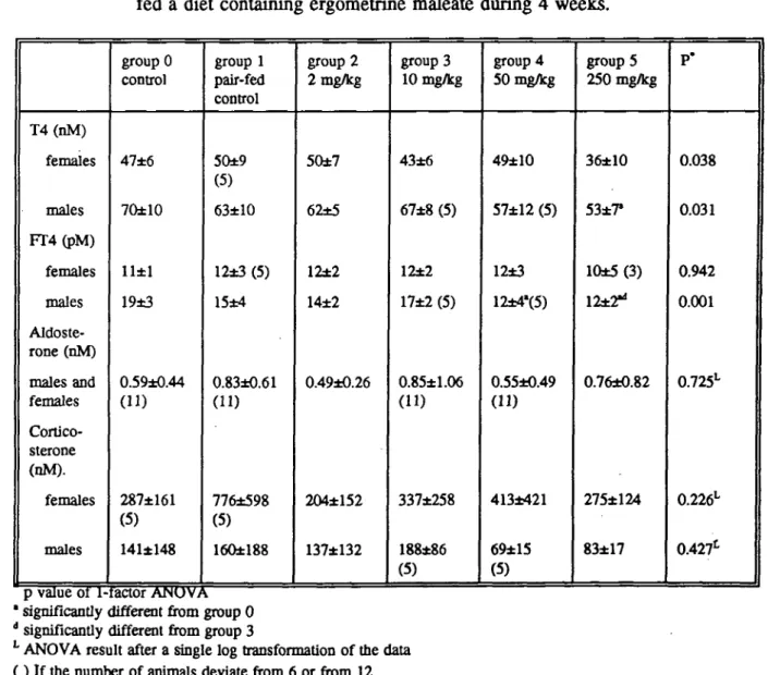
No treatment related effects on haematological parameters were seen. Plasma glucose levels were significantly decreased in females at 50 mg/kg and 250 mg/kg. In males also a tendency to decreased glucose levels was observed, but the effect was not significant with the multiple-comparison test (table 6a). In male rats, T4 levels were decreased at 250 mg/kg, and FT4 at both 50 and 250 mg/kg. In female rats there was a tendency to decreased T4 levels at 250 mg/kg, but this effect was not statistically significant. The
ultra-individual variation in serum corticosterone and aldosterone levels was large. The results are shown in Table 6b. Eight months after finishing the experiment, prolactin was determined in serum samples that were stored at -20 "C. The number of samples was too low for statistical analysis of the results. However, it may be obvious that serum prolactin levels were decreased at 50 and 250 mg/kg in both males and females (Table 6c). Ergometrine maleate had no influence on kidney function parameters like creatinine or urea clearance.
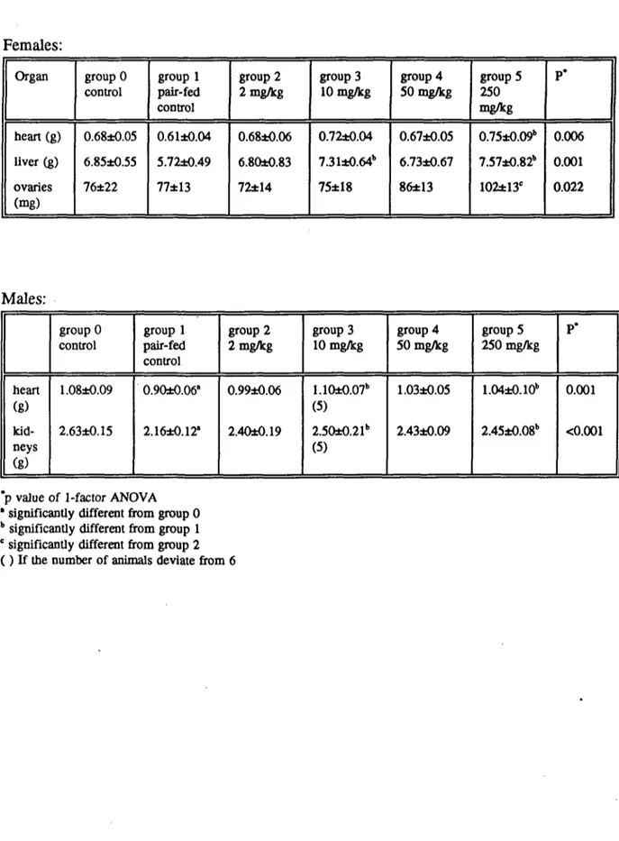
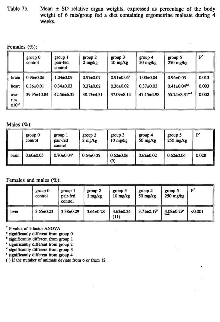
In males and females, absolute heart weight was increased at 250 mg/kg compared to the pair-fed control group, and at 10 (but not at 50 mg/kg) in males. The relative heart weight was also increased in female rats treated with 250 mg/kg, but not in males. In male rats the relative brain weight was increased in the pair-fed control group as compared to the control group. Absolute liver weight was increased in females at 10 and 250 mg/kg (but not at 50 mg/kg), and relative liver weight was increased at 50 and 250 mg/kg in both males and females. Furthermore, the absolute and relative weight of the ovaries was increased at 250 mg/kg. In males absolute kidney weight was increased at 10 and 250 mg/kg. Results are expressed in the Tables 7a and 7b.
Macroscopic examination revealed a slight dose-related increase in the incidence of enlarged mediastinal lymph nodes in males and, to some extent, of enlarged parathymal lymph nodes in males. The enlargement occurred also in pair-fed controls. In females there was no evidence of any dose-related differences in the size of these lymph nodes between the groups.
Treatment-related histopathological changes were observed in the liver only. Histopatholo-gical hepatic changes occurred in high-dose males and females and were characterized by relatively large and occasionally swollen hepatocytes with rarefied cytoplasmic staining in the central part of the cell and clumping of stainable cytoplasm at the cellular wall. This hepatocellular aspect was observed in varying degrees in all groups, but was distinctly most pronounced in high dose males and females (marked degree is statistically
significantly different from the controls, P= 0.03). Although the picture was indicative of glycogen storage, the glycogen content of most of these cells appeared to be relatively low (PAS-staining). This counted both for the hepatocytes from the high-dose animals and for those from controls that occasionally also showed the typical aspect of glycogen storage. With higher magnification, PAS-positive membraneous structures were discerned within the pale central part of the cells. These structures became PAS-negative upon diastase digestion. Similar pale cells, periferally in the sample (underneath the liver capsule), were frequently laden with granular PAS-positive material. These features suggested artificial loss of glycogen in the major part of the liver sample during the storage in formalin or at the histological processing. It should be stressed that the histological picture did not resemble that of fatty vacuolation or hydropic degeneration. Therefore, the pale swollen cells in the high dose group were interpreted as increased glycogen storage. There was no evidence of ergometrine-induced hepatocellular necrosis or other liver abnormalities at the dose levels used.
The mediastinal lymph nodes that were macroscopically enlarged generally showed slight degrees of reactive hyperplasia, seen as an increased lymphocytic population in cortex and/or paracortex, and occasionally germinal center formation. The degree of activation
was within the limits that are considered normal for untreated controls. Although special attention was paid to the microscopy of the other lymphoid organs, no treatment-related abnormalities were observed. Neither was there evidence of changes that might point to effects on the immune system.
The kidneys of most animals showed moderate to severe mineralization in the intercorticomedullary area, accompanied by various degrees of tubular changes. In mild cases the tubular changes were restricted to the local presence of a few basophilic proximal tubules, especially in the outer cortex. Other animals showed tubular nephrosis, defined as clusters of basophilic proximal tubules, peritubular fibrosis, tubular dilatation and occasionally infiltration of a few mononuclear inflammatory cells. The renal changes were most pronounced in females. They occurred in all groups, including the controls, without indications of a treatment-relationship.
Microscopy of the other organs and tissues did not provide distinct and convincing indications for differences between the groups. The increased relative weight of the heart and ovaries were not accompanied by histopathological changes.
4 DISCUSSION
On certain days, the pair-fed animals did not eat all the food they were provided with. One might argue that it could have been technically difficult to eat all the food to the bottom of the food-box. However, when this hypothesis is assumed, the difference in food intake between the pair-fed control group and the highest dose group should have been more structural. The findings are probably coincidental.
Plasma glucose levels were significantly decreased in females at 50 mg/kg and 250 mg/kg. In males also a tendency to decreased glucose levels was observed, but no significance was reached by multiple-comparison (Table 6a). No effect was seen on insulin. In the liver increased glycogen storage was seen. Apparently, ergometrine maleate has influence on the carbohydrate mechanism, but the precise mechanism is not known.
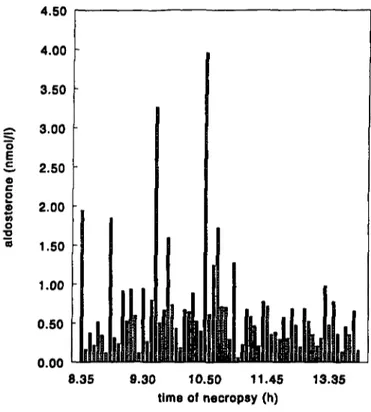
The intra-individual variation in serum corticosterone and aldosterone levels was large (table 7). From the literature it is known that certain hormones, for example corticosterone, have a diurnal rithm (Ottenweller, 1978; Wong, 1983). In this study, these hormones were determined in blood that was collected during necropsy, which lasted from ca 8:00 am to 2:30 p.m. each day. So, the time of necropsy might have influenced the hormone concentrations. However, in this study, the large intraindividual variation of corticosterone and aldosterone appeared not to be caused by variation in time the animals were necropsied (Figure la and lb). Corticosterone levels were higher in females than in males. This is well known from the literature (Tausk, 1975). Serum prolactin levels were reduced in males and females treated with 50 and 250 mg/kg/day. This prolactin lowering effect is well known from ergometrine and other ergot alkaloids (Begley, 1990).
In male rats the relative brain weight was increased in the pair-fed control group as compared to the control group. This is explained by the fact that brain weight only changes under extreme conditions and not in case of a slight decrease in food intake as the body weight does.
Although the increase in relative heart weight was not accompanied by biochemical or histopathological changes, pharmacodynamic actions of ergometrine may have influenced
the heart weight. Ergot alkaloids have vasocontrictive properties. Ergometrine may provoke coronary spasm (Taylor, 1985; Murad, 1990).
Renal mineralization (nephrocalcinosis) and nephrosis seen in untreated and treated rats was unrelated to administration of ergometrine maleate. The effects were most pronounced in females.
Ergometrine maleate is registered as a therapeutic drug in the Netherlands (Ergometrini Maleas, tablets 0.3 mg, RVG 51269 and RVG 53000) for the indications blood loss during partus or abortion, and menorrhagia. Advised dose levels are 1-2 tablets two or three times daily (College ter Beoordeling van Geneesmiddelen, 1993). The maximum daily dose is therefore 1.8 mg daily, this is ca 0.03 mg/kg body weight for a man weighing 60 kg. Doses used in animal studies can be extrapolated to doses used in man, by means of differences in metabolic rate between animals and man. This extrapolation factor is based on differences in body surface between animals and man. Generally, this way of extrapola-tion is more adequate than extrapolaextrapola-tion on a mg/kg basis (Voisin, 1990). Administraextrapola-tion of an oral dosis that is five times higher than the therapeutic oral dose in man, results in similar exposure in rat and man (Peters-Volleberg et al, 1994). In this study, the dosis in rats that is equivalent to the therapeutic dose in man is therefore ca 0.15 mg/kg body weight So, the lowest dose group (2 mg/kg diet) corresponds to the therapeutic dose in man (see Table 5).
The no-observed-adverse-effect-level (NOAEL) in this study is 10 mg ergometrine maleate/kg diet, or ca 1.0 mg ergometrine maleate/kg body weight, corresponding with 0.74 mg ergoraetrine/kg body weight. According to the general tolerable daily intake (TDI) or acceptable daily intake (ADI) concept, this implies 7.4 ug/kg ergometrine maleate/kg body weight for man. A 10-fold uncertainty factor is included to account for variation in susceptibility in humans and a 10-fold uncertainty factor to extrapolate from animal data to human data (Beck et al, 1989). The average human daily consumption of cereal products is estimated at 200 gram per person. The maximal concentration of total ergot alkaloids in rye, wheat and oats was reported to be 2.36 mg/kg (Besting, 1984). Following this assumption the maximal intake of the ergot alkaloids for humans (60 kg body weight) would be 7.8 ug ergot alkaloids/kg body weight.
In our Institute sub-acute toxicity studies have been performed with two compounds: ergotamine tartrate and ergometrine maleate. These compounds fall in different categories of alkaloids (Schoch, 1985; Speijers, 1989). Furthermore, a sub-chronic study with ergotamine tartrate has been performed (Speijers, 1993). In the near future, a sub-acute toxicity study in rats is planned with ergocryptine. Ergocryptine is an ergot-alkaloid from a third category: the sub-class of ergotoxins (Schoch, 1985; Speijers, 1989). The toxicological profile of compounds in different classes will be compared then. The estimated human maximal daily intake of ergot alkaloids of 7.8 ug/kg body weight (Speijers, 1993) will be evaluated with regard to the toxicological profile of the ergot alkaloids.
8
5. COMPARISON OF THE SUBACUTE TOXICITY OF ERGOMETRINE MALEATE AND ERGOTAMINE TARTRATE IN THE RAT
Recently, also sub-acute and sub-chronic studies with ergotamine tartrate have been performed in the Laboratory of Toxicology, (Speijers, 1992 and 1993). The subactue toxicity of ergotamine tartrate and the subacute toxicity of ergometrine maleate (both in the Sprague Dawley rat) are compared. In the subacute study with ergotamine tartrate, daily dose levels were 0, 4, 20, 100 and 500 mg/kg diet Dose levels in the study with ergometrine were half the dose levels of ergotamine. The dose levels were based on acute oral toxicity studies in rats (Griffith, 1978) indicating a higher toxicity of ergometrine.
One of the findings of the subacute study with ergotamine was diffuse degeneration and slight fibrosis of the tail tips in the high dose group(s) (100mg/kg for males only, and 500 mg/kg for both sexes). This finding was not observed with ergometrine. The fact that the tail necrosis is not seen in this experiment does not necessarily imply that ergometrine has no vasoconstrictive effect, even at a higher dose level. Nevertheless as with ergotamine tartrate this necrosis of the tail was seen at a dose level of 100-500 mg/kg, ergometrine maleate seems to have less vasoconstrictive potency. It is also possible that the effect on the tail is specific for ergotamine, and not for other ergot alkaloids.
With ergotamine, activation of the ileacal lymph nodes was observed. In males treated with ergometrine, there was a slight dose-related increase in the incidence of enlarged mediastinal lymph nodes, and to some extent, of enlarged parathymal lymph nodes. The enlargement occurred also in pair-fed controls. Histopathological examination revealed slight degrees of reactive hyperplasia. The effect in males was considered within the limits that are considered normal for untreated rats. In females no effect was observed. Other lymphoid organs appeared normal as well. The enlargement might very well represent a fortuitous finding, unrelated to treatment
The decrease in haematological parameters seen with ergotamine was not observed with ergometrine. Decreased thyroxine levels, increased relative weights of the heart, liver and ovaries, and (to some extend) enlarged lymph nodes were seen with both compounds. Nephrotoxicity and necrosis of the tail tips was specific for ergotamine tartrate. Indications for disturbance of the carbohydrate metabolism with increased glycogen storage in the liver was seen with ergometrine only (Table 10).
6 REFERENCES
Beck, B.D., Calabrese E.J. and Anderson, P.D. (1989). The use of toxicology in the regulatory prcess. In: Principles and methods of toxicology. (Hayes, A.W., ed). Raven Press, New York, pp 1-28.
Begley, CM. (1990). The effect of ergometrine on breast feeding. Midwifery 6, 60-72. Besting, J.R. and J.H. van der Haar (1984). Moederkoren alkaloïden in graanproducten.
Mycotoxinenrapport 84-2, Keuringsdienst van Waren, Rotterdam. Bove, F.J., (1970). The story of ergot Karger Verlag , Basel, New York.
Bradford, M.M. (1976). A rapid sensitive method for quantitation of microgram quantities of protein utilizing the principle of protein dye binding. Anal. Biochem. 72: 248-254.
College ter Beoordeling van Geneesmiddelen. (1993). Lijst van Farmaceutische Producten; derde kwartaal 1993. Rijswijk, the Netherlands.
Elvers, L.H, Somers, H.H.J, and Loeber, J.G. (1986). Het bepalen van de plasma renine activiteit door middel van de radioimmunochemische bepaling van Angiotensine I. RTVM report number 368102001, Bilthoven, the Netherlands.
Griffith, R.W., Grauwiler, J., Hodel, Ch., Leist, K.H, and Matter, B. (1978). Toxicologie considerations. In: Ergot alkaloids and related compounds (Berde P. and Schild H.O., eds). Springer-Verlag, Berlin, pp 805-851.
Guggisberg, G (1964). Mutterkom als Gift. Karger Verlag, Basel, New York.
Janssen, G.B., Leeuwen, F.X.R. van, en Speijers, G.J.A. (1988). De HDL-cholesterol-bepaling en de veranderingen in het hpoproteinpatroon bij de rat onder verschillen-de dieet omstandigheverschillen-den. Rijksinstituut voor Volksgezondheid en Milieuhygiëne. Rapport nummer 618471.001. Bilthoven, Nederland.
Murad, F, (1990). Drugs used for the treatment of angina: organic nitrates,
calcium-channel blockers, and P-adrenergic antagonists. In: Goodman and Oilman's The pharmacological basis of therapeutics. 8th edition. Pergamon Press, New York. Opitz, K (1984). Der Ergotismus und seine heutige Bedeutung. Getreide, Mehl und Brot,
38: 281-284.
Ottenweller, J.E., Meier, A.H, Ferrell, B.R., Horseman, N.D. and Proctor, A. (1978). Extrapituitary regulation of the circadian rhythm of plasma corticosteroid concen-tration in rats. Endocrinology, 103(5): 1875-1879.
Peters-Volleberg, G.W.M., de Waal, E.J. de, and van der Laan, J.W. (1994). Interspecies extrapolation in safety evaluation of human medicines in the Netherlands
(1990-1992). Submitted for publication.
Rail, T.W. (1990). Oxytocin, prostaglandins, ergot alkaloids, and other drugs: toxolytic agents. In: The pharmacological basis of therapeutics. Goodman Gilman, A., Rail, T.W., Nies, A.S. and Taylor, P (eds). Pergamon Press, New York, pp. 933-954. Schoch, U. and Schlatter, Ch, (1985). Gesundheitsrisiken durch Mutterkom aus Getreide.
Mitt Gebiete Lebensm. Hyg. 76, 631-644.
Smith, P.J., Elvers, L.H. and Loeber, J.G. (1987). Het bepalen van prolactine in ratteserum met behulp van een radioummunochemische methode. RTVM report number 368307003, Bilthoven, the Netherlands.
Speijers, G.J.A. (1989). Toxicologisch onderzoek van moederkorenalkaloïden. De Ware(n)-Chemicus 19, 189-194.
10
Speijers, G.J.A., Krajnc-Franken, M.A.M., Van Leeuwen, F.X.R., Danse, L.H.J.C, Loeber, J.G., Elvers, L.H., Hoejenbos-Spithout H.H.M. and Janssen G.B. (1992). Subactue toxicity experiments with rats fed a diet containing ergotamine-tartrate. 1) General toxicology. National Institute of Public Health and Environmental Protection. Report number 618312001. Bilthoven, the Netherlands.
Speijers, G.J.A., Wester, P.M., van Leeuwen, F.X.R. de la Fonteyne-Blankestijn, L., Post W., van Egmond, H.P., Sizoo, E.A. and Janssen, G.B. (1993). Subchronic toxicity experiment with rats fed a diet containing ergotamine-tartrate. National Institute of Public Health and Environmental Protection. Report number 618312002. Bilthoven, the Netherlands.
Tausk, M. (1975). Pharmacology of hormones. Georg Thieme Publishers, Stuttgart
Taylor, G.J., and Cohen, B, (1985). Ergonovine-induced coronary artery spasm and myo cardial infarction after normal delivery. Obstetrics & Gynecology, 66(6): 821-822. Voisin, E.M, Ruthsatz, M., Collings, J.M, and Hoyle, P.C. (1990). Extrapolation of animal
toxicity to humans: Interspecies comparison in drug development Regul. Toxicol. Pharmacol. 12: 107-116.
Wong, C.C, Döhler, K.D., and Geerlings, H. (1983). Influence of age, strain and season on circadian periodicity of pituitary, gonadal and adrenal hormones in the serum of male laboratory rats. Hormone Res. 17: 202-215.
Young, J.C. (1979). Ergot contamination of feedstuffs. Feedstuffs Feature Section 24: 31-33.
11 7 FIGURES o E c a c o e « o •o 0.50 • 0.00 8.35 9.30 10.50 11.45 time of necropsy (h) 13.35
Figure la. Serum aldosterone concentrations versus time of necropsy.
o E c e c o o «5 o _o r o u 10UU 1200 800 400
luUllli
u i l i Lt nllyililb
ik
mi
8.35 9.30 10.50 11.45 time of necropsy (h) 13.3512 8 TABLES
Table la. Results of homogeneity studies of ergometrine maleate in SSP-tox standard diets, performed in week 1.
target concentration (mg ergometrine maleate/kg diet) 2 10 50 250 number of test portions analysed 10 2 2 10 mean concentration found (mg/kg) 1.7 9.5 47.5 251 SD 0.08 0.08 0.64 4
cv
% 4.4 0.8 1.3 1.5Table lb. Results of stability studies of ergometrine maleate in SSP-tox standard diets, after 5 weeks storage at 4 °C in the dark.
target concentration (mg ergometrine maleate/kg diet) 0 2 10 50 250 number of test portions analysed 4 2 2 2 2 mean concentration found (mg/kg) <0.4 1.5 8.8 46.1 246 SD -0.03 0.1 0.8 2.1 CV % -1.8 1.5 1.7 0.9
13
Table 2. Mean ± SD body weight (g) of 6 rats/group fed a diet containing ergometrine maleate during 4 weeks.
Females: week -2 week 1 week 2 week 3 week 4 week 5 group 0 control 79±6 127±9 145±8 164±13 177±13 19Q±12 group 1 pair-fed control 78±5 130±7 147±6 157±6 175±10 177±9 group 2 2 mg/kg 79±5 128±8 148±10 166±13 177±15 188±!9 group 3 10 mg/kg 78±6 133±9 154±10 175±14 191±16 203±12b group 4 50 mg/kg 78±6 127±7 143±7 162±10 175±10 180±12 groupS 250 mg/kg 78±6 131±8 140±7 160±8 173±11 183±7 P* 0.997 0.663 0.085 0.120 0.154 0.016 Males: week -2 week 1 week 2 week 3 week 4 week 5 group 0 control 82±6 170±9 221±15 263±17 300±19 324±27 group 1 pair-fed control 78±9 161±9 191*10* 231±14' 258*17" 278±18' group 2 2 mg/kg 83±8 164±10 210±12 247±18 282±23 301±28 group 3 10 mg/kg 80±6 168±9 216±14b 260±17* 300±20b 314±22 (5) group 4 50 mg/kg 80±6 165±7 (5) 212±7 247±17 279±11 300±12 group 5 250 mg/kg 83±9 169±8 207±8 248±10 281±I4 298±15 P* 0.905 0.471 0.002 0.010 0.003 0.022
* p value of 1-factor ANOVA * significantly different from group 0
b significantly different from group 1
14
Table 3. Mean ± SD body weight gain per period (g) of 6 rats/group fed a diet containing ergometrine maleate during 4 weeks.
Females: week -2-1 week 1-2 week 2-3 week 3-4 week 1-5 group 0 control 48±10 18±1 19±5 13±6 63±8 group 1 pair-fed control 51±6 17±3 11±4 18±5 47±9' group 2 2 mg/kg 49±7 20±3 18±4 11±4 60±11 group 3 10 mg/kg 56±5 21±2 21±6b 17±4 70±4b group 4 50 mg/kg 49±7 16±3d 19±5 13±4 53±9d group 5 250 mg/kg 53±8 8±2"be* 20±4 13±5 51±5d P* 0.474 <0.001 0.013 0.196 <0.001 Males: week -2-1 week 1-2 week 2-3 week 3-4 week 1-5 group 0 control 88±13 51±7 42±7 37±5 154±19 group 1 pair-fed control 82±10 30±4" 40±4 27±5' 118±14' group 2 2 mg/kg 82±4 45±6b 38±I0 35±7 137±27 group3 10 mg/kg 88±5 48±7b 44±4 40±3 145±21 (5) group 4 50 mg/kg 87±8 (5) 45±4b(5) 35±6 32±4 135±12 (5) groupS 250 mg/kg 86±5 39±4a 41±5 33±5 129±13 P* 0.601 <0.001 0.186 0.003 0.044
p value of 1-factor ANOVA * significantly different from group 0
b significantly different from group 1 c significantly different from group 2 d significantly different from group 3
* significantly different from group 4
15
Table 4. Mean ± SD food intake per period (g) of 6 rats/group fed a diet containing ergometrine maleate during 4 weeks.
Females: week 1 week 2 week 3 week 4 group 0 control llOtlO 110±13 113±14 108±11 group 1 pair-fed control 90±1' 97±2 98±2 88±1' group 2 2 mg/kg 109±5b 109±10 109±12 101±12 group 3 10 mg/kg 106±15 114±11 117±12b 107±9b group 4 50 mg/kg 96±6 96±7d 100±6 86±8"1 groups 250 mg/kg 99±9 100±6 101±8 88±7"i P* 0.002L 0.004L 0.008L <0.001L Males: week 1 week 2 week 3 week 4 group 0 control 160±15 166±13 174±9 162±14 group 1 pair-fed control 118±5' 146±6' 148±8' 134±12 group 2 2 mg/kg 149±llb 150±12 157*13 140±11 group 3 10 mg/kg 158±9b 162±7 173±12b 131±43 group 4 50 mg/kg 147±5b 147±10* ISteS*1 134±6 groupS 250 mg/kg 142*7"* 156±5 159±9 147±14 P* <0.001 0.003 <0.001 0.273 p value of 1-factor ANOVA
significantly different from group 0 significantly different from group 1 significantly different from group 3
16
Table 5. Mean ± SD daily intake of ergometrine maleate (mg/kg body weight/day) of 6 rats/group fed a diet containing ergometrine maleate during 4 weeks.
Females: wk 1 w k 2 w k 3 wk 4 group 2 2 mg/kg 0.25±0.01 0.21±0.01 0.19±0.01 0.16±0.02 group 3 10 mg/kg 1.13±0.12 1.06±0.05 O.96±0.09 0.80±0.06 group 4 50 mg/kg 5.41±0.28 4.79±0.31 4.41±0.16 3.53±0.35 group 5 250 mg/kg 26.80±0.99 25.62±1.29 22.60±1.80 18.19±0.99 Males: wk 1 w k 2 w k 3 w k 4 group 2 2 mg/kg 0.26±0.02 0.21±0.01 0.18±0.00 0.14±0.01 group3 10 mg/kg 1.35±0.07 1.07±0.06 0.96±0,09 0.62±0.21 group 4 50 mg/kg 6.39±0.20 (5) 4.96*0.35 4.38±0.17 3.44±0.15 groupS 250 mg/kg 30.17±2.30 26.98±1.33 22.85±0.89 18.63±1.43 ( ) the number of animals if deviating from 6
17
Table 6a. Mean ± SD plasma glucose levels (mM) of 6 rats/group fed a diet containing ergometrine maleate during 4 weeks.
females males group 0 control 8.0±1.0 7.8±1.4 group 1 pair-fed control 7.5±0.6 8.8±0.7 group 2 2 mg/kg 7.0±0.6 8.6±1.1 group 3 10 mg/kg 7.3±0.4 8.3±0.5 (5) group 4 50 mg/kg 6.7±0.5' 6.9±1.5 (5) group 5 250 mg/kg 6.8±0.6' 7.1±1.0 P* 0.013 0.021
* p value of 1-factor ANOVA * significantly different from group 0 ( ) If the number of animals deviate from 6
Table 6b. Mean ± SD values of endocrinological parameters in serum of 6 rats/group fed a diet containing ergometrine maleate during 4 weeks.
T4(nM) females males FT4(pM) females males Aldoste-rone (nM) males and females Cortico-sterone (nM). females males group 0 control 47±6 70±10 11±1 19±3 0.59±0.44 (ID 287±161 (5) 141±148 group 1 pair-fed control 50±9 (5) 63±10 12±3 (5) 15±4 0.83±0.61 (11) 776±598 (5) 160±188 group 2 2 mg/kg 50±7 62±5 12±2 14±2 0.49±0.26 204*152 137*132 group 3 10 mg/kg 43±6 67±8 (5) 12±2 17±2 (5) 0.85±1.06 (11) 337*258 188±86 (5) group 4 50 mg/kg 49±10 57±12 (5) 12±3 12±4'(5) 0.55±0.49 413±421 69±15 (5) group 5 250 mg/kg 36±10 53*7" 10±5(3) 12*2" 0.76±0.82 275±124 83±17 P' 0.038 0.031 0.942 0.001 0.725L 0.226L 0.427L
p value of l-factor ANOVA " significantly different from group 0 * significantly different from group 3
L ANOVA result after a single log transformation of the
( ) If the number of animals deviate from 6 or from 12 data
18
Table 6c. Individual serum prolactin levels (ug/1) of rats fed a diet containing ergometrine maleate during 4 weeks.
females number 232 235 236 237 238 239 242 243 244 247 248 249 251 252 253 255 257 258 259 261 262 267 group/dose control control control control pair-fed control pair-fed control pair-fed control pair-fed control 2 mg/kg 2 mg/kg 2 mg/kg 2 mg/kg 10 mg/kg 10 mg/kg 10 mg/kg 10 mg/kg 50 mg/kg 50 mg/kg 50 mg/kg 50 mg/kg 250 mg/kg 250 mg/kg prolactin 26 38 25 61 36 >40 31 21 >40 4 23 40 38 38 35 24 1 7 7 38 7 1 number 268 269 271 272 273 274 275 277 278 279 280 281 283 284 285 286 287 289 290 291 292 295 296 298 301 303 males group/dose control control control control control pair-fed control pair-fed control pair-fed control pair-fed control pair-fed control 2 mg/kg 2 mg/kg 2 mg/kg 2 mg/kg 2 mg/kg 10 mg/kg 10 mg/kg 10 mg/kg 10 mg/kg 10 mg/kg 50 mg/kg 50 mg/kg 50 mg/kg 250 mg/kg 250 mg/kg 250 mg/kg prolactin 7 4 8 7 5 7 6 30 21 11 3 5 10 4 8 18 24 10 3 17 <0.6 <0.6 1 <0.6 <0.6 <0.6
19
Table 7a. Mean ± SD absolute organ weights of 6 rats/group fed a diet containing ergometrine maleate during 4 weeks.
Females: Organ heart (g) liver (g) ovaries (mg) group 0 control 0.68*0.05 6.85*0.55 76*22 group 1 pair-fed control 0.61*0.04 5.72*0.49 77*13 group 2 2 mg/kg 0.68*0.06 6.80*0.83 72*14 group 3 10 mg/kg 0.72*0.04 7.31±0.64b 75*18 group 4 50 mg/kg 0.67*0.05 6.73*0.67 86*13 group 5 250 mg/kg 0.75*0.09" 7.57*0.82" 102±13e P* 0.006 0.001 0.022 Males: heart (g) kid-neys (g) group 0 control 1.08*0.09 2.63*0.15 group 1 pair-fed control 0.90*0.06' 2.16*0.12' group 2 2 mg/kg 0.99*0.06 2.40*0.19 group 3 10 mg/kg 1.10±0.07b (5) 2.50*0.21" (5) group 4 50 mg/kg 1.03*0.05 2.43*0.09 group 5 250 mg/kg 1.04*0.10b 2.45±0.08b P* 0.001 <0.001
*p value of 1-factor ANOVA • significantly different from group 0
b significantly different from group 1 c significantly different from group 2
20
Table 7b. Mean ± SD relative organ weights, expressed as percentage of the body weight of 6 rats/group fed a diet containing ergometrine maleate during 4 weeks. Females (%): brain heart ova-ries xlO"3 group 0 control 0.96*0.06 0.36*0.01 39.95*10.84 group I pair-fed control 1.04*0.09 0.34*0.03 42.56*6.35 group 2 2 mg/kg 0.97*0.07 0.37*0.02 38.13*4.51 group 3 10 mg/kg 0.91±0.05b 0.36*0.02 37.09*8.14 group 4 50 mg/kg 1.00*0.04 0.37*0.02 47.15*4.98 groups 250 mg/kg 0.96*0.03 0.41±0.04M 55.24*8.31** P* 0.013 0.003 0.002 Males (%): brain group 0 control 0.60*0.05 group 1 pair-fed control 0.70*0.04' group 2 2 mg/kg 0.64*0.05 group 3 10 mg/kg 0.62*0.06 (5) group 4 50 mg/kg 0.62*0.02 groupS 250 mg/kg 0.62*0.06 P* 0.028
Females and males (%):
liver group 0 control 3.65*0.23 group 1 pair-fed control 3.38*0.29 group 2 2 mg/kg 3.64*0.28 group3 10 mg/kg 3.63*0.24 (11) group 4 50 mg/kg 3.71±0.19b groupS 250 mg/kg 4.08*0.29* b e * P ' <0.001
' P value of 1-factor ANOVA * significantly different from group 0 " significantly different from group 1
c significantly different from group 2 4 significantly different from group 3 e significantly different from group 4
21
Table 8. Some macroscopic findings of 6 rats/sex/group fed a diet containing ergometrine maleate during 4 weeks.
Group Sex mg/kg 0 M 0 1 M 0 2 M 2 3 M 10 4 M 50 5 0 M F 250 0 1 F 0 2 F 2 3 F 10 4 5 F F 50 250 Mediastinal lymph node
Number examined Enlarged
Small haemorrhagic focus
Parathymal lymph node Number examined Enlarged 6 0 0 6 0 6 1 0 6 1 6 1 1 6 1 6 2 0 6 0 6 3 0 6 2 6 4 0 6 3 6 1 0 6 0 6 3 0 6 2 6 3 0 6 1 6 3 0 6 0 6 3 0 6 1 6 3 0 6 0
22
Table 9. Some microscopic findings of 6 rats/group fed a diet containing ergometrine maleate during 4 weeks.
Group Sex mg/kg Kidney Number examined Nephrosis minimal slight moderate marked Mineralization minimal slight moderate marked severe
Parathymal lymph node Number examined Not remarkable Moderate reactive hyperplasia Slight medullary plasmacytosis Liver Number examined glycogen storage minimal slight moderate marked Pancreas Number examined Not remarkable Thyroid+parathyroid Number examined Not remarkable Ultimobranchial remnant 0 M 0 6 0 0 0 0 0 5 2 2 0 1 0 0 0 0 0 6 2 1 0 1 0 6 6 6 5 1 1 M 0 6 0 0 0 0 0 3 0 3 0 0 0 0 0 0 0 6 3 0 3 0 0 0 0 0 0 0 2 M 2 6 0 0 0 0 0 4 1 3 0 0 0 0 0 0 0 6 4 2 2 0 0 0 0 0 0 0 3 M 10 6 0 0 0 0 0 6 1 3 2 0 0 0 0 0 0 6 2 2 0 0 0 0 0 0 0 0 4 M 50 6 0 0 0 0 0 2 0 2 0 0 0 0 0 0 0 6 5 1 3 0 1 0 0 0 0 0 5 M 250 6 1 1 0 0 0 4 1 0 3 0 1 3 0 2 1 6 6 0 0 2 4 6 6 6 5 1 0 F 0 6 4 0 0 4 0 6 0 0 1 5 0 1 1 0 0 6 3 1 2 0 0 6 6 6 4 2 1 F 0 6 5 0 3 2 0 6 0 1 4 0 0 0 0 0 0 6 0 0 0 0 0 0 0 0 0 0 2 F 2 6 5 0 3 1 1 6 0 1 3 2 2 0 0 0 0 6 3 2 1 0 0 0 0 0 0 0 3 F 10 6 5 0 4 1 0 6 0 0 3 3 2 0 0 0 0 6 3 1 2 0 0 0 0 0 0 0 4 F 50 6 5 0 4 1 0 6 0 0 6 0 0 0 0 0 0 6 4 3 1 0 0 0 0 0 0 0 5 F 250 6 6 0 2 4 0 6 0 0 1 5 0 0 0 0 0 6 5 1 1 3 0 6 6 6 4 2
Other organs were not shown because no treatment related histological or biochemical alterations were observed in these other organs.
23
Table 10. Comparison of subacute toxicity of ergometrine maleate and ergotamine tartrate in the Sprague Dawley rat.
Haeraatoloev R B C i H b i P C V i M C V i M C H l Clinical chemistry glucose i T 4 i T S H i urea T urine volume T urine creatinine -t urine protein i Orean weights heart rel. t liver rel. t ovaries rel. t Macroscoov
Mediastinal lymph nodes enlarged Iliacal lymph nodes enlarged Thyroid: pale Histopatholoav Ergometrine (mg/kg diet) -_ -50(?), 250(?) 250(d") -250(?) 50, 250 250 10(<f),50(<f).250(cP) -Ergotamine (mg/kg diet) 500(?) 100(?), 500 100(?), 500 500 500(<f) - . 500 100(<f). 500(cT) 500(?) 100(eP),500(eP) 100(<f),500(eT) 100(<f),500(cf) 20(?), 100, 500 20(?), 100, 500 100, 500 i -500 100, 500
Liver: picture of glycogen storage Kidney: regeneration and
degeneration
Iliacal lymph nodes: activation Tail: degeneration and fibrosis
250
500 500
100(eP),500
Dose levels are expressed at which a significant difference with the control group was observed. In brackets if the effect was significant in one sex only.
• I
fPMIWhï
onderzoek in dienstvan mens en milieu RIJKSINSTITUUT VOOR VOLKSGEZONDHEID EN MILIEUHYGIËNE
Aan geadresseerde Bilthoven Ons kenmerk Uw kenmerk Onderwerp Bijlage(n) Fax 17 december 1994 0510/94 LFT i.o. GS/MvG Rapport 388802 009 1 (030) 29 41 72
Hierbij bied ik u aan een exemplaar van het rapport nr 388802 009, getiteld: "Toxicity of ergot alkaloids: ergometrine maleate".
In dit rapport wordt beschreven hoe Sprague Dawley (SD) ratten (6 dieren/geslacht/groep) gedurende 4 weken werden blootgesteld aan ergometrine maleaat, in doseringen van 0, 2, 10, 50 of 250 mg/kg voer. Er zijn twee controle groepen meegenomen: één controle groep kreeg het SSP-tox voer ad libitum, en één controle groep (groep 1) werd 'pair-fed' gevoerd met de hoogste doseringsgroep (groep 5). In week 4 zijn bloed monsters verzameld voor hemato-logische en klinisch-chemische bepalingen, en werd gedurende 24 uur urine verzameld. In week 5 werden de dieren opgeofferd waarbij bloed is verzameld voor biochemische en endocrinologische bepalingen. Een aantal organen zijn gewogen, gefixeerd en er is een uitgebreid histopathologisch onderzoek verricht.
Het hoofd van het Laboratroium voor Farmacologie en Toxicologie
^4t^r\.
Dr W.H. Könemann
Antonie van Leeuwenhoeklaan 9, Postbus 1,3720 BA BILTHOVEN, Telefoon: 030 - 74 91 11, Telex: 47215 rivm nl, Telefax: 030 - 74 29 71 Bereikbaar zowel vanaf CS. Utrecht als Station Bilthoven met bus 57