This investigation has been performed by order and for the account of The Food and Consumer Product Safety Authority (VWA) within the framework of project V/340320: Gezondheidsbevorderende Voedingsmiddelen
RIVM, P.O. Box 1, 3720 BA Bilthoven, the Netherlands telephone: 31-30-2749111 RIVM report 340320005/2006
Long-term Immune effects of Lactobacillus casei
Shirota during lactation
J. Ezendam and H. van Loveren
Contact: J.Ezendam
Laboratory for Toxicology, Pathology and Genetics (TOX) E-mail: Janine.Ezendam@rivm.nl
HET RAPPORT IN HET KORT
Effecten van vroege blootstelling aan probiotica op het immuunsysteem
Verkennend onderzoek naar de toediening van probiotica aan zuigelingen duidt op de mogelijke ontwikkeling van allergie en autoimmuniteit later in het leven.
Probiotica worden in reclameboodschappen ook wel ‘goede bacteriën’ genoemd. Fabrikanten claimen een positief effect op de darmflora van de consument, op zijn weerstand en op mogelijk preventie van allergieën. Echter, zowel werkzaamheid als veiligheid van dit soort producten is wetenschappelijk niet onderbouwd. Momenteel is er nog geen regelgeving op het gebied van probiotica. Producten zoals zuigelingenvoeding worden als veilig beschouwd. De effecten van vroege blootstelling aan probiotica zijn bestudeerd met behulp van twee experimentele proefdiermodellen. De waargenomen effecten zijn gering, maar duiden wel op een potentieel risico van probiotica.
Om meer inzicht te verkrijgen in de negatieve effecten van probiotica is verder onderzoek nodig.
Trefwoorden: Lactobacillus, probiotica, respiratoire allergie, autoimmuniteit, immuunmodulatie, lactatie
ABSTRACT
Effects of early administration of probiotics on the immune system
Exploratory studies on potential effects of administration of probiotics to infants have revealed a potential induction of allergy and autoimmunity later in life. Advertisements for probiotics claim their beneficial effects on gut flora, resistance and allergies. However, neither beneficial effects nor adverse effects have been proven and no legislation for probiotic use is currently in place. Probiotics in baby foods are, however, generally regarded as safe bacteria. Studies described here show the effects of probiotic administration during lactation in two animal models. Effects, even when small, were found to indicate a potential hazard. For a broader view of the effects on early administration, different types of probiotics will need to be studied in the models used.
Trefwoorden: Lactobacillus, probiotics, respiratory allergy, autoimmunity, immuunmodulation, lactation
CONTENS
SUMMARY ... 4
1. INTRODUCTION 5
2. MATERIALS AND METHODS ... 7
2.1 Bacteria ...7
2.2 Animals ...7
2.3 Experimental design respiratory allergy ...7
2.4 Bronchoalveolar lavage ...8
2.5 Culture of spleen cells...9
2.6 Experimental Design EAE...9
2.7 Culture of spleen and mesenteric lymph node cells...10
3. ELISA PROTOCOLS ... 11
3.1 ELISA specific for ovalbumin IgE and IgG1 ...11
3.2 Mouse IL-4, IL-10 and IFN-γ ELISA...11
3.3 Rat IL-4, IL-12 and IFN-γ ELISA ...12
4. STATISTICAL ANALYSIS ... 13
5. EFFECTS OF EARLY ADMINISTRATION OF LCS ON A MURINE MODEL FOR RESPIRATORY ALLERGY ... 14
5.1 Airway response to methacholine...14
5.2 Bronchoalveolar lavage: cell counts and differentiation ...14
5.3 Cytokine production...15
5.4 Ovalbumin-specific IgG1 and IgE levels...17
6. EFFECTS OF EARLY ADMINISTRATION OF LCS ON A EXPERIMENTAL AUTOIMMUNE ENCEPHALOMYELITIS ... 19
6.1 Body weight gain ...19
6.2 Clinical symptoms ...20
6.3 Cytokines ...21
7. DISCUSSION ... 23
ACKNOWLEDGEMENTS ... 25
SUMMARY
Several studies have shown an association between microflora and allergies and this has brought up the idea that supplementation of infant formulas with probiotics might beneficially influence the immune system and reduce the risk on allergies. Probiotics are non-pathogenic bacteria that are considered to be health promoting. Intake of probiotics has been linked to beneficial effects on both gut flora and immune system. These effects have predominantly been studied in experimental animal studies, but there is not much evidence from clinical trials. Furthermore, most studies focus on assessment of efficacy, whereas safety issues are not studied. It is unknown if intake of probiotics by infants is safe. The immune system of infants is still in development and therefore more susceptible for immunomodulation. To investigate if early intake of probiotics could induce unwanted immune effects two experimental animal models were used: a murine model for respiratory allergy and a rat model for experimental autoimmune encephalomyelitis (EAE). In both experiments administration of the probiotic Lactobacillus casei Shirota (LcS) started when the animals were two weeks old (lactation phase) and allergy or autoimmunity were induced when the animals were 6-7 weeks old. In the allergy model, LcS increased the influx of eosinophils and lymphocytes after challenge with ovalbumin and slightly but not significantly enhanced IgG1 and IgE serum levels. Stimulation of immune responses was also observed in the
autoimmunity model. Clinical symptoms of EAE were more pronounced, duration of symptoms was longer and also the incidence was higher. In conclusion, LcS had stimulatory effects on both experimental allergy and autoimmunity. Although these effects were small and not always significant, they could implicate that there is a potential hazard. Except for the aggravation of inflammatory lung responses in the allergy model, there were no differences between immune effects induced by administration early in life compared to adult exposure. To obtain more insight in effects of probiotics, similar experiments will be performed with a different probiotic strain: Bifidobacterium breve and experimental data from both probiotic strains will be used in the future for further hazard identification.
1. INTRODUCTION
At birth the gastrointestinal tract is sterile, but it becomes rapidly colonized by microbes. The microflora changes in the first weeks after birth and is influenced by the way of birth and feeding. Formula-fed infants have a complex mixture of anaerobic strains, such as bacteroides and clostridia, while breast-fed infants are colonized predominantly with bifidobacteria and lactobacilli (Kalliomaki et al., 2001a). After weaning, the composition of microflora
resembles the adult flora (Salminen et al., 1998). It has been shown in several studies that the intestinal bacterial flora plays a crucial role in the generation of an appropriate functioning immune system (Sudo et al., 1997; Sudo et al., 2002; Macpherson and Harris, 2004).
Furthermore, an association between intestinal microflora and allergies has been observed in a study comparing microflora from allergic and non-allergic children. Allergic children were less often colonized with lactobacilli and bifidobacteria than non-allergic children (Bjorksten et al., 1999). This has led to the hypothesis that modulation of gut microflora early in life might beneficially influence maturation of the immune system and reduce the risk on allergies.
The association between microflora and allergies had brought up the idea that
supplementation of infant formulas with probiotics might beneficially influence the immune system and reduce the risk on allergies. Probiotics are non-pathogenic bacteria that are considered to be health promoting. Intake of probiotics has been linked to beneficial effects on both gut flora and immune system (Meydani and Ha, 2000, Isolauri, 2001, Perdigon et al., 2003). Probiotics seem to influence the immune system via several mechanisms, including improvement of the mucosal barrier (Rosenfeldt et al., 2004), skewing the immune balance towards Th1 immunity (Matsuzaki et al., 1998; Kato et al., 1999; Pohjavuori et al., 2004) and activation of regulatory T cells (Chapat et al., 2004; Smits et al., 2005).
In experimental animal models it has been shown that probiotics can enhance resistance against infections (Alvarez et al.,2001; de Waard et al., 2001; de Waard et al., 2003; De Vrese et al., 2004), reduce allergic responses (Murosaki et al., 1998; Kim et al., 2005) and improve autoimmune disorders (Matsuzaki et al., 1997a; Matsuzaki et al., 1997b). Evidence for these beneficial immune effects from clinical studies in humans is scarce, however (Montrose and Floch, 2005). Improvement of allergic diseases has only been shown in infants with atopic eczema (Isolauri et al., 2000; Kalliomaki et al., 2001b; Kalliomaki et al., 2003; Rosenfeldt et al., 2003), where no improvement was observed in patients with asthma (Wheeler et al., 1997) or birch-pollen allergy (Helin et al., 2002). Effects of probiotics on other immune-mediated diseases have not been studied yet in large clinical trials.
Probiotics are promoted as being beneficial for health. However, stimulation of immune responses may also cause unwanted effects. Stimulation of Th1 responses can alleviate allergic symptoms, but might also aggravate Th1-mediated autoimmune disorders and contact hypersensitivity. Furthermore, it is unknown if intake of probiotics early in life can induce (long-term) adverse effects. In adults, colonization resistance prevents permanent colonization of probiotic strains in the gut, but colonization resistance is lower in infants. So, probiotics might colonize the gut and effects of this are unknown. Furthermore, the infant’s gut is not completely closed yet and probiotics may translocate and induce systemic effects.
Additionally, the immune system is still developing in infants and is more susceptible to immunomodulation. Together, these factors could imply that infants are a group at risk. Although some studies have studied safety of probiotics in infants (Kullen and Bettler, 2005) long-term effects have not been studied yet.
To investigate effects of early intake of probiotics on the immune system, two experimental animal models were used: a murine model for respiratory allergy and a rat model for experimental autoimmune encephalomyelitis (EAE). Respiratory allergy is Th2 mediated, whereas EAE is Th1 mediated. The probiotic Lactobacillus casei Shirota (LcS) was chosen because it has immunomodulatory effects that have been previously demonstrated with in
vitro studies (Kato et al., 1999; Cross et al., 2004) and experimental animal studies
(Matsuzaki et al., 1997a; Matsuzaki et al., 1997b; Kato et al., 1998; Matsuzaki, 1998, Matsuzaki et al., 1998; De Waard et al., 2001; De Waard et al., 2002; Shida et al., 2002; Herias et al., 2005). LcS was daily administrated from the lactation phase (2 weeks of age) until the end of the experiment. Allergy and autoimmunity were induced when the animals were young adults (6-7 weeks old). To assess if administration of LcS during the lactation phase influences the development of allergy and autoimmunity the results from these experiments will be compared to results from similar experiments that were performed in adult animals (Ezendam et al., 2006).
2. MATERIALS AND METHODS
2.1 Bacteria
Lactobacillus casei Shirota strain (LcS; isolated from a commercially available LcS
containing drink) was cultured for 72 hours at 30°C under anaerobic conditions in Man Rogosa Sharpe (MRS) broth (CM359; Oxoid, Haarlem, the Netherlands). Thereafter, bacteria were washed twice with saline (0.9% NaCl) containing 1 mg/ml peptone (saline/peptone) and resuspended in saline/peptone to a final concentration of 2*109 CFU/ml. The number and viability of the lactobacilli were determined by aerobic culturing on MRS plates (Oxoid CM361) for 72 hours.
2.2 Animals
Pregnant BALB/c mice were obtained from our own breeding colony. Mice were bred specific pathogen free and kept under conventional conditions. Mice were fed Hope Farms chow pellets (Woerden, NL) and water ad libitum. The breeding colony of the animals was pre-screened/monitored for endogenous pathogenic viruses and bacteria and was found negative.
Pregnant specific pathogen-free Lewis rats (LEW/HanHsD) were obtained from Harlan (Horst, The Netherlands). Rats were fed Hope Farms chow pellets (Woerden, NL) and water
ad libitum.
The experimental setup of the studies was examined and agreed upon by the Ethical Committee on Experimental Animals.
2.3 Experimental design respiratory allergy
After birth, mice were randomly divided over the mothers. Each nest contained the same amount of pups with a equal male/female ratio. Oral administration of LcS started when the mice were two weeks old. Mice received 2-4*108 CFU LcS or saline/peptone alone (controls)
daily in a volume of 100 μl, except for the first week when this dose was administered in 50 μl. At the age of three weeks mice were taken away from their mothers and housed in the experimental groups (Table 1).
Sensitization and challenged was performed as described earlier (Smit et al., 2003) with some minor modifications. Sensitization of the mice started when they were six weeks old (day 0). Mice were sensitized twice on day 0 and day 14 by two i.p. injections with 10 mg ovalbumin
(grade V, Sigma) adsorbed on to 2.25 mg aluminum hydroxide (AlumInject, Pierce,
Rockford, IL, USA) in saline or with saline alone. Mice were challenged on day 35, 38 and 41 by inhalation of ovalbumin or saline aerosols in a plexiglass exposure chamber for 20
minutes. Aerosols were generated by nebulizing a solution with 10 mg/ml ovalbumin in saline or saline alone using a nebulizer.
Airway responsiveness to inhaled nebulized methacholine (Sigma) was determined 24 hours after the final challenge, in conscious, unrestrained mice using whole body plethysmography (BUXCO, EMKA, Paris, France). At day 43 mice were sacrificed and blood was collected, clotted and serum was collected for determination of ovalbumin specific immunoglobulines. Spleens were collected and cultured for cytokine measurements.
Table 1: Experimental groups respiratory allergy
Sensitization/challenge LcS Sex Number
Ovalbumin LcS female 8
Ovalbumin vehicle female 8
vehicle LcS female 4
vehicle vehicle female 4
Ovalbumin LcS male 8
Ovalbumin vehicle male 8
vehicle LcS male 4
vehicle vehicle male 4
2.4 Bronchoalveolar lavage
Bronchoalveolar lavage (BAL) was performed by flushing the lungs with 1 ml sterile PBS. BAL fluid was centrifuged at 1200 rpm for 10 minutes. Cell pellets were used for
determination of total cell number and for cytospin preparations. Cytospins were stained with May-Grünwald Giemsa and on each preparation 400 cells were counted.
Figure1: Experimental setup respiratory allergy model
dissection -28 LcS 2 weeks Age 6 weeks ip sensitization OVA 0 14 intranasal challenge 35 38 41 42 B U X C O 43 LcS dissection -28 LcS 2 weeks
Age 2 weeks 6 weeks
Age 6 weeks ip sensitization OVA 0 14 intranasal challenge 35 38 41 42 B U X C O 43 LcS
2.5 Culture of spleen cells
Spleens were collected and single-cell suspensions were prepared under aseptic conditions by pressing the spleen through a sterile 70 μm nylon cell strainer. Cells were washed in 5% FCS medium (10 min., 4oC, 300g) and resuspended in 1 ml standard medium with 10% FCS. The
concentration of the cell suspensions was adjusted to 4*106 cells/ml. Spleen cells (1 ml/well; 4*106 cells) were stimulated with 5 μg/ml ConA (1 ml/well) for 48 hours in 6 well plates.
Supernatants were collected for cytokine measurements.
2.6 Experimental Design EAE
After birth, rats were randomly divided over the mothers. Each nest contained the same amount of pups with a equal male/female ratio. Oral administration of LcS started when the rats were two weeks old. Rats received 1-2*109 CFU daily in a volume of 500 μl. Control rats
received 500 μl saline/peptone daily. At the age of three weeks rats were taken away from their mothers and housed in the experimental groups (Table 2).
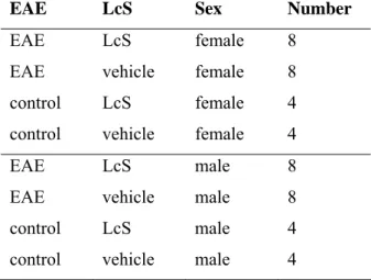
Table 2: Experimental groups EAE
EAE LcS Sex Number
EAE LcS female 8
EAE vehicle female 8
control LcS female 4
control vehicle female 4 EAE LcS male 8
EAE vehicle male 8
control LcS male 4
control vehicle male 4
Acute EAE was induced at the age of 7 weeks as described previously (Hendriks et al., 2004). Rats were injected subcutaneously in the left ankle with 100 μl of an emulsion containing 20 μg guinea pig myelin basic protein (MBP; Sigma), 500 μg Mycobacterium tuberculosis type H37RA (Difco, Detroit, MI), 50 μl complete Freund’s adjuvant (CFA, Difco)
supplemented with saline (0.9% NaCl) to reach a volume 100 μl. EAE injections were performed under isoflurane anesthesia. After induction of EAE body weight was recorded daily. Also, neurological signs were scored daily and graded from 1 to 5; 0, no clinical signs; 0.5, loss of tonicity in distal half of tail; 1, flaccid tail; 1.5, unsteady gait; 2, partial hind limb paralysis; 2.5, complete hind limb paralysis; 3, paralysis of the complete lower part of the body up to the diaphragm; 4, paraplegia; and 5, death due to EAE. Rats were sacrificed
27 days after induction of EAE.
Figure 2: Experimental design EAE study
2.7 Culture of spleen and mesenteric lymph node cells
Spleen and mesenteric lymph nodes (MLN) were collected and single-cell suspensions were prepared by pressing the organs through a sterile 70 μm nylon cell strainer. Cells were washed in 5% FCS medium (10 min., 4oC, 300g) and resuspended in 1 ml standard medium
with 10% FCS. The concentration of the cell suspensions was adjusted to 4*106 cells/ml.
For cytokine measurements spleen and MLN cell suspensions (100 μl/well; 4*105 cells) were
stimulated with 5 μg/ml ConA (100 μl/well) or 5 μg/ml LPS (100 μl/well) for 48 hours in 96 well plates. -35 LcS 2 weeks Age Induction EAE 7 weeks 0 LcS 28 dissection Body weight and clinical score
-35 LcS 2 weeks Age Induction EAE 7 weeks 0 7 weeks 0 LcS 28 dissection Body weight and clinical score
3. ELISA PROTOCOLS
3.1 ELISA specific for ovalbumin IgE and IgG1
Specific ovalbumin IgE and IgG1 titers in sera were determined by ELISA. Incubations were followed by extensive washing on an automatic plate washer with PBS containing 0.1% Tween-20. To measure ovalbumin-specific IgE 96-wells plates (Nunc-Immuno Plate, Denmark) were coated overnight at 4oC with 2 μg/ml rat-anti-mouse IgE (rαm IgE, Zymed,
04-7000) diluted in sodium carbonate buffer (pH 9.6) and incubated overnight at 4°C. Plates were blocked by adding 0.05M Tris buffered saline with 1% BSA, pH 8 (Sigma) for 1 hour at 37°C. Thereafter, serial dilutions of mouse serum samples and a pooled positive reference serum were incubated for 1 hour at 37°C. All dilutions were done in blocking buffer plus 0.05% Tween-20. Then, wells were incubated for 1 h at 37°C with DIG-conjugated ovalbumin. The coupling of ovalbumin to DIG (molar mixture 1:10) was performed
according to the manufacturer's instructions (Roche Diagnostics). Then, wells were incubated with anti-DIG Fab fragments labeled with peroxidase (PO) (Roche Diagnostics) for 2 hours at 37°C. Plates were incubated with TMB substrate and the enzyme reaction was stopped with 2 M H2SO4 and absorbance was read at 450 nm.
To measure ovalbumin-specific IgG1 wells were coated overnight at 4°C with 10 μg/ml ovalbumin/ml PBS (grade V, Sigma). Blocking buffer was added and wells were incubated for 1 hour at 37°C. Thereafter, serial dilutions of mouse serum samples and a pooled positive reference serum were added to the wells and incubated for 2 hours at RT. Biotinylated rat-anti-mouse IgG1 (Zymed Laboratories, San Francisco, CA) was added and wells were incubated for 1.5 hour at RT, followed by incubation with poly-horseradish peroxidase labeled streptavidin for 45 minutes at RT. Then plates were incubated with TMB substrate and the enzyme reaction was stopped with 2 M H2SO4 and absorbance was read at 450 nm.
Extinction values of the positive reference serum were used to calculate the amount of IgG1 and IgE in the samples and extinction values were expressed as arbitrary units.
3.2 Mouse IL-4, IL-10 and IFN-
γ
ELISA
IL-4, IL-10 and IFN-γ concentrations in supernatants were determined by enzyme-linked immunosorbent assay (ELISA). Monoclonal antibodies for IL-4 and IFN-γ were obtained from BD Biosciences Pharmingen (San Diego, CA, USA) and for IL-10 from R&D Systems (Minneapolis, MN) and ELISA’s were performed according to the manufacturer’s
3.3 Rat IL-4, IL-12 and IFN-
γ
ELISA
IL-4 was determined with a rat IL-4 cytoset (Biosource) and IL-12 was determined with a rat IL-12 + p40 cytoset (Biosource) according to the manufacturer’s instruction. IFN-γ was detected with a rat IFN-γ OptEIA set (BD Biosciences Pharmingen) according to the protocol provided by the manufacturer.
4. STATISTICAL ANALYSIS
In the respiratory allergy experiment statistical differences between the four experimental groups were determined by one-way ANOVA followed by Scheffe’s post hoc test. Differences in ovalbumin-specific IgE and IgG1 levels were determined with a two-tailed Student’s t-test comparing the two experimental groups that were sensitized and challenged with ovalbumin. Statistical differences in the EAE experiment between the experimental groups that were immunized with EAE were determined with a Wilcoxon test. Differences in cytokine production between all experimental groups were assessed by a one-way ANOVA followed by Scheffe’s post hoc test.
5. EFFECTS OF EARLY ADMINISTRATION OF LCS ON
A MURINE MODEL FOR RESPIRATORY ALLERGY
5.1 Airway response to methacholine
Airway responsiveness was assessed by exposing the mice to increasing concentrations of methacholine 24 hours after the last challenge. Airway responses are expressed as enhanced pause (Penh) in figure 3A (females) and B (males). Airway responses to methacholine increased dose-dependently, but responses in ovalbumin sensitized and challenged mice were not higher than those of non-sensitized animals. LcS did not affect airway responses either.
Figure 3: Airway responses to increasing concentrations of methacholine in female (A) and male (B) mice. Mice were sensitized and challenged with ovalbumin (OVA) or saline (vehicle) and treated orally with LcS or saline/peptone (vehicle).
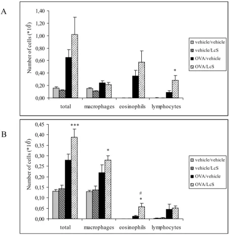
5.2 Bronchoalveolar lavage: cell counts and differentiation
Cell counts after lung lavage showed that lungs of both female and male mice that were sensitized and challenged with ovalbumin had higher numbers of total cells than controls. This increase was predominantly due to an increase in eosinophils and lymphocytes. Influx of0 1 2 3 4 5 6 0 2 4 7,5 15 30 Methacholine (mg/ml) Pe nh OVA/vehicle OVA/LcS vehicle/vehicle vehicle/LcS 0 1 2 3 4 5 6 0 2 4 7,5 15 30 Methacholine (mg/ml) Pe nh OVA/vehicle OVA/LcS vehicle/vehicle vehicle/LcS B A
eosinophils and lymphocytes after challenge with ovalbumin was more pronounced in female mice than in male mice (Figure 4). LcS administration further enhanced the number of eosinophils and lymphocytes in females and the number of lymphocytes in males.
Figure 4: Number of cells in lung lavage fluid in female (A) and male (mice). Mice were sensitized and challenged with ovalbumin (OVA) or saline (vehicle) and orally treated with LcS or saline/peptone (vehicle). Cell number is expressed in 106 cells. Statistical significance was determined by a one-way ANOVA with Scheffe’s post hoc test. Statistically difference compared to vehicle/vehicle group: * p<0.05, *** p<0.001 and to OVA/vehicle group:
#
p<0.05.
5.3 Cytokine production
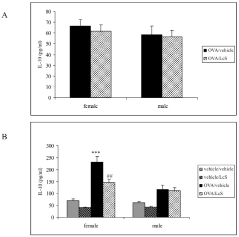
Spleen cells were stimulated with ovalbumin for 96 hours to measure specific cytokine production and with ConA for 48 hours to measure mitogen-induced cytokine production. Culture of spleen cells with ovalbumin resulted in IL-10 production (Figure 5A), but LcS administration did not affect this. After ConA stimulation IL-10 production was higher in mice that were sensitized with ovalbumin than in controls. Female mice had significantly higher IL-10 levels than male mice. LcS reduced IL-10 production significantly in sensitized females, whereas in males LcS did not affect IL-10 production (Figure 5B).
A 0,00 0,20 0,40 0,60 0,80 1,00 1,20 1,40
total macrophages eosinophils lymphocytes
N um ber o f cel ls (* 10 6 ) vehicle/vehicle vehicle/LcS OVA/vehicle OVA/LcS * 0,00 0,05 0,10 0,15 0,20 0,25 0,30 0,35 0,40 0,45
total macrophages eosinophils lymphocytes
N um be r of c ells (* 10 6 ) vehicle/vehicle vehicle/LcS OVA/vehicle OVA/LcS *** * * # B
Figure 5: IL-10 production after stimulation of spleen cells with ovalbumin for 96 hours (A) or ConA for 48 hours (B). IL-10 levels in supernatants are expressed in pg/ml. Statistical significance was determined by a one-way ANOVA with Scheffe’s post hoc test. Statistically difference compared to vehicle/vehicle group: *** p<0.001 and to OVA/vehicle group:
##
p<0.01.
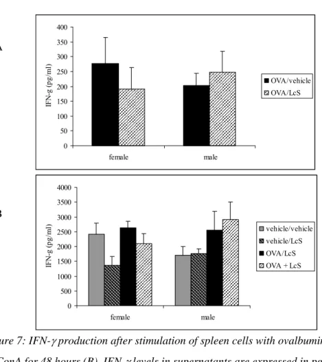
Only a few samples had detectable IL-4 after ovalbumin stimulation. IL-4 was measured in two females and two males that were sensitized and challenged with ovalbumin and orally treated with vehicle. Mice that were received LcS had no detectable IL-4 levels. After ConA stimulation IL-4 production was detected in all samples and levels were significantly higher in females than in males (Figure 6). LcS reduced IL-4 production in sensitized and non-sensitized female mice and this difference was almost significant for the mice that were not sensitized (p<0.058). In non-sensitized male mice, the IL-4 production was significantly lower after LcS treatment, whereas in sensitized mice LcS had no effect on IL-4 production. LcS did not affect IFN-γ production after in vitro ovalbumin stimulation. ConA-induced IFN−γ production was decreased in non-sensitized females that received LcS (not significant). In males IFN-γ production was higher in sensitized mice. LcS did not affect IFN-γ production in males (Figure 7) 0 10 20 30 40 50 60 70 80 female male IL -10 (pg/ m l) OVA/vehicle OVA/LcS A 0 50 100 150 200 250 300 female male IL -10 (pg/ m l) vehicle/vehicle vehicle/LcS OVA/vehicle OVA/LcS *** ## B
Figure 6: IL-4 production after stimulation of spleen cells with ConA for 48 hours. IL-4 levels in supernatants are expressed in pg/ml. Statistical significance was determined by a one-way ANOVA with Scheffe’s post hoc test. Statistically difference compared to vehicle/vehicle group: *** p<0.001.
Figure 7: IFN-γ production after stimulation of spleen cells with ovalbumin for 96 hours (A) or ConA for 48 hours (B). IFN-γ levels in supernatants are expressed in pg/ml.
5.4 Ovalbumin-specific IgG1 and IgE levels
Serum samples were used to detect ovalbumin-specific IgG1 (Figure 8) and IgE (Figure 9) levels in mice that were sensitized and challenged with ovalbumin. Mice that were sensitized
0 20 40 60 80 100 120 140 160 180 200 female male IL -4 (p g/ m l) vehicle/vehicle vehicle/LcS OVA/vehicle OVA/LcS *** 0 50 100 150 200 250 300 350 400 female male IFN-g (p g/ m l) OVA/vehicle OVA/LcS 0 500 1000 1500 2000 2500 3000 3500 4000 female male IFN-g (p g/ m l) vehicle/vehicle vehicle/LcS OVA/LcS OVA + LcS A B
and challenged with vehicle served as a negative control and serum of these mice did not contain ovalbumin-specific IgG1 and IgE (not shown). Ovalbumin-specific IgG1 levels were comparable in males and females, but ovalbumin-specific IgE levels were higher in females than in males. Oral administration of LcS did not significantly affect specific IgG1 and IgE levels, but in both females and males IgG1 serum levels were slightly higher after LcS administration. In females that received LcS, IgE levels were also slightly higher.
Figure 8: Serum levels of ovalbumin-specific IgG1 measured by ELISA. Mice were sensitized and challenged with ovalbumin and treated orally with LcS or saline/peptone (vehicle). Serum levels are expressed in arbitrary units that were calculated by using reference serum with ovalbumin-specific IgG1.
Figure 9: Serum levels of ovalbumin-specific IgE measured by ELISA. Mice were sensitized and challenged with ovalbumin and treated orally with LcS or saline/peptone (vehicle). Serum levels are expressed in arbitrary units that were calculated by using reference serum with ovalbumin-specific IgG1.
0 10000 20000 30000 40000 50000 60000 70000 80000 90000 female male Ig G1 (a rb it ra ry u ni ts (* 10 3 )) OVA/vehicle OVA/LcS 0 10000 20000 30000 40000 50000 60000 70000 female male OV A-s pe ci fi c Ig E (a rb it ra ry u ni ts ) OVA/vehicle OVA/LcS
6. EFFECTS OF EARLY ADMINISTRATION OF LCS ON
A EXPERIMENTAL AUTOIMMUNE
ENCEPHALOMYELITIS
6.1 Body weight gain
Body weight gain from the day of immunization with MBP is depicted in figure 10. In male control rats that received either vehicle or LcS no effects on body weight gain were observed, whereas in rats that were immunized with MBP effects on body weight gain were observed. Some individual rats suffered from slight weight loss. Overall, group averages show that male rats with EAE that received vehicle do not gain weight from day 12 until day 16 and rats with EAE that received LcS display reduced growth from day 17 until day 21. Final body weight gain was lower in rats with EAE compared with control rats. In females, normal growth was observed in control rats. In rats with EAE that received vehicle a growth reduction from day 6 until 9 was observed. Rats with EAE that received LcS did not gain body weight from day 12 until 15.
Figure 10: Body weight gain in male (A) and female Lewis rats (B) after EAE induction (day 0). Body weight is expressed as increase in body weight compared to body weight on day 0.
0 20 40 60 80 100 120 140 1 3 5 7 9 11 13 15 17 19 21 23 25 27
Days after EAE induction
In cr ea se c om pa re d to bw t da y 0 (g ) EAE/vehicle EAE/LcS Control/vehicle Control/LcS -10 0 10 20 30 40 50 60 1 3 5 7 9 11 13 15 17 19 21 23 25 27
Days after EAE induction
In cr eas e c om par ed to b w t d ay 0 (g ) EAE/vehicle EAE/LcS Control/vehicle Control/LcS t A B
6.2 Clinical symptoms
Both male and female rats developed EAE. In males the onset of EAE symptoms varied slightly between rats. Loss of tonicity in the tail was observed in male Lewis rats that received vehicle from day 14 onward. Day of onset varied between day 14 and 18. In male Lewis rats that received LcS first symptoms were observed after 15 days and day of onset was in the range of 15-21 days after induction. Figure 11A shows the course of cumulative clinical score in male rats. LcS did not affect mean day of onset, but increased incidence (Table 3). All male rats that received LcS developed EAE, whereas in controls only 6 out of 8 rats developed symptoms. Other parameters, such as clinical score per rat, duration of symptoms and clinical disease index, were not affected by LcS.
Figure 11: Time course of mean cumulative score per day on EAE in male (A) and female (B) Lewis rats. Clinical disease symptoms were scored daily and added over time per animal.
In females first clinical signs were observed after 12 days in rats that received vehicle and after 11 days in rats that received LcS. Day of onset differed in individual rats. The day of onset was in the range of 12-18 days in rats that received the vehicle. For rats that received
A B 0 1 2 3 4 5 6 7 8 9 11 12 13 14 15 16 17 18 19 20 21 22 23 24 25 26 27
Days after induction EAE
M ean cu mu la ti ve s c o re EAE/vehicle EAE/LcS 0 1 2 3 4 5 6 11 12 13 14 15 16 17 18 19 20 21 22 23 24 25 26 27
Days after induction EAE
M e a n cu m u la ti ve s c o re EAE/vehicle EAE/LcS
LcS this range was 11-18 days after EAE induction. There was no difference between mean day of onset (Table 3). Figure 11B shows the course of cumulative clinical score and shows that clinical score was higher in rats that received LcS. This also resulted in higher clinical score per animal and cumulative disease index in rats that received LcS. Symptoms lasted longer in females that received LcS, resulting in a significant increase of duration of symptoms from 3.4 to 6.6 days. Incidence was also increased by LcS. All females rats that received LcS developed clinical symptoms, while 6 out of 8 rats that received vehicle developed EAE (Table 3).
Table 3: Summary of EAE clinical symptoms
Group Day of onset Incidence Clinical
score per rat
Duration symptoms Cumulative disease index Males EAE/vehicle 15.7 ± 0.6 6/8 4.6 ± 2.9 4.6 ± 1.2 2.6 EAE/LcS 17.5 ± 0.8 8/8 4.0 ± 1.2 5.0 ± 1.0 2.6 Females EAE/vehicle 15.7 ± 0.8 6/8 2.9 ± 1.5 3.4 ± 1.4 1.6 EAE/LcS 15.3 ± 0.9 8/8 4.2 ± 1.8 6.6 ± 1.0* 2.2
Clinical symptoms of EAE are summarized in this table. Day of onset is the first day a rat displayed clinical symptoms and is expressed as mean ± SE of all rats with symptoms. Incidence is the number of rats with clinical symptoms. Clinical score per rat is the cumulative clinical scores per rat at the end of the experiment expressed as group average ± SE. Duration of symptoms is the number of days an animal displayed symptoms expressed as group average ± SE. Cumulative disease index is the sum of the daily clinical scores for a group over a given number of days divided by the number of days. Asterisks denotes
significance (p<0.05) compared to the EAE/vehicle group.
6.3 Cytokines
Spleen cells were cultured with LPS for 48 hours to assess IL-12 production and with ConA for 48 hours to detect IL-4 and IFN-γ production. In both male and female rats that were immunized with MBP IL-12 levels were higher than in controls (Figure 12A). In females treatment with LcS reduced IL-12 levels significantly. IFN-γ production in males was not different between experimental groups. In females, IFN-γ production was significantly higher in rats with EAE than in control rats. Control rats that received LcS had higher IFN-γ levels compared to controls that received vehicle (p=0.074). There was no difference in IFN-γ production between rats with EAE that received vehicle or LcS (Figure 12B). IL-4 could not
be detected in any of the samples and is either not produced or produced below the detection limit.
Figure 12: IL-12 (A) and IFN-γ (B) production after stimulation of spleen cells with LPS (5 μg/ml) to detect IL-12 and with ConA (5 μg/ml) to detect IFN-γ for 48 hours. IL-12 and IFN-γ are both expressed as pg/ml. Asterisk denotes significance from the control group (vehicle/vehicle), * p<0.05. Significantly different from the EAE/vehicle group: # p<0.05.
0 20 40 60 80 100 120 males females IL -1 2 (p g/ m l) Control/vehicle Control/LcS EAE/vehicle EAE/LcS * # 0 5000 10000 15000 20000 25000 30000 males females IF N -g a mma ( p g /ml ) vehicle/vehicle vehicle/LcS EAE/vehicle EAE/LcS * * A B
7. DISCUSSION
We have studied the effects of administration of the probiotic LcS during the lactation phase on the development of allergy and autoimmunity. Until now, no experiments have been performed that have studied effects of early probiotic administration on allergic and autoimmune responses.
In the respiratory allergy model LcS increased the influx of eosinophils and lymphocytes after challenge with ovalbumin. Systemic responses appeared to be also slightly stimulated, since ovalbumin-specific IgG1 and IgE levels were somewhat enhanced. However, cytokine production assessed in the spleen after mitogen stimulation are not in line with this. LcS decreased the Th2 cytokines IL-4 and IL-10, but did not affect the Th1 cytokine IFN-γ. Thus, Th1/Th2 cytokine profiles seem not predictive for the observed stimulation of lung and systemic responses induced by ovalbumin. Furthermore, results from this study are not in line with previous studies that were performed with LcS or other probiotics in allergy models. LcS has been shown to induce a shift in the Th1/Th2 balance towards Th1, thereby reducing ovalbumin-specific IgE levels (Shida et al., 1998; Shida et al., 2002). Similar results have also been found for other probiotics (Kim et al., 2005; Ohno et al., 2005). However, not all
probiotic strains stimulated Th1 responses and suppressed Th2 responses. In a respiratory allergy model Lactobacillus rhamnosus HN001 invoked a mixed cytokine profile, with higher levels of both Th1 and Th2 cytokines. In these mice serum IgE levels and inflammatory lung responses were not assessed and it is therefore unknown what the effects of this
immunostimulation were on these parameters (Cross et al., 2002).
Effects of LcS on respiratory allergy have also been studied in a similar experiment in adult mice (Ezendam et al., 2006). In sensitized females that received LcS the number of
eosinophils was slightly decreased, whereas the number of lymphocytes was not affected. In sensitized males, inflammatory lung responses were not affected after LcS administration. In line with this were the observed lower levels of Th2 cytokines IL-4, IL-5, IL-10 and IL-13 in the BAL fluid of sensizited females that received LcS. In females, LcS slightly enhanced both serum IgE and IgG1 levels and in males LcS slightly increased IgE levels. Cytokine profiles assessed in the spleen after specific stimulation with ovalbumin also show increased levels of both Th2 and Th1 cytokines, indicatory of a general immunostimulatory effect. Effects of LcS in adults are not completely comparable with effects observed in mice that received LcS from lactation phase onward. Effects on lung inflammatory responses are different, since LcS had beneficial effects in adult females and no effects in males. Effects on systemic responses appear to be comparable. The increases of serum IgE and IgG1 levels after LcS
Thus, administration of LcS early after birth did stimulate inflammatory lung responses, a phenomenon not observed in adults. However, systemic stimulation of the immune responses was observed in both studies.
Stimulation of immune responses was also observed in female Lewis rats with EAE. After LcS administration clinical symptoms of EAE were more pronounced, duration of symptoms was longer and also the incidence was higher. The latter was also observed in male rats, but clinical symptoms and duration of these symptoms were not affected. Effects of probiotics on EAE have also been studied in adult mice. This study has shown that lactobacillus strains can induce differential effects. Lactobacillus reuteri aggravated the clinical symptoms,
Lactobacillus plantarum had no effects, whereas Lactobacillus casei and Lactobacillus murines inhibited severity of the disease. These strain differences can probably be explained
by cytokine profiles induced by the different strains, which were assessed in a different model. Lactobacillus reuteri induced a Th1-like profile (TNF-α and IL-2), whereas
Lactobacillus casei induced a regulatory profile (IL-10 and TGF-β) (Maassen et al., 1998).
Cytokines that were measured in our study seem not predictive for the effects on clinical symptoms. LcS increased IFN-γ levels in control females, but not in females with EAE. Furthermore, LcS decreased IL-12 production in females with EAE compared to vehicle treated females with EAE. It is unclear how LcS aggravated EAE in females, but it cannot be explained by the cytokines we have measured.
Effects of LcS in this study are in line with effects observed in a similar study performed in adult male rats (Ezendam et al., 2006). In this experiment LcS administration resulted in an earlier onset, higher incidence, higher clinical score and longer duration of symptoms. Early intake of LcS, however, does not seem to induce more pronounced effects of EAE.
In conclusion, LcS has stimulatory effects on both experimental allergy and autoimmunity and these effects are similar after adult and juvenile administration, except for inflammatory lung responses that were enhanced after juvenile administration and alleviated after adult administration. Although these effects are small and not always significant, they could implicate that there is a potential hazard. Newly marketed products, such as infant formulas that are supplemented with probiotics, are considered to be GRAS (generally regarded as safe) because of long-term use in the adult population. Although we only demonstrated a difference in the inflammatory lung responses after administration of LcS early in life or at the adult age, this does not mean that these products are safe. Maybe the timing of the experiments was not optimal and administration of LcS administration should have started earlier. Furthermore, since probiotic effects appear to be strain-dependent, no general conclusion on supplementation of baby formulas can be drawn yet. Therefore, similar experiments will be performed with a different probiotic strain: Bifidobacterium breve and data from both probiotic strains will be used in the future for further hazard identification.
ACKNOWLEDGEMENTS
We would like to acknowledge Arja de Klerk, Eric Gremmer, Bert Verlaan. Bianca Matthee, Liset de la Fonteyne and Yvonne Wallbrink from the Laboratory for Toxicology, Pathology and Genetics of the RIVM for excellent technical assistance. We thank Hans Strootman, Dirk Elberts, Piet van Schaaik, Christine Soputan and Jouke van de Siepkamp from PMP
REFERENCES
Alvarez, S., Herrero, C., Bru, E., and Perdigon, G. (2001). Effect of Lactobacillus casei and yogurt administration on prevention of Pseudomonas aeruginosa infection in young mice. J Food Prot 64, 1768-1774.
Bjorksten, B., Naaber, P., Sepp, E., and Mikelsaar, M. (1999). The intestinal microflora in allergic Estonian and Swedish 2-year-old children. Clin Exp Allergy 29, 342-346. Chapat, L., Chemin, K., Dubois, B., Bourdet-Sicard, R., and Kaiserlian, D. (2004).
Lactobacillus casei reduces CD8+ T cell-mediated skin inflammation. Eur J Immunol
34, 2520-2528.
Cross, M. L., Ganner, A., Teilab, D., and Fray, L. M. (2004). Patterns of cytokine induction by gram-positive and gram-negative probiotic bacteria. FEMS Immunol Med
Microbiol 42, 173-180.
Cross, M. L., Mortensen, R. R., Kudsk, J., and Gill, H. S. (2002). Dietary intake of Lactobacillus rhamnosus HNOO1 enhances production of both Th1 and Th2 cytokines in antigen-primed mice. Med Microbiol Immunol (Berl) 191, 49-53.
De Vrese, M., Rautenberg, P., Laue, C., Koopmans, M., Herremans, T., and Schrezenmeir, J. (2004). Probiotic bacteria stimulate virus-specific neutralizing antibodies following a booster polio vaccination. Eur J Nutr.
De Waard, R., Claassen, E., Bokken, G. C., Buiting, B., Garssen, J., and Vos, J. G. (2003). Enhanced immunological memory responses to Listeria monocytogenes in rodents, as measured by delayed-type hypersensitivity (DTH), adoptive transfer of DTH, and protective immunity, following Lactobacillus casei Shirota ingestion. Clin Diagn Lab
Immunol 10, 59-65.
De Waard, R., Garssen, J., Bokken, G. C., and Vos, J. G. (2002). Antagonistic activity of Lactobacillus casei strain shirota against gastrointestinal Listeria monocytogenes infection in rats. Int J Food Microbiol 73, 93-100.
De Waard, R., Garssen, J., Snel, J., Bokken, G. C., Sako, T., Veld, J. H., and Vos, J. G. (2001). Enhanced antigen-specific delayed-type hypersensitivity and immunoglobulin G2b responses after oral administration of viable Lactobacillus casei YIT9029 in Wistar and Brown Norway rats. Clin Diagn Lab Immunol 8, 762-767.
Ezendam, J., Baken K., and van Loveren, H. 2006. Immune effects of Lactobacillus casei Shirota. RIVM report 340320004.
Helin, T., Haahtela, S., and Haahtela, T. (2002). No effect of oral treatment with an intestinal bacterial strain, Lactobacillus rhamnosus (ATCC 53103), on birch-pollen allergy: a placebo-controlled double-blind study. Allergy 57, 243-246.
Hendriks, J. J., Alblas, J., van der Pol, S. M., van Tol, E. A., Dijkstra, C. D., and de Vries, H. E. (2004). Flavonoids influence monocytic GTPase activity and are protective in experimental allergic encephalitis. J Exp Med 200, 1667-1672.
Herias, M. V., Koninkx, J. F., Vos, J. G., Huis In't Veld, J. H., and van Dijk, J. E. (2005). Probiotic effects of Lactobacillus casei on DSS-induced ulcerative colitis in mice. Int J Food Microbiol 103, 143-155.
Isolauri, E. (2001). Probiotics in human disease. Am J Clin Nutr 73, 1142S-1146S. Isolauri, E., Arvola, T., Sutas, Y., Moilanen, E., and Salminen, S. (2000). Probiotics in the
management of atopic eczema. Clin Exp Allergy 30, 1604-1610.
Kalliomaki, M., Kirjavainen, P., Eerola, E., Kero, P., Salminen, S., and Isolauri, E. (2001a). Distinct patterns of neonatal gut microflora in infants in whom atopy was and was not developing. J Allergy Clin Immunol 107, 129-134.
Kalliomaki, M., Salminen, S., Arvilommi, H., Kero, P., Koskinen, P., and Isolauri, E. (2001b). Probiotics in primary prevention of atopic disease: a randomised placebo-controlled trial. Lancet 357, 1076-1079.
Kalliomaki, M., Salminen, S., Poussa, T., Arvilommi, H., and Isolauri, E. (2003). Probiotics and prevention of atopic disease: 4-year follow-up of a randomised placebo-controlled trial. Lancet 361, 1869-1871.
Kato, I., Endo-Tanaka, K., and Yokokura, T. (1998). Suppressive effects of the oral administration of Lactobacillus casei on type II collagen-induced arthritis in DBA/1 mice. Life Sci 63, 635-644.
Kato, I., Tanaka, K., and Yokokura, T. (1999). Lactic acid bacterium potently induces the production of interleukin-12 and interferon-gamma by mouse splenocytes. Int J Immunopharmacol 21, 121-131.
Kim, H., Kwack, K., Kim, D.-Y., and Ji, G. E. (2005). Oral probiotic bacterial administration suppressed allergic responses in an ovalbumin-induced allergy mouse model. FEMS Immunology and Medical Microbiology 45, 259-267.
Kullen, M. J., and Bettler, J. (2005). The delivery of probiotics and prebiotics to infants. Curr Pharm Des 11, 55-74.
Maassen, C. B., van Holten, J. C., Balk, F., Heijne den Bak-Glashouwer, M. J., Leer, R., Laman, J. D., Boersma, W. J., and Claassen, E. (1998). Orally administered
Lactobacillus strains differentially affect the direction and efficacy of the immune response. Vet Q 20 Suppl 3, S81-83.
Macpherson, A. J., and Harris, N. L. (2004). Interactions between commensal intestinal bacteria and the immune system. Nat Rev Immunol 4, 478-485.
Matsuzaki, T. (1998). Immunomodulation by treatment with Lactobacillus casei strain Shirota. Int J Food Microbiol 41, 133-140.
Matsuzaki, T., Nagata, Y., Kado, S., Uchida, K., Hashimoto, S., and Yokokura, T. (1997a). Effect of oral administration of Lactobacillus casei on alloxan-induced diabetes in mice. Apmis 105, 637-642.
Matsuzaki, T., Nagata, Y., Kado, S., Uchida, K., Kato, I., Hashimoto, S., and Yokokura, T. (1997b). Prevention of onset in an insulin-dependent diabetes mellitus model, NOD mice, by oral feeding of Lactobacillus casei. Apmis 105, 643-649.
Matsuzaki, T., Yamazaki, R., Hashimoto, S., and Yokokura, T. (1998). The effect of oral feeding of Lactobacillus casei strain Shirota on immunoglobulin E production in mice. J Dairy Sci 81, 48-53.
Meydani, S. N., and Ha, W. K. (2000). Immunologic effects of yogurt. Am J Clin Nutr 71, 861-872.
Montrose, D. C., and Floch, M. H. (2005). Probiotics used in human studies. J Clin Gastroenterol 39, 469-484.
Murosaki, S., Yamamoto, Y., Ito, K., Inokuchi, T., Kusaka, H., Ikeda, H., and Yoshikai, Y. (1998). Heat-killed Lactobacillus plantarum L-137 suppresses naturally fed antigen-specific IgE production by stimulation of IL-12 production in mice. J Allergy Clin Immunol 102, 57-64.
Ohno, H., Tsunemine, S., Isa, Y., Shimakawa, M., and Yamamura, H. (2005). Oral Administration of Bifidobacterium bifidum G9-1 Suppresses Total and Antigen Specific Immunoglobulin E Production in Mice. Biol Pharm Bull 28, 1462-1466.
Perdigon, G., Locascio, M., Medici, M., Pesce de Ruiz Holgado, A., and Oliver, G. (2003). Interaction of bifidobacteria with the gut and their influence in the immune function. Biocell 27, 1-9.
Pohjavuori, E., Viljanen, M., Korpela, R., Kuitunen, M., Tiittanen, M., Vaarala, O., and Savilahti, E. (2004). Lactobacillus GG effect in increasing IFN-gamma production in infants with cow’s milk allergy. J Allergy Clin Immunol 114, 131-136.
Rosenfeldt, V., Benfeldt, E., Nielsen, S. D., Michaelsen, K. F., Jeppesen, D. L., Valerius, N. H., and Paerregaard, A. (2003). Effect of probiotic Lactobacillus strains in children with atopic dermatitis. J Allergy Clin Immunol 111, 389-395.
Rosenfeldt, V., Benfeldt, E., Valerius, N. H., Paerregaard, A., and Michaelsen, K. F. (2004). Effect of probiotics on gastrointestinal symptoms and small intestinal permeability in children with atopic dermatitis. J Pediatr 145, 612-616.
Salminen, S., Bouley, C., Boutron-Ruault, M. C., Cummings, J. H., Franck, A., Gibson, G. R., Isolauri, E., Moreau, M. C., Roberfroid, M., and Rowland, I. (1998). Functional food science and gastrointestinal physiology and function. Br J Nutr 80 Suppl 1, S147-171. Shida, K., Makino, K., Morishita, A., Takamizawa, K., Hachimura, S., Ametani, A., Sato, T.,
antigen-induced IgE secretion through regulation of cytokine production in murine splenocyte cultures. Int Arch Allergy Immunol 115, 278-287.
Shida, K., Takahashi, R., Iwadate, E., Takamizawa, K., Yasui, H., Sato, T., Habu, S., Hachimura, S., and Kaminogawa, S. (2002). Lactobacillus casei strain Shirota suppresses serum immunoglobulin E and immunoglobulin G1 responses and systemic anaphylaxis in a food allergy model. Clin Exp Allergy 32, 563-570.
Smit, J. J., van Loveren, H., Hoekstra, M. O., Nijkamp, F. P., and Bloksma, N. (2003). Influence of the macrophage bacterial resistance gene, Nramp1 (Slc11a1), on the induction of allergic asthma in the mouse. Faseb J 17, 958-960.
Smits, H. H., Engering, A., van der Kleij, D., de Jong, E. C., Schipper, K., van Capel, T. M. M., Zaat, B. A. J., Yazdanbakhsh, M., Wierenga, E. A., van Kooyk, Y., and Kapsenberg, M. L. (2005). Selective probiotic bacteria induce IL-10-producing regulatory T cells in vitro by modulating dendritic cell function through dendritic cell-specific intercellular adhesion molecule 3-grabbing nonintegrin. Journal of Allergy and Clinical Immunology
115, 1260-1267.
Sudo, N., Sawamura, S., Tanaka, K., Aiba, Y., Kubo, C., and Koga, Y. (1997). The requirement of intestinal bacterial flora for the development of an IgE production system fully susceptible to oral tolerance induction. J Immunol 159, 1739-1745.
Sudo, N., Yu, X. N., Aiba, Y., Oyama, N., Sonoda, J., Koga, Y., and Kubo, C. (2002). An oral introduction of intestinal bacteria prevents the development of a long-term Th2-skewed immunological memory induced by neonatal antibiotic treatment in mice. Clin Exp Allergy 32, 1112-1116.
Wheeler, J. G., Shema, S. J., Bogle, M. L., Shirrell, M. A., Burks, A. W., Pittler, A., and Helm, R. M. (1997). Immune and clinical impact of Lactobacillus acidophilus on asthma. Ann Allergy Asthma Immunol 79, 229-233.