Published by:
National Institute for Public Health and the Environment
P.O. Box 1 | 3720 BA Bilthoven The Netherlands
Molecular detection and typing of
Coxiella burnetii
RIVM Letter report 330291002/2009 A. de Bruin | I. Janse | B.J. van Rotterdam
Colofon
© RIVM 2009
Parts of this publication may be reproduced, provided acknowledgement is given to the 'National Institute for Public Health and the Environment', along with the title and year of publication.
Keywords:
Coxiella burnetii, Q fever, qPCR, Molecular typing, Molecular detection
Trefwoorden:
Coxiella burnetii, Q-koorts, qPCR, Moleculaire typering, Moleculaire detectie
Arnout de Bruin (Researcher), RIVM
Ingmar Janse (Researcher), RIVM
Bart van Rotterdam (Project Leader), RIVM
Contact:
Bart van Rotterdam
Laboratory for Zoonoses and Environmental Microbiology
bart.van.rotterdam@rivm.nl
This investigation has been performed by order and for the account of the Food and Consumer Product Safety Authority, within the framework of Deelproject 9.2.3. D Coxiella in kennisvraag liverstock-borne zoonoses.
Contents
1 Introduction—5
1.1 Q fever outbreaks in the Netherlands—5
1.2 Diagnosis of Q fever: Serology versus DNA based methods—6 1.3 Report Outline—8
2 Molecular detection of Coxiella burnetii—9
2.1 Sampling environmental and animal sources—9 2.2 DNA extraction procedures—9
2.3 Detection of C. burnetii DNA by quantitative multiplex real-time PCR—9 2.4 Source finding investigations during Q fever outbreak seasons—10 2.5 qPCR assay comparison during ring trials—11
2.6 qPCR assay optimization and validation—12
2.6.1 Sensitivity assessment of the multiplex qPCR assays—13 2.6.2 Specificity of the multiplex qPCR assays—14
2.6.3 Efficiency of the multiplex qPCR assays—15
3 Molecular typing of Coxiella burnetii—17
3.1 Multi-locus Variable number of tandem repeats Analysis—17
3.1.1 Whole Genome Amplification in environmental & animal samples—19 3.1.2 Optimization of the MLVA assay—20
3.2 Multi-spacer Sequence Typing (MST)—21
4 Conclusions—23
1
Introduction
1.1 Q fever outbreaks in the Netherlands
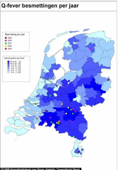
In 2007, 2008, and 2009, large community outbreaks of Q fever have occurred in a rural area of the Netherlands. Two years before the first documented outbreak in the Netherlands in 2007 (1), large abortion waves were reported on (primarily goat) farms in the same region (Figure 1.1 & 1.2). This implicated (goat) farms as potential sources for human Q fever infection. However, the contribution of various transmission routes of Q fever relevant for the Dutch situation was not well understood.
Figure 1.1: Q fever on dairy goat farms in 2005-2009, not taking bulk milk screening into account. (Animal Health Service)
Coxiella burnetii infection in humans can occur via close contact with infected
animals, or contaminated animal products. In addition, C. burnetii can persist for long periods of time in the environment and transmission to animals and
humans by inhalation of contaminated aerosols is thought to be the primary route (2, 3). Infected animals, like goats, sheep, and cattle, often show no
clinical signs of infection except for abortions or stillbirths that may occur due to infection of the placenta. When animals are infected, the main sources of C.
burnetii shedding to the environment are manure, urine, milk, and most
importantly birth materials like amnion fluid and placenta (4, 5).
Figure 1.2: Q fever in humans in 2008 (left) and 2009 (right), RIVM.
1.2 Diagnosis of Q fever: Serology versus DNA based methods
Diagnosis of Q fever, both in humans and animals, is still mainly based on serology. Serological methods often used for the detection of C. burnetii are: indirect immunofluorescence, complement fixation and enzyme-linked
immunosorbent assays (ELISA) (18, 20). For the detection of C. burnetii in the environment, in order to investigate possible routes of dissemination and transmission, serology cannot be applied.
DNA (or RNA) based detection and quantification methods, like the polymerase chain reaction (PCR), are aimed to detect organisms directly by targeting one or more specific sequences in the genome of the organism of interest, in this case
C. burnetii. A number of conventional PCR diagnostic assays, have been
developed for the detection of C. burnetii DNA in (primarily) clinical samples (10, 21, 24). More recently, other types of PCR assays, like nested PCRs (23, 25), or quantitative real-time PCRs (qPCR) (3, 4, 8) have been developed, sometimes in combination with high-throughput capabilities (14).
The specific sequences within a genome of an organism, targeted by PCR, can occur in single copy, or in multiple copies within the genome. A single copy marker is a specific sequence, which occurs only once in the genome of an organism. A multicopy marker is a specific sequence that occurs more often within the genome. A single copy marker can be used for quantification of the number of organisms in a sample (one copy=one genome=one organism), and a multicopy marker is valuable for quick detection of an organism in a given sample, especially when the organism is present in very low concentrations. Multicopy markers are detected much quicker in PCR assays because of their higher initial starting concentration in the genome and the process of
Until now almost all PCR based methods for C. burnetii were designed as singleplex assays, in which each specific region in the genome of C. burnetii is amplified in a separate reaction volume. Combining primers and probes,
targeting different sequences in the genome in a single reaction volume is called multiplexing and saves both time and consumables. Most multiplex PCR based assays however, require extensive (re)design of primers and probes using sophisticated software, helping to avoid cross reaction between primers and probes for the different target sequences. One successful multiplex PCR for the detection of C. burnetii was developed by incorporating the IS1111 element and the bovine CD18 gene as internal control (5). Quantification using this multiplex PCR assay is not possible, however, because the marker targeting C. burnetii DNA (IS1111) is a multicopy marker, which can be present within the genome between 7 and 110 copies (8). A robust PCR based method for detection and quantification of C. burnetii should include at least one single copy marker for quantification, and a multicopy target for sensitive detection, preferentially in multiplex (simultaneous detection) format including an internal control marker. In PCR reactions, it is advantageous to add external DNA template to the sample other than to DNA one wishes to detect, as an internal PCR control. This internal control marker is essential for determining the level of inhibition on the PCR assay by substances present in environmental matrices.
For the current study we developed a quantitative multiplex real-time PCR assay (qPCR) in which three genomic targets commonly used for the detection of C.
burnetii (icd, com1 & IS1111) are combined into a single assay. In addition, an
internal control target (B. thuringiensis gene cry 1) was added to the assay to investigate possible inhibition on the qPCR assay, by the complex environmental matrices. In DNA extraction protocols, a specific amount of Bacillus thuringiensis spores is added to the samples as an internal control for DNA extraction. Next to being an extraction control, the DNA isolated from the B. thurigiensis spores serves as a positive control during qPCR.
For matrices, which inhibit the qPCR assay, DNA extraction protocols have to be optimised such that as much as possible qPCR inhibiting substances are
removed to be able to obtain a high yield and quality of C. burnetii DNA for accurate detection and quantification. Modification of DNA extraction protocols does not only ensure a better insight in the quantification of C. burnetii DNA in environmental and animal samples, but is also necessary for robust molecular typing methods.
The complete genome of C. burnetii Nine Mile RSA phase I (RSA493) is
sequenced (19), and this sequence was used in the development of a molecular typing method for C. burnetii, referred to as Multi-Locus Variable number of tandem repeats Analysis, or MLVA (2, 22). This typing method is based on the variety in particular parts of the C. burnetii genome between strains, called tandem repeats.
This molecular typing method was tested on a large number of reference strains and isolates, and compared to another molecular typing method called
Multispacer Sequence typing (MST) (2, 6). However, this method has never been tested on complex environmental or animal samples. Most molecular typing methods require target DNA of high quality and yield for a robust result. This is often achieved by cultivation of the organism of interest from the clinical, environmental, or animal source. Cultivation of C. burnetii is described using a variety of methods (1, 11-13, 16), however, it is a difficult and laborious process and requires cultivation facilities in a BioSafety Level 3 laboratory (BSL-3). Cultivation of C. burnetii within this project will become feasible at a later stage.
1.3 Report Outline
This report describes the final results and recommendations of our investigations regarding the development of a quantitative DNA based detection method and high resolution typing methods for Coxiella burnetii. This report is an extended and modified version of the report entitled: ‘Molecular detection and typing of
Coxiella burnetii ‘, which was a product within the framework of livestock-borne
zoonoses, project 9.2.3.D Coxiella in 2008.
The design and validation of a molecular detection method (qPCR), and the optimisation of molecular typing methods (MLVA & MST) for C. burnetii are described. The validation of the qPCR assay is described in Chapter 2, and includes: (i) refinements in DNA extraction protocols for recovery of high quality and high yield DNA from complex environmental and animal matrices, and (ii) specificity, sensitivity, and efficiency analyses of the qPCR detection assay. In chapter 3, refinements to two molecular typing methods (MLVA & MST) and the application of these methods in C. burnetii source finding investigations is described.
2
Molecular detection of Coxiella burnetii
2.1 Sampling environmental and animal sources
Environmental and animal samples were collected by employees from the Food and Consumer Product Safety Authority (VWA) on human-linked Q fever affected (goat) farms in the province of Noord-Brabant, the Netherlands in 2008 and 2009. Environmental samples included surface area swabs from within stables, and animal samples were represented by (bulk)milk and vaginal swabs. Infected placenta materials were obtained from the Animal Health Service (GD) in 2008.
2.2 DNA extraction procedures
DNA was extracted from environmental and animal samples using a modified Nuclisens Magnetic Extraction Kit (Biomerieux, France). Modifications to the manufacturer’s guidelines for DNA isolation were developed for liquid samples (milk and water), manure, and surface area swabs and animal vaginal swabs. For the processing of liquid samples, 1 ml of liquid samples was added to 10 ml of NucliSens lysisbuffer. Surface area swabs obtained from stable areas and vaginal swabs obtained from goats or sheep were added to 10 ml of NucliSens lysisbuffer. Processing manure samples was carried out by adding goat droppings to Phosphate-buffered Saline (PBS) in 50 ml Greiner tubes (Greiner Bio-one, the Netherlands), using a 1:1 ratio of manure and PBS. This sample was homogenized for about 2 hours on a rotating tube holder at 10 rpm. Greiner tubes were centrifuged (Varifuge 3.2RS, Heraeus) at 2000 rpm for 10 minutes. The supernatant was transferred to a new Greiner tube, and 1 ml of supernatant was added to 10 ml of NucliSens lysisbuffer. To all samples, 1.2 x 10⁵ spores of
Bacillus thuringiensis were added as internal control for DNA extraction. In
addition, 50 µl of magnetic beads are added to each sample and samples were placed at room temperature for one hour to complete lysis and hybridization of DNA to the magnetic beads. After lysis, samples were placed in a magnetic holder, which can accommodate 15 ml Greiner tubes, for 1 minute and the supernatant was removed. Further steps in DNA extraction were carried out according the manufacturer’s protocol. DNA from (positive) placenta materials was extracted under BSL-3 conditions using a QIAamp DNA Mini Kit according the manufacturer’s protocol in the QIAamp DNA Blood Mini Kit Handbook (September 2001).
2.3 Detection of C. burnetii DNA by quantitative multiplex real-time PCR
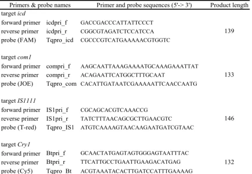
Detection of C. burnetii DNA was initially carried out by a quantitative multiplex real-time PCR assay (qPCR) developed for the project: Molecular detection and typing of Coxiella burnetii ‘, which was a product within the framework of livestock-borne zoonoses, project 9.2.3.D Coxiella in 2008. In short, three genomic targets that are most frequently used for detection of C. burnetii DNA were incorporated into a single qPCR assay (Table 2.1). Targets selected were: the isocitrate dehydrogenase gene (icd), an outer membrane protein coding gene (com1) and an insertion element (IS1111). In addition, an internal control target (B. thuringiensis gene cry 1) was added to the assay to investigate possible inhibition on the qPCR assay by the complex environmental and animal samples.
A colour compensation experiment for hydrolysis (Taqman) probes was carried out according the Roche LightCycler 480 manual, to compensate for possible overlap between fluorescence channels.
Primer and probe sequences (5'-> 3') Product length target icd
forward primer icdpri_f GACCGACCCATTATTCCCT
reverse primer icdpri_r CGGCGTAGATCTCCATCCA
probe (FAM) Tqpro_icd CGCCCGTCATGAAAAACGTGGTC
target com1
forward primer compri_f AAGCAATTAAAGAAAATGCAAAGAAATTAT
reverse primer compri_r ACAGAATTCATGGCTTTGCAAT
probe (JOE) Tqpro_com CACATTGATAATCGAAAAATTCAACCAATG
target IS1111
forward primer IS1pri_f CGCAGCACGTCAAACCG
reverse primer IS1pri_r TATCTTTAACAGCGCTTGAACGTC
probe (T-red) Tqpro_IS1 ATGTCAAAAGTAACAAGAATGATCGTAAC
target Cry1
forward primer Btpri_f GCAACTATGAGTAGTGGGAGTAATTTAC
reverse primer Btpri_r TTCATTGCCTGAATTGAAGACATGAG
probe (Cy5) Tqpro_Bt ACGTAAATACACTTGATCCATTTGAAAAG
Primers & probe names
139
133
146
132
Table 2.1. Primers and probes for each target developed in Visual Omp 6 for the multiplex qPCR for C. burnetii. Primer and probe sequences, and product
lengths obtained are given for C. burnetii targets icd, com1, IS1111, and for B.
thuringiensis target cry1.
2.4 Source finding investigations during Q fever outbreak seasons
The new molecular detection method for C. burnetii was used in source finding investigation in 2008 and 2009. The findings of these source finding
investigations are reported more extensively in report: ‘A Query for Q fever using qPCR in environmental and veterinary matrices’.
In 2008, the Q fever outbreak started in the province of Noord-Brabant, around week 19 in the month May. The Municipal Health Service (GGD) ‘Hart voor Brabant’ started an active source finding study to pinpoint potential sources of
C. burnetii infection in collaboration with the Food and Consumer Product Safety
Authority (VWA).
Employees of the VWA obtained primarily animal matrices (vaginal swabs) on diary goat farms, because goat excretion products are considered a potential source for human Q fever infection. Samples were transported to the National Institute for Public Health and the Environment (RIVM), which conducted the actual screening for C. burnetii DNA by multiplex quantitative real-time PCR (qPCR).
Between May and December of 2008, 409 samples divided over 30 farms were screened by the Laboratory for Zoonosis and Environmental Microbiology (LZO) of RIVM.
Samples screened were primarily animal matrices: vaginal swabs of goats, sheep & cattle. Environmental matrices were represented by surface area swabs.
Seven out of 30 locations (23%) were unlikely sources for the human cases in their near vicinity as none of the samples tested positive. In 23 locations (77%), at least one sample scored positive and in 16 locations (53%), 50% or more of the samples tested positive. The total number of positive samples was found to be 190 (46%).
In 2009, the Q fever outbreak started in the month of April around week 14. Again, most cases started to emerge in the province of Brabant. The Municipal Health Service ‘Hart voor Brabant’ (and other Municipal Health Services) started active source finding again, in close collaboration with the Food and Consumer Product Safety Authority (VWA) and RIVM.
Between April and October of 2009, 1008 samples divided over 57 farms were screened by LZO. The same matrices were sampled and screened as in 2008. Sixteen locations (28%) were unlikely sources for nearby human cases as none of the samples tested positive. In 41 locations (72%), at least one sample scored positive and in 30 locations (53%), 50% or more of the samples tested positive. The total number of positive samples was found to be 488 (48%). In most farms, positive results for vaginal swabs in goats and/or sheep were accompanied by positive results for the surface area swabs taken on the same farm, and vice versa, if animal matrices scored negative in the assay, surface area swabs on the same farm also scored negative. These results indicate that both animal matrices like vaginal swabs and environmental matrices like surface area swabs are good indicators for the presence of C. burnetii DNA. Screening results were forwarded by the Food and Consumer Product Safety Authority (VWA) to the Municipal Health Services (GGD’s).
2.5 qPCR assay comparison during ring trials
Our qPCR assay for C. burnetii DNA detection was compared to other PCR assays for C. burnetii DNA detection in a variety of ring trials in 2009. In two ring trials, both DNA extraction and PCR assays were tested, and in a third ring trial only the PCR assays were compared between laboratories.
In January 2009, our qPCR assay was tested in a ring trial investigation
organized by the Veterinary Laboratories Agency in Weybridge, United Kingdom. Seven laboratories participated in the ring-trial including: Veterinary
Laboratories Agency, Weybridge, UK; Health Protection Agency, Porton Down, UK; Royal Hospitals, Belfast, UK; National Institute for Public Health and the Environment (RIVM), Bilthoven, the Netherlands; Bundesintitut für
Risikobewertung, Berlin, Germany; National Veterinary Institute (SVA), Uppsala, Sweden; and Central Veterinary Institute (CVI), Lelystad, the Netherlands. The findings were reported in a paper submitted to the journal of Veterinary Microbiology as a short communication in September 2009. Out of the seven participating laboratories, two laboratories incorporated target icd in their PCR assay, three institutes incorporated target com1, and all laboratories
incorporated target IS1111 in their PCR assays for C. burnetii. A clear ranking of institutes, based on PCR sensitivity, could not be established because it is not clear what volume of DNA template was used in each PCR assay.
When results between targets within assays are compared, it becomes clear that target IS1111 is the most sensitive target of all targets incorporated in PCR assays. This result confirms other studies, and our own findings in, for example, source finding investigations.
A second ring trial was facilitated by the ‘Canisius-Wilhemina Hospital’ (CWZ), Nijmegen, the Netherlands. Two departments of the National Institute for Public Health and the Environment (RIVM) participated in this trial: the Laboratory for Infectious Diseases and Screening (LIS) and the Laboratory for Zoonoses and
Environmental microbiology (LZO). In addition, 5 other (primarily hospital) laboratories participated, located in ‘s Hertogenbosch, Tilburg, Nijmegen (Radboud), Nieuwegein en Amsterdam (VU).
In this ring trial, 4 DNA samples and 10 human serum samples were tested. The serum samples had to be processed using in-house DNA extraction protocols by the participating laboratories. The DNA samples could be tested directly using in-house PCR assays for C. burnetii DNA. In the ringtrial protocol it was stated that the amount of DNA template to be tested should not exceed 5 µl. The LZO department was able to participate only partially in this ring trial, because we were notified of this exercise only days before the actual trial started and the limited amount of material available for testing. However, we were able to test 3 µl of DNA template of all samples, kindly provided by the LIS department of our institute. Again, results indicated that PCR assays incorporating the multicopy insertion element IS1111 were among the most sensitive. Results also indicated that PCR assays targeting relatively short sequences obtained very good results. In general, PCR amplification is more efficient when the target sequence for primers is short, resulting in small sized PCR products.
Based on the results above it becomes clear that the aim of ring trials is not always defined very well. In the set up of a ring trial the aim of the comparison should be stated, for instance, DNA extraction efficiency, PCR assay sensitivity, or both. A good ring trial setup for an inter-laboratory comparison of detection methods (for instance for C. burnetii) should include not only DNA templates for testing PCR assays, but also raw materials to test DNA extraction protocols. This can be achieved by providing the participating laboratories with (i)
homogeneous positive and negative samples (of human, veterinary, or environmental origin) to be processed with in-house DNA extraction methods, and (ii) DNA samples obtained from the same positive and negative samples for PCR assay sensitivity tests. In addition, the initial quantities of starting material for DNA extraction and DNA template for PCR assays should be stated in the ring trial protocol. Finally, the minimal number of replicates should be stated in the ring trial protocol to correct for variation within and between laboratories in statistical analyses of the results.
2.6 qPCR assay optimization and validation
Based on the results of the ring trials, we decided to improve the sensitivity of the current 4-target qPCR assay for C. burnetii DNA. We decided to drop target
icd from the PCR assay. Primer and probes developed for this target in the
4-target PCR assay are very specific for C. burnetii, however, the gene it encodes is not and occurs in other species as well. This makes the whole qPCR assay more prone to false positive results.
One single copy target in the qPCR assay for C. burnetii proved to be sufficient in our source finding investigations and other experimental procedures. Therefore, we chose to incorporate only one single copy gene into a new 3-target qPCR assay: com1.
This single copy target occurs only in the C. burnetii genome. The new 3-target qPCR assay includes single copy target com1, multicopy target IS1111, and the internal control target cry1.
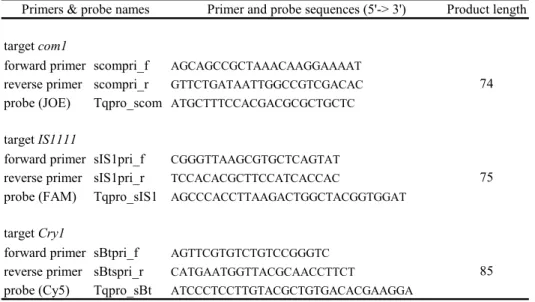
For targets com1 and IS1111, shorter primers and new (hydrolysis) probes were designed using Visual OMP 6 for simultaneous detection (see Table 2.2). For each target, probes were labeled with a different fluorescent label. For target
com1, the probe was labeled with (JOE), like in the 4-target qPCR assay. For
target IS1111, the probe was labelled with FAM. This dye is more fluorescent than dye CFR590, which was used to label the probe for IS1111 in the 4-target
qPCR. The probe for the internal control target Cry 1 was labelled with Cy5, like in the 4-target qPCR assay. All probes were additionally labeled with Black Hole Quencher 1 (BHQ-1).
Primer and probe sequences (5'-> 3') Product length target com1
forward primer scompri_f AGCAGCCGCTAAACAAGGAAAAT reverse primer scompri_r GTTCTGATAATTGGCCGTCGACAC probe (JOE) Tqpro_scom ATGCTTTCCACGACGCGCTGCTC target IS1111
forward primer sIS1pri_f CGGGTTAAGCGTGCTCAGTAT reverse primer sIS1pri_r TCCACACGCTTCCATCACCAC
probe (FAM) Tqpro_sIS1 AGCCCACCTTAAGACTGGCTACGGTGGAT target Cry1
forward primer sBtpri_f AGTTCGTGTCTGTCCGGGTC reverse primer sBtspri_r CATGAATGGTTACGCAACCTTCT
probe (Cy5) Tqpro_sBt ATCCCTCCTTGTACGCTGTGACACGAAGGA
85 74
75 Primers & probe names
Table 2.2. Primers and probes for each target developed in Visual Omp 6 for the 3-target multiplex qPCR for C. burnetii. Primer and probe sequences and product lengths obtained are given for C. burnetii targets com1, IS1111, and B.
thuringiensis target cry1.
To test the new qPCR assay, genomic DNA of C. burnetii was used as DNA template (starting material). DNA of C. burnetii strains: Nine Mile RSA phase I (RSA493), and isolates from mouse spleen (EP3, Russia, 1958, Apodemus
flavicollis), and tick (EP5, Slovakia, 1968, Dermacentor marginatus) were used
in these experiments.
2.6.1 Sensitivity assessment of the multiplex qPCR assays
The minimal number of copies per target that can be detected with a 95% probability was determined using probit analysis. Standard curves for targets
icd, com1, and IS1111 were derived from a conventional PCR assay using the C. burnetii Nine Mile RSA493 (RSA phase I) strain as template. Target sequences
were amplified with primers described in Table 2.3. Primers for each target were designed using Visual OMP 6, and were chosen to include the target sequences amplified using both multiplex real-time PCR assays.
Sequence (5'-> 3') Product length target icd
forward primer icdtrg_f CGGAGTTAACCGGAGTATCCA
reverse primer icdtrg_r CCGTGAATTTCATGATGTTACCTTT
target com1
forward primer comtrg_f CCCTGCAATTGGAACGAAG
reverse primer comtrg_r GTTCTGATAATTGGCCGTCGACA
target IS1111
forward primer IS1trg-f AGAATTTCTATTTTCAAAAAAAGGAGAAG
forward primer IS1trg-r CGGTTCAACAATTCGGTATACAAACAA
Primers & probes
738
775
605 Table 2.3. Primers developed in Visual Omp 6 for conventional PCR, which will be used to derive standard curves for sensitivity assessment by probit analysis. Primer and probe sequences and product lengths obtained are given for C.
burnetii targets icd, com1, and IS1111.
PCR products for each of the three targets were cleaned using the Qiaquick PCR Purification Kit (Qiagen, Venlo, the Netherlands). PCR products were visualized on a 2% agarose gel electrophoresis using SYBR-GOLD. The concentration of cleaned PCR products was measured using the Nanodrop 1000 (Isogen Life Science, De Meern, The Netherlands), and the number of copies per µl was calculated for each target. A two-fold dilution series containing 102 to 100 copies of each target per reaction was used in three separate multiplex real-time PCR runs. In each run, eight replicates for each dilution series were used and the number of successes was scored for each target for all dilutions. The
minimal number of genome equivalents per reaction that could be detected with a 95 % probability for each target was established by probit analysis using the statistical software package SPSS v.15.
For the 4-PCR assay, the proportion of PCR successes for each of the three targets was scored for each dilution. For each target, the detection probability was calculated from the proportion of positive PCRs observed. For the 4-target qPCR assay, the minimal number of genome equivalents per reaction that could be detected with a 95% probability by real-time PCR was 5.0 for target icd, 4.5 for target com1, and 3.7 for target IS1111. Sensitivity analysis for the 3-target qPCR assay will be performed in 2010 and published together with results of the 4-target qPCR PCR in a scientific paper.
2.6.2 Specificity of the multiplex qPCR assays
Both multiplex qPCR assays were tested on a large panel of non-target
organisms to verify any cross reaction with other (closely related) species. These non target organisms include: Bacillus thuringiensis subsp. kurstaki, B.
thuringiensis subsp. aizawai, B. thuringiensis var. galleriae, B. thuringiensis
Berliner 1915 AL (ATCC 10792), B. cereus Frankland 1887 AL (ATCC 14579), B.
cereus, B. mycoides Flügge 1886 AL (ATCC 6462), B. mycoides (3 isolates), Yersinia pseudotuberculosis (3 isolates), Y. agglumerans, (3 isolates), Y.
frederiksenii, Y. enterocolitica (6 isolates), Klebsiella pneumonite, Pseudomonas aeruginosa (ATCC 15442), Legionella pneumophila serotype 1 (ATCC 33152), L. bozemonii ATCC 33217), L. longbeachae (ATCC 33462), L. micdadei (ATCC
33218), L. dumoffii (ATCC 33279), L. anisa (ATCC 35292), Rickettsia
heilongjiangii, R. akari (ATCC VR-148), R. Helvetica, R. typhi (ATCC VR-142), R. monacensis, R. africae, R. conorii (ATCC VR-613), R. honei, and R. prowazekii.
No cross reactions were observed with any of these species, using both qPCR assays for C. burnetii DNA.
2.6.3 Efficiency of the multiplex qPCR assays
The efficiency curve of a qPCR assay provides information on the amplification performance of the assay. The efficiency of a qPCR assay is calculated from a standard curve, in which the concentrations of standard samples (known DNA concentrations) are plotted against the crossing points of the samples (output of qPCR analyses Software). The X axis of the standard curve represents the log of the initial DNA concentration, and the Y axis represents crossing point values in cycles. The slope of this standard curve is referred to as the efficiency of the curve. A perfect amplification reaction would produce a standard curve with an efficiency of “2”, because the amount of target DNA would double with each amplification cycle. In reality, reactions often have a lower efficiency.
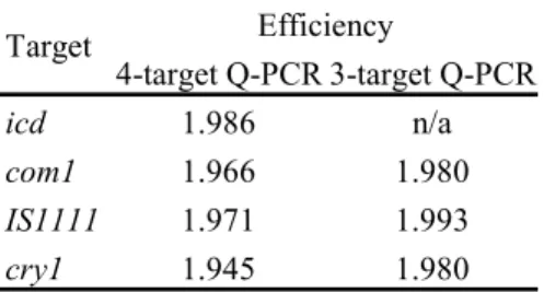
For both 4-target and newly developed 3-target qPCR assays for C. burnetii, the efficiency for each target was calculated from the standard curves. A dilution series of 107-100 copies per reaction for each target was plotted against the crossing point values. The slope for each target was calculated by linear
regression, indicating the efficiency for that target. Results can be found in table 2.4. The efficiency of both qPCR assays was very high, just below ‘2’. The efficiency for all targets was marginally higher for the new developed 3-target qPCR assay. 4-target Q-PCR 3-target Q-PCR icd 1.986 n/a com1 1.966 1.980 IS1111 1.971 1.993 cry1 1.945 1.980 Target Efficiency
Table 2.4. Efficiency of the 4-target qPCR and 3-target qPCR assays, calculated from standard curves.
3
Molecular typing of Coxiella burnetii
Genes or regions within the genome selected for detection assays are optimally highly conserved between strains, for it is important to detect any strain of the organism of interest. Genes or regions within the genome selected for molecular typing should vary among strains. Distribution patterns of strain types are very important in source finding, establishment of transmissions routes, or
epidemiological studies. A specific region within the (C. burnetii) genome is called a locus (plural loci). Two different molecular typing methods have been developed for C. burnetii: multi-locus variable number of tandem repeats analysis (MLVA) as described by Svraka et al 2006 and Arricau-Bouvery et al. 2006, and multi locus sequence typing (MLST) as described by Glazunova et al. 2006.
3.1 Multi-locus Variable number of tandem repeats Analysis
The molecular (PCR based) typing technique, called Multi-Locus Variable number of tandem repeats Analysis (MLVA) is based on the variety between C. burnetii strains in specific loci, which contain so called tandem repeats. These tandem repeats are sequences that contain a repetitive element (repeats) of about 6-10 bases, aligned next to each other. C. burnetii strains may differ in the number of repeats within a specific locus, and a number of different loci can be combined to obtain specific MLVA types.
A MLVA type can be assigned to a specific strain. A MLVA assay for C. burnetii has been developed at RIVM in 2006 by Svraka et al., in which sixteen C.
burnetii isolates and five passage history/laboratory variants were characterized
using 7 loci (22). Analyses of complex environmental and animal samples without prior cultivation however have never been attempted. This is of great importance in source finding investigation, and insights on the impact of the various transmission routes of C. burnetii.
Another more extensive MLVA assay was developed by Arricau-Bouvery et al in 2006. This MLVA assay uses 17 loci, of which the 7 loci developed by Svraka et al. 2006 are incorporated.
As typing requires more and higher quality DNA than for detection, MLVA was only attempted in our experiments on clear positive samples. These samples originate from three different sources: (1) from placenta materials tested positively by the GD using methods based on histology, (2) DNA from three C.
burnetii strains typed by Svraka et al. 2006, and (3) qPCR positive samples from
source finding investigations in 2008.
MLVA PCR assays, for each of the seven loci, were carried out on a PCR-express machine (Thermo-Scientific, Breda, the Netherlands). The separation of PCR fragments was performed on an ABI 3700 DNA sequencer (Applied Biosystems, Foster City, CA) using the standard GeneScan module. The GeneScan data were imported into Genemarker and Bionumerics 4.0 software package (Applied Maths) for analysis.
We obtained exactly the same results as Svraka et al. 2006 in typing DNA of the three C. burnetii strains (177, 180 & 182) still available at RIVM, and all three strains showed different MLVA types.
In addition, two C. burnetii positive goat placenta’s (1040 & 1050) provided by the GD were also successfully typed, and showed identical MLVA patterns, but differed from the three C. burnetii strains. Finally, nineteen C. burnetii positive vaginal swab samples from the source finding investigation in 2008 were
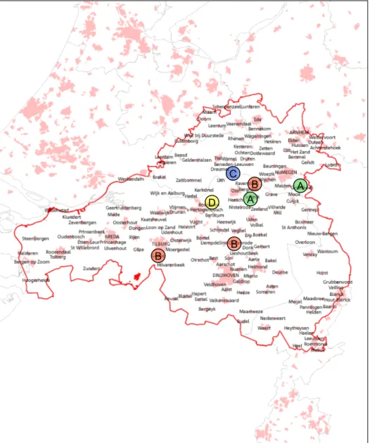
selected for molecular typing using MLVA. Results are summarised in Table 3.1. Unfortunately, many of the samples failed to produce robust MLVA patterns for all markers. Markers which did not produce reliable results are shown by -. MLVA typing of these 19 samples using the 7 MLVA markers revealed that the number of variant alleles per locus varied between 1 and 3. Based on these 7 loci, 4 different types (A-D) within the 19 samples were found with not more than 1 type per location (Table 3.1 & Figure 3.1).
Location Sample number Locus 1 Locus 2 Locus 3 Locus 4 Locus 5 Locus 6 Locus 7 Type:
1 8 4 3 4 3 - 3 A 2 8 4 3 4 3 - 3 A 1 7 - 3 - - - - B 2 7 4 3 - 3 - 3 B 3 7 - 3 - - - 3 B 4 7 4 3 - 3 - 3 B 5 7 4 3 - - - - B 6 7 - 3 - - - 3 B 7 7 4 3 - - - 3 B 1 7 - 3 - - - 3 B 2 7 4 3 - 3 - 3 B 1 - - 3 - - - 3 B 2 - - 3 - - - - ? Schaijk (Dhs5) 1 8 4 3 - - - - A 1 7 - 3 - - - 2 C 2 8 - 3 - - - - ? 3 - - 3 - - - - ? 1 2 - 3 - - - 2 D 2 - 4 3 - - - 2 ?
Goat Placenta 1040 n/a 7 3 4 3 3 9 3 E
Goat Placenta 1050 n/a 7 3 - 3 3 9 3 E
C. burnetii strain 177 (NM) n/a 5 4 4 9 6 8 5 F C. burnetii strain 180 n/a 3 3 18 2 3 3 2 G C. burnetii strain 182 n/a 3 3 13 2 3 3 5 H
# of variants: 5 2 4 4 1 3 3 Oijen Rosmalen Groesbeek Herpen St. Oedenrode Goirle Table 3.1. Results of MLVA typing of 19 positive vaginal swab samples from 7 different locations in the Netherlands revealed four different MLVA types. Not classified types are indicated by symbol ‘?’. Only a single type was found per location.
In an earlier report, we described the difficulties encountered when especially environmental samples were used for molecular typing of C. burnetii via MLVA. It became clear that typing of C. burnetii in environmental samples using MLVA is hampered by (1) inhibiting substances in environmental and animal matrices, (2) low amounts of C. burnetii DNA (for typing) and most importantly (3) background DNA in the matrix material originating from other organisms than C.
burnetii that can bind to the selected primers. To increase the amount of C. burnetii DNA for more robust typing we applied a technique called: Whole
A A B B B D C A A B B B D C
Figure 3.1. Locations of farms selected for molecular typing. Four different MLVA types were found, indicated by A (green), B (red), C (blue), and D (yellow).
3.1.1 Whole Genome Amplification in environmental & animal samples
In Whole Genome Amplification (WGA), the total amount of DNA in a sample is amplified. This can increase the robustness of molecular typing if enough C.
burnetii target DNA is amplified within a sample for MLVA. The method is based
on Multiple Displacement Amplification (MDA) technology, which carries out isothermal genome amplification utilizing a uniquely processive DNA polymerase (Phi29). This DNA polymerase is a “high processivity” polymerase, which
remains attached to template DNA far longer than typical Taq polymerases. Additionally, accurate loci and allele representation is maintained due to 3’→5’ exonuclease proofreading activity. The Qiagen Repli-g Midi Kit is developed for (whole genome) amplification of human genomic DNA, however, the application in our lab is to increase C. burnetii DNA for robust MLVA analysis from complex environmental samples. To test his, positive samples were
subjected to a whole genome amplification procedure, using the Qiagen Repli-g Midi Kit according the manufacturer’s protocol. Samples were tested before and after WGA by qPCR to investigate if C. burnetii DNA was successfully increased. This procedure was described in an earlier report and results showed that not all environmental and animal samples showed an increase in C. burnetii content. An increase of C. burnetii DNA was detected in some samples obtained from a Q fever affected farm, however, only DNA of C. burnetii in 2 environmental samples (swabs from a milk unit filter & milk unit) was increased sufficiently for molecular typing using all 7 loci.
All four successfully typed samples showed a different MLVA type, which is quite unlikely for a single location. The variability of circulating C. burnetii strains in the Netherlands is not yet known, however, further testing and optimization of the MLVA assay for typing from environmental and animal samples is necessary. As mentioned earlier, cultivation of the organism of interest from clinical,
environmental, or animal sources is often used as a first step in order to isolate pure and high quality target DNA prior to molecular typing. Our results show that unbiased amplification of DNA from environmental and animal matrices by means of WGA does not always solve all the challenging aspects of robust MLVA-typing. However, a large number of C. burnetii positive vaginal swabs obtained during source finding investigations will be used in WGA procedures, before the cultivation of C. burnetii from environmental and animal samples will be
attempted. Cultivation of C. burnetii is laborious, requires BSL- 3 conditions, and is therefore costly.
3.1.2 Optimization of the MLVA assay
The current procedure for MLVA typing (22) is quite cumbersome. Typing 7 MLVA loci requires 7 different primers sets with corresponding annealing temperatures, and thus 7 different PCR reactions. In 2009 we became involved in another Q fever project, in which we were able to participate in the
development of a multiplex MLVA assay for C. burnetii. Klaassen et al 2009 (26), developed 2 multiplex MLVA PCR assays, in which 5 MLVA markers developed by Svraka et al. 2006 and 1 MLVA marker developed by Arricau-Bouvery et al. 2006 are incorporated. The first multiplex MLVA PCR assay (Q6) incorporates markers Cox 1, Cox 2, and Cox 5 developed by Svraka et al. 2006. The second multiplex MLVA PCR (Q7) incorporates markers Cox 4, and Cox 6 developed by Svraka et al. 2006 and marker MS 33 developed by Arricau-Bouvery et al. 2006 (see table 3.2). This new MLVA assay will be used, together with WGA
procedures, to investigate the number of different C. burnetii strain present on sheep and goat farms in the Netherlands.
Marker Labeled primer sequence (5’-3’) Unlabeled primer sequence (5’-3’) µM Location Ms27 HEX*-TCTTTATTTCAGGCCGGAGT GAACGACTCATTGAACACACG 0.5 838421-828508 Ms28 TAMRA*-AGCAAAGAAATGTGAGGATCG GCCAAAGGGATATTTTTGTCCTTC 0.5 839703-839812 Ms34 FAM*-TTCTTCGGTGAGTTGCTGTG GCAATGACTATCAGCGACTCGAA 0.1 1471800-1471899 Ms23 HEX*-CGCMTAGCGACACAACCAC GACGGGCTAAATTACACCTGCT 0.2 197619-197750 Ms24 FAM*-TGGAGGGACTCCGATTAAAA GCCACACAACTCTGTTTTCAG 0.7 259515-259775 Ms33 TAMRA*-TCGCGTAGCGACACAACC GTAGCCCGTATGACGCGAAC 1.0 1435025-1435128 Multiplex Q6 Multiplex Q7
Table 3.2. Primer sets for typing of C. burnetii, using 6 loci in 2 multiplex MLVA PCR assays, as developed by Klaassen et al. 2009.
3.2 Multi-spacer Sequence Typing (MST)
In molecular typing using MST, sequence variation in non-coding intergenic spacer regions within the genome is used to investigate the similarity (or dissimilarity) among C. burnetii strains. This method uses the same type of molecular information as in a related molecular typing method: multi-locus sequence typing (MLST). The non-coding intergenic spacer regions, however, are thought to be under less selection pressure in comparison to the more conserved and protein coding house-keeping genes used in MLST.
In MST, 10 intergenic spacer regions are used to investigate possible
relationships between C. burnetii strains. Another advantage of this sequence based typing method is that sequence information is more robust than PCR fragment length analysis methods, like MLVA. Molecular typing methods can be hampered by the same factors as MLVA: PCR inhibiting substances, low amounts of C. burnetii DNA, and background DNA in the matrix material originating from other organisms.
In 2005 and 2006, the only available whole genome sequence of C. burnetii available was the Nine Mile RSA phase I (RSA493) strain. Sequence information of this strain was used to set up the MLVA methods by Svraka et al. (2006) and Arricau-Bouvery et al. (2006). Since then, four other whole genome sequences of C. burnetii became available and an in silico analysis was performed on the 5 genomes available to investigate if there was a difference in resolution between the two different molecular typing methods.
Using the 7 MLVA markers, developed by Svraka et al 2006, the 5 C. burnetii strains revealed 5 different MLVA types. Individual markers showed differences in resolution.
In addition, sequence information revealed that mismatches occur in some primer binding sites. For instance, in marker Cox 2, a mismatch was found for the reverse primer in one C. burnetii genome. For marker Cox 3, a mismatch was found for the forward primer in another C. burnetii genome. Finally, for marker Cox 6, mismatches were found in the forward primer for 3 C. burnetii genomes. In our typing experiments with DNA of the three C. burnetii strains, all strains revealed different MLVA patterns.
Using the 10 MST markers, in silico analyses revealed that the 5 available genomes of C. burnetii could also be distinguished into 5 different MST types. A mismatch was found for the forward primer of MST marker MST 56 in one of the
C. burnetii genomes.
Two mismatches for MST marker MST 61 were observed in the forward primer for another C. burnetii genome. In our typing experiments with DNA of the three
C. burnetii strains, only two strains could be distinguished showing different MST
types.
In 2010, further refinements of MLVA and MST primer design and experimental testing will be carried out to investigate the applicability of MLVA and MST in typing the available C. burnetii strains within RIVM, and C. burnetii positive environmental or animal matrices obtained during Q fever source finding investigations.
4
Conclusions
Coxiella burnetii DNA can be successfully detected in environmental and animal
matrices using our developed 4-target multiplex real-time PCR assay. Validation of this assay is completed.
The development of a (more sensitive) 3-target qPCR assay for C. burnetii will be completed in 2010.
Molecular typing of C. burnetii is at the moment hampered by the lack of a cultivation procedure at RIVM to obtain pure and high quality DNA of C. burnetii. Whole Genome Amplification of DNA obtained from environmental and animal samples prior to MLVA analysis does not improve results sufficiently.
The setup of BSL-3 facilities at RIVM, required for cultivation of C. burnetii, were completed in the beginning of 2010. Training procedures have started and cultivation of C. burnetii is planned to commence before June 2010.
5
Literature
1. Aboudharam, G., M. Drancourt, and D. Raoult. 2004. Culture of C. burnetii from the dental pulp of experimentally infected guinea pigs. Microb Pathog 36:349-50. 2. Arricau-Bouvery, N., Y. Hauck, A. Bejaoui, D. Frangoulidis, C. C. Bodier, A. Souriau,
H. Meyer, H. Neubauer, A. Rodolakis, and G. Vergnaud. 2006. Molecular characterization of Coxiella burnetii isolates by infrequent restriction site-PCR and MLVA typing. BMC Microbiol 6:38.
3. Boulos, A., J. M. Rolain, M. Maurin, and D. Raoult. 2004. Measurement of the antibiotic susceptibility of Coxiella burnetii using real time PCR. Int J Antimicrob Agents 23:169-74.
4. Brennan, R. E., and J. E. Samuel. 2003. Evaluation of Coxiella burnetii antibiotic susceptibilities by real-time PCR assay. J Clin Microbiol 41:1869-74.
5. Edingloh, M., C. C. Merck, and E. Manz. 1999. [Multiplex PCR for the diagnostic detection of Coxiella burnetii in cow's milk]. Berl Munch Tierarztl Wochenschr 112:5-9.
6. Glazunova, O., V. Roux, O. Freylikman, Z. Sekeyova, G. Fournous, J. Tyczka, N. Tokarevich, E. Kovacava, T. J. Marrie, and D. Raoult. 2005. Coxiella burnetii genotyping. Emerg Infect Dis 11:1211-7.
7. Guatteo, R., F. Beaudeau, A. Joly, and H. Seegers. 2007. Coxiella burnetii shedding by dairy cows. Vet Res 38:849-60.
8. Klee, S. R., J. Tyczka, H. Ellerbrok, T. Franz, S. Linke, G. Baljer, and B. Appel. 2006. Highly sensitive real-time PCR for specific detection and quantification of
Coxiella burnetii. BMC Microbiol 6:2.
9. Madariaga, M. G., K. Rezai, G. M. Trenholme, and R. A. Weinstein. 2003. Q fever: a biological weapon in your backyard. Lancet Infect Dis 3:709-21.
10. Mallavia, L. P., L. L. Whiting, M. F. Minnick, R. Heinzen, D. Reschke, M. Foreman, O. G. Baca, and M. E. Frazier. 1990. Strategy for detection and differentiation of
Coxiella burnetii strains using the polymerase chain reaction. Ann N Y Acad Sci
590:572-81.
11. Miller, J. D., A. T. Curns, and H. A. Thompson. 2004. A growth study of Coxiella
burnetii Nine Mile Phase I and Phase II in fibroblasts. FEMS Immunol Med Microbiol
42:291-7.
12. Muhlemann, K., L. Matter, B. Meyer, and K. Schopfer. 1995. Isolation of Coxiella
burnetii from heart valves of patients treated for Q fever endocarditis. J Clin
Microbiol 33:428-31.
13. Ormsbee, R. A. 1952. The growth of Coxiella burnetii in embryonated eggs. J Bacteriol 63:73-86.
14. Panning, M., J. Kilwinski, S. Greiner-Fischer, M. Peters, S. Kramme, D. Frangoulidis, H. Meyer, K. Henning, and C. Drosten. 2008. High throughput detection of Coxiella
burnetii by real-time PCR with internal control system and automated DNA
preparation. BMC Microbiol 8:77.
15. Parker, N. R., J. H. Barralet, and A. M. Bell. 2006. Q fever. Lancet 367:679-88. 16. Raoult, D., G. Vestris, and M. Enea. 1990. Isolation of 16 strains of Coxiella burnetii
from patients by using a sensitive centrifugation cell culture system and establishment of the strains in HEL cells. J Clin Microbiol 28:2482-4.
17. Rodolakis, A., M. Berri, C. Hechard, C. Caudron, A. Souriau, C. C. Bodier, B. Blanchard, P. Camuset, P. Devillechaise, J. C. Natorp, J. P. Vadet, and N. Arricau-Bouvery. 2007. Comparison of Coxiella burnetii shedding in milk of dairy bovine, caprine, and ovine herds. J Dairy Sci 90:5352-60.
18. Rousset, E., B. Durand, M. Berri, P. Dufour, M. Prigent, P. Russo, T. Delcroix, A. Touratier, A. Rodolakis, and M. Aubert. 2007. Comparative diagnostic potential of three serological tests for abortive Q fever in goat herds. Vet Microbiol 124:286-97. 19. Seshadri, R., I. T. Paulsen, J. A. Eisen, T. D. Read, K. E. Nelson, W. C. Nelson, N.
L. Ward, H. Tettelin, T. M. Davidsen, M. J. Beanan, R. T. Deboy, S. C. Daugherty, L. M. Brinkac, R. Madupu, R. J. Dodson, H. M. Khouri, K. H. Lee, H. A. Carty, D. Scanlan, R. A. Heinzen, H. A. Thompson, J. E. Samuel, C. M. Fraser, and J. F. Heidelberg. 2003. Complete genome sequence of the Q fever pathogen Coxiella
burnetii. Proc Natl Acad Sci U S A 100:5455-60.
20. Slaba, K., L. Skultety, and R. Toman. 2005. Efficiency of various serological techniques for diagnosing Coxiella burnetii infection. Acta Virol 49:123-7.
21. Stein, A., and D. Raoult. 1992. Detection of Coxiella burnetti by DNA amplification using polymerase chain reaction. J Clin Microbiol 30:2462-6.
22. Svraka, S., R. Toman, L. Skultety, K. Slaba, and W. L. Homan. 2006. Establishment of a genotyping scheme for Coxiella burnetii. FEMS Microbiol Lett 254:268-74. 23. To, H., N. Kako, G. Q. Zhang, H. Otsuka, M. Ogawa, O. Ochiai, S. V. Nguyen, T.
Yamaguchi, H. Fukushi, N. Nagaoka, M. Akiyama, K. Amano, and K. Hirai. 1996. Q fever pneumonia in children in Japan. J Clin Microbiol 34:647-51.
24. Willems, H., D. Thiele, R. Frolich-Ritter, and H. Krauss. 1994. Detection of Coxiella
burnetii in cow's milk using the polymerase chain reaction (PCR). Zentralbl
Veterinarmed B 41:580-7.
25. Zhang, G. Q., S. V. Nguyen, H. To, M. Ogawa, A. Hotta, T. Yamaguchi, H. J. Kim, H. Fukushi, and K. Hirai. 1998. Clinical evaluation of a new PCR assay for detection
of Coxiella burnetii in human serum samples. J Clin Microbiol 36:77-80.
26. Klaassen, C.H.W., Nabuurs-Franssen, M.H., Tilburg, J.J.H.C., Hamans, M.A.W.M. and Horrevorts, A.M. 2009. Multigenotype Q Fever Outbreak, the Netherlands. Emerg Infect Dis 15(4): 613–614.
Parts of this publication may be reproduced, provided acknowledgement is given to the 'National Institute for Public Health and the Environment', along with the title and year of publication.
Published by:
National Institute for Public Health and the Environment
P.O. Box 1 | 3720 BA Bilthoven The Netherlands