Nanotechnologies in medical devices
Colophon
© RIVM 2015
Parts of this publication may be reproduced, provided acknowledgement is given to: National Institute for Public Health and the Environment, along with the title and year of publication.
R.E. Geertsma (author), RIVM M.V.D.Z. Park (author), RIVM C.F. Puts (author), RIVM B. Roszek (author), RIVM R. van der Stijl (author), RIVM W.H. de Jong (author), RIVM Contact:
Robert Geertsma
Centre for Health Protection Robert.Geertsma@rivm.nl
This investigation has been performed by order and for the account of the Health Care Inspectorate and the Ministry of Health, Welfare and Sport
This is a publication of:
National Institute for Public Health and the Environment
P.O. Box 1 | 3720 BA Bilthoven The Netherlands
Publiekssamenvatting
Nanotechnologie in medische hulpmiddelen
Nanotechnologie wordt steeds meer gebruikt voor medische
hulpmiddelen. Talrijke medische disciplines profiteren van de innovaties die nanotechnologie mogelijk maakt. Ook neemt de kennis over hoe je de veiligheid van nanotechnologie moet beoordelen toe. Recente wetenschappelijke leidraden geven aan waarop moet worden gelet als nanotechnologie wordt gebruikt bij de fabricage van een medisch hulpmiddel. Kennis en leidraden vormen daarmee een goede basis om de risicobeoordeling van nanomedische hulpmiddelen uit te voeren. Dit blijkt uit een overzicht van het RIVM van het gebruik van
nanotechnologie voor medische hulpmiddelen.
Een van de belangrijkste trends is het gebruik van nanocoatings op allerlei implantaten. Hierdoor integreert het implantaat beter met het omliggende weefsel, wat de kans op afstoten of complicaties verkleint. Onder andere de cardiologie (coating op stents), orthopedie (coatings op heupimplantaat) en tandheelkunde (tandheelkundige implantaten) profiteren hiervan. Verder worden antimicrobiële eigenschappen van nanomaterialen gebruikt in coatings, voor wondverzorging en medisch textiel.
Nanomaterialen kunnen ook natuurlijke weefsels nabootsen. Met behulp van nanotechnologie kunnen bij implantaten optimale biologische, fysische en mechanische eigenschappen worden gerealiseerd. Een derde trend hangt samen met de elektrische en magnetische eigenschappen van nanomaterialen. Deze worden vooral gebruikt in medische hulpmiddelen voor neurologie en cardiologie, bijvoorbeeld om hartritmestoornissen beter te verhelpen. Ook kunnen batterijen met een langere levensduur worden ontwikkeld voor implantaten.
Een specifieke toepassing van nanotechnologie is oncologie. Voorbeelden zijn testen om kanker vroegtijdig op te sporen en
hulpmiddelen om de grenzen te bepalen van tumoren of uitzaaiingen te detecteren tijdens een chirurgische ingreep. Ook kunnen nanomaterialen door lokale temperatuurverhogingen het effect van chemotherapie of bestraling versterken, of zelfs direct tumorcellen doden.
Net als bij alle medische producten moet de risicobeoordeling van nanomedische hulpmiddelen per product worden uitgevoerd. De kans dat een nanomateriaal vrij beschikbaar komt in het lichaam, bepaalt hoe diepgaand de ‘nano’ risicobeoordeling moet zijn.
Kernwoorden: nanotechnologie, nanomateriaal, medisch hulpmiddel, klinisch nut, risicobeoordeling
Synopsis
Nanotechnologies in medical devices
The application of nanotechnologies in medical devices is a growing area and numerous medical disciplines benefit from innovative features enabled by nanotechnologies. Knowledge about the safety evaluation of nanotechnology is also evolving. Recently, scientific guidance has become available, specifying considerations to be taken into account when nanotechnology is used for the manufacture of a medical device. The combination of knowledge and guidance forms a suitable basis for the risk assessment of nanomedical devices. These are the main conclusions of an overview performed by RIVM on applications of nanotechnology in medical devices.
One of the most important types of nanotechnological applications is nanocoatings, which increase biocompatibility and thus improve
integration with the surrounding tissues of a variety of medical implants used, for example, in cardiology (stent coating), orthopaedics (coating on joint replacement implants) and dentistry (dental implants). In addition, antimicrobial properties of nanomaterials are used in coatings, and also in wound care and medical textiles.
Another clear trend is the use of nanomaterials to mimic naturally occurring structures. This leads to optimal biological, physical, and mechanical characteristics of implants.
A third trend of applications is related to the electrical and magnetic properties of materials on the nanoscale. This is especially relevant to medical devices used in neurology and cardiology, for instance to improve the treatment of cardiac arrhythmia. Furthermore,
nanotechnologies enable the development of batteries with greatly increased lifetime for use in active implantable medical devices. A number of nanotechnology applications are specific to oncology. Examples include diagnostic tests used in the early detection of cancer, and devices for the identification of the boundaries of a tumour or metastases during surgical interventions. Nanomaterials can also enhance the effect of therapies like chemotherapy or radiation therapy through locally increased temperature, or they can kill tumour cells directly at high temperature.
Like all medical products, the risk assessment of nanomedical devices needs to be performed on a case-by-case basis. The potential for release, leading to a higher or lower exposure to nanomaterials, is considered the most important feature driving the extent of the “nano” risk assessment.
Keywords: nanotechnology, nanomaterial, medical device, clinical benefits, risk assessment
Contents
Summary—9 1 Introduction—11
2 Definition of nanotechnologies in medical devices—13 3 Methods—15
3.1 Literature study—15
3.2 US Food and Drug Administration and Health Canada medical device
databases—15
3.3 Clinical trial databases—15
3.4 Patent search—16
4 Medical devices enabled by nanotechnologies—19
4.1 Cardiology—19 4.2 Dentistry—29 4.3 Neurology—36 4.4 Oncology—41 4.5 Orthopaedics—46 4.6 Surgery—50
4.7 Textiles and wound care products—53
4.8 Overall analyses of clinical trials and patents—59 5 Risk assessment considerations—63
6 Discussion and conclusions—69 References—73
Summary
The RIVM conducted an investigation to provide insights into and an overview of the field of medical devices using nanotechnologies, for products already on the market and for those expected within five years. This investigation was performed at the request of the Dutch Health Care Inspectorate and the Dutch Ministry of Health, Welfare and Sport. In order to obtain an overview of medical devices using
nanotechnologies, the following sources were used: scientific literature (retrieved using Pubmed and Scopus), databases on medical devices including the 510(k) Premarket Notification database of the US Food and Drug Administration (FDA) and the Health Canada Medical Device
database, clinical trial databases such as ClinicalTrials.gov and the International Clinical Trials Registry Platform of the World Health Organization, and the database of the European Patent Office. In addition, European and international guidance documents of regulatory bodies on nanomaterials and nanotechnologies were consulted.
The size range of approximately 1 nm to 100 nm is commonly used to define nanomaterials and nanotechnologies. This is an arbitrary choice, with no clear scientific argumentation. In this report, the size range was not restricted to the upper limit of 100 nm, but included applications based on structures up to 1000 nm. This is in line with the approach followed by regulatory bodies like the European Medicines Agency, FDA, and Health Canada.
The application of nanotechnologies in medical devices is a growing area and numerous medical disciplines benefit from innovative features enabled by nanotechnologies.
A number of general trends can be identified with regard to the application of nanotechnologies in medical devices and their benefits. One of the most important types of applications is nanocoatings and/or surface modifications which create an increased biocompatibility and thus improved integration with surrounding tissues of a variety of medical implants used, for example, in cardiology (e.g. stents, catheter balloons), orthopaedics (e.g. joint replacement implants) and dentistry (e.g. dental implants). In addition, the antimicrobial properties of
nanomaterials are used in coatings as well as in wound care and medical textiles.
Another clear trend is the use of nanomaterials to mimic naturally occurring structures. This feature is applied in dentistry and in orthopaedics for both dental and bone filler materials, but also for applications in cardiology, neurology and wound care. It yields optimal biological, physical, mechanical properties, and for dental fillers it also improves aesthetic characteristics.
Especially relevant to neurology and cardiology, a third trend of
nanotechnological applications is related to the electrical and magnetic properties of materials at the nanoscale. Future nanosized applications may provide a significantly improved bioelectrical interface between a
device and the surrounding neural tissue. Furthermore,
nanotechnologies enable the development of batteries with greatly increased lifetime for use in active implantable medical devices such as pacemakers and cardioverter defibrillators.
A number of nanotechnology applications are specific to oncology. Examples include diagnostic tests used in the early detection of cancer, and devices used for the identification of the boundaries of a tumour or metastases during surgical interventions. Nanomaterials can enhance the effect of cancer therapies like chemotherapy or radiation therapy when injected into a tumour and placed in an alternating magnetic field, generating an increase in temperature (hyperthermia). A high increase in temperature damages cell structures, resulting in cancer cell
destruction (thermoablation).
At the moment, knowledge is evolving on the safety evaluation of nanomaterials in general, and thus also when used in medical devices. Current state-of-the-art guidance provides a suitable base for
performing the risk assessment of nanomedical devices. Nanomaterials exhibit specific characteristics that may or may not lead to toxic effects. As for all medical products, risk assessment of nanomedical products needs to be performed on a case-by-case basis. The potential for release, leading to a higher or lower exposure to nanomaterials, is considered the most important feature driving the extent of the risk assessment to be performed.
1
Introduction
It is widely anticipated that innovative medical applications of
nanotechnologies will have a profound impact on health care in the near future (Bleeker et al., 2015). New opportunities will become available for the diagnosis, treatment, monitoring and prevention of disease. These nanotechnology applications relate to both medicinal products and medical devices. This paper focuses on nanotechnology applications for medical devices.
Nanotechnology applications in the field of medical devices span a wide range of extremely diverse products, technologies and application areas. Their intended use can be for therapy, diagnosis, monitoring or
prevention of disease. Devices can be non-invasive or invasive,
contacting any kind of tissue. Nanomedical devices can involve the use of nanomaterials, however, nanotechnologies also enable innovative devices without using nanomaterials, for example by applying nano-electronics or lab-on-a-chip technologies (Bleeker et al., 2015). All medical disciplines are benefiting from nanomedical devices, especially orthopaedics, dentistry, oncology, neurology and cardiology. In addition, a number of innovations in clinical chemistry laboratories are enabled by nanotechnology (Hermsen et al., 2013).
While the advantages are highly desirable, the emergence of innovative nanomedical products also gives rise to questions whether currently used risk assessment strategies and testing methods provide a sound scientific basis for an adequate evaluation of the quality, safety and efficacy of these products within the current regulatory framework (SCENIHR, 2015).
For the Dutch government, it is strategically important to be aware of new and emerging technologies, and the benefits as well as the risks they bring. Nanotechnologies are used in a large number of medical devices, however, there is no comprehensive overview of these products. The Ministry of Health, Welfare and Sports and the Health Care Inspectorate asked the RIVM to provide insights into and an overview of the field of medical devices using nanotechnologies, for products already on the market and for those expected within 5 years.
2
Definition of nanotechnologies in medical devices
The current regulatory framework for medical devices contains no specific provisions for nanotechnologies. Currently, a revision process of the regulatory framework for medical devices is ongoing. Partly based on recommendations made by the European Commission Working Group on New and Emerging Technologies in Medical Devices (NET WG, 2007), it was decided that specific provisions are warranted in relation to nanomaterials, but not to nanotechnologies in general. The European Commission has published a proposal for a revision of the medical devices legislation (EC, 2012) which includes a definition of
nanomaterial taken from Commission Recommendation 2011/969/EU on the definition of nanomaterial (EC, 2011): ‘Nanomaterial’ means a natural, incidental or manufactured material containing particles, in an unbound state or as an aggregate or as an agglomerate and where, for 50% or more of the particles in the number size distribution, one or more external dimensions is in the size range 1 nm–100 nm.
The size range of approximately 1 nm to 100 nm is commonly used in various other working definitions or descriptions of nanotechnology proposed by the regulatory and scientific community (ISO/TS 80004-1:2010; OECD, 2011). There is, however, general consensus that the upper limit of 100 nm is not based on scientific arguments, and that size-related phenomena can also occur at sizes above 100 nm (Rauscher
et al., 2015; SCENIHR, 2009). At the present time, the available
scientific information does not establish a uniform upper boundary above 100 nm where novel properties and phenomena similar to those seen in materials with dimensions in the nanoscale range cease to occur for all potential materials or end products. For this reason, the US Food and Drug Administration (FDA) finds it reasonable to consider evaluation of materials or end products engineered to exhibit properties or
phenomena attributable to dimensions up to 1000 nm, as a means to screen materials for further examination and to determine whether these materials exhibit properties or phenomena attributable to their dimension(s) and associated with the application of nanotechnology (FDA, 2014). Also according to Health Canada's working definition for nanomaterial, the term "nanoscale" means 1 nm to 100 nm inclusive. However, individual Canadian regulatory programs may request
information above the 100 nm size range to an upper limit of 1000 nm in order to maintain flexibility when assessing potential nanomaterials, including suspected nanoscale properties and phenomena (Health Canada, 2011). In Europe, the European Medicines Agency (EMA) currently states that ‘nanotechnology is the use of tiny structures - less than 1000 nm across - that are designed to have specific properties’. Based on these considerations, this report is not restricted to the use of structures with an upper limit of 100 nm, but also includes applications with structures up to 1000 nm.
3
Methods
3.1 Literature study
An electronic literature search of PubMed and Scopus was performed using relevant keywords for each particular medical specialism and medical device category, combined with nano, nanotechnology and nanomaterial. Relevant reviews and research articles were used to identify current products and potential short-term future developments within the different categories. Internet searches using Google were performed to find
additional information for products found using other sources, e.g. to determine whether the product was actually enabled by nanotechnologies, whether the product was on the market, or whether it was in clinical investigation. Furthermore, the internet was searched for available commercial market reports with overviews of nanomedical products. Two reports were selected and purchased based on publication date and inclusion of data on nanomedical devices in addition to nanomedicinal products.
3.2 US Food and Drug Administration and Health Canada medical device databases
The 510(k) Premarket Notification database (FDA) was accessed on April 13, 2015 to search for medical devices containing nanotechnology. Two search strategies were employed. First, a search for “nano” in the search field “Device Name” resulted in 65 hits. Second, a search for “nano” in the search field “Applicant” resulted in 45 hits. All other fields were left empty. The hits were exported to MS Excel. All hits were manually checked via internet searches to confirm the use of nanotechnology within the product. The FDA confirmed that their 510(k) Premarket Notification database is their only database that enables a broad search on nanomedical devices, and that our approach was the best method to extract data on nanomedical devices from the database [FDA, personal communication].
The Medical Device Active Licence Listing database (Health Canada) was accessed on April 14, 2015 to search for medical devices containing nanotechnology. The database was searched for “nano” in the search option “Device Name”, resulting in 119 hits. The hits were exported to MS Excel. All hits were manually checked via internet searches to confirm the use of nanotechnology within the product.
3.3 Clinical trial databases
A clinical trial web search was conducted using the databases
ClinicalTrials.gov of the US National Institute of Health (ClinicalTrials.gov), International Standard Randomised Controlled Trial Number (ISRCTN), and International Clinical Trials Registry Platform of the World Health Organization (ICTRP WHO). The ICTRP WHO database includes several databases, including the ClinicalTrials.gov database. All databases were accessed on April 2, 2015. The search term “nano” was used for the search in the ClinicalTrials.gov and ISRCTN databases. In the ICTRP WHO database “nano*” was used (search period 2002 - March 2015). The web search resulted in 81, 3, and 510 clinical trials in the ClinicalTrials.gov,
ISRCTN, and ICTRP WHO databases, respectively. Information on these trials was downloaded to exclude non-medical device trials such as drug trials or radiopharmaceutical trials, duplicate trials, and trials that did not actually involve nanotechnology. Using the trial identification number, each trial was retrieved from the corresponding database and the information was assessed manually. In total, 115 clinical trials were identified. Selected clinical trials were assessed to add a medical speciality, e.g. dentistry, oncology, orthopaedics etc., for each trial. Subsequently, the resulting information was analysed using IBM SPSS Statistics (IBM Corporation, USA; version 22).
3.4 Patent search
The database of the European Patent Office (Espacenet) was used to search for nanomedical device patents in the European patent (EP) collection, including full text of European published patent applications. Searching for European patents, instead of worldwide patents, was chosen because of a focus on European nanomedical devices and due to time constraints. It is likely that searching in the worldwide database (Espacenet worldwide collection of 90+ countries) would have resulted in more patents found, however this result would also include patents that are not valid in any of the European member states. The Espacenet EP collection was searched per nanomedical device category using the following four search strategies:
1. International Patent Classification (IPC) symbols for medical device category combined with IPC classification symbol for nanotechnology;
2. IPC classification symbol for nanotechnology combined with keywords of medical device category in title or abstract;
3. IPC classification symbols for medical device category combined with keywords related to nanotechnology in title or abstract; 4. keywords of medical device category in title or abstract combined
with keywords related to nanotechnology in title or abstract. IPC classification codes were obtained from the World Intellectual Property Organization (WIPO) by searching for keyword terms. A
keyword search of the title and abstract, instead of full text, was used to obtain specificity. The term “medical” (in full text) was used in certain categories to obtain specificity for medical applications. Espacenet has a limit of 10 keywords and 1000 wildcard entries. Search results were exported in XLS, subsequently combined, and duplicates were removed based on publication number. Withdrawn, lapsed or non-nanomedical device category patents were removed after manual examination. Patents that fit another nanomedical device category were moved accordingly. These different strategies allowed the capture of a large number of patents related to nanomedical devices. However, as the focus was only on patents related to a medical application, more general patents that could in theory be used in nanomedical devices were
excluded when this was not stated in the patent application description or claims (e.g. general material patents). Patents were included in only one category. It was difficult to determine whether patents were already being used in actual products. Manufacturers do not often state which patents are used in their products, and reversely, patent applications do not state in which products they are used. In addition, patents can be
concerned with more general technologies that can be used by a multitude of manufacturers in a different range of products.
4
Medical devices enabled by nanotechnologies
Many medical disciplines benefit from medical devices enabled by nanotechnologies, also indicated as “nanomedical devices”. This chapter provides an overview of the use of nanomedical devices in cardiology, dentistry, neurology, oncology and orthopaedics. In addition, applications related to surgery as well as textiles and wound care products are
included. Lab-on-a-chip devices for clinical diagnostics, which are often enabled by nanotechnology, are not included as a previous RIVM report was specifically dedicated to this category of medical devices (Hermsen et
al., 2013).
At the end of this chapter, analyses of the data resulting from the clinical trial databases and patent searches are included in order to provide additional insights in trends related to the various medical disciplines and recent developments.
4.1 Cardiology
Different categories of implantable medical devices contribute to the treatment of cardiovascular disease, structural heart disease, and cardiac arrhythmia. Examples of such devices that are the mainstay of (interventional) cardiology, including cardiac surgery are stents, ventricular assist devices, and pacemakers. In the following section examples of endovascular medical devices for the treatment of
peripheral artery disease are also given. Treatment of peripheral artery disease is usually performed by an interventional radiologist and not by an interventional cardiologist.
Current
Stents
Coronary stents are one of the most used implantable medical devices in the USA (Allen, 2011). Coronary stents are tubular devices placed by a catheter within a coronary blood vessel for the treatment of patients with coronary artery disease. Stents support a segment of a blood vessel or any other anatomical lumen so as to preserve or regain its patency. Stents are also used for the treatment of patients with
peripheral artery disease, e.g. blood vessel narrowing in the lower and upper leg. Several types of stents are currently on the market, such as bare metal, drug-eluting, and bioresorbable vascular scaffolds.
Nanotechnology holds great promise for these medical devices: several nanocoated bare metal and drug-eluting stents for the treatment of patients with coronary as well as peripheral artery disease are currently available (Table 4.1.1).
Diamond-like carbon is a class of material with excellent biological, mechanical, and tribological properties. It can be deposited on many substrates such as body fluid contacting parts of implantable medical devices. Coating technologies such as Carbofilm™ (coating thickness ≤500 nm), iCarbofilm™ (also known as Bio Inducer Surface coating, <300 nm) or the diamond-like carbon coating (average thickness 35 nm) used by Japan Stent Technology Co Ltd (Japan) bring similarity to
the diamond structure of pure carbon and its exceptional
biocompatibility and haemocompatibility. The haemocompatibility of diamond-like carbon coatings is attributed to their hydrophobicity and surface smoothness. A modified coating technology is the Dylyn™ technology, which is a diamond-like nanocomposite-coating containing Si:O. The major limitation of diamond-like carbon coatings is the microcracks that can form on the surface of metal substrates.
The Inert Carbon Technology (ENDOCOR GmbH, Germany) is a surface modification created by high speed bombardment of carbon ions under vacuum conditions onto the stent’s surface. The carbon ions are
implanted within the metal lattice under the stent surface. The depth of implantation of carbon ions is 50 nm. The Inert Carbon Technology creates a barrier for migrating heavy metal ions such as chromium, molybdenum and nickel, which would be ideal for patients suffering from metal allergy.
Polyzene®-F surface technology from CeloNova Biosciences, Inc (USA) is an advanced surface modification that is thrombo-resistant, promotes rapid endothelialisation. Polyzene®-F is a nano-thin (≤50 nm) polymer coating applied to bare metal stents, helping physicians to achieve improved efficacy without the burden of long-term anti-platelet therapy. The Camouflage® nanocoating (100 nm) developed by eucatech AG (Germany) is a modified heparine. It is a synthetic glycocalyx covalently bonded to the stent surface. The Camouflage® nanocoating leads to masking of the stent surface which prevents pathological reactions triggered by foreign surfaces and provides biomimicry of the arterial glycocalyx.
A proprietary process has been developed by Hexacath (France) to coat titanium-nitride-oxide on the surface of the stent, based on a plasma technology using the nanosynthesis of a prespecified gas mixture of nitrogen and oxygen and metal. This plasma-enhanced vapour
deposition of titanium in a vacuum chamber can be applied to different metals, for instance stainless steel, cobalt-chromium-cobalt or nickel-titanium alloys. This coating is extremely dense and hard, making titanium-nitride-oxide coated stents extremely well adapted for direct stenting.
Supercritical fluid-based techniques are applied to the production of nanoparticles, nanofibres, nanowires, nanotubes, nanofilms and nanostructured materials (Fulton et al., 2003) and result in
nanomaterials with potentially better performances. Supercritical fluid technology is used to apply to the electrostatic coating process, resulting in the deposition of dry powder, crystalline hydrophilic drugs, e.g.
sirolimus, onto the stent surface. Because the drug is never dissolved in solvent, the crystal structure is maintained during application of the coating. Drug and polymer are layered onto the stent and each layer is sintered to fuse the coating into a smooth, conformal, well-adhered film. Maintaining the crystalline structure of sirolimus within the coating confers enhanced drug stability.
The ProPass™ stent is a bare metal coronary stent system with a platinum coating (250 nm) and is marketed by Vascular Concepts Ltd (UK). The platinum coating ensures microsmooth surface architecture.
Table 4.1.1 Cardiac medical device categories and examples of CE-marked devices utilizing nanotechnologies (non-exhaustive)
Manufacturer Medical device Coating Application
Bare metal stent
Alvimedica, Turkey¹ Chrono Carbofilm™ CAD
Avantgarde iCarbofilm™ CAD
Blue Medical Devices BV,
Netherlands² XTRM-FIT Dylyn™ CAD
CeloNova Biosciences Inc, USA CATANIA™ Polyzene®-F CAD
COBRA PzF™ Polyzene®-F CAD
eucatech AG, Germany CCFLex
ProActive
Camouflage® CAD
ENDOCOR GmbH, Germany³ GHOST™ Inert Carbon
Technology CAD
Hexacath, France⁴ Titan2® PEVP technology CAD
Titan Optimax PEVP technology CAD Japan Stent Technology Co Ltd,
Japan MOMO® diamond-like carbon CAD
Vascular Concepts Ltd, UK ProPass™ platinum CAD
Drug-eluting stent
Alvimedica, Turkey5 Optima Jet Carbofilm™ CAD
Cre8™ Bio Inducer Surface CAD
ENDOCOR GmbH, Germany SEQUENCE™ Inert Carbon
Technology CAD
Micell Technologies Inc, USA MiStent® SCF technology CAD
Drug-coated balloon
Eurocor GmbH, Germany DIOR®6 nanoporous shellac7 CAD
MAGICAL™8 nanoporous shellac CAD
Pacemaker lead
Biotronik SE & Co KG , Germany Safio9 fractal coating CRM
Implantable cardioverter defibrillator lead
Biotronik SE & Co KG , Germany Linox smart fractal coating CRM
Cardiac resynchronization lead
Biotronik SE & Co KG , Germany Corox OTW BP10 fractal coating CRM
Abbreviations: CAD – coronary artery disease, CRM – cardiac rhythm management, PEVP – plasma-enhanced vapour deposition, SCF – supercritical fluid.
¹ The endovascular (renal, iliac, and femoropopliteal region) portfolio of Alvimedica includes bare metal stents with Carbofilm™ coating (Radix 2) and iCarbofilm™ or Bio Inducer Surface coating (Easy HiFlype, Isthmus Logic, Easy Flype, and Inperia Advance). The Chrono and Avantgarde coronary stent systems were developed by CID Srl (Italy).
² Blue Medical Devices BV went bankrupt and was taken over by Wellinq Holding BV in 2013. XTRM-FIT is not available anymore on the website.
³ ENDOCOR GmbH commercializes GHOST PV™ a balloon expandable peripheral stent system with Inert Carbon Technology.
4 Hexacath’s endovascular portfolio includes bio active stents with titanium nitride oxide coating
for the treatment of peripheral artery disease (HeliFlexTi) and renal artery disease (HeliosSD). 5 The Optima Jet drug-eluting coronary stent system was developed by CID Srl (Italy). The
Optima Jet has reservoirs of tracolimus coated with Carboflim™. Alvimedica’s endovascular portfolio (below the knee region (BTK)) includes Cre8™ BTK with Bio Inducer Surface coating.
6 Eurocor’s portfolio includes the second generation peripheral drug-eluting dilatation catheters
FREEWAY™ 014 and FREEWAY™ 035.
8 MAGICAL™ is a GENIUS® MAGIC CC bare metal stent delivered by the DIOR® drug-coated
balloon.
9 Biotronik’s pacemaker lead portfolio includes Selox, Setrox, Siello and Solia leads. 10 Biotronik’s cardiac resynchronization lead portfolio includes Sentus OTW Quadripolar and
Sentus OTW Bipolar.
Drug-coated balloons
The DIOR® coronary balloon dilatation catheter (Eurocor GmbH, Germany) is coated with a nanoporous matrix consisting of shellac, a natural resin, and paclitaxel. In contact with body liquid, i.e. blood, the hydrophilic shellac network of the nanoporous composite swells and opens the structure for pressure-induced fast release of paclitaxel on the inflated balloon. Drug-coated balloons have already proven effective in clinical trials for the treatment of in-stent restenosis. Its coronary application may potentially be widened to complex coronary de novo lesion subsets, such as small diameter vessels, diabetes, and diffuse lesions, where the use of stents may be hampered by suboptimal results (Loh and Waksman, 2012). Drug-coated balloons are also combined with bare metal stents. For example, the MAGICAL® drug-eluting stent system (Eurocor Gmbh, Germany) represents state-of-the-art stent technology of the GENIUS® MAGIC CC bare metal stent delivered by the DIOR® paclitaxel-eluting coronary balloon dilatation catheter.
Ventricular assist devices
Ventricular assist devices were born out of a need to support clinically deteriorating patients for whom organ shortage precludes immediate transplantation. Current implantable left ventricular assist devices support the blood stream adequately. Ventricular assist devices are now used to treat patients with terminal heart failure, not only as a bridge to transplantation, but also as a bridge to recovery or destination therapy in selected patients. The VentrAssist™ (Ventracor Ltd, Australia) is a third generation pump (i.e., magnetically-elevated pump impeller) with a diamond-like carbon coating on the blood-contacting surfaces which minimizes thrombosis. VentrAssist™ was implanted in more than 400 patients worldwide before the company collapsed in 2009. Ventracor’s intellectual property was sold to Thoratec Corporation (USA). No data were identified on the structural dimensions of the diamond-like coatings. However, given the manufacturing technology, this can be considered a nanotechnology-enabled device.
Cardiac rhythm management devices
Fractal coating of implantable pacing leads (i.e. the tip of the electrode) for bradycardia therapy, tachyarrhythmia therapy, and cardiac
resynchronisation optimises the electrically active surface area of the lead. The electrochemical area of the tip is dramatically increased by tissue fluid penetrating the space between the micro/nanoporous surface of the electrode, leading to lower polarization and increased sensing capabilities. In addition, a small geometric surface and therefore a low stimulation threshold can be achieved. Fractal surface coating of the tip improves its electrical sensing and pacing properties (Safak et al., 2013). The electrode is coated with a thin layer of iridium using physical vapour deposition technology creating a cauliflower-like
structure. Biotronik SE & Co KG (Germany) is the forerunner in this field, and is the only manufacturer of fractal coated leads (Table 4.1.1). Fractal-coated temporary pacing leads can be also be used for treatment and diagnosis of arrhythmias following open heart surgery procedures (Mellert et al., 2008).
St. Jude Medical, Inc (USA) has been using giant magnetoresistive-based high-speed communication systems for its pacemakers since 2001 (NVE Corporation, 2001). Giant magnetoresistive sensors are made of sandwiches of thin films consisting of alternating nanometre thick layers of magnetic and nonmagnetic layers. Giant magnetoresistive
components are highly stable and sensitive magnetic sensors that replace the reeds of conventional pacemakers. Pacemakers must be tuned to the specific needs of each person’s body. Physicians use magnetics to tune the pacemaker from outside the body. The device that responds to the magnetic signals is usually the reed switch. Giant magnetoresistive sensors are an order of magnitude more sensitive than reed sensors, and they are solid-state devices, not mechanical. These sensors allow the pacemaker to communicate information about heart function and to receive new instructions faster and more reliably.
Future perspectives
Stents
Technologies to modify stent surfaces can be broadly classified as
nanopatterned stent surfaces/coatings, which may or may not be loaded with a drug, and nanocarriers on stents, which are based on
encapsulating drugs in nanocarriers, later coated onto stents (Arsiwala
et al., 2014).
Microporous and nanoporous stent surfaces are intriguing because of their capacity to increase drug loading and influence drug release kinetics without the need for a polymer coating. Bare metal stents with a microporous surface are, for example, Cobal+C and Cobal+CE (Relisys Medical Devices Ltd, India) and the Yukon® Choice PC (Translumina, Germany). An example of a drug-eluting stent with microporous surface is the Yukon® Choice DES (Translumina, Germany ), and an example of a nanothin microporous surface is the VESTAsync™ (MIV Therapeutics, USA). The VESTAsync™ stent is coated with hydroxyapatite (thickness 300-1000 nm) with a porosity of 40-60% in volume. The stent showed promising results in a first-in-man clinical trial (Costa et al., 2009) and a randomised clinical trial (Costa et al., 2014). A novel concept is the Nano+™ Polymer-free Sirolimus-eluting Coronary Stent System (Lepu Medical Technology Co Ltd, China) which utilises nano-sized pores on the stent surface. The nanoporous cavity serves as a drug carrier, and more than 80% surface porosity guarantees firm adhesion of the drug. The pore diameter is 400 nm (Suwannasom et al., 2015). Currently, the Nano+™ is under clinical investigation (Table 4.1.2). The Bicare™ stent (Lepu Medical Technology Co Ltd, China) has a platform identical to the Nano+™ stent, but with a dual drug elution of sirolimus and probucol. In a first-in-man study, the Bicare™ stent demonstrated absence of early adverse safety events (Yu et al., 2014). The BuMA Supreme stent (Sino Medical Sciences Technology Inc, China) is a novel (second generation) biodegradable polymer drug-eluting stent (Table 4.1.2). An
electro-grafting (eG™) base layer (poly(n-butyl methacrylate) coating) is added between the biodegradable polymer drug carrier and the metal (cobalt-chromium) stent strut. The eG™ layer (thickness 100-200 nm) secures the adhesion of the biodegradable polymer coating through
interdigitation and prevents cracking and delamination upon stent expansion. It also has the benefit of supressing corrosion and ion release from metal substrates which could contribute to a lower local inflammation response in vivo. The patented eG™ coating technology was acquired from AlchiMedics SA (France). Currently, the BuMA
Supreme stent is in clinical trial in Europe. The first generation (stainless steel) BuMA™ sirolimus-eluting stent has been approved in China. The creation of nanotopography, such as nanopillars and nanopits, on the metal struts of the stent, using radiofrequency plasma surface texturing, influences the growth and proliferation of endothelial cells. Of note, surfaces with a defined nanopatterned grid demonstrated a higher percentage of endothelial coverage, compared to responses observed on random nanostructured surfaces. Interestingly, nanostructured surfaces, when compared to microstructured surfaces, appear to afford greater adhesion of endothelial cells, lead to higher cell densities, and to enhanced adhesion and spreading.
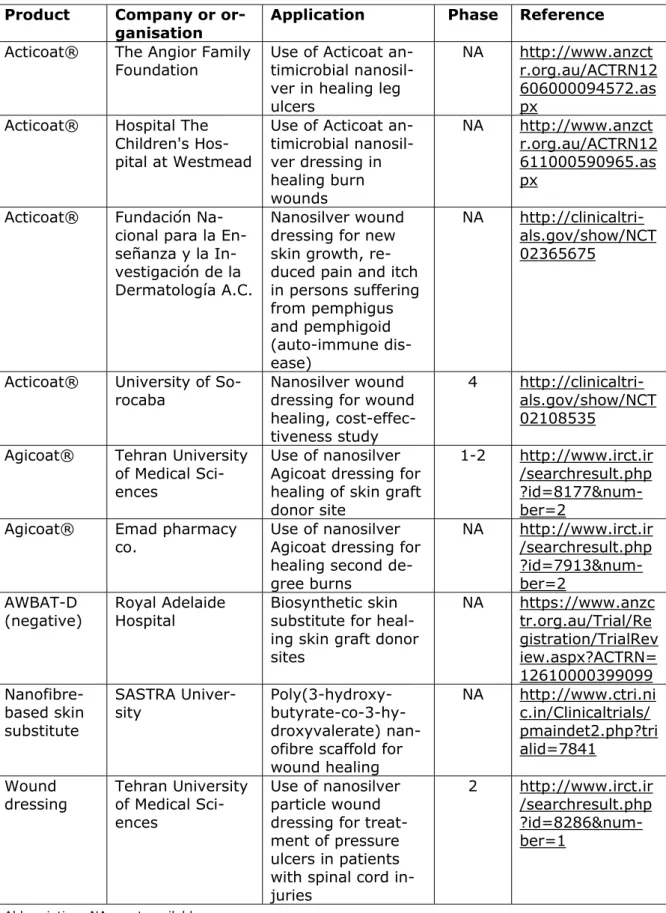
Table 4.1.2. Registered clinical trials on new medical devices Product Company or organisation Appli-cation Phase Reference Nano+
Sirolimus-Eluting Stent Lepu Medical Technology (Beijing) Co Ltd, China CAD NA http://clinicaltr ials.gov/show/ NCT01925027 BuMA Supreme Sirolimus-Eluting Stent Sino Medical Sciences Technology Inc, China CAD NA http://clinicaltr ials.gov/show/ NCT02236975 FOCUS np™ Sirolimus
Based Nano Carrier Eluting Coronary Stent System
Envision Scientific
Pvt Ltd, India CAD NA http://www.ctri.nic.in/ Clinicaltrials/p maindet2.php? trialid=3165 Magic Touch™ Nano
Sirolimus Drug Coated Balloon Catheter Concept Medical Research Pvt Ltd, India CAD NA http://www.ctr i.nic.in/ Clinicaltrials/p maindet2.php? trialid=3971 Plasmonic
photothermal and stem cell therapy (trial has been completed)
Ural State Medical
Academy, Russia CAD NA http://clinicaltrials.gov/show/ NCT01270139
Abbreviations: CAD – coronary artery disease ; NA – not available
Sub-micron rough and nanometre rough stent surfaces lead to a higher endothelial cell attachment with lesser platelet adhesion compared to stents without these features. A technology like this may even bypass the need for long-term (dual) antiplatelet therapy (Arsiwala et al., 2014).
Nanoparticles have been extensively applied in various drug delivery systems. However, few studies have reported the results of stent surfaces coated with nanoparticles. For example, an active coating of nanoparticles was deposited on the surfaces of metallic stents via cationic electrodeposition technology. Preclinical studies in animal models evaluated the feasibility of this nanoparticle-eluting stent system. It was concluded that the nanoparticle-eluting stent is a potential innovative platform exhibiting unique aspects in vascular compatibility and an efficient drug delivery system compared with the dip-coated polymer-eluting stent (Nakano et al., 2009). Another
interesting nanoparticle-mediated drug delivery system is composed of endothelial cells loaded with magnetic nanoparticles. Instead of being coated directly on the stent surface, endothelial cells were loaded first with magnetic nanoparticles and then injected into rats with stainless steel stents placed in their carotid arteries. When a magnetic field was applied, these magnetic nanoparticle-loaded cells were preferentially driven to the stented area and remained attached (Polyak et al., 2008). Although promising results have been reported, further evaluation of this technology in animal studies and clinical trials is required.
A patented nanocarrier technology (Nanoactive™) developed by Envision Scientific Pvt Ltd (India) enables encapsulation of a drug at nanometre size and can be used for hydrophilic (e.g. sirolimus) as well as
hydrophobic (e.g. paclitaxel) drugs. The NanoActive™ technology has been applied to a novel stent platform, the FOCUSnp™ Stent System (Envision Scientific Pvt Ltd). Phospholipid bilayer nanoparticles (mean diameter ~200 nm) containing sirolimus are sprayed on the polymer-free cobalt-chromium FOCUSnp™ stent after it has been crimped onto the balloon delivery catheter. A calcium phosphorous-based component is added to the nanoparticles, making them sensitive to subtle variations in pH, thus controlling the release of the drug content. Thus, the
FOCUSnp™ Stent System delivers sirolimus through nanoparticles from the (abluminal) stent strut area and the balloon. Promising results were obtained in an animal study (Takimura et al., 2015). Currently, the FOCUSnp™ is under clinical investigation (Table 4.1.2).
Using a technique called layer-by-layer (polymer) self-assembly for tunable multiple drug release in thin films (Tan et al., 2013) is another way to achieve controlled and sustained drug release. The film
architecture is precisely designed (up to 1 nm precision) and can be controlled through fabrication parameters. Layer-by-layer coatings on stents with both anti-proliferative and anti-thrombogenic drugs can mitigate problems like in-stent restenosis and stent thrombosis inherent to current bare metal and drug-eluting stents. The layer-by-layer
coating can be fine-tuned to match blood vessel healing times. In
addition, multilayers can be functionalized with bioactive molecules such as nitric oxide to reduce platelet adhesion, antibodies to encourage endothelialisation, and DNA for gene therapy. The layer-by-layer coating technique is currently in the experimental phase. No animal studies or clinical trials have yet been conducted. Issues like maintaining batch-to-batch consistency and the advantage of this technique over conventional spray coating or dip coating still has to be demonstrated.
Covered stents / endovascular stent grafts / vascular grafts
Complications associated with bare metal and drug-eluting stents, such as injury, rupture or perforation of the vessel wall and embolization of dislodged atherosclerotic plaque fragments during stent placement, have led to the development of covered stents. Covered stents usually have a (ultra-)thin (micrometre range thickness) synthetic membrane sleeve that either covers the interior lumen or the outside surface of the metallic scaffold of the stent, or completely covers the stent in a
sandwich like manner (Farhatnia et al., 2013). They can be used to treat perforations, heavy thrombus burden, and pathological conditions such as congenital vascular disease and fistulae. Covered stents used to treat aneurysms, a weak spot in an artery, are also known as endovascular stent grafts. Physicians typically use endovascular stent grafting to treat abdominal aortic aneurysms, thoracic aortic aneurysms and, less
commonly, aneurysms at other locations.
Commonly, (ultra-)thin synthetic polymers are used as covering material, for example, polytetrafluoroethylene (PTFE), polyethylene terephthalate (PET) and polyurethane (PU). More recent developments and approaches use nanomaterials to develop synthetic nanocomposite materials. For example, polyhedral oligomeric silsesquioxane (POSS) nanoparticles (~1.5 nm diameter) can be incorporated into
poly(carbonate-urea) urethane (PCU) through covalent modification (Ghanbari et al., 2011). POSS-PCU nanocomposite has shown excellent haemocompatibility with anti-inflammatory and anti-thrombogenic surface properties. Incorporation of POSS nanocage structures change the morphology of the PCU polymer, and provides a surface roughness on a nanometre scale which is more favourable for endothelial cell interactions and cellular behaviour through adhesion, growth, and proliferation than is a smoother surface profile. In addition, the functional groups of the POSS nanocage structure within POSS-PCU have the potential to be further modified by incorporation of bioactive peptides, growth factors, receptor ligands and/or antibodies with the polymeric matrix.
In recent years, other nanocomposites utilising nanofibrous bacterial cellulose, silk fibroin, carbon nanotubes and iron oxide magnetic nanoparticles have been explored for cardiovascular stent and graft applications (Vellayappan et al., 2015). Bacterial cellulose inclusion in a material was found to improve the mechanical strength and blood
compatibility of the nanocomposite. Silk fibroin improves the mechanical strength of the matrix material. Carbon nanotube inclusion improves haemocompatibility, endothelialisation and mechanical strength. When iron oxide magnetic nanoparticles are incorporated in the matrix
material, they are found to improve the antibacterial activity of the host. The company Nano4Imaging GmbH (Germany) (Nano4Imaging) has developed a proprietary technology able to modify most existing medical devices such as biopsy needles, catheters and guide wires with small markers (often metal nanoparticles), creating optimal visibility and enable navigation in multiple imaging systems including MRI, CT, X-ray, ultrasound. In 2014 they received the CE mark for an MRI-conditional guide wire which is safe to use in 1.5 and 3.0 Tesla for real-time interventions.
Electrospinning is an emerging polymer processing technique. It was discovered in the early 1900s and allows the creation of polymer nanofibres with thicknesses ranging from nanoscale to microscale. Proper selection of the processing parameters has also been shown to create polymer fibres characterised by regular structure, i.e. nanopores and nanopits (Bognitzki et al., 2001). Electrospinning offers a way to form a non-woven fabric and fabrics with complex shapes. Electrospun materials have high surface-to-weight and volume ratios, which make these materials candidates for controlled biological interactions. If biodegradable materials are used, the scaffolding is eventually absorbed.
Electrospun polymeric nanofibres are promising materials for vascular grafts, as they provide topographical and biochemical cues that resemble the extracellular matrix and constitute aligned architectures that guide cell growth and spreading. In particular, aligned nanofibrous scaffolds positively affect various endothelial cell behaviours (Du et al., 2011; Feng et al., 2010). Combining different micro- and
nanotopographic cues by complementary soft lithography and electrospinning technologies provides an interesting perspective for engineered vascular replacement constructions (Moffa et al., 2014). In general, these promising strategies consist of seeding endothelial cells in the graft lumen before implantation. Thus, potential products will not be regulated as medical devices.
Drug-coated balloons
Concept Medical Pvt Ltd (India) developed the Magic Touch™ Nano Sirolimus Drug Coated Balloon Catheter based on the patented Nanoluté™ Nanocarrier Technology. The Nanocarrier term refers to a biological excipient carrier with drug, where both the components are at nanometre size range (50-300 nm). Currently, the Magic Touch™ is in clinical trials (Table 4.1.2) and is available for in-stent restenosis, small vessels, bifurcation coronary artery lesions, and other lesions difficult to treat with stent implantation.
Plasmonic photothermal therapy and stem cells
Investigators hypothesise that plasmonic photothermal therapy using gold nanoparticles with silica-iron oxide shells in stem cells can resolve coronary atherosclerosis (Table 4.1.2). Gold nanoparticles with silica-iron oxide shells promise high-energy plasmonic photothermic burning or melting effects under the near-infrared laser irradiation, and stem cells promise restoration of the vessel wall. The results of the first-in-man trial showed that plasmonic photothermal therapy using silica-gold nanoparticles was associated with significant regression of coronary atherosclerosis (Kharlamov et al., 2015).
The efficacy of nanoshell technologies is limited by gaps in current understanding of the thermal interactions between nanoshell particles and laser light pulses or continuous waves in the context of complex biological environments. Irradiation, even with moderate pulses of energy, can induce melting, evaporation, and fragmentation of nanoparticles. These events can drastically alter the intended
therapeutic effects and lead to the formation of vapour bubbles (Lapotko, 2009).
Heart valves
Heart valve concepts are under development for the treatment of patients with structural heart disease. Several materials/techniques have been explored.
In situ tissue engineering is a promising route to create living heart
valves within the body at the site of destination allowing integration into the body (Mol et al., 2009). In this innovative approach, cell-free
electrospun scaffolds gradually transform into living substitutes that replace the affected valve. Repopulation of scaffolds in situ is most likely to occur due to a combination of cell migration from adjacent tissues and cell capture from the blood. Compared to classical in vitro heart valve tissue engineering, this technology offers off-the-shelf availability at substantially reduced costs. In addition, regulatory complexity is reduced because the scaffolds can be considered as medical devices at the time of implantation (Bouten et al., 2012).
Several other materials for heart valve leaflets have been explored, including POSS-PCU nanocomposite (Kidane et al., 2009) and bacterial cellulose-based nanocomposite (Mohammadi, 2011). It is hypothesised that synthetic heart valve leaflets fabricated from these fibre-reinforced composite materials which mimic native valve leaflet structure, will optimise leaflet stresses and decrease tears and perforations.
Cardiac rhythm management devices
Currently, pacemaker batteries last seven to ten years on average (or three to six years for an implantable cardioverter defibrillator), requiring frequent replacements which can expose patients to potential risks involved in medical procedures such as infections or severe bleeding. Enhancing battery lifetime is thus a critical issue to assure longer working time of the implanted medical device, and increase the
replacement cycle. Using advanced nanotechnology, a research team in Korea has developed a self-powered cardiac pacemaker that is operated by a flexible single crystalline piezoelectric energy harvester
(“nanogenerator”) (Hwang et al., 2014). Tested in an animal model, the nanogenerator stimulated the heart using electrical energy converted from body movements. The flexible energy harvester could lead to a robust method to enable longer operating time as well as miniaturization of batteries, because they could be readily recharged by cyclic
deformation behaviours from biomechanical energy sources such as the heartbeat or diaphragm elevation. Another research team in the USA also developed a prototype device using polymer-based piezoelectric nanoribbons that can be used to harvest power from the beating heart or to harness the motion of other organs to recharge batteries for medical devices requiring power (Dagdeviren et al., 2014).
The application of nanotechnology and nanomaterials can improve lithium-ion battery technology used for implantable medical devices such as cardiac rhythm management devices and neuromodulation devices. Today’s implantable cardioverter defibrillators use lithium vanadium oxide batteries. Nanostructured vanadium oxides, such as
vanadium oxide nanotubes/nanowires/nanorods, silver vanadium oxide nanowires, are found to have higher capacity and are considered to be the most promising cathode materials. However, they still suffer from fast capacity fading during high-rate discharge/charge. The challenge is to improve the cycling stability of vanadium oxides at a high rate. A leading supplier and developer of implantable batteries for cardiac rhythm management devices is Greatbatch Medical Inc (USA). Breath metabolomics, the study of the complex mixture of volatile organic compounds in exhaled breath, represents a new frontier in medical diagnostics. Standard volatile-compound detection methods using spectrometry and spectroscopy techniques have shown the potential for diagnosing illnesses via breath tests; this includes heart disease, cancer, and even more. Unfortunately, this approach requires expensive equipment and high levels of expertise to operate the
necessary instruments, and the tests must be done quickly, all of which impede its adoption. Nanomaterial-based sensors are likely to become a clinical and laboratory diagnostic tool because they are significantly smaller, easier-to-use, and less expensive than spectrometry or spectroscopy. Studies have demonstrated that a breath print derived from a select group of volatile organic compounds, could accurately discriminate, for instance, between heart failure patients and control subjects (Samara et al., 2013), patients with gastric cancer and other gastric disease (Xu et al., 2013), or patients with multiple sclerosis and healthy subjects (Ionescu et al., 2011). Breath analysis platforms using Bluetooth-enabled nanosensors are under development which are capable of transmitting breath print data to a smartphone for analysis and upload (Vantage mHealtcare Inc, USA). A breath print has the potential to offer patients a non-invasive and potentially inexpensive way of detecting diseases in a point-of-care setting.
Breath testing is a complex process involving numerous steps, each of which has several possible technological alternatives with advantages and drawbacks that might affect the performance of the nanomaterial-based sensors in a breath-testing system.
4.2 Dentistry Current
Dentistry is one of the few fields of medical devices in which
nanotechnology has long been used. Since the 1970s nanosized filler materials have been used in dental composites, although at that time they were called “microfilled composites” (Besinis et al., 2015).
Currently the range of dental products in which nanotechnology is used has widened (Table 4.2.1). According to an overview from FIDE, the European Dental Industry organisation, it is estimated that there are close to 3500 dental products containing nanomaterials in Europe. The main product groups reported are (>100 products estimated for the European market ordered from high to low) (Stock, 2014):
Impression materials; Dental composites; Denture base resins; Veneering materials;
Accessories for verification of occlusion; Bonding agents;
Plastic materials for bite registration; Materials for crowns and bridges; Final luting cements;
Cements; Artificial teeth.
The publicly available medical device databases from the FDA and
Health Canada report 7 and 42 dental medical devices, respectively, that have “nano” in their name (FDA; Health Canada). The 46 unique
products (3 duplicates) consist of 27 dental composites, 11 dental implants coated with NanoTite™, 4 bonding agents and 1 bone graft material. This corresponds with a recent literature review which states that fillers in dental composites and dental implants are the main application fields of nanomedical devices in dentistry (Besinis et al., 2015). The same trend can be seen in European patents with regard to dental nanomedical devices. The European patent collection contains 21 patents on dental nanomedical devices, of which 11 are related to dental composites and four to dental implants (Espacenet). Interestingly, the FIDE overview shows that impression materials are the largest group of dental medical devices containing nanotechnology, while none of these products feature in the FDA, Health Canada and Espacenet databases. Although dental composites are abundant, dental implants (or artificial teeth) are much lower on the list.
Dental composites are restorative materials used to fill cavities or reconstruct parts of a tooth. They consist of three main components: organic matrix, inorganic matrix (filler) and a coupling agent. Most of the current conventional composites have a filler particle size in the range of 40-700 nm. New and advanced composites contain nanofillers with a particle size of 1-100 nm. The most important breakthrough is not the particle size, but the increased percentage of filler present within the composite (Besinis et al., 2015). Nanofillers improve the mechanical properties of dental composites by increasing the microtensile bond strength, flexural strength, flexural modulus, fracture strength and material hardness (Besinis et al., 2015). Nanoparticles have also been used to match the radiopacity of dental adhesives, and to reduce the working and setting times for dental resins (Besinis et al., 2015).
Furthermore, these so-called nanocomposites offer aesthetic advantages in terms of smoothness, polishibility, and modified translucency to improve colour matching with the patients’ natural teeth (Besinis et al., 2015). However, there appears to be an optimum for the percentage of nanofiller, after which the mechanical properties do not improve further or even deteriorate (Atai et al., 2009; Prentice et al., 2006).
Table 4.2.1. Examples of nanomedical devices in dentistry (non-exhaustive)
Application Company Product
Bonding agent 3M Health Care Adper™
Bonding agent Pentron Clinical Technologies NANO-BOND
Cement GC Fuji IX GP
Dental composite Cosmodent Renamel NANO
Dental composite Dentsply Ceram-X®
Dental composite DMP Nanoceram Bright
Dental composite Ivoclar Vivadent Tetric EvoCeram
Dental composite Kerr Premise™
Dental composite S&C Polymer, Silicone- und Composite Spezialitäten GmbH
Zircore Flow Nano
Dental composite Tokuyama Dental Palfique® Estelite
®
Dental composite Voco GrandiO®
Dental composite 3M ESPE Filtek™ Supreme XT
Dental composite 3M ESPE Ketac™
Dental material Dental Nanotechnology S.A. NanoCare Gold Dental material Panasonic Healthcare Co. Ltd. NANOZR milling
blank
Implant surface coating Astra Tech OsseoSpeed™
Implant surface coating Biomet 3i NanoTite™
Implant surface coating Brånemark Integration AB BioHelix™
Implant surface coating NanoMech MultiCare™
NanoSpray
Implant surface coating Straumann SLActive®
Implant surface coating Intra-Lock® International Ossean®
Implant surface coating Promimic HAnano
Impression material Zhermack Nanotech Elite H-D
Root canal sealer Dentsply AH plus™
Root canal sealer BUSA® Dental
Instrumentation BC Sealer™
Veneering material GC G-Coat PLUS
Veneering material GC OPTIGLAZE
Sources: Besinis et al. (2015), FDA, Health Canada, Jain (2015), Khurshid et al., (2015)
Dental implants ideally have a high success rate and longevity. The current failure rates of 5-10% are mainly due to poor osseointegration1, infection, or rejection (Besinis et al., 2015). The success of a dental implant is determined by the inflammatory response and the behaviour of the tissue at the tissue-implant interface. As a result, the implant surface chemistry and topography are of major importance (Besinis et
al., 2015). This is where nanotechnology can make a difference, as the
body’s cells and proteins interact with the implant on a nanometre scale. Often ceramic nanomaterials, such as hydroxy apatite, bioactive glass, and other calcium phosphate compounds, are used to create coatings on metallic implants (Besinis et al., 2015). These coatings increase the biocompatibility of implants and encourage increased cell adherence.
1 Osseointegration is the direct structural and functional connection between living bone and the surface of a
However, there have also been studies that show no significant benefits of modifying an implant surface with nanoparticles. It is currently unclear whether the improved characteristics of nanocoated dental implants are due to the coating’s chemistry or due to the introduction of nanoscale surface roughness (Besinis et al., 2015). In addition, it has been suggested that particle size and morphology (e.g. rod-shaped, prism) may play a role in bone-cell adhesion and bone formation
(Mankani et al., 2001; Shi et al., 2009). This fits with current knowledge of nanoparticles, where size and morphology can have a large impact on functionality and particle properties.
In addition to these two major product groups of composites and dental implants, nanotechnology is also used in other dental product categories including, for example, bonding agents, dental materials, impression materials, root canal sealers and veneering materials (Table 4.2.1):
Bonding agents are used to adhere dental composites and other materials to the natural substance of teeth. The use of
nanotechnology ensures product homogeneity and better mixing in dental adhesive solutions (Arora and Kapoor, 2014).
The dental material NanoCare® Gold contains gold and silver nanoparticles which have antibacterial properties and, as the manufacturer claims, should hereby prevent secondary caries formation on previously treated teeth (Bednarski et al., 2013; Nanotec Endo).
Impression materials are used to make a negative imprint of hard and soft tissues in the mouth, with the goal of forming a cast at a later stage. The commercially available Elite HD+ A-silicone contains nanofillers that provide high tear, distortion and heat resistance, and an instant set to reduce movement-induced errors (Arora and Kapoor, 2014).
Root canal sealers, together with root canal fillers, seal the root canal after a root canal treatment. Sealing the root canal is important to prevent (re)infection with bacteria. Nanoparticles, composed of calcium silicate, calcium phosphate, calcium
hydroxide or zirconia used in root canal sealers provide improved handling and physical properties. The nanosized particles provide a better fit to the irregular tooth and root canal surfaces. In addition, the formation of a hydroxyapatite nanostructure in the root canal provides antimicrobial properties due to its highly alkaline pH (Khurshid et al., 2015).
Veneering materials serve as a tooth coating to improve aesthetics or to protect the tooth’s surface from damage. Nanotechnology is used to improve the aesthetic qualities, prevent discoloration, and increase the wear resistance of veneering materials.
Future perspectives
As nanotechnology in dental medical devices is already a common occurrence, what can be expected in the near future? Developments in material science will most likely have a strong impact on the dental industry, as materials and their fine-tuned properties play a major role in many dental products. This development will most likely result in increased performance of existing product types like dental composites
and implants. For example, single walled carbon nanotubes have
recently been used in a laboratory setting as fillers in dental composites due to their improved mechanical properties, high strength and unique dimensional distribution (Besinis et al., 2015). New materials are also being studied to improve osseointegration of dental implants, mainly in
in vitro studies (Arsiwala et al., 2014). There are currently 27 registered
clinical trials on dental nanomedical devices, with 17 of them starting from 2010 onwards (Table 4.2.2). From the 10 clinical trials performed prior to 2010, the products NanoTite and Ostim® (see orthopaedics) are now available on the market. For the remaining clinical trials it was not possible to determine if they resulted in or will lead to a commercial product.
The clinical trials after 2010 do not show a particular trend in application area. However, most of them are investigating the possible superior properties of nanomaterials in existing dental applications, such as increased wear resistance of nano-hybrid denture teeth or improved tissue response to nanostructured dental implants. Several clinical trials are being conducted on existing products (Ketac Nano, Venus Pearl, NANOZR), as manufacturers try to broaden the number of conditions their products can be used for. Furthermore, the clinical trial from Indiana Nanotech LLC has resulted in the development of several
Clinpro™ products, such as anti-cavity toothpaste and sealant, produced by 3M ESPE. These products contain fluoride and tri-calcium phosphate to promote dental mineral growth and thereby prevent or treat caries. Whether tri-calcium phosphate is present as a nanoparticle is not clear. Furthermore, it is uncertain whether Clinpro™ toothpaste should be considered a medical device or a medicinal product.
The oral microbiota2 strongly determines oral health or disease. Oral microorganisms play a role in dental caries formation, periodontal disease, and dental implant or reconstructed tooth infections. Therefore, preventing biofilm3 formation or removing pathogenic bacteria is
important for dentists to ensure patient health. Dental nanomedical devices that contain antimicrobial properties are a field of interest and more products are likely to enter the market in the near future. From the first use of antimicrobial nanoparticles in wound dressings, it is a relatively small step to applying this concept in dental products (Ge et
al., 2014). The abovementioned commercially available product
NanoCare® Gold already applies nanosilver and nanogold particles for this purpose. Furthermore, researchers have used nanosilver particles incorporated in dental composites to kill bacteria, which is an example of a drug/device combination product (Cheng et al., 2015; Ge et al.,
2014).
2 The oral microbiota is the ecological community of commensal, symbiotic and pathogenic microorganisms
living in the oral cavity.
3 A biofilm is a group of microorganisms living together on a surface, often embedded within a self-produced
Table 4.2.2. Registered clinical trials on dentistry nanomedical devices (2010 onwards)
Product Company or organisation
Application Phase Reference
Acrylic base plate of orthodontic removable re-tainer Hamedan Uni-versity of Medical Sci-ences Antimicrobial effect of nanosilver parti-cles for dental caries
NA http://www.irct.ir/se archresult.php?id=9 086&number=2 Ketac Nano Tanta
Univer-sity Filling cement for caries in primary teeth
NA http://clinicaltrials.g ov/show/NCT020930 91
Venus Pearl Heraeus Kulzer
GmbH Dental nanohybrid composite for dental restoration
NA http://clinicaltrials.g ov/show/NCT019250 40
Dental composite 3M Clinical perfor-mance of nano-composite CAD/CAM milled dental restoration NA http://clinicaltrials.g ov/show/NCT014642 94
Ceramic coating University of
North Carolina Reduced sliding re-sistance of ortho-dontic archwires
NA http://clinicaltrials.g ov/show/NCT023114 91
Toothpaste Indiana
Nan-otech, LLC Anti-caries tooth-paste NA http://clinicaltrials.g ov/show/NCT010942 10
Toothpaste Federal Uni-versity of São Paulo
Desensitizing toothpaste for cer-vical dentin hyper-sensitivity
NA http://clinicaltrials.g ov/show/NCT020187 83
Denture teeth University of
California Nano hybrid com-posite acrylic for denture teeth
NA http://clinicaltrials.g ov/show/NCT011882 26
Dental implant University
Ghent Nanostructured calcium-phosphate-coated implant abutment NA http://clinicaltrials.g ov/show/NCT016209 18 T3 implant
sys-tem Biomet 3i Inc Dental implant NA http://clinicaltrials.gov/show/NCT021618 74 NANOZR Universitäts-klinikum Tü-bingen, Pana-sonic Healthcare Co. Ltd. NANOZR-based ceramic material in fixed partial den-tures/crowns
NA http://www.drks.de/ DRKS00003348
Toothpaste Coswell S.p.A. Zinc-carbonate-hydroxyapatite nanocrystals for remineralisation of enamel surfaces and desensitising dentifrices NA http://www.anzctr.or g.au/ACTRN1261200 0272897.aspx