21
stEURL-Salmonella
interlaboratory comparison
study (2016) on typing of
Salmonella spp.
RIVM Report 2017-0082
W.F. Jacobs-Reitsma et al.
21
stEURL-Salmonella interlaboratory
comparison study (2016) on typing of
Salmonella spp.
Colophon
© RIVM 2018
Parts of this publication may be reproduced provided acknowledgement is given to: National Institute for Public Health and the Environment, along with the title and year of publication.
DOI 10.21945/RIVM-2017-0082
This is a publication of:
National Institute for Public Health and the Environment
P.O. Box 1│3720 BA Bilthoven The Netherlands
www.rivm.nl/en
W.F. Jacobs-Reitsma (author), RIVM A. Verbruggen (author), RIVM E. Bouw (author), RIVM K.A. Mooijman (author), RIVM Contact:
W.F. Jacobs-Reitsma
cZ&O Centre for Zoonoses and Environmental Microbiology wilma.jacobs@rivm.nl
This investigation has been performed by order and for the account of the European Commission, Directorate General for Health and Food Safety (DG-Sante), within the framework of RIVM project number E/114506/16 European Reference Laboratory for Salmonella
Synopsis
21st EURL-Salmonella interlaboratory comparison study (2016)
on typing of Salmonella spp.
The National Reference Laboratories (NRLs) of all 28 European Union (EU) Member States performed well in the 2016 quality control test on Salmonella typing. Overall, the EU-NRLs were able to assign the correct name to 99% of the strains tested.
In addition to the standard method for typing Salmonella (serotyping), fifteen laboratories performed typing at DNA level using Pulsed Field Gel Electrophoresis (PFGE). This more detailed typing method is sometimes needed to trace the source of a contamination. For quality control, participants received another ten strains of Salmonella to be tested by this method. Thirteen of the fifteen participating laboratories were suitably equipped to use the PFGE method.
Since 1992, the NRLs of the EU Member States are obliged to participate in annual quality control tests which consist of interlaboratory
comparison studies on Salmonella. Each Member State designates a specific laboratory within their national boundaries to be responsible for the detection and identification of Salmonella strains in animals and/or food products. These laboratories are referred to as the National Reference Laboratories (NRLs). The performance of these NRLs in Salmonella typing is assessed annually by testing their ability to identify 20 Salmonella strains. NRLs from countries outside the European Union occasionally participate in these tests on a voluntary basis. The EU-candidate-countries Former Yugoslav Republic of Macedonia and Serbia, and EFTA countries Iceland, Norway and Switzerland took part in the 2016 assessment.
The annual interlaboratory comparison study on Salmonella typing is organised by the European Union Reference Laboratory for Salmonella (EURL-Salmonella). The EURL-Salmonella is located at the National Institute for Public Health and the Environment (RIVM), Bilthoven, the Netherlands.
Keywords: EURL-Salmonella, Salmonella, serotyping, molecular (PFGE) typing, interlaboratory comparison study
Publiekssamenvatting
Eenentwintigste EURL-Salmonella ringonderzoek (2016) voor de typering van Salmonella spp.
De Nationale Referentie Laboratoria (NRL’s) van de 28 Europese
lidstaten scoorden in 2016 goed bij de kwaliteitscontrole op Salmonella-typering. Uit de analyse van alle NRL’s als groep bleek dat de laboratoria aan 99 procent van de geteste stammen de juiste naam konden geven. Vijftien laboratoria hebben, behalve de standaardtoets (serotypering) op Salmonella, extra typeringen op DNA niveau uitgevoerd met behulp van de zogeheten PFGE-typering (Pulsed Field Gel Electroforese). Deze preciezere typering kan soms nodig zijn om de bron van een besmetting op te sporen. Om de kwaliteit ervan te toetsen moeten de laboratoria tien extra stammen met deze methode typeren. Dertien van de vijftien deelnemende laboratoria waren daartoe in staat.
Sinds 1992 zijn de NRL’s van de Europese lidstaten verplicht om deel te nemen aan jaarlijkse kwaliteitstoetsen, die bestaan uit zogeheten ringonderzoeken voor Salmonella. Elke lidstaat wijst een laboratorium aan, het Nationale Referentie Laboratorium (NRL), dat namens dat land verantwoordelijk is om Salmonella in monsters van levensmiddelen of dieren aan te tonen en te typeren. Om te controleren of de laboratoria hun werk goed uitvoeren moeten zij onder andere twintig Salmonella-stammen op juiste wijze identificeren. Soms doen ook landen van buiten de Europese Unie vrijwillig mee. In 2016 waren dat de
kandidaat-lidstaten Macedonië en Servië, en de EFTA-landen IJsland, Noorwegen en Zwitserland. EFTA staat voor European Free Trade Association. De organisatie van het ringonderzoek is in handen van het Europese Unie Referentie Laboratorium (EURL) voor Salmonella
(EURL-Salmonella), dat is ondergebracht bij het RIVM in Nederland.
Kernwoorden: EURL-Salmonella, Salmonella, serotypering, moleculaire (PFGE) typering, vergelijkend laboratoriumonderzoek
Contents
Summary — 9
1 Introduction — 11
2 Participants — 13
3 Materials and methods — 15
3.1 Design of the interlaboratory comparison study — 15 3.1.1 Laboratory codes — 15
3.1.2 Protocol and test report — 15 3.1.3 Transport — 15
3.2 Serotyping part of the study — 15 3.2.1 Salmonella strains for serotyping — 15 3.2.2 Evaluation of the serotyping results — 16 3.2.3 Follow-up study serotyping — 17
3.3 PFGE typing part of the study — 17 3.3.1 Salmonella strains for PFGE typing — 17 3.3.2 Evaluation of the PFGE gel image — 18
3.3.3 Evaluation of the analysis of the PFGE gel in Bionumerics — 18
4 Results and Discussion — 21
4.1 Technical data interlaboratory comparison study — 21 4.1.1 General — 21 4.1.2 Accreditation — 22 4.1.3 Transport of samples — 22 4.2 Serotyping results — 22 4.2.1 General — 22 4.2.2 Biochemical testing — 23
4.2.3 Use of PCR for confirmation — 23
4.2.4 Background information on the PCR methods used — 24 4.2.5 Serotyping results per laboratory — 24
4.2.6 Performance of the participants — 26 4.2.7 Serotyping results per strain — 27 4.2.8 Results follow-up study — 28
4.2.9 Trend analysis of the serotyping results of the EU NRLs — 28 4.3 PFGE typing results — 29
4.3.1 General — 29
4.3.2 Technical data PFGE typing — 29
4.3.3 Results on the evaluation of the PFGE gel image — 30
4.3.4 Results on the evaluation of the analysis of the gel in BioNumerics — 32
5 Conclusions — 35
5.1 Serotyping — 35 5.2 PFGE typing — 35
Annex 1 PulseNet Guidelines on quality grading of PFGE images — 43
Annex 2 Evaluation of gel analysis of PFGE images in BioNumerics — 46
Annex 3 Serotyping results per strain and per laboratory — 47 Annex 4 Details of serotyping results for strains S1 and S21 — 50 Annex 5 Details of strains that caused problems in
serotyping — 54
Annex 6 Example of an individual laboratory evaluation report on serotyping results — 55
Annex 7 Historical overview on the results of the EURL-Salmonella serotyping studies — 57
Annex 8 Evaluation of PFGE images per participant and per parameter — 59
Annex 9 Evaluation of the analysis of the gel in BioNumerics per participant and per parameter — 60
Annex 10 Examples of PFGE images obtained by the participants — 61
Annex 11 Example of an individual laboratory evaluation report on PFGE typing results — 62
Summary
In November 2016, the 21st interlaboratory comparison study on the
typing of Salmonella was organised by the European Union Reference Laboratory for Salmonella (EURL-Salmonella, Bilthoven, the
Netherlands). The study’s main objective was to evaluate whether the typing of Salmonella strains by the National Reference Laboratories (NRLs-Salmonella) in the European Union was carried out uniformly, and whether comparable results were being obtained.
A total of 29 NRLs-Salmonella of the 28 Member States of the European Union participated, supplemented by the NRLs of the EU-candidate-countries Former Yugoslav Republic of Macedonia (FYROM) and Serbia, and the EFTA countries Iceland, Norway and Switzerland.
All 34 laboratories performed serotyping. A total of twenty obligatory Salmonella strains plus one optional Salmonella strain were selected by the EURL-Salmonella for serotyping. The strains had to be typed
according to the method routinely used in each laboratory, following the White-Kauffmann-Le Minor scheme (Grimont and Weill, 2007). The laboratories were allowed to send strains for serotyping to another specialised laboratory in their country if this was part of their usual procedure.
Overall, nearly 100% of the strains were typed correctly for the
O-antigens, 99% of the strains were typed correctly for the H-antigens and 99% of the strains were correctly named by the participants. In 2007, criteria for ‘good performance’ with regard to serotyping were defined (Mooijman, 2007). Using these criteria, 32 participants achieved good results. The 2 participants that did not achieve the level of good performance were no NRLs within the EU, and therefore their
participation in a follow-up study including ten additional strains for serotyping was not obligatory.
Fifteen participating laboratories also performed additional typing at DNA level using Pulsed Field Gel Electrophoresis (PFGE).
The participants received another ten strains of Salmonella to be tested by this method. Thirteen (87%) of the fifteen participating laboratories were able to produce a PFGE gel of sufficient quality to enable a profile determination suitable for use in inter-laboratory database comparisons. Ten participants also processed their gel in the dedicated software BioNumerics, and all of them were able to analyse their PFGE profiles in this computer program.
1
Introduction
This report describes the 21st interlaboratory comparison study on the
typing of Salmonella spp. organised by the European Union Reference Laboratory for Salmonella (EURL-Salmonella, Bilthoven, the
Netherlands) in November 2016.
According to EC Regulation No. 882/2004 (EC, 2004), one of the tasks of the EURL-Salmonella is to organise interlaboratory comparison studies for the National Reference Laboratories for Salmonella (NRLs-Salmonella) in the European Union. The main objectives for the typing of Salmonella strains are that the typing should be carried out uniformly in all Member States, and that comparable results should be obtained. The implementation of typing studies started in 1995.
A total of 34 laboratories participated in this study. These included 29 NRLs-Salmonella in the 28 EU Member States, 2 NRLs in EU-candidate countries and 3 NRLs in EFTA countries. The main objective of this study was to check the performance of the NRLs in serotyping Salmonella spp. and to compare the results of the serotyping of Salmonella spp. among the NRLs-Salmonella. All NRLs performed serotyping of the 20 obligatory strains and all but four of the participants serotyped the optional 21st
strain. Any NRLs of EU Member States that do not achieve the defined level of good performance for serotyping have to participate in a follow-up study, in which 10 additional strains have to be serotyped.
For the fourth time, the typing study also included PFGE typing. Fifteen NRLs participated in this part of the study by PFGE typing 10 designated Salmonella strains and submitting images for evaluation. Ten of these participants also used a pre-configured database, provided by the EURL-Salmonella, to analyse the profiles on their gel in the dedicated
2
Participants
Country City Institute
Austria Graz IMED Graz/AGES
Belgium Brussels CODA-CERVA
Bulgaria Sofia NDRVI
Croatia Zagreb Croatian Veterinary Institute
Cyprus Nicosia Cyprus Veterinary Services
Czech Republic Prague State Veterinary Institute Prague
Denmark Søborg National Food Institute
Estonia Tartu Veterinary and Food Laboratory
Finland Kuopio Finnish Food Safety Authority Evira
France
Maisons-Alfort ANSES (Laboratoire de Sécurité des Aliments)
Germany Berlin Federal Institute for Risk Assessment
(BFR)
Greece Chalkida Veterinary Laboratory of Chalkis
Hungary Budapest National Food Chain Safety Office,
Food and Feed Safety Directorate
Iceland Reykjavik Landspitali University Hospital,
Dept. of Clinical Microbiology
Ireland Celbridge Central Veterinary Research
Laboratories
Italy Legnaro Istituto Zooprofilattico Sperimentale
delle Venezie
Latvia Riga Institute of Food Safety, Animal
Health and Environment (BIOR)
Lithuania Vilnius National Food and Veterinary Risk
Assessment Institute
Luxembourg Dudelange Laboratoire National de Santé
Macedonia,
FYR of Skopje Faculty of Veterinary Medicine – Food Institute
Malta Valletta Malta Public Health Laboratory
Netherlands Bilthoven National Institute for Public Health and the Environment (RIVM), Center for Infectious Diseases Research, Diagnostics and Screening (IDS)
Norway Oslo Norwegian Veterinary Institute
Poland Pulawy National Veterinary Research
Institute, Department of Microbiology
Portugal Oeiras INIAV-Instituto Nacional de
Investigação Agrária e Veterinária
Romania Bucharest Institute for Diagnosis and Animal
Country City Institute
Serbia Belgrade Institute of Veterinary Medicine of
Serbia
Slovak Republic Bratislava State Veterinary and Food Institute
Slovenia Ljubljana UL, Veterinary Faculty
Spain Algete-Madrid Laboratorio Central de Veterinaria
Sweden Uppsala National Veterinary Institute (SVA)
Switzerland Bern Institute of Veterinary Bacteriology
(ZOBA)
United Kingdom Addlestone Animal and Plant Health Agency (APHA)
3
Materials and methods
3.1 Design of the interlaboratory comparison study
3.1.1 Laboratory codes
Each NRL-Salmonella was randomly assigned a laboratory code between 1 and 34.
3.1.2 Protocol and test report
Three weeks before the start of the study, the NRLs received the protocol by email. As usual, the study used web-based test report forms: a form for serotyping and a separate form for PFGE typing. Instructions for the completion of these test report forms and data entry were sent to the NRLs in week 45, 2016.
The protocol and test report forms can be found on the EURL-Salmonella website:
http://www.eurlsalmonella.eu/Proficiency_testing/Typing_studies
3.1.3 Transport
The parcels containing the strains for serotyping and PFGE typing were sent by the EURL-Salmonella in week 45, 2016. All samples were packed and transported as Biological Substance Category B (UN-3373) and transported by a door-to-door courier service.
3.2 Serotyping part of the study
3.2.1 Salmonella strains for serotyping
A total of 20 Salmonella strains (coded S1–S20) had to be serotyped by the participants. As decided at the 21st EURL-Salmonella Workshop in
St. Malo (Mooijman, 2016), a less common strain (S21) was additionally included in the study. Testing this strain was optional and results were not included in the evaluation.
The Salmonella strains used for the study on serotyping originated from the National Salmonella Centre collection in the Netherlands. The strains were verified by the Centre before distribution. The complete antigenic formulas of the 21 serovars, in accordance with the most recent White-Kauffmann-Le Minor scheme (Grimont & Weill, 2007), are shown in Table 1. However, participants were asked to report only those results on which the identification of serovar names was based.
Seven strains (S3, S4, S6, S9, S12, S15, S19) represented serovars included in the EURL-Salmonella serotyping studies for the first time.
Table 1. Antigenic formulas of the 21 Salmonella strains according to the White-Kauffmann-Le Minor scheme used in the 21st EURL-Salmonella typing study
Strain
code O-antigens H-antigens H-antigens (phase 1) (phase 2) Serovar
S1a) 1,4,[5],12 i - 1,4,[5],12:i:- S2 6,8,20 e,h 1,2 Newport S3b) 28 z 10 e,n,x Umbilo S4b) 16 k 1,2 Szentes S5 6,7,14 r 1,5 Infantis S6b) 1,4,12,27 d e,n,z 15 Duisburg S7 3,{10}{15}{15,34} y 1,5 Orion S8 1,4,[5],12 e,h 1,2 Saintpaul S9b) 6,7,14 i 1,2 Augustenborg
S10 1,4,[5],12 e,h e,n,x Chester
S11 6,7,14 b l,w Ohio S12b) 1,4,[5],12 a e,n,x Bispebjerg S13 1,4,[5],12 i 1,2 Typhimurium S14 1,9,12 g,m - Enteritidis S15b) 1,4,[5],12 e,h 1,5 Reading S16 6,8 z10 e,n,x Hadar S17 6,7,14 f,g - Rissen S18 1,4,[5],12 z10 1,2 Haifa S19b) 6,7,14 y e,n,z 15 Mikawasima S20 6,7,14 r 1,2 Virchow S21c) 60 r z 60:r:z
a) Typhimurium, monophasic variant as determined by PCR.
b) First time represented in an EURL-Salmonella serotyping study.
c) Salmonella enterica subspecies diarizonae (optional strain).
3.2.2 Evaluation of the serotyping results
The evaluation of the various serotyping errors mentioned in this report is presented in Table 2.
Table 2. Evaluation of serotyping results
Results Evaluation
Auto-agglutination or,
Incomplete set of antisera (outside range of antisera) Not typable Incomplete set of antisera or,
Part of the formula (for the name of the serovar) or, No serovar name
Partly correct Wrong serovar or,
Mixed sera formula Incorrect
In 2007, criteria for ‘good performance’ during an interlaboratory comparison study on serotyping were defined (Mooijman, 2007). Penalty points are given for the incorrect typing of strains, but a distinction is made between the five most important human
health-related Salmonella serovars (as indicated in EU legislation) and all other strains:
• 4 penalty points: incorrect typing of S. Enteritidis,
S. Typhimurium (including the monophasic variant), S. Hadar, S. Infantis or S. Virchow, or assigning the name of one of these five serovars to another strain;
• 1 penalty point: incorrect typing of all other Salmonella serovars. The total number of penalty points is calculated for each
NRL-Salmonella. The criterion for good performance is set at less than four penalty points. All EU Member State NRLs not meeting the criterion of good performance (four penalty points or more) have to participate in a follow-up study.
3.2.3 Follow-up study serotyping
The follow-up study for serotyping consisted of typing an additional set of 10 Salmonella strains. The strains selected for the follow-up study are presented in Table 3.
Table 3. Antigenic formulas of the 10 Salmonella strains according to the White-Kauffmann-Le Minor scheme used in the follow-up part of the 21st
EURL-Salmonella typing study
Strain O-antigens H-antigens (phase 1) H-antigens (phase 2) Serovar
SF-1 1,4,[5],12 e,h 1,2 Saintpaul
SF-2 1,4,[5],12 e,h e,n,x Chester
SF-3 6,7,14 b l,w Ohio SF-4 1,9,12 g,m - Enteritidis SF-5 1,4,[5],12 i 1,2 Typhimurium SF-6 3,{10}{15}{15,34} e,h 1,5 Muenster SF-7 6,8 d e,n,z15 Herston SF-8 1,3,19 i z6 Taksony SF-9 6,8 z10 e,n,x Hadar SF-10 3,{10}{15}{15,34} l,v 1,7 Give
3.3 PFGE typing part of the study
3.3.1 Salmonella strains for PFGE typing
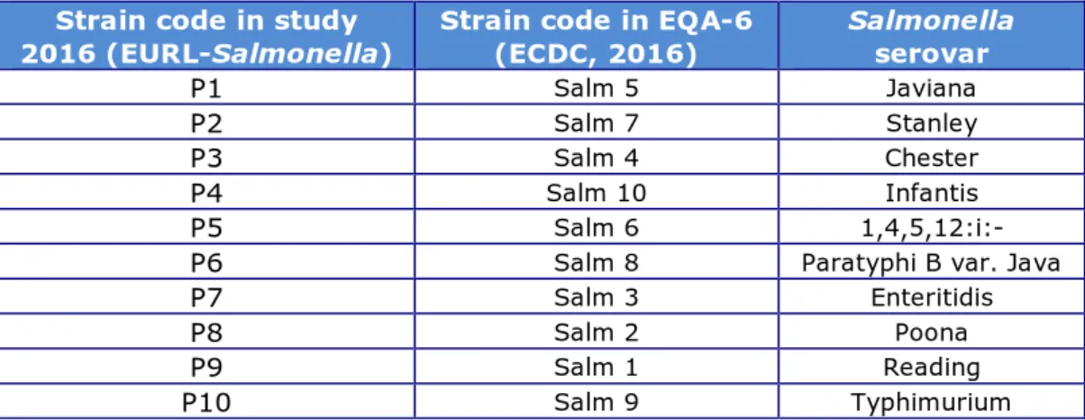
A total of 10 Salmonella strains (coded P1–P10) were included in the study on PFGE typing.
After consultation with the Statens Serum Institut (SSI), Copenhagen, Denmark, the same strains were used as in the External Quality
Assessment EQA-7 on PFGE typing, organised by the SSI for the Food- and Water-borne Diseases and Zoonoses Laboratories Network (FWD laboratories network) (ECDC, 2016). Background information on the strains is given in Table 4. Additionally, the reference image and its analysis in BioNumerics was kindly provided by SSI. In this way,
performance of both the NRLs network and the FWD laboratory network can be compared in the future.
Table 4. Background information on the Salmonella strains used for PFGE typing in 2016
Strain code in study
2016 (EURL-Salmonella) Strain code in EQA-6 (ECDC, 2016) Salmonella serovar
P1 Salm 5 Javiana
P2 Salm 7 Stanley
P3 Salm 4 Chester
P4 Salm 10 Infantis
P5 Salm 6
1,4,5,12:i:-P6 Salm 8 Paratyphi B var. Java
P7 Salm 3 Enteritidis
P8 Salm 2 Poona
P9 Salm 1 Reading
P10 Salm 9 Typhimurium
3.3.2 Evaluation of the PFGE gel image
Participants were asked to test the strains using their own routine PFGE method (XbaI digestion) and to give details of the method in the
electronic test report. However, the EURL-Salmonella-recommended method can be found in EFSA supporting publication 2014:EN-703 (Jacobs et al., 2014). Annex C of this publication describes the Standard PulseNet protocol Salmonella PFGE (PulseNet, 2013).
The PFGE gel images were to be emailed as uncompressed 8-bit grey scale Tagged Image File Format (TIFF) files to the EURL-Salmonella, and had to include the laboratory code in the filename.
Evaluation of the PFGE results was based on the quality of the PFGE images. Quality was assessed on seven parameters in accordance with the PulseNet guidelines (www.pulsenetinternational.org), as given in Annex 1. To comply with these guidelines the reference strain S. Braenderup H9812 must be run in every 6 lanes as a minimum. Each parameter is given a score of up to 4 points, where a poor result equals 1 point and an excellent result equals 4 points.
In general, an acceptable quality should be obtained for each parameter as a low quality score in just one category can still have a large impact on the suitability to further analyse the image and compare it to other profiles.
3.3.3 Evaluation of the analysis of the PFGE gel in Bionumerics
For the second time, the evaluation of the (optional) analysis of the PFGE gel in the bioinformatics software application BioNumerics was included.
In short, this included the following actions by the participants: • start a new database in BioNumerics,
• import the pre-configured database set-up as sent by email on 10 November 2017,
• import the TIFF image and analyse the gel (also see the protocol EURL-Salmonella typing study-2016 for further reference), • export the analysed data in either XML plus TIFF files (BN 6.0
and below) or in one .ZIP file (BN 7),
• email the correctly named files in a zipped format to the EURL-Salmonella.
Evaluation of the analysis of the gel in BioNumerics was done according to the guidelines used in the EQAs for the FWD laboratories (Annex 2). These guidelines use 5 parameters, which are scored with 1 (poor), 2 (fair/good) or 3 (excellent) points.
4
Results and Discussion
4.1 Technical data interlaboratory comparison study
4.1.1 General
A total of 34 laboratories participated in this study (Chapter 2). These included 29 NRLs-Salmonella in the 28 EU Member States, 2 NRLs in EU-candidate countries and 3 NRLs in EFTA countries.
The frequency of serotyping of Salmonella at the participating
laboratories and the number of strains that were serotyped and PFGE typed in 2016 are summarised in Table 5.
Table 5. Frequency and number of strains serotyped, and number of strains PFGE typed (for all 34 participants)
Lab
code frequency in Serotyping 2016 No. of strains serotyped in 2016 No. of strains PFGE typed in 2016 8 Daily 150 300 22 Daily 198 6 Daily 200 16 Daily 300 22 3 Daily 317 4 Daily 400 5 Daily 410 24 Daily 460 11 12 Daily 500 0 15 Daily 500 30 33 Daily 550 19 Daily 600 31 Daily 740 7 Daily 900 15 23 Daily 1200 200 21 Daily 1300 80 29 Daily 1500 50 10 Daily 1750 100 26 Daily 2500 32 Daily 3300 150 1 Daily 3500 200 25 Daily 3800 40 18 Daily 4500 9 Daily 5500 170 13 Thrice a week 150 14 Thrice a week 400 17 Twice a week 63 2 Twice a week 80 34 Twice a week 190 40 28 Twice a week 208
Lab
code frequency in Serotyping 2016 No. of strains serotyped in 2016 No. of strains PFGE typed in 2016 20 Once a week 13 27 Once a week 3000 n=34 39789 1408 4.1.2 Accreditation
Of the 34 participants, 32 are accredited for serotyping Salmonella, mainly according to ISO 17025, and in some cases according to ISO/TR 6579-3. The other two laboratories noted that they were working on their accreditation of Salmonella serotyping.
One laboratory is accredited for serotyping of all serovars except S. Typhi, and one laboratory is accredited for serotyping S. Enteritidis, S. Tyhpimurium, S. Infantis, S. Hadar, and S. Virchow; all other
laboratories stated that they are accredited for all Salmonella serovars. 4.1.3 Transport of samples
All but one of the participants received their package in the same week as sent (week 45 of 2016). The remaining parcel was delivered in week 46. All packages were received in good condition.
The participants used a variety of media from various manufacturers for sub-culturing the Salmonella strains. Non-selective nutrient agar was the most commonly used medium.
4.2 Serotyping results
4.2.1 General
One participant (lab code 16) sent the additional strain S21 to another laboratory for further serotyping or confirmation. Another participant (lab code 10) sent strain S3 to another laboratory, because of a lack of antisera needed for this strain. All other laboratories tested the 20 obligatory strains in their own laboratory.
Details on the number and the source of the sera as used by the participants are summarised in Table 6 and Table 7.
Table 6. Number of laboratories using sera from various manufacturers
Manufacturer Number of NRLs (n=34) Biorad 15 Microgen 1 Own preparation 5 Pro-Lab 6 Reagensia 2 Remel 1 Sifin 20
Table 7. Number of laboratories using sera from one or more manufacturers and/or in-house prepared sera
Number of manufacturers from which sera are obtained
(including in-house preparations) Number of NRLs (n=34)
1 9
2 12
3 9
4 4
4.2.2 Biochemical testing
Twenty-eight participants confirmed the use of biochemical tests. Twenty-one participants used a variety of biochemical tests on the optional strain S21, uncommon serovar 60: r, z (S. enterica subsp. diarizonae). Eighteen participants confirmed strain S12
(1,4,[5],12;a:e,n,x) to be a S. enterica enterica strain (Bispebjerg) by biochemical testing, most often by using malonate.
4.2.3 Use of PCR for confirmation
A total of 19 laboratories reported using PCR for the confirmation of serotyped strains. Seventeen of the laboratories use this PCR routinely, and the number of samples tested by PCR in 2016 are summarised in Table 8.
Three laboratories used PCR to confirm all the strains. Sixteen
laboratories used PCR to confirm strain S1, the monophasic variant of S. Typhimurium 1,4,[5],12:i:-, and seven of these also used PCR to confirm strain S13, S. Typhimurium. Strains S12 (1x), S14 (3x), S17 (2x) and S21 (2x) were also reported to have been confirmed using PCR.
Table 8. Number of strains routinely tested by PCR in 2016
Laboratory code Number of strains tested by PCR in 2016
12 2 22 4 5 13 16 17 24 20 31 22 8 30 28 38 10 80 11 80 20 120 33 148 23 150 29 700 27 750 26 2000 7 Unknown
4.2.4 Background information on the PCR methods used
PCR testing is mainly done to confirm monophasic (Typhimurium) strains. Eight laboratories mentioned the following reference:
• EFSA Journal, 2010.
Other references mentioned, sometimes in combination with others, were: • Aabo et al., 1993; • Barco et al., 2011; • Bugarel et al., 2012; • Lee et al., 2009; • Park et al., 1993 • Prendergast et al., 2013; • Tennant et al., 2010.
References regarding molecular serotyping in general were: • Fitzgerald et al., 2007 and McQuiston et al., 2011.
4.2.5 Serotyping results per laboratory
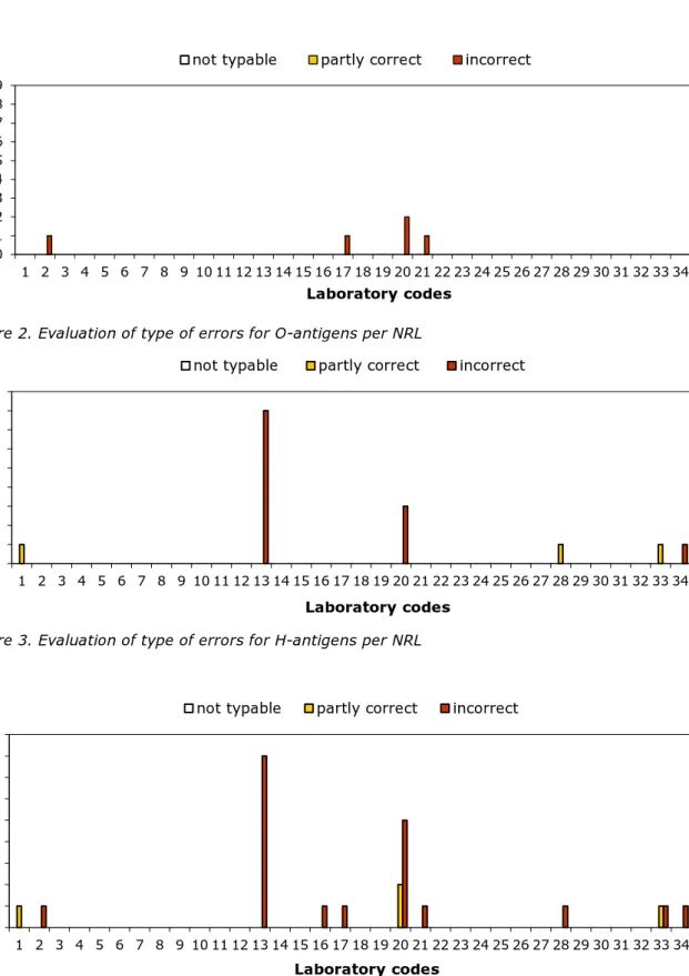
The percentages of correct results per laboratory are shown in Figure 1. The evaluation of the type of errors for O- and H-antigens and
identification of the strains are shown in Figures 2, 3 and 4.
The O-antigens were typed correctly by 30 of the 34 participants (88%). This corresponds to nearly 100% of the total number of strains. The H-antigens were typed correctly by 28 of the 34 participants (82%), corresponding to 99% of the total number of strains. A total of 24 participants (71%) gave the correct serovar names, corresponding to 99% of all strains evaluated.
Figure 1. Percentages of correct serotyping results 0 20 40 60 80 100 1 2 3 4 5 6 7 8 9 10 11 12 13 14 15 16 17 18 P er cen et ag e c o rr ec tn es s Laboratory codes
O-antigens H-antigens Serovar names
0 20 40 60 80 100 19 20 21 22 23 24 25 26 27 28 29 30 31 32 33 34 All P er ce n ta g e c o rr ec tn es s Laboratory codes
Figure 2. Evaluation of type of errors for O-antigens per NRL
Figure 3. Evaluation of type of errors for H-antigens per NRL
Figure 4. Evaluation of type of errors in the identification of serovar names 4.2.6 Performance of the participants
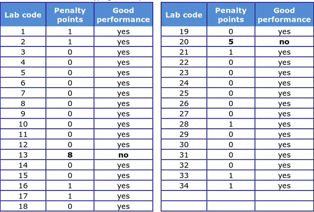
The number of penalty points was determined for each NRL using the guidelines described in Section 3.2.2. Table 9 shows the number of penalty points for each NRL and indicates whether the level of good
0 1 2 3 4 5 6 7 8 9 1 2 3 4 5 6 7 8 9 10 11 12 13 14 15 16 17 18 19 20 21 22 23 24 25 26 27 28 29 30 31 32 33 34 N u m be r o f st ra in s Laboratory codes
not typable partly correct incorrect
0 1 2 3 4 5 6 7 8 9 1 2 3 4 5 6 7 8 9 10 11 12 13 14 15 16 17 18 19 20 21 22 23 24 25 26 27 28 29 30 31 32 33 34 N u m be r o f st ra in s Laboratory codes
not typable partly correct incorrect
0 1 2 3 4 5 6 7 8 9 1 2 3 4 5 6 7 8 9 10 11 12 13 14 15 16 17 18 19 20 21 22 23 24 25 26 27 28 29 30 31 32 33 34 N u m be r o f st ra in s Laboratory codes
performance was achieved (yes or no). Two participants, both from a non-EU country, did not meet the level of good performance at this stage of the study and, in this case a voluntary, follow-up study was organised.
An example of an individual laboratory evaluation report on serotyping results is given in Annex 6.
Table 9. Evaluation of serotyping results per NRL
Lab code Penalty points performance Good Lab code Penalty points performance Good
1 1 yes 19 0 yes 2 1 yes 20 5 no 3 0 yes 21 1 yes 4 0 yes 22 0 yes 5 0 yes 23 0 yes 6 0 yes 24 0 yes 7 0 yes 25 0 yes 8 0 yes 26 0 yes 9 0 yes 27 0 yes 10 0 yes 28 1 yes 11 0 yes 29 0 yes 12 0 yes 30 0 yes 13 8 no 31 0 yes 14 0 yes 32 0 yes 15 0 yes 33 1 yes 16 1 yes 34 1 yes 17 1 yes 18 0 yes
4.2.7 Serotyping results per strain
The results found per strain and per laboratory are given in Annex 3, except for the more complicated strains S1 and S21; these are reported separately in Annex 4.
A completely correct identification was obtained for ten Salmonella serovars: Infantis (S5), Duisburg (S6), Bispebjerg (S12), Typhimurium (S13), Enteritidis (S14), Reading (S15), Hadar (S16), Rissen (S17), Mikawasima (S19), and Virchow (S20).
Most problems occurred with the serovar Umbilo (S3). Six laboratories had difficulties assigning the correct serovar name to this strain, mostly due to problems with the O-antigens. Details of the strains that caused problems in serotyping are shown in Annex 5.
The reported serovar names for strain 1,4,[5],12:i:- (S1) are shown in Annex 4. Nineteen participants used a PCR method to confirm this strain to be a monophasic Typhimurium strain.
In the evaluation of the results obtained by the participants, mistakes in typing the five designated Salmonella serovars (Enteritidis,
Typhimurium, Hadar, Infantis and Virchow) are more severely judged than errors in typing the other Salmonella serovars. This ‘Salmonella top
serovars, though one NRL reported the 1,4,[5],12:i:- strain as a Typhimurium strain (no PCR confirmation available).
Details of the additional and optional strain S21 are given in Annex 4. All but four participants tried to serotype strain S21, a Salmonella enterica subsp. diarizonae (IIIb). However, not all laboratories had access to the required antisera to finalise this (60:r:z).
4.2.8 Results follow-up study
Two participants, both non-EU-NRLs, did not achieve the level of good performance (Table 9; Lab code 13 and Lab code 20) and one of them participated in a follow-up study, receiving 10 additional strains for serotyping in week 18, 2017. The other laboratory did an extensive internal investigation to find out about any possible mistakes in the serotyping process, but had to decide to await the next interlaboratory study to test the improvements made due to lack of human resources at the time of the follow-up study.
Also for the follow-up study, the number of penalty points was determined using the guidelines described in Section 3.2.2. Table 10 shows the results of the follow-up study for participant 20, which again did not achieve the level of good performance. Unfortunately, the
communication on the results and the way these were produced was quite difficult, and thereby hampering the improvement of the serotyping results for the moment.
Table 10. Evaluation of serotyping results per NRL in the follow-up study
Lab code Penalty points Good performance
20 9 No
4.2.9 Trend analysis of the serotyping results of the EU NRLs
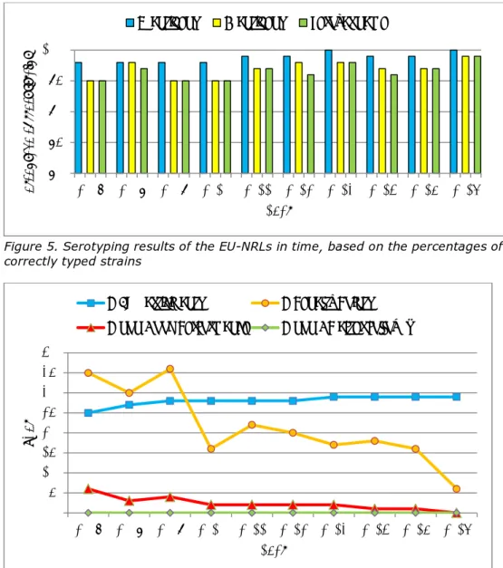
The historical data of the EURL-Salmonella interlaboratory comparison studies on the serotyping of Salmonella are given in Annex 7, in Table A7-1 for EU-NRLs only and in Table A7-2 for all participants per study. The data on the EU-NRLs only are also visualised in Figure 5, showing the percentages of correctly typed strains, and in Figure 6, showing the number of Penalty Points and non-Good Performance in time.
The percentages of correctly typed strains have remained stable over time, usually showing a better performance for the O-antigens than for the H-antigens.
The number of Penalty Points has clearly declined, from 35 points at the start of this system in 2007, to 6 points in the 2016 study. In line with this, the number of EU-NRLs with a non-Good Performance is low: two in the period 2010 – 2013, only one in the 2014 and 2015 studies and none in the 2016 study.
Figure 5. Serotyping results of the EU-NRLs in time, based on the percentages of correctly typed strains
Figure 6. Serotyping results of the EU-NRLs in time, based on the number of Penalty Points and non-Good Performance
4.3 PFGE typing results
4.3.1 General
A total of 15 NRLs participated in the fourth study on PFGE typing. Five participants in the 2015-study did not participate in the 2016 study, and four participants were new, compared to the 2015 study.
Ten participants reported using the Standard PulseNet Protocol
Salmonella PFGE (PulseNet International, 2013)/the EURL-Salmonella SOP (Jacobs et al., 2014). Five participants use this Standard protocol with modifications.
4.3.2 Technical data PFGE typing
Details on the manufacturer of the XbaI Enzyme, on the electrophoresis system and on the gel documentation system are summarised in Table
80 85 90 95 100 2007 2008 2009 2010 2011 2012 2013 2014 2015 2016 Per cen ta ge co rr ec t s tr ai ns Year
O-antigens H-antigens Serovar name
0 5 10 15 20 25 30 35 40 2007 2008 2009 2010 2011 2012 2013 2014 2015 2016 N ub er N Year
N EU participants N Penalty Points
Table 11. Manufacturers of the enzyme XbaI used by the participants
Manufacturer Number of NRLs
New England BioLabs 2
Promega 2
Roche Diagnostics 6
Thermo Scientific 5
Table 12. Electrophoresis system used by the participants
Electrophoresis system Number of NRLs
Bio-Rad CHEF Mapper (XA) 3
Bio-Rad CHEF-DR III System 10
Bio-Rad CHEF-DR II System 2
Table 13. Gel documentation system used by the participants
Gel documentation system Number of NRLs
Chemi Doc XR, Bio-Rad 1
G:Box (Syngene) 1 GelDoc 1 GelDoc XR 2 GelDoc XR+ 5 GeneGenious (Syngene) 1 Image Lab 5.2.1 1 Kodak Digital 1 Proxima Geldoc 2000 1
UVP EC3 Chemi HR Imaging System 1
Note: Different names may have been used for the same instruments.
One participant used Sybr Safe for staining the gel; all other participants used Ethidium Bromide. The duration of the staining varied between 15 minutes (1x) and 90 minutes (1x), but most participants used 30 minutes (8x). De-staining was even more diverse, varying between 5 minutes and 2 hours, a majority of participants used up to 60 minutes. Eight participants used a comb with narrow teeth, and seven
participants used one with wide teeth.
4.3.3 Results on the evaluation of the PFGE gel image
The scores per NRL (n=15), broken down across the seven parameters of evaluation (Annex 1), are given in Annex 8. The overall scores per parameter are shown in Figure 7.
Figure 7. Evaluation of the quality of the PFGE images in scores per parameter, 2016 study
The quality of the produced PFGE gel images results was generally good, though, as in former studies, some variation in results between the participants was seen (Annex 10).
Overall, 90% of the scores were Good or Excellent. However, two of the 15 images resulted in a Poor score on at least one of the seven
parameters, one for “Bands” and one for “Restriction” (Figure 7). This indicates that these two images are not suitable for use in inter-laboratory database comparison of these PFGE profiles.
Most problems were seen in the parameter “Gel background”, with 5 participants scoring only Fair. Fewer problems were seen in the parameters “Lanes” and “DNA degradation”, in which all participants scored Good or Excellent.
Eight out of the 15 participants (53%) scored Good or Excellent for each of the 7 parameters as evaluated.
An example of an individual laboratory evaluation report is given in Annex 11.
Figure 8 shows the results of the evaluation of the TIFF images from the studies 2013 - 2016. Improvements in time are clearly seen in the reduction of red (Poor) results in 2013 and 2014 compared to 2016.
0 2 4 6 8 10 12 14 16 P F G E P F G E P F G E P F G E P F G E P F G E P F G E N u m be r o f N R Ls
Scores per Parameter 2016
P = Poor F = Fair G = Good E = Excellent
Image Acquisition/
Running Conditions
Cell
Figure 8. Evaluation of the quality of the PFGE images in scores per parameter, 2013-2016 studies
4.3.4 Results on the evaluation of the analysis of the gel in BioNumerics For the second time the evaluation of the (optional) analysis of the gel in BioNumerics was included in the study as well. The participants all used the pre-configured database provided by the EURL-Salmonella, and therefore used identical experimental settings in BioNumerics. A total of 10 participants sent in their analysed gel data for evaluation. The scores per participating NRL, broken down across the five parameters of
evaluation (Annex 2), are given in Annex 9. The summarised scores per parameter are shown in Figure 9.
Overall, 68% of the scores were Excellent. Only one participant scored a Poor for one of the parameters. This concerned “position of gel frame”, and was due to wrongly included wells when placing the frame. This will be easy to correct in future analysis.
All ten participants scored a Fair/Good for the parameter “Band assignment”. For all of them this was due to occasionally assigning double bands as single bands; less frequently single bands were assigned as double bands. Three participants were noted to assign bands under 33 kb, thereby not following the protocol. As an example, band assignment results for strain P3 are given in Figure 10.
Figure 9. Evaluation of the analysis of the gel in BioNumerics in scores per parameter, 2016 study
Figure 10. PFGE profiles with band assignment in BioNumerics by 10 participants for strain P3. 0 2 4 6 8 10 12 P F/G E P F/G E P F/G E P F/G E P F/G E N um be r o f N R Ls
Scores per Parameter 2016
P = Poor F/G = Fair/Good E = Excellent
Position of
gel frame Strips Curves Normalisation assigmentBand
Labcode
2016-07
2016-24
2016-01
2016-08
2016-09
2016-32
2016-29
2016-10
2016 REF EQA7
2016-23
2016-12
Figure 11. Evaluation of the analysis of the gel in BioNumerics in scores per parameter, 2015-2016 studies
5
Conclusions
5.1 Serotyping
• Overall results for all 34 participating laboratories are:
– Nearly 100% of the strains were typed correctly for the O-antigens.
– 99% of the strains were typed correctly for the H-antigens. – 99% of the strains were correctly named.
• Serotyping of S. Umbilo caused the most problems in this study (six participants).
• All participants correctly serotyped the ‘top 5’ strains S. Enteritidis, S. Hadar, S. Infantis, S. Typhimurium and S. Virchow.
• All 29 EU-NRLs directly achieved the defined level of good performance.
• Two non-EU-NRLs initially did not achieve the defined level of good performance and were offered a follow-up study, typing an additional set of 10 strains. Only one non-EU-NRL participated, but was not able to improve itself.
5.2 PFGE typing
• Thirteen (87%) of the fifteen participating laboratories were able to produce a PFGE gel of sufficient quality to enable a profile determination suitable for use in inter-laboratory database comparisons.
• Ten participants also processed their gel in BioNumerics, and all of them were able to analyse their PFGE profiles in this computer program.
List of abbreviations
BN BioNumerics
DG-SANTE Directorate General for Health and Food Safety ECDC European Centre for Disease prevention and Control EFTA European Free Trade Association
EQA External Quality Assessment
EU European Union
EURL-Salmonella European Union Reference Laboratory for Salmonella FWD Food- and Water-borne Diseases and Zoonoses
Programme
NRL-Salmonella National Reference Laboratory for Salmonella PCR Polymerase Chain Reaction
PFGE Pulsed Field Gel Electrophoresis
RIVM National Institute for Public Health and the Environment (Bilthoven, The Netherlands) SSI Statens Serum Institut (Copenhagen, Denmark) TIFF Tagged Image File Format
References
Aabo, S., O.F. Rasmussen, L. Rossen, P.D. Sørensen, and J.E. Olsen (1993). Salmonella identification by the polymerase chain reaction. Mol. Cell Probes 7(3):171-178.
Barco, L., A.A. Lettini, E. Ramon, A. Longo, C. Saccardin, M.C. Pozza and A. Ricci (2011). Rapid and sensitive method to identify and differentiate Salmonella enterica serotype Typhimurium and Salmonella enterica serotype 4,[5],12:i:- by combining traditional serotyping and multiplex polymerase chain reaction. Foodborne Pathog. Dis. 8(6): 741–743.
Bugarel, M., M.L. Vignaud, F. Moury, P. Fach and A. Brisabois (2012). Molecular identification in monophasic and nonmotile variants of Salmonella enterica serovar Typhimurium. Microbiology Open. doi:10.1002/mbo3.39
EC (2004). European Regulation EC No 882/2004 of the European Parliament and of the Council of 29 April 2004 on official controls performed to ensure the verification of compliance with feed and food law, animal health and animal welfare rules. Official Journal of the European Union L 165: 30 April 2004.
http://eur-lex.europa.eu/LexUriServ/LexUriServ.do?uri=OJ:L:2004:165:0001:01
41:EN:PDF (accessed 17/1/2018).
European Centre for Disease Prevention and Control ECDC (2016). Seventh external quality assessment scheme for Salmonella typing. Stockholm: ECDC; doi 10.2900/147851
EFSA Panel on Biological Hazards (BIOHAZ) (2010) Scientific Opinion on monitoring and assessment of the public health risk of ‘Salmonella Typhimurium-like’ strains. EFSA Journal 8(10): 1826.
http://www.efsa.europa.eu/en/efsajournal/pub/1826.htm (accessed
17/1/2018).
Fitzgerald, C., M. Collins, S. van Duyne, M. Mikoleit, T. Brown and P. Fields (2007) Multiplex, bead-based suspension array for molecular determination of common Salmonella serogroups. J. Clin. Microbiol. 45(10): 3323–3334.
Grimont, P.A.D. and F.-X. Weill (2007) Antigenic formulae of the
Salmonella serovars, 9th ed. WHO Collaborating Centre for Reference and Research on Salmonella. Institute Pasteur, Paris, France.
https://www.pasteur.fr/sites/default/files/veng_0.pdf (accessed
17/1/2018).
ISO/TR 6579-3:2014. Microbiology of the food chain -- Horizontal method for the detection, enumeration and serotyping of Salmonella -- Part 3: Guidelines for serotyping of Salmonella spp.. International Organization for Standardization, Geneva.
ISO/IEC 17025. General requirements for the competence of testing and calibration laboratories. International Organization for
Jacobs, W., S. Kuiling, K. van der Zwaluw, 2014. Molecular typing of Salmonella strains isolated from food, feed and animals: state of play and standard operating procedures for pulsed field gel electrophoresis (PFGE) and Multiple-Locus Variable number tandem repeat Analysis (MLVA) typing, profiles interpretation and curation. EFSA supporting publication 2014:EN-703, 74 pp.
http://www.efsa.europa.eu/sites/default/files/scientific_output/files/
main_documents/703e.pdf (accessed 17/1/2018).
Lee, K., T. Iwata, M. Shimizu, T. Taniguchi, A. Nakadai, Y. Hirota and H. Hayashidani (2009) A novel multiplex PCR assay for Salmonella subspecies identification. J. Appl. Microbiol. 107(3): 805–811.
McQuiston, J.R., R.J. Waters, B.A. Dinsmore, M.L. Mikoleit and P.I. Fields (2011) Molecular determination of H antigens of Salmonella by use of a microsphere-based liquid array. J Clin Microbiol, 49(2): 565–573. Mooijman, K.A. (2007) The twelfth CRL-Salmonella Workshop; 7 and 8
May 2007, Bilthoven, the Netherlands. National Institute for Public Health and the Environment, Bilthoven, the Netherlands. RIVM Report no.: 330604006.
http://www.eurlsalmonella.eu/Publications/Workshop_Reports
(accessed 17/1/2018).
Mooijman K.A., 2016. The 21th EURL-Salmonella workshop; 9 June 2016, St. Malo, France. National Institute for Public Health and the Environment, Bilthoven, the Netherlands. RIVM Report no.: 2016-0045. http://www.eurlsalmonella.eu/Publications/Workshop_Reports
(accessed 17/1/2018).
Park S.H. et al., 2009, Protocol „Identificarea Salmonella enterica subspecia I, Salmonella enterica Typhimurium, Enteritidis şi Typhi utilizând metoda multiplex PCR”, FEMS Microbiol Lett, 301: 137-146. Prendergast, D.M., D. Hand, E. Nί Ghallchóir, E. McCabe, S. Fanning, M.
Griffin, J. Egan and M. Gutierrez (2013) A multiplex real-time PCR assay for the identification and differentiation of Salmonella enterica serovar Typhimurium and monophasic serovar 4,[5],12:i:-. Int. J. Food Microbiol. 16;166(1): 48–53.
PulseNet international (2013) Standard Operating Procedure for
PulseNet PFGE of Escherichia coli O157:H7, Escherichia coli non-O157 (STEC), Salmonella serotypes, Shigella sonnei and Shigella flexneri. PNL05, effective date 03-04-2013. Available at:
http://www.pulsenetinternational.org/assets/PulseNet/uploads/pfge/P
NL05_Ec-Sal-ShigPFGEprotocol.pdf (accessed 17/1/2018).
Tennant, S.M., S. Diallo, H. Levy, S. Livio, S.O. Sow, M. Tapia, P.I. Fields, M. Mikoleit, B. Tamboura, K.L. Kotloff, J.P. Nataro, J.E. Galen and M.M. Levine (2010) Identification by PCR of non-typhoidal Salmonella enterica serovars associated with invasive infections among febrile patients in Mali. PLoS. Negl. Trop. Dis 4(3): 621.
Acknowledgements
The authors would like to thank Sjoerd Kuiling and Kim van der Zwaluw (Centre for Infectious Diseases, Diagnostics and Screening, RIVM, Bilthoven, The Netherlands) for their expert contribution to the evaluation of the PFGE typing results.
Annex 1 PulseNet Guidelines on quality grading of PFGE
images
From www.pulsenetinternational.org :
STANDARD OPERATING PROCEDURE FOR TIFF QUALITY GRADING
CODE: PNQ01
Effective Date:
5 09 2005
1. PURPOSE: To describe guidelines for the quality of TIFF images
submitted to the PulseNet national databases.
2. SCOPE: This applies to all TIFF images submitted to PulseNet,
thereby allowing comparison of results with other PulseNet laboratories.
3. DEFINITIONS/TERMS:
3.1 TIFF: Tagged Image File Format
3.2 TIFF Quality: The grading of the appearance and ease of analysis of a TIFF, according to the TIFF Quality Grading Guidelines within this SOP. This is a main component of the evaluation of a TIFF submitted for certification or proficiency testing.
3.3 SOP: Standard Operating Procedure
4. RESPONSIBILITIES/PROCEDURE:
Parameter TIFF Quality Grading Guidelines
Excellent Good Fair Poor
Image Acquisition and Running Conditions By protocol, for example: - Gel fills whole TIFF - Wells included on TIFF - Bottom band of standard 1-1.5 cm from bottom of gel - Gel doesn’t fill whole TIFF but band finding is not affected
Not protocol; for example, one of the following:
- Gel doesn’t fill
whole TIFF and band finding is affected
- Wells not included
on TIFF - Bottom band of standard not 1-1.5 cm from bottom of gel - Band spacing of standards
doesn’t match global standard
Not protocol; for example, >1 of the following:
- Gel doesn’t fill
whole TIFF and this affects band finding
- Wells not included
on TIFF - Bottom band of standard not 1-1.5 cm from bottom of gel - Band spacing of standards
doesn’t match global standard
Cell
Suspensions The cell concentration is approximately the same in each lane 1-2 lanes contain darker or lighter bands than the other lanes - >2 lanes contain darker or lighter bands than the other lanes, or
- At least 1 lane is
much darker or lighter than the other lanes,
The cell
concentrations are uneven from lane to lane, making the gel impossible to
Bands Clear and distinct
all the way to the - Slight band distortion in 1 lane - Some band distortion (e.g., nicks) in 2-3 lanes but still - Band distortion that makes analysis difficult bottom of the
gel but doesn’t interfere analyzable - Very fuzzy bands. with analysis - Fuzzy bands - Many bands too
thick to - Bands are
slightly - Some bands (e.g., 4-5) are distinguish fuzzy and/or
slanted too thick - Bands at the bottom of the - A few bands
(e.g., - Bands at the bottom of the gel too light to distinguish :S3) difficult to
see gel are light, but analyzable clearly (e.g., DNA overload), especially at bottom of gel
Lanes Straight - Slight smiling(higher bands in the - Significant smiling - Slight curves on the outside - Smiling or curving that interferes with analysis outside lanes vs. the lanes
inside) - Still analyzable - Lanes gradually run longer toward the right or left - Still analyzable Restriction Complete restriction in all lanes - One to two faint shadow bands on gel
- One lane with
many shadow bands
- A few shadow
bands spread out over several lanes
- Greater than 1
lane with
several shadow bands
- Lots of shadow
bands over the whole gel Gel
Background
Clear - Mostly clear
background - Minor debris present that doesn’t affect analysis - Some debris present that may or may not make analysis difficult (e.g., auto
- Lots of debris present that may or may not make analysis difficult (i.e., auto band search finds too
many band search finds too many
bands) bands)
- Background caused by
photographing a gel with very
light bands (image contrast
was “brought up” in photographing gel-makes
DNA
Degradation (smearing in the lanes)
Not present - Minor background (smearing) in a few lanes but bands are clear
- Significant
smearing in 1-2 lanes that may or may not make analysis difficult - Minor background (smearing) in many lanes - Significant smearing in >2 lanes that may or may not make analysis difficult - Smearing so that a lane is not analyzable (except if untypeable [thiourea required]) 1. FLOW CHART: 2. BIBLIOGRAPHY: 3. CONTACTS: 4. AMENDMENTS:
Annex 2 Evaluation of gel analysis of PFGE images in
BioNumerics
Evaluation of gel analysis of PFGE images in BioNumerics according to the EQAs for the FWD laboratories (European Centre for Disease
Prevention and Control. Seventh external quality assessment scheme for Salmonella typing. Stockholm: ECDC; 2016. Available at:
http://ecdc.europa.eu/en/publications/Publications/salmonella-typing-seventh-external-quality-assessment.pdf
(accessed on 17-1-2018)
Parameter Poor [1] Grade [score in points]Fair [2] Excellent [3]
Position of
Gel Frame - placing the frame Wells wrongly included when
- Gel is not inverted.
- The frame is positioned too low.
- Too much space framed at the bottom of the gel.
- Too much space framed on the sides of the gel.
Excellent placement of frame and gel is inverted.
Strips Lanes incorrectly defined. - Lanes are defined too narrowly (or widely).
- Lanes are defined outside profile.
- A single lane is not correctly defined.
All lanes correctly defined.
Curves Curve set so that artefacts will
cause wrong band assignment. Curve extraction is defined either too narrowly or including almost the whole lane.
1/3 or more of the lane is used for averaging curve extraction.
Normali-zation - reference lanes. Many bands not assigned in the
- The references were not included when submitting the data.
- Assignment of band(s) in reference lane(s) to incorrect size(s).
- Bottom bands <33kb are not assigned in some or all of the reference lanes.
- Some bands wrongly assigned in reference lane(s).
All bands correctly assigned in all reference lanes
Band
Assignment Incorrect band assignment making inter-laboratory comparison impossible.
- Few double bands assigned as single bands or single bands assigned as double bands.
- Few shadow bands are assigned.
- Few bands are not assigned.
Excellent band assignment with regard to the quality of the gel.
Note that the EFSA supporting publication 2014:EN-703 (recommended SOP) states:
When using the S. Braenderup H9812 reference, visible bands of test isolates should be marked down to ~33 kb (third band from the bottom of the H9812 reference), but not below (referring to Band Assigment). In Normalisation, all bottom bands (also < 33 kb) in all reference lanes are assigned.
2 Newport Djibouti Szentes Infantis Duisburg Orion Saintpaul Augustenborg Chester 3 Newport Umbilo Szentes Infantis Duisburg Orion Saintpaul Augustenborg Chester 4 Newport Umbilo Szentes Infantis Duisburg Orion Saintpaul Augustenborg Chester 5 Newport Umbilo Szentes Infantis Duisburg Orion Saintpaul Augustenborg Chester 6 Newport Umbilo Szentes Infantis Duisburg Orion Saintpaul Augustenborg Chester 7 Newport Umbilo Szentes Infantis Duisdurg Orion Saintpaul Augustenborg Chester 8 Newport Umbilo Szentes Infantis Duisburg Orion Saintpaul Augustenborg Chester 9 Newport Umbilo Szentes Infantis Duisburg Orion Saintpaul Augustenborg Chester 10 Newport Umbilo Szentes Infantis Duisburg Orion Saintpaul Augustenborg Chester 11 Newport Umbilo Szentes Infantis Duisburg Orion Saintpaul Augustenborg Chester 12 Newport Umbilo Szentes Infantis Duisburg Orion Saintpaul Augustenborg Chester 13 Cremieu Moroto Maumee Infantis Duisburg Langensalza Chester Stuttgart Chartres 14 Newport Umbilo Szentes Infantis Duisburg Orion SaintPaul Augustenborg Chester 15 Newport Umbilo Szentes Infantis Duisburg Orion Saintpaul Augustenborg Chester 16 Newport Luckenwalde Szentes Infantis Duisburg Orion Saintpaul Augustenborg Chester 17 Newport Djibouti Szentes Infantis Duisburg Orion Saintpaul Augustenborg Chester 18 Newport Umbilo Szentes Infantis Duisburg Orion Saintpaul Augustenborg Chester 19 Newport Umbilo Szentes Infantis Duisburg Orion Saintpaul Augustenborg Chester 20 8:e,h:2 Albert OMC:k:2 Infantis Duisburg Muenster Sandiego Aberden Chester 21 Newport Telhashomer Szentes Infantis Duisburg Orion Saintpaul Augustenborg Chester 22 Newport Umbilo Szentes Infantis Duisburg Orion Saintpaul Augustenborg Chester 23 Newport Umbilo Szentes Infantis Duisburg Orion Saintpaul Augustenborg Chester 24 Newport Umbilo Szentes Infantis Duisburg Orion Saintpaul Augustenborg Chester 25 Newport Umbilo Szentes Infantis Duisburg Orion Saintpaul Augustenborg Chester 26 Newport Umbilo Szentes Infantis Duisburg Orion Saintpaul Augustenborg Chester 27 Newport Umbilo Szentes Infantis Duisburg Orion Saintpaul Augustenborg Chester 28 Newport Umbilo Szentes Infantis Duisburg Orion Saintpaul Oritamerin Chester 29 Newport Umbilo Szentes Infantis Duisburg Orion Saintpaul Augustenborg Chester 30 Newport Umbilo Szentes Infantis Duisburg Orion Saintpaul Augustenborg Chester 31 Newport Umbilo Szentes Infantis Duisburg Orion Saintpaul Augustenborg Chester
Ohio Bispebjerg Typhimurium Enteritidis Reading Hadar Rissen Haifa Mikawasima Virchow REF
Ohio Bispebjerg Typhimurium Enteritidis Reading Hadar Rissen Haifa Mikawasima Virchow 1 Ohio Bispebjerg Typhimurium Enteritidis Reading Hadar Rissen Haifa Mikawasima Virchow 2 Ohio Bispebjerg Typhimurium Enteritidis Reading Hadar Rissen Haifa Mikawasima Virchow 3 Ohio Bispebjerg Typhimurium Enteritidis Reading Hadar Rissen Haifa Mikawasima Virchow 4 Ohio Bispebjerg Typhimurium Enteritidis Reading Hadar Rissen Haifa Mikawasima Virchow 5 Ohio Bispebjerg Typhimurium Enteritidis Reading Hadar Rissen Haifa Mikawasima Virchow 6 Ohio Bispebjerg Typhimurium Enteritidis Reading Hadar Rissen Haifa Mikawasima Virchow 7 Ohio Bispebjerg Typhimurium Enteritidis Reading Hadar Rissen Haifa Mikawasima Virchow 8 Ohio Bispebjerg Typhimurium Enteritidis Reading Hadar Rissen Haifa Mikawasima Virchow 9 Ohio Bispebjerg Typhimurium Enteritidis Reading Hadar Rissen Haïfa Mikawasima Virchow 10 Ohio Bispebjerg Typhimurium Enteritidis Reading Hadar Rissen Haifa Mikawasima Virchow 11 Ohio Bispebjerg Typhimurium Enteritidis Reading Hadar Rissen Haifa Mikawasima Virchow 12 Adime Bispebjerg Typhimurium Enteritidis Reading Hadar Rissen Haifa Mikawasima Virchow 13 Ohio Bispebjerg Typhimurium Enteritidis Reading Hadar Rissen Haifa Mikawasima Virchow 14 Ohio Bispebjerg Typhimurium Enteritidis Reading Hadar Rissen Haifa Mikawasima Virchow 15 Ohio Bispebjerg Typhimurium Enteritidis Reading Hadar Rissen Haifa Mikawasima Virchow 16 Ohio Bispebjerg Typhimurium Enteritidis Reading Hadar Rissen Haifa Mikawasima Virchow 17 Ohio Bispebjerg Typhimurium Enteritidis Reading Hadar Rissen Haifa Mikawasima Virchow 18 Ohio Bispebjerg Typhimurium Enteritidis Reading Hadar Rissen Haifa Mikawasima Virchow 19 Ohio Bispebjerg Typhimurium Enteritidis Reading Istanbul Rissen Shubra Mikawasima Virchow 20 Ohio Bispebjerg Typhimurium Enteritidis Reading Hadar Rissen Haifa Mikawasima Virchow 21 Ohio Bispebjerg Typhimurium Enteritidis Reading Hadar Rissen Haifa Mikawasima Virchow 22 Ohio Bispebjerg Typhimurium Enteritidis Reading Hadar Rissen Haifa Mikawasima Virchow 23 Ohio Bispebjerg Typhimurium Enteritidis Reading Hadar Rissen Haifa Mikawasima Virchow 24 Ohio Bispebjerg Typhimurium Enteritidis Reading Hadar Rissen Haifa Mikawasima Virchow 25 Ohio Bispebjerg Typhimurium Enteritidis Reading Hadar Rissen Haifa Mikawasima Virchow 26 Ohio Bispebjerg Typhimurium Enteritidis Reading Hadar Rissen Haifa Mikawasima Virchow 27 Ohio Bispebjerg Typhimurium Enteritidis Reading Hadar Rissen Haifa Mikawasima Virchow 28 Ohio Bispebjerg Typhimurium Enteritidis Reading Hadar Rissen Haifa Mikawasima Virchow 29 Ohio Bispebjerg Typhimurium Enteritidis Reading Hadar Rissen Haifa Mikawasima Virchow 30 Ohio Bispebjerg Typhimurium Enteritidis Reading Hadar Rissen Haifa Mikawasima Virchow 31 Ohio Bispebjerg Typhimurium Enteritidis Reading Hadar Rissen Haifa Mikawasima Virchow 32
remark X = number of deviating laboratories per strain partly correct (no penalty points)
incorrect (1 penalty point)
Strain
code O-antigens
H-antigens H-antigens
Serovar confirmed PCR- Lab code
(phase 1) (phase 2)
S-1 1,4,[5],12 i - 1,4,[5],12:i:- yes REF
S-1 4,5 i 2 Typhimurium no 1
S-1 4,5,12 i - 1,4,5,12:i:- no 2
S-1 4,5,12 i - 4,5,12:i:- no 3
S-1 4,5,12 i - 4,5,12: i : - . Typhimurium monophasic variant. no 4
S-1 4,5,12 i - 4,5,12:i:- yes 5 S-1 4, 5, 12 i - 4,5,12:i:- no 6 S-1 4,12 i - 4,12 : i : - yes 7 S-1 4,5,12 i - 4,5,12:i:- yes 8 S-1 1,4,5,12 i - 1,4,5,12:i:- yes 9 S-1 4,5,12 i - 4,5,12 : i : - yes 10 S-1 4,5,12 i - 4,5,12:i:- yes 11
S-1 4,5,12 i - Typhimurium, monophasic (4,5,12:i:-) yes 12
S-1 4,5,12 i - 4,5,12:i:- no 13
S-1 4,5,12 i - 4,5,12:i:- no 14
S-1 4,5 i - 4,5:i:- no 15
S-1 4 i - Typhimurium monophasic variant yes 16
S-1 4,5,12 i - 4,5,12:i:- no 17
S-1 4,5,12 i - 4,5,12:i:- (Typhimurium-like monophasic variant) no 18
S-1 4,5,12 i - 4,5,12:i:- no 19
S-1 4,5 i - 4,5:i:- S. Typhimurium monophasic yes 20
S-1 1,4,5,12 i - 1,4,5,12:i:- yes 21
S-1 1,4,5,12 i - 1,4,5,12:i:- yes 22
S-1 1, 4, 5 i - Monophasic S. Typhimurium yes 23
S-1 4,5,12 i - 1,4,[5],12:i:- yes 24
code O-antigens (phase 1) (phase 2) Serovar confirmed Lab code
S-1 4,5,12 i - 4,5,12:i:- yes 26
S-1 4,5 i - 4,5,12:i:- yes 27
S-1 4,5,12 i - 4,5,12:i:- yes 28
S-1 1,4,5,12 i - Monophasic variant S.Typhimurium yes 29
S-1 1,4,5,12 i - 1,4,5,12:i:- no 30
S-1 4,5,12 i - monofasisk subspI=4,5:i:- yes 31
S-1 4,5,12 i - 4,5,12:i:- no 32
S-1 4,5,12 i - monophasic Typhimurium yes 33
S-1 4,5,12 i - 4,5,12:i:- no 34
Reference remark
partly correct; in the naming: no penalty points incorrect; in the naming: 1 penalty point
code O-antigens (phase 1) (phase 2) Serovar Lab code
S-21 60 r z 60:r:z REF
S-21 60 r z S. IIIb 60 : r : z 1
S-21 - - - 2
S-21 60 r z Salmonella enterica subsp. diarizonae ( III b) 60:r:z 3
S-21 60 r z S. enterica subsp. diarizonae 60:r:z 4
S-21 60 r z III b diarizonae 5
S-21 6
S-21 60 r - 60 : r : - IIIb 7
S-21 60 r z 60:r:z 8
S-21 60 r z S. IIIb 60:r:z 9
S-21 Salmonella Subspecies II (salamae) 10
S-21 60 r z 60:r:z 11
S-21 60 r z Salmonella enterica subsp.diarizonae ser. 60 : r : z 12
S-21 13
S-21 60 r - 60:r:- 14
S-21 ? r z ?:r:z 15
S-21 60 r - 60 : r : - (enterica subsp. diarizonae) 16
S-21 60 r z S. enterica subsp. diarizonae /IIIb/ 17
S-21 60 r z SGIIIb 60:r:z 18
S-21 60 r z 60:r:z 19
S-21 OMG r - OMG:r:- 20
S-21 60 r z53 60:r:z53 21
S-21 60 r z 60:r:z 22
S-21 60 r z S. enterica subsp. diarizonae 60:r:z 23
S-21 60 r z IIIb 60:r:z 24 S-21 60 r z 60:r:z 25 S-21 60 r z IIIb 60:r:z 26 S-21 60 r z SIII 60:r:z 27 S-21 - r - -:r:- 28 S-21 60 r z subsp. Diarizonae 29 S-21 60 r z 60:r:z 30
code O-antigens (phase 1) (phase 2) Serovar Lab code
S-21 60 r z S.SubspIIIb=60:r:z 31
S-21 60 r z 60:r:z 32
S-21 33
S-21 60 r z III b 34
S-21: Salmonella enterica subspecies diarizona (IIIb), optional strain reference