Implications of not detecting
non-genotoxic carcinogens in the
absence of carcinogenicity tests under
REACH guidelines
Report 340700003/2008
RIVM Report 340700003/2008
Implications of not detecting non-genotoxic carcinogens
in the absence of carcinogenicity tests under REACH
guidelines
L.G. Hernandez J. van Benthem M. Luijten H. van Steeg Contact:Jan van Benthem
Laboratory for Health Protection Research jan.van.benthem@rivm.nl
This investigation has been performed by order and for the account of the Ministry of Health, Welfare and Sports of the Netherlands (VWS), Directorate General for Public Health (DGV), Directorate for Food, Health Protection and Prevention (VGP), within the framework of project V/340700, Knowledge Base Carcinogenicity, Mutagenicity and Reproduction Toxicology.
© RIVM 2008
Parts of this publication may be reproduced, provided acknowledgement is given to the 'National Institute for Public Health and the Environment', along with the title and year of publication.
Abstract
Implications of not detecting non-genotoxic carcinogens in the absence of carcinogenicity tests under REACH guidelines
Research performed by the RIVM has demonstrated that the European legislation does not comply with a specific small group of carcinogens, the so-called non-genotoxic carcinogens, which induce cancer via a mechanism other than the introduction of DNA damage. In view of these findings, several recommendations for alternative test methods are given including the use of innovative genomics techniques for the detection of these compounds.
REACH, which stands for Registration, Evaluation, Authorization and Restriction of Chemicals (REACH), was implemented on July 2008. The policy forces companies to collect, assess and distribute information on qualities and risks of chemicals among users to safely manage these compounds.
Under REACH test batteries for carcinogenicity of a chemical are in vitro and in vivo genotoxicity assays. Carcinogenicity tests are only performed for compounds with production volumes greater than 1000 tonnes per annum and/or positive in the previously mentioned genotoxicity tests. Given that non-genotoxic carcinogens are negative for non-genotoxicity, they will not be detected under REACH.
Ten to twenty % of the known, probable or possible human carcinogens appear to be non-genotoxic carcinogens. For one third of these compounds, exposure is high enough that an increased risk for cancer can not be excluded. Nevertheless, the expected number of non-genotoxic carcinogens among newly registered compounds is assumed to be very low. Moreover, REACH requires data on
carcinogenicity for compounds with a high production volume and a probable higher human exposure. Key words: non-genotoxic carcinogens, carcinogenicity, risk assessment, REACH, toxicogenomics
Rapport in het kort
De gevolgen van de invoering van REACH op de identificatie van niet-genotoxische kankerverwekkende stoffen
De Europese wetgeving REACH voldoet niet voor een bepaalde groep chemische stoffen die
kankerverwekkend zijn. Het gaat hierbij om zogeheten niet-genotoxische kankerverwekkende stoffen die kanker veroorzaken via een ander mechanisme dan door effecten op het DNA te introduceren. Het gaat echter om een kleine hoeveelheid stoffen. Dit blijkt uit onderzoek van het RIVM. Het instituut reikt een aantal voorstellen voor alternatieve testmethoden aan, waaronder het gebruik van innovatieve genomicstechnieken.
REACH staat voor Registratie, Evaluatie, Autorisatie en Beperking van Chemische stoffen en is in juli 2008 van kracht geworden. De wetgeving verplicht bedrijven om informatie over eigenschappen en risico’s van chemische stoffen te verzamelen, te beoordelen en te verspreiden onder gebruikers om veilig met de stoffen te kunnen omgaan.
Onder REACH bestaan de teststrategieën om kankerverwekkende chemische stoffen te herkennen uit genotoxiciteitstesten. Testen naar kankerverwekkende eigenschappen worden alleen gedaan voor chemicaliën met een productievolume van meer dan duizend ton per jaar en/of als ze mutaties in genen veroorzaken. Omdat niet-genotoxische kankerverwekkende stoffen negatief scoren in deze testen, worden ze onder de REACH-wetgeving niet herkend.
Tien tot twintig procent van de stoffen waarvan vaststaat dat ze voor de mens kankerverwekkend zijn of zouden kunnen zijn, blijken niet-genotoxisch. Bij ongeveer een derde van deze stoffen is de blootstelling potentieel dusdanig dat een verhoogd risico op kanker niet kan worden uitgesloten. Daar staat tegenover dat onder de nieuw te registreren stoffen het percentage niet-genotoxische
kankerverwekkende stoffen naar verwachting erg klein is. Bovendien eist REACH voor stoffen met een hoog productievolume en dus mogelijke hogere blootstelling, altijd gegevens over de carcinogeniteit. Trefwoorden: niet-genotoxische kankerverwekkende stoffen, kanker, risicoschatting, REACH, toxicogenomics
Contents
List of Abbreviations 9
Summary 11
1 Background 13
2 Identification of non-genotoxic carcinogens 15
2.1 Weight of evidence approach 15
2.2 Registration, Evaluation, Authorization of Chemicals (REACH) 18
3 Human cancer risk assessment 19
3.1 Non-genotoxic carcinogens in IARC 20
3.2 Margin of Eexposure (MOE) 21
3.3 Reference Dose (RfD) 23
3.4 Implications 24
4 Alternative methods for detecting non-genotoxic carcinogens 29 4.1 Structure-Activity Relationship (SAR) 29
4.2 Replicative DNA Synthesis (RDS) 30
4.3 In vitro cell transformation assay 31
4.4 Toxicogenomics 33
5 Conclusions 37
6 Recommendations for future research 39
Acknowledgements 41 References 43 Appendix 1 References for Table 3 49
List of Abbreviations
Ah Aryl hydrocarbon
ARNT Aryl hydrocarbon Receptor Nuclear Translocator BMD Benchmark dose
DES Diethylstilbestrol
DDT Dichlorodiphenyltrichloroethane DEHP Di(2-ethylhexyl)phthalate
DMEL Derived minimal effect level DNEL Derived no-effect level E2 17β-Estradiol
EPA Environmental Protection Agency
GJIC Gap Junction Intercellular Communication HSP Heat Shock Protein
IARC International Agency for Research on Cancer IRIS Integrated Information System
LD Lethal Dose
LTD Lower-bound Toxic Dose mg/kg bw milligrams/kilogram body weight MF Modifying Factors
MOE Margin of Exposure MP Methapyrilene
NOAEL No Observed Adverse Effect Level
OECD Organization for Economic Cooperation and Development 2-OHE 2,2-hydroxyestradiol
4-OHE 2,4-hydroxyestradiol PB Piperonylbutoxide PCB Polychlorinated Biphenyl PCNA Proliferating Cell Nuclear Receptor
PPAR Peroxisome Proliferator Activated Receptor Q(SAR) Quantitative Structure-Activity Relationship
REACH Registration, Evaluation, Authorization and Restriction of Chemicals RDS Replicative DNA Synthesis
ROS Reactive Oxygen Species
SAR Structure-Activity Relationship SHE Syrian Hamster Embryo
TCDD 2,3,7,8-tetrachlorodibenzo-p-dioxin TD Toxic Dose
tpa tone per annum
UF Uncertainty Factors
Summary
Non-genotoxic carcinogens are substances that induce cancer through indirect stimulation of hyperplastic responses, without altering DNA, chromosome number or structure. Non-genotoxic carcinogens have a wide variety of mechanisms of cancer induction including tumor promoting, endocrine modifying, receptor mediating, cytotoxicity, immunosuppression, or inducing tissue-specific toxicity and inflammatory responses. The diversity of modes of action for each non-genotoxic
carcinogen, the tissue specificity, and the absence of genotoxicity makes their identification and characterization very challenging. In Europe, human cancer risk assessment of carcinogens involves a three-step process. First, there is an assessment of the genotoxic effects elicited by the compound, which includes hazard identification and dose-response evaluation. Second, there is an exposure assessment which estimates the levels to which human populations are exposed to. Finally, there is a risk characterization which involves the estimation of the incidence and severity of the adverse effects likely to occur in human populations due to actual or predicted exposure to a substance (EC, 2003). For non-genotoxic carcinogens, the assessment of the dose-response relationship involves an evaluation within the range of tumor observations to identify the toxicological effects that result in carcinogenesis and the extrapolation to lower dose levels to mimic human exposure levels. In many instances,
mechanistic studies are necessary to better understand the mode of action leading to carcinogenesis and the human relevance of animal tumor findings. Thus, in the absence of genotoxicity, hazard
identification and dose-response evaluations are not possible unless there is carcinogenicity data available. For non-genotoxic carcinogens, it is generally assumed that their modes of actions can be associated with threshold doses, and it is possible to identify no-effect levels for the underlying toxic effects of concern that lead to tumor formation. In view of the various epigenetic mechanisms of action of non-genotoxic carcinogens, human cancer risk assessment is done on a case-by-case basis using a weight of evidence approach from 90-day sub-chronic toxicity studies, toxicokinetic and disposition studies, and/or 2-year cancer bioassays in rodents.
In this report, the non-genotoxic carcinogens in the International Agency for Research and Cancer (IARC) were analyzed. Results indicated that non-genotoxic carcinogens were present in 23% (16/71) of known human carcinogens (Group 1), in 5% (3/57) of probable human carcinogens (Group 2A), and in 12% (28/234) of possible human carcinogens (Group 2B). Importantly, many non-genotoxic
carcinogens are known human carcinogens (Group 1) and are a public health concern. Human cancer risk assessment of non-genotoxic carcinogens is dependent on the lowest no observed adverse effect level (NOAEL) obtained from toxicity and/or carcinogenicity studies. The NOAEL is important because it is used for the calculation of the margin of exposure (MOE) and a reference dose (RfD);
both important parameters in human cancer risk assessment. The MOE and RfD were evaluated in IARC groups 1, 2A and 2B non-genotoxic carcinogens. The MOE is the ratio of the NOAEL or lower-bound toxic dose (LTD10) to the average daily human exposure. An MOE lower than 100 for
consumers or lower than 50 for workers represents a high concern from a public health point of view with a high priority for risk management actions. Our analysis demonstrated that when the MOE was derived from the NOAEL, 7 out of 18 (39%) non-genotoxic carcinogens analyzed had an MOE lower than 50. When the MOE was derived from the LTD10, 4 out of the 14 (29%) non-genotoxic carcinogens analyzed had an MOE less than 50. In addition to the MOE, the RfD was also used to analyze non-genotoxic carcinogens. An RfD is a benchmark dose (BMD) estimated, with uncertainty factors, of a daily exposure of possible non-genotoxic carcinogens to the human population that is likely to be without appreciable risk of deleterious effects during a lifetime (i.e. NOAEL or BMD/uncertainty factors). Any dose level above the RfD is considered to be unacceptable with the possibility of adverse effects. Results showed that the average daily human exposure was higher than the RfD in 3 out of 10 (30%) of the non-genotoxic carcinogens analyzed. These results suggest that the magnitude by which the NOAEL exceeds the estimated human exposure dose levels needs to be re-examined with the inclusion of additional safety factors. The implications of these findings for the European policy for Registration, Evaluation, Authorization and Restriction of Chemicals (REACH) guidelines are an urgent need for the development of alternative methods for the early detection of non-genotoxic carcinogens for proper human cancer risk and hazard assessment.
Although the expected number of non-genotoxic carcinogens among newly registered compounds is very low, there is a growing concern that under REACH, non-genotoxic carcinogens will not be detected because the test batteries for carcinogenicity are genotoxic endpoints. Further, 2-year
carcinogenicity assays are rarely performed under REACH guidelines. Given that some non-genotoxic carcinogens are known human carcinogens and the potential hazard associated with them, there is a need for the development of alternative methods for the early detection of non-genotoxic carcinogens. Structure-activity relationships (SARs) and (Q)SARs, replicative DNA synthesis assay, and/or the in
vitro cell transformation assay are possible alternative methods for detecting non-genotoxic
carcinogens but none provide any information on the mode of action and have limited applicability. Finally, several recommendations were given including the development of SAR models for proper (Q)SAR analysis and the use of transcriptomics to analyze multiple pathway-specific gene expression profiling of known non-genotoxic carcinogens. These improvements should lead to a better
identification of potential alerts/markers of putative non-genotoxic carcinogens. In terms of REACH guidelines, additional tests need to be considered for the detection of non-genotoxic carcinogens, in the absence of a 2-year carcinogenicity assay.
1
Background
A genotoxic carcinogen is an agent that induces tumors though mechanisms involving genotoxic events such as DNA mutations and/or chromosomal aberrations. Genotoxic carcinogens can either directly react with DNA to induce mutations or can induce chromosomal effects in the absence of mutagenic action, as in the case of non-DNA reactive genotoxins such as aneugens, topoisomerase and spindle poisons. In contrast, non-genotoxic carcinogens do not react with DNA or alter chromosome number or structure but rather induce neoplastic transformation through an indirect stimulation of hyperplastic responses. Non-genotoxic carcinogens induce carcinogenic effects in a threshold manner, similar to non-DNA reactive genotoxic carcinogens (Foth et al., 2005). It has been suggested that proper evaluation of putative non-genotoxic carcinogens can only be done with information on their
mechanism of action (Jackson et al., 1993). Most non-genotoxic carcinogens elicit their carcinogenic effects by acting as tumor promoters (phenobarbital; 1,4-dichlorobenzene), endocrine-modifiers (hormones including estrogens such as 17β-estradiol), receptor-mediators (peroxisome proliferators such as ciprofibrate, clofibrate and 2,3,7,8-tetrachlorodibenzo-p-dioxin), cytotoxicants (d-limonene), immunosuppressors (cyclosporin) or inducers of tissue-specific toxicity and inflammatory responses (minerals such as asbestos and metals such as arsenic, beryllium, cadmium) (Melnick et al., 1996; Williams, 2001). Non-genotoxic carcinogens are, for the most part, species- and tissue- specific (Williams and Whysner, 1996), and for adequate human risk assessment, it is important to understand all the biological processes that may lead to carcinogenesis, to examine various species, to include adequate routes of exposure and assay all possible target tissues.
In Europe, a new policy is implemented which deals with Registration, Evaluation, Authorization and Restriction of Chemicals (REACH) and tests their carcinogenic potential by using various genotoxicity endpoints depending on the production volume of the chemical (EU, 2003). This regulation requires for every chemical with a production volume greater than 1 tone per annum (tpa) to be evaluated. The test battery for carcinogenicity of REACH is based on genotoxic endpoints including the Ames
mutagenicity test in bacteria, genotoxicity in mammalian cells under in vitro and in vivo conditions, and germ cell mutagenicity tests. The test strategy for carcinogens adopted by REACH consists of a three-step process. First, there is an assessment of the genotoxic effects elicited by the compound, which includes hazard identification and dose-response evaluation. Second, there is an exposure assessment which estimates the levels to which human populations are exposed to. Finally, there is a risk characterization which involves the estimation of the incidence and severity of the adverse effects likely to occur in human populations due to actual or predicted exposure to a substance (EC, 2003). Thus, in the absence of genotoxicity, hazard identification and dose-response evaluations are not
possible unless there is carcinogenicity data available. Further, carcinogenicity tests under REACH are only performed for chemicals with production volumes greater than 1000 tpa and positive for most of the previously mentioned genotoxicity tests. Given that non-genotoxic carcinogens are negative for genotoxicity, alternative methods need to be developed for their identification. The aim of this report is to assess the human risk associated with non-genotoxic carcinogens and to identify alternative methods to recognize these compounds on the basis of their modes of action.
2
Identification of non-genotoxic carcinogens
Under REACH, non-genotoxic carcinogens will not be detected because the main test battery is genotoxicity. Before REACH, the majority of non-genotoxic carcinogens have been identified using a variety of tests including 90-day subchronic toxicity studies in rats and mice, toxicokinetic and
disposition studies, and/or 2-year cancer bioassays in both rats and mice. Results from these studies are used to determine the human risk associated with non-genotoxic carcinogens based on a weight-of-evidence evaluation (Olin et al., 1997) and assessed on a case-by-case basis according to the mode of action of the substance (Williams, 2001). Further, extrapolation of animal data to assess human cancer risk requires an understanding of the similarities and differences between humans and animals in regards to carcinogenic processes (Olin et al., 1997).
2.1 Weight of evidence approach
A weight of evidence approach relies on information on the mode of action of non-genotoxic carcinogens obtained from 90-day toxicity studies in rats and mice, toxicokinetic and disposition studies, and/or 2-year cancer bioassays in both rats and mice. These studies are vital to our
understanding on the mechanisms of non-genotoxic carcinogens and are essential for proper human cancer risk assessment. Importantly, each non-genotoxic carcinogen has a unique mechanism of action and human risk assessment varies in each case. The weight of evidence approach has been used to assess dose-level differences between rodents and humans. For instance, sodium saccharin is a low calorie artificial sweetener that, in very high doses (30,000 mg/kg body weight (bw)-75,000 mg/kg bw), induces bladder tumors in male rats but not in female rats or mice. The species- and sex-difference is possibly due to the formation of sodium saccharin-induced crystals in male rats when saccharin is given at high doses. Saccharin binds to silicates and proteins causing cell damage in the urinary bladder, which induces cellular proliferation and tumor formation. Humans, on the other hand, consume very low levels of saccharin (5 mg/kg bw/day) and for this reason no cancer risk would be predicted (Whysner and Williams, 1996; Williams et al., 1996).
An example of species differences between rodents and humans is the food additive d-limonene which induces α2µ-globulin accumulation and renal carcinogenesis in F344 male rats but not in female rats or mice. The binding of d-limonene and the metabolite d-limonene-1,2-oxide to α2µ-globulin induces hyaline droplet formation in renal proximal tubules. Tumor formation in F344 male rats is associated with hyaline droplet-induced nephropathy that leads to atypical hyperplasia and atypical tubules. Similarly, 1,2-dichlorobenzene induces renal carcinogenesis only in male rats. Male F344 rats are very
proteinuric in comparison to humans and other species (Purchase, 1994). Further, no human urinary protein similar to α2µ-globulin has been identified. Taken together, the mechanism of d-limonene- and 1,2-dichlorobenzene-induced renal tumors is unique to male rats and no human cancer risk would be expected with either compound (Melnick et al., 1996; Purchase, 1994; Williams and Whysner, 1996). Other examples of species differences include peroxisome proliferators di(2-ethylhexyl)phthalate (DEHP) and Wyeth-14643 which induce liver tumors in rats and mice (Melnick et al., 1996).
Peroxisomes are subcellular organelles containing oxidative enzymes that generate hydrogen peroxide and catalase. It has been suggested that peroxisome proliferation alone does not account for the high incidence of liver tumors given that rats treated with DEHP and Wyeth-14643 that produce similar levels of peroxisome proliferation, have different carcinogenic responses. Tumors induced by Wyeth-14643 have a shorter latency period and higher tumor multiplicity (Melnick et al., 1996). Studies have shown that the murine peroxisome proliferator activated receptor (PPAR)α increases the mRNA expression of PCNA, cMYC, cJUN, CDK4 and other cyclins. Consequently, murine
hepatocarcinogenesis has been postulated to involve the downregulation of the microRNA let-7c gene, resulting in elevated levels of c-myc (Gonzalez and Shah, 2008). In humans, peroxisome proliferators such as clofibrate, ciprofibrate and gemfibrozil are used to treat cardiovascular disease (Milionis et al., 2000). The World Health Organization cooperative trial using clofibrate demonstrated an excess of deaths from non-malignant diseases of the liver, gall bladder and intestine in the clofibrate group in comparison to controls (WHO, 1984). Therefore, peroxisome proliferators are generally not associated with human hepatocarcinogenesis. PPARα-humanized mice treated with Wy-14643 showed no downregulation of let-7c as observed with murine PPARα, and showed resistance to peroxisome proliferator-induced cell proliferation and cancer (Gonzalez and Shah, 2008). Species differences in response to peroxisome proliferators between mice and human PPARα appear to be associated with their ability to downregulate let-7c. Overall, not all rodent carcinogens are human carcinogens and human risk assessment should be made on a case-by case basis taking into account the mechanism of action of each non-genotoxic carcinogen.
In other instances, similar reactivity between rodents and humans is detected as in the case of the non-genotoxic carcinogen 2,3,7,8-tetrachlorodibenzo-p-dioxin (TCDD) and the aryl hydrocarbon (Ah) receptor, a ligand-dependent transcription factor. TCDD binds and activates the Ah receptor by forming a heterodimer with the Ah receptor nuclear translocator (ARNT). The Ah-TCDD-ARNT complex binds to regulatory sequences and alters their expression (Whitlock, 1993). Treatment with TCDD has been associated with increased expression of ras, erbA, c-fos, c-jun and other cylcin-dependent kinases (Knerr and Schrenk, 2006). Further, TCDD has been shown to induce CYP1A1, glutathione-S-transferase Ya subunit, aldehyde dehydrogenase and quinine reductase (Whitlock, 1993).
The potent induction of CYP1A1 by TCDD also results in the generation of reactive oxygen species. In terms of carcinogenicity, TCDD induces tumors in the liver, thyroid, oral cavity and lungs in rats, and in the liver, thymus and skin in mice (Knerr and Schrenk, 2006). Human epidemiological studies indicate an increased risk in lung cancer, soft tissue sarcoma, non-Hodgkin lymphoma and several other malignant neoplasms in herbicide producers exposed to TCDD (IARC, 1997). For this reason, the International Agency for the Research on Cancer (IARC) has classified TCDD as carcinogenic to humans (IARC, 1997). Other examples of similar reactivity between rodents and humans are endocrine modifiers. For example, dichlorodiphenyltrichloroethane (DDT) is a pesticide that disrupts the
endocrine system, promotes hormone-dependent pathology (Kavlock et al., 1996), and induces CYP2B and CYP3A in a sex-related manner suggestive of a possible role of cytochrome P450s in endocrine disruption (Sierra-Santoyo et al., 2000). DDT also reduces gap-junctional intercellular communications (GJICs) in rat and mouse hepatocytes (IARC, 1999). In humans, there are elevated incidences of non-Hodgkin's lymphoma and lung cancer in workers exposed to DDT (IARC, 1999). Due to the limited epidemiological data, DDT is considered a possible human carcinogen (IARC, 1999). Another example of endocrine modifiers are polychlorinated biphenyls (PCBs) used in coolants and insulating fluids for transformers and capacitors. PCBs modify the endocrine system by binding directly to thyroid hormone receptors, inhibiting deiodinase, or displacing thyroxine from serum-binding protein transthyretin (Zoeller, 2007). PCBs also generate reactive oxygen species (ROS) through the induction of lipid peroxidation (Gurer-Orhan et al., 2006), and inhibit GJICs in rat epithelial cells (Machala et al., 2003). In humans, epidemiological data demonstrates increases in hepatobilary cancers, lymphomas, and melanomas from occupational exposure to PCBs; thus PCBs are considered probable human carcinogens (IARC, 1998). Of particular importance are estrogenic hormones such as 17β-estradiol (E2), along with its hydroxylated metabolites 2-hydroxyestradiol (2-OHE2) and 4-hydroxyestradiol (4-OHE2), which are potent mitogens and are carcinogenic in rodents and induce proliferation in human MCF-7 breast cancer cells (Kim et al., 2005). In humans, breast and endometrial cancers are estrogen-responsive tumors (Rosen et al., 2005), and for this reason estrogenic hormones are
considered known human carcinogens (IARC, 1987). Overall, human cancer risk is unique to each non-genotoxic carcinogen and assessment is made on a case-by-case basis according to the mode of action. The weight of evidence evaluation is dependent on data obtained from carcinogenicity studies, toxicity studies in rats and mice, toxicokinetic and disposition studies, and, when available, human
epidemiological data from exposed individuals. Given that these studies are extremely costly, time consuming, require large number of animals and are not performed under REACH, alternative short-term tests for the detection of non-genotoxic carcinogens need to be developed.
2.2 Registration, Evaluation, Authorization of Chemicals (REACH)
Non-genotoxic carcinogens have distinct modes of action, some require binding with a variety of different types of receptors and others involve epigenetic changes coupled with cellular proliferation. In REACH, the in vitro gene mutation assay in bacteria (Ames test) is the minimum requirement for all substances manufactured or imported in quantities of 1 tpa or greater. Other mutagenicity studies are prompted when a positive result is obtained in the gene mutation assay. If substances are produced in quantities of 10 tpa or more, in vitro cytogenicity studies are performed in mammalian cells. In vivo mutagenicity studies are only performed when a positive result is obtained in any in vitro genotoxicity assay. The fact that the carcinogenic activity of non-genotoxic carcinogens cannot be detected by standard genotoxicity assays is a major regulatory problem. In view of this, there is a growing concern of the human cancer risk associated with non-genotoxic carcinogens given that in REACH, the test battery for carcinogenicity are genotoxic endpoints; thus non-genotoxic carcinogens are not recognized. The human cancer risk associated with non-genotoxic carcinogens will be investigated.
3
Human cancer risk assessment
Human cancer risk assessment is based on low-dose extrapolation of the risk of chemical carcinogens based on the mode of action:
1. Genotoxic carcinogens – DNA reactive (direct) Æ Clearly DNA reactive and initiating Æ No threshold Æ Linear approach for risk assessment
2. Non-DNA reactive carcinogens (indirect) (i.e. aneugens, spindle and topoisomerase poisons) Æ True threshold likely
3. Non-genotoxic carcinogens Æ True/perfect threshold likely (Bolt and Degen, 2004; Foth et al., 2005).
Cancer risk assessment is performed by comparing a measured or estimated human dose to a dose associated with a carcinogenicity endpoint, such as the no observed adverse effect level (NOAEL) or a benchmark dose (BMD), after adjustment for safety factors such as extrapolation from animals to humans and/or inter-individual variation in sensitivity (Bolt and Degen, 2004). In Europe, an important aspect of cancer risk assessment involves the evaluation of available data to obtain information on the mode of action leading to carcinogenicity. This analysis is important for the assessment of human relevance, existence of thresholds and comparability with other structurally related carcinogens (EC, 2003). For direct (DNA-reactive) carcinogens, non-threshold dose-response curves are used for risk assessment (EC, 2003). For indirect (non-DNA reactive) and non-genotoxic carcinogens, with
sufficient mechanistic information a NOAEL and safety factor of 100 is justified (EC, 2003; Foth et al., 2005). The European Chemical Agency uses the derived no-effect level (DNEL) or the derived
minimal effect level (DMEL) as their risk assessment parameters. The DNEL is the level of exposure to a substance below which no adverse effects are expected to occur. The DNEL is considered a derived exposure because it is calculated from the NOAEL or BMD. The DMEL is applied for non-threshold effects with the underlying assumption that a no-effect-level cannot be established and it represents an exposure level corresponding to a low, possibly theoretical, risk which is considered a tolerable risk. In general, dose-response assessment of non-genotoxic carcinogens involves a two-step process. First, there is an evaluation within the range of tumor observations to identify the
toxicological effects responsible for carcinogenesis. This process involves an analysis and evaluation of the risk estimates across all tumor types, an overview of the available information on the mode of action, and an evaluation of the anticipated human relevance of each tumor type. Once the most sensitive tumor endpoint is selected, it is used for the second step of dose-response assessment which is the extrapolation to lower dose levels that mimic human exposures (EC, 2003).
3.1 Non-genotoxic carcinogens in IARC
In order to estimate the percentage of non-genotoxic carcinogens that would not be recognized under REACH guidelines, we analyzed the monographs from the International Agency for Research on Cancer (IARC). IARC classifies chemicals as follows:
• Group 1: Carcinogenic to humans (102 agents)
o 55 chemicals, 9 viruses or pathogens (i.e. HIV), 16 mixtures (ie. coal-tars) and 22 exposure circumstances (ie. chimney sweeping).
• Group 2A: Probably carcinogenic to humans (69 agents)
o 50 chemicals, 2 viruses or pathogens, 7 mixtures and 10 exposure circumstances. • Group 2B: Possibly carcinogenic to humans (246 agents)
o 221 chemicals, 4 viruses or pathogens, 13 mixtures and 8 exposure circumstances. • Group 3: Not classifiable as to its carcinogenicity to humans (516 agents), and
• Group 4: Probably not carcinogenic to humans (1 agent).
Agents in Group 1 are known human carcinogens and 23% of chemicals and mixtures in this group were classified as non-genotoxic carcinogens based on the criteria that compounds had to be negative for mutagenicity in the National Toxicology Program (http://ntp.niehs.nih.gov/) and have negative genotoxicity data in the IARC monographs (http://monographs.iarc.fr/) (Table 1). Similarly, 5% and 12% of chemicals and mixtures in Group 2A and 2B, respectively, were categorized as non-genotoxic carcinogens.
Overall, it was estimated that ~13% of chemicals in Groups 1, 2A and 2B were non- Table 1: Distribution of non-genotoxic carcinogens in IARC monographs
% of non-genotoxic carcinogens IARC Group
Chemicals Mixtures Total
1 29% (16/55) 0% (0/16) 23% (16/71)
2A 4% (2/50) 14% (1/7) 5% (3/57)
2B 12% (27/221) 8% (1/13) 12% (28/234)
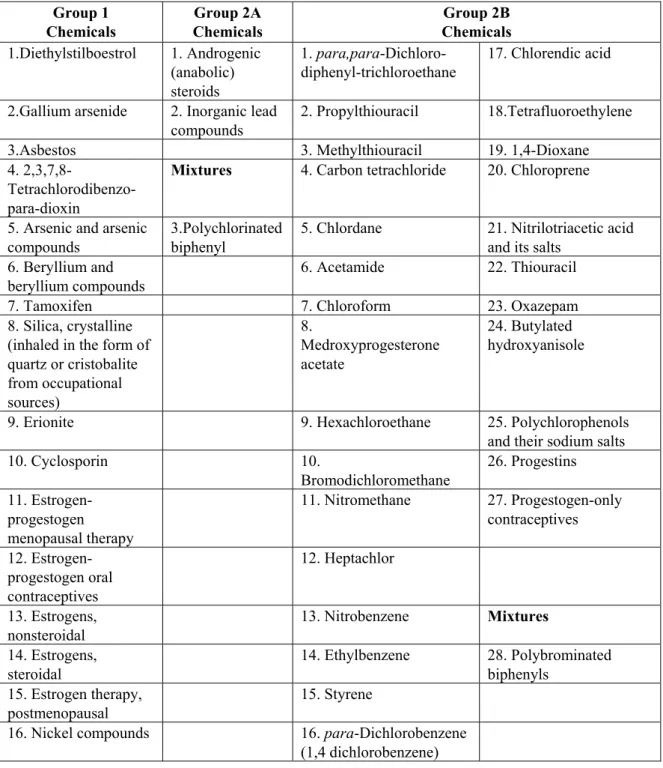
genotoxic carcinogens and would therefore not be recognized in REACH (Table 1). All the non-genotoxic carcinogens in IARC are listed in Table 2.
3.2 Margin of Eexposure (MOE)
In a non-linear (threshold) dose response, as in the case of non-genotoxic carcinogens, the margin of exposure (MOE) may be used for human risk assessment (Barlow et al., 2006). The TD10, a dose associated with an extra lifetime tumor risk of 10%, has been selected as the benchmark dose by the U.S. Environmental Protection Agency because it is the lowest estimate of statistically significant increases in tumor incidence (Andersen et al., 2000; Gold et al., 2003). Thus, the MOE is the ratio between the Lower-bound Toxic Dose (LTD10), a lower 95% confidence limit on the Toxic Dose (TD10) and the anticipated human exposure level. An estimate of the LTD10 may be obtained by using the TD50/10.2 (Gold et al., 2003). In most instances, the carcinogenic potency database was used to obtain TD50 values from standardized chronic, long-term animal cancer tests (Gold et al., 2005). The carcinogenic potency value (TD50) was defined as the daily dose rate in mg/kg body weight/day to
induce tumors in half of test animals that would have remained tumor-free without treatment. In Europe, the MOE is generally derived from the ratio between the NOAEL and the anticipated human exposure levels. In this report, the MOE was derived from both the NOAEL and LTD10 for
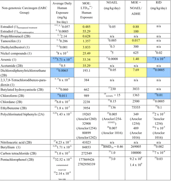
comparison. A possible human hazard is associated with non-genotoxic carcinogens that have an MOE lower than 100 for consumers or an MOE lower than 50 for workers (Table 3). Only 18 out of the 47 non-genotoxic carcinogens in groups 1, 2A and 2B of the IARC monographs were analyzed (Table 3) because either the average daily human exposure (mg/kg bw/day) and/or rodent TD50 values were not available for the MOE calculation.
Our analysis showed that when the MOE was derived from the LTD10, 7 out of the 18 (39%) non-genotoxic carcinogens analyzed had an MOE lower than 50, in comparison to 4 out of the 14 (29%) non-genotoxic carcinogens when the MOE was derived from the NOAEL (Table 3). The NOAEL was not available in 4 out of the 18 non-genotoxic carcinogens analyzed and thus, only 14 agents were analyzed. Thus, under REACH guidelines, between 29 and 39% of non-genotoxic carcinogens of high risk will not be detected. It is important to point out that estradiol, propylthiouracil,
Table 2: List of non-genotoxic carcinogens in IARC monographs Group 1 Chemicals Group 2A Chemicals Group 2B Chemicals 1.Diethylstilboestrol 1. Androgenic (anabolic) steroids 1. para,para-Dichloro-diphenyl-trichloroethane 17. Chlorendic acid
2.Gallium arsenide 2. Inorganic lead compounds
2. Propylthiouracil 18.Tetrafluoroethylene 3.Asbestos 3. Methylthiouracil 19. 1,4-Dioxane 4.
2,3,7,8- Tetrachlorodibenzo-para-dioxin
Mixtures 4. Carbon tetrachloride 20. Chloroprene
5. Arsenic and arsenic compounds
3.Polychlorinated biphenyl
5. Chlordane 21. Nitrilotriacetic acid and its salts
6. Beryllium and beryllium compounds
6. Acetamide 22. Thiouracil
7. Tamoxifen 7. Chloroform 23. Oxazepam
8. Silica, crystalline (inhaled in the form of quartz or cristobalite from occupational sources) 8. Medroxyprogesterone acetate 24. Butylated hydroxyanisole
9. Erionite 9. Hexachloroethane 25. Polychlorophenols and their sodium salts
10. Cyclosporin 10. Bromodichloromethane 26. Progestins 11. Estrogen-progestogen menopausal therapy 11. Nitromethane 27. Progestogen-only contraceptives 12. Estrogen-progestogen oral contraceptives 12. Heptachlor 13. Estrogens, nonsteroidal 13. Nitrobenzene Mixtures 14. Estrogens, steroidal 14. Ethylbenzene 28. Polybrominated biphenyls 15. Estrogen therapy, postmenopausal 15. Styrene
16. Nickel compounds 16. para-Dichlorobenzene (1,4 dichlorobenzene)
tamoxifen and diethylstilbestrol are drugs and are not included under REACH guidelines because human risk assessment is based on a risk/benefit approach. If these are eliminated, the MOE was lower than 50 in 3 out of 14 (21%) non-genotoxic carcinogens when derived from the LTD10, in comparison to 2 out of 11 (18%) when the MOE was derived from the NOAEL. These results suggest that the NOAEL and the LTD10 result in almost equivalent number of non-genotoxic carcinogens that are possible human hazards with an MOE lower than 50 (Table 3). Further, it is recommended that in the availability of human exposure levels, human cancer risk parameters should be adjusted accordingly in order for the MOE to be greater than 100 through additional safety factors, in addition to the current intra- and inter-species uncertainty factors (i.e. 10 x 10 = 100) and efforts need to be made to minimize human carcinogen exposure.
3.3 Reference Dose (RfD)
The RfD represents a reference dose (with uncertainty factors) of a daily exposure to the human population (including sensitive sub-groups) that is likely to be without appreciable risk of deleterious effects during a lifetime.A question is raised as to whether the reference dose for chronic oral exposure (RfD) for non-genotoxic carcinogens is sufficient to protect against human carcinogenesis or if there is a greater risk than can be covered by the RfD. The National Toxicology Program lead by the U.S. Environmental Protection Agency (EPA) has developed an Integrated Information System (IRIS) to assess human cancer risk associated with chemical exposure. The RfD is estimated from the NOAEL after adjustment for uncertainty factors (UF) and modifying factors (MF) based on professional judgment of the entire data base of the chemical (RfD = NOAEL/[UF x MF]). The EPA considers any dose level above the RfD to be unacceptable with the possibility of adverse effects. In order to address whether RfD is a good estimate of a safe daily human dose, the average daily human exposure was compared to RfD values, where available in IRIS (Table 3). Results indicated that 3 out of 10 of the chemicals analyzed had higher average daily human exposure levels than covered by the RfDs, as in the case of arsenic, dichlorodiphenyltrichloroethane and chloroform (Table 3). The RfD was only available in 10 out of the 18 non-genotoxic carcinogens analyzed. These results suggest that if
unknown carcinogens are evaluated based on RfD values, in approximate 30% of the cases, the average daily exposure of carcinogens will be higher than predicted. In view of these results, additional safety factors may be needed when dealing with non-genotoxic carcinogens and efforts need to be made to reduce human exposure to non-genotoxic carcinogens.
3.4 Implications
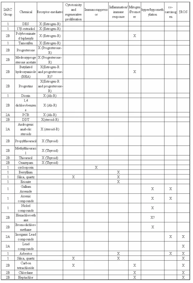
Overall, our analysis demonstrated that 13% (47/362) of chemicals and mixtures in IARC Groups 1, 2A and 2B chemicals were considered non-genotoxic carcinogens; with the highest proportion (23%, 16/71) in Group 1, which are known human carcinogens. The non-genotoxic carcinogens in Groups 1, 2A and 2B have distinct plausible modes of action leading to carcinogenicity (Table 4). Currently, test that detect some of these modes of action are used as alerts for potential carcinogenicity. For example, further tests are prompted if an agent is positive in the cell transformation, endocrine disruption or intercellular gap junction communication assay. Analysis of the MOE, determined from the NOAEL or LTD10 indicated that 29 and 39%, respectively of non-genotoxic carcinogens were associated with a potential human hazard. Similar results were obtained when evaluating RfD values obtained from NOAEL and a potential human hazard was detected in 3 out of 10 of non-genotoxic carcinogens analyzed (i.e. average human daily exposure > RfD). The low MOE values found in some of the non-genotoxic carcinogens evaluated suggests that the magnitude by which the NOAEL exceeds the
estimated human exposure dose needs to be re-examined with the inclusion of additional safety factors. In terms of risk, which is the likelihood of harm induced by non-genotoxic carcinogens, our results show a potential risk associated with 29-39% of non-genotoxic carcinogens in IARC groups 1, 2A and 2B; thus justifying the need for further research to identify novel methods for their detection in the absence of carcinogenicity studies. In terms of hazard which is the potential of an agent to cause harm, proper safety measures cannot be implemented if non-genotoxic carcinogens are not detected and classified as carcinogens. The implications of these findings for REACH guidelines are an urgent need for the development of alternative methods for the early detection of non-genotoxic carcinogens for proper human cancer risk and hazard assessment.
Table 3: The MOE and RfD of Non-genotoxic carcinogens
Non-genotoxic Carcinogen (IARC Group) Average Daily Human Exposure (mg/kg bw/day) MOE: LTD10**/ Human Exposure NOAEL (mg/kg/day) MOE = NOAEL/ ADHE RfD (mg/kg/day)
Estradiol (1)menopausal treatment Estradiol (1)oral contraceptive
***, #, 1 0.057 #, 10.0005 0.485 55.29 2 0.05 0.88 100 n/a
Propylthiouracil (2B) #, 32.14 0.628 n/a n/a n/a
Tamoxifen (1) #, 40.286 1.357 50.005 0.017 n/a
Diethylstilbestrol (1) #, 60.001 3.833 70.3 300 n/a
Nickel compounds (1) 88 x 10-3 25.49 95 625 90.02
Arsenic (1) #,105.71 x 10-4 33.34 110.0008 1.40 113 x 10-4
Acetamide (2B) 120.5 35.29 n/a n/a n/a
Dichlorodiphenyltrichloroethane (2B)
130.0065 193.1 140.05 7.69 140.0005
2,3,7,8-Tetrachlorodibenzo-para-dioxin (1)
#, 156 x 10-9 384 n/a n/a n/a
Butylated hydroxyanisole (2B) #, 160.060 662 17230 3833 n/a
Chloroform (2B) 180.011 989 19LOAEL = 15 1363 190.01
Chlordane (2B) 206.0 x 10⁻⁵ 2238 210.15 2500 210.0005
Ethylbenzene (2B) 221.8 x 10⁻³ 3954 23136 75555 230.1
Polychlorinated biphenyls (2A) #,241.43 x 10-5 19265
(Aroclor1260) 32908 (Aroclor1254) 80899 (Aroclor1242) 250.005 (Aroclor1254-LOAEL) 260.007 (Aroclor 1016) 349 (Aroclor 1254) 489 (Aroclor 1016) 252 x 10-5 (Aroclor 1254) 267 x 10-5 (Aroclor 1016)
Nitriloacetic acid (2B) 274.23 x 10⁻³ 41023 n/a n/a n/a
Beryllium (1) #,281.71 x 10-6 84853 29BMD10 = 0.46 269005 290.002 Carbon tetrachloride (2B) 301.0 x 10⁻⁵ 272549 311.0 100000 317 x 10-4 Pentachlorophenol (2B) 3232.52 x 10-9 contaminated reservoir 322.14 x 10⁻⁹ low risk 177869426 2702950339 333.0 9.2 x 10⁸ 1.4 x 10⁹ 330.03
ADHE = Average daily human exposure; NOAEL = no observed adverse effect level; RfD = reference dose for chronic oral exposure based on the assumption that thresholds exist for certain toxic effects; LOAEL = lowest observed adverse effect level; BMD10 = benchmark dose10 is the dose corresponding to a 10% increase in incidence of these effects compared with
controls, n/a = values not available in IRIS (Integrated Risk Information System).
*Data was only available in 18/47 non-genotoxic carcinogens in groups 1, 2A and 2B in the IARC monographs
(http://monographs.iarc.fr/ENG/Classification/ListagentsCASnos.pdf).
**LTD
10 = TD50/10.2 (Gold et al., 2003); TD50 values were obtained from http://potency.berkeley.edu/. ***Refer to Appendix 1 for references.
#Values were calculated in mg/day and divided by 70kg (average body weight of a person).
4
Alternative methods for detecting non-genotoxic
carcinogens
Justification for the development of alternative methods for the early detection of non-genotoxic carcinogens include their overall presence (13%) in Groups 1, 2A and 2B of IARC, their presence (23%) in the known human carcinogens (IARC Group 1), the potential risk associated with them (30% had average daily exposures > RfD values), and the low MOE when using either LTD10 (39% had MOE < 50) or NOAEL (29% had MOE < 50). When searching for alternative methods for detecting non-genotoxic carcinogens, consideration needs to be made to their wide spectrum of modes of action including mitogenic induction, inhibition of gap-junctional intercellular communications, endocrine modifiers, generation of reactive oxygen species, and/or DNA methylation (Table 4). Several
alternative methods have been suggested for the detection of non-genotoxic carcinogens. Among these include activity relationships (SARs) of non-genotoxic carcinogens and quantitative structure-activity relationships ((Q)SARs), measurements of replicative DNA synthesis as an indicator of cell proliferation, the in vitro cell transformation assays, and the use of gene expression profiles with mechanistic networks for the identification of potential markers of non-genotoxic carcinogens.
4.1 Structure-Activity Relationship (SAR)
Structure-activity relationship (SAR) models for detection of non-genotoxic carcinogens have used several markers of in vitro cell toxicity including inhibition of gap-junctional intercellular
communications, modulation of apoptosis, and induction of cellular proliferation. In this approach, the structural features of the non-genotoxic carcinogen associated with toxicity or ligand binding, as in the case of estrogen, peroxisome proliferators and tubulin protein receptors, were analyzed (Combes, 2000). This information was used for quantitative structure-activity relationship ((Q)SAR) where chemical structure was quantitatively correlated with a defined process, such as biological activity or chemical reactivity. A major assumption in this model is that similar molecules have similar activities. For regulatory analysis, (Q)SAR models must satisfy the principles outlined by the Organization for Economic Cooperation and Development (OECD, 2003; OECD, 2004). Briefly, (Q)SAR models must have a defined endpoint, an unambiguous algorithm, and measures of goodness-of-fit, robustness, predictivity, and applicability domain should be known to the regulator.
A SAR analysis has been done for a variety of natural, synthetic and environmental estrogens. Structural comparisons of endogenous 17β-estradiol with xenoestrogens showed that a rigid ring
structure and the 3-position H-bonding ability of phenols is a significant requirement for estrogen receptor binding (Fang et al., 2001). This study provided distinguishing criteria that can be used for the identification of potential estrogens.
A SAR analysis of 10 000 chemicals representative of the ‘universe of chemicals’ investigated mechanistic similarities between toxicological phenomena such as inhibition of gap junctional intercellular communications (GJIC), binding to aryl hydrocarbon receptor, α2µ-globulin-mediated nephropathy, inhibition of CYP2D6, skin permeability, Salmonella mutagenicity, SOS DNA repair, rodent carcinogenicity, developmental toxicity in humans, developmental toxicity in hamsters, allergic contact dermatitis, eye irritation, sensory irritation, respiratory hypersensitivity, rat LD50, cell toxicity in HeLa cells and cell toxicity in BALB/c-3T3 cells. Results indicated that inhibition of GJIC is involved in cancer induction by non-genotoxic carcinogens through non-genotoxic mechanisms such as inflammation, cell toxicity, cell proliferation, inhibition of cell differentiation and apoptosis
(Rosenkranz et al., 2000). This study suggests that inhibition of GJIC may be used a putative marker for the identification of non-genotoxic carcinogens.
Given that non-genotoxic carcinogens have a wide range of modes of action, development of SAR models identifying various models from various laboratories are needed (Combes, 2000). For instance, if regulators want to use a (Q)SAR model to identify endocrine modifiers, SARs need to be developed to identify endocrine modifiers that bind to 1) estrogen receptors, 2) thyroid hormone receptors, 3) aryl hydrocarbon receptors, 4) androgen receptors, 5) IP3 receptor/channel pores, or 6) progesterone receptors. Thus, just to identify endocrine modifiers, at least six SARs are needed, demonstrating that (Q)SAR predictive models are only efficient for very specific group of chemicals with specific mechanisms.
Although SAR models offer an in silico approach for detecting non-genotoxic carcinogens, the wide spectrum of mechanism of actions suggests that a large number of SAR models need to be developed for proper (Q)SAR analysis.
4.2 Replicative DNA Synthesis (RDS)
As presented in Table 4, many non-genotoxic carcinogens are mitogenic inducers resulting in an increase in cellular proliferation. In vivo studies using rat hepatocytes indicate that many
hepatocarcinogens (genotoxic and non-genotoxic) have accelerated hepatocyte division (Uno et al., 1999; Uno et al., 1992a; Uno et al., 1992b; Yoshikawa, 1996). A comprehensive screening analysis by
Yoshikawa (1996) showed that measurement of replicative DNA synthesis (RDS) resulted in a positive response in 82.5% (52/63) of the non-genotoxic and genotoxic heptatocarcinogens while a negative result was obtained in 77% (24/31) of the non-carcinogens. The major advantages of the RDS test are the in vivo response to the agent in question and short-term nature of the assay. The major
disadvantages include the high doses required to attain responses, the false-positives obtained because of regenerative cell proliferation due to acute toxicity, and the false-negative results obtained if the liver is not the primary target for the agent in question (Yoshikawa, 1996). Given the high degree of false positives and negatives, the RDS test should be performed in conjunction with other short-term tests for validation of results. The correlation between significant increases in RDS and carcinogenesis may be enhanced by measurements of apoptotic cell rate (Elcombe et al., 2002).
The RDS assay may be used in conjunction with other test for the detection of putative non-genotoxic carcinogens. Results need to be evaluated with caution because of the high incidence of false positives and negatives. This assay may be improved by measurements of apoptotic cell rate and mechanistic justification of results.
4.3 In vitro cell transformation assay
The in vitro cell transformation assays in BALB/c 3T3, C3H10T1/2, or Syrian Hamster Embryo (SHE) cells, detect the carcinogenic potential of a chemical by their ability to cause morphologic
transformation (OECD, 2001). Correlation between in vivo carcinogenicity and the in vitro
transformation of BALB/c 3T3, C3H10T1/2 and SHE cells are demonstrated in Table 5. Validation studies indicate that SHE > C3H10T1/2 > BALB/c 3T3 cells in relation to sensitivity, specificity, predictivity and validity for the detection of non-genotoxic carcinogens (Table 5). Overall, the SHE transformation assay offers many advantages over other test systems including the normality of the cells, low spontaneous transformation, capacity for metabolic activation, rapidity of phenotypic changes, and reproducibility of results with the same chemical (OECD, 2001). The major disadvantages include the inter- and intralaboratory variation due to subjectivity in the scoring of transformed cells, the periodic preparation of primary cultures because cells from various preparations differ in transformation capacity, variation in cloning efficiency and transformation frequency due to composition of fetal calf serum, and the use of an initiation-promotion protocol in order to enhance the transformation frequency (OECD, 2001).
Given that most non-genotoxic carcinogens are mitogenic inducers, treatment with an initiating agent is sometimes necessary to detect the promoting effects of a chemical. The use of initiated cells
(v-Ha-ras-transfected BALB/c 3T3 cell line, Bhas 42 cells) shortens the experimental period from 4-6 weeks to 2.5 to 3 weeks after cell inoculation because there is no need to treat cell with an initiating agent nor the subsequent cultivation for an expression period (Ohmori et al., 2004). In an interlaboratory study of non-genotoxic carcinogens using the Bhas transformation assay, several issues regarding the protocol were identified. Medium selection and freshness was an issue between laboratories because foci were smaller in old medium and labs using M10F medium yielded low transformation responses (Ohmori et al., 2005). Although scoring transformed cells is obvious from their basophilic staining and spindle-shape, there were inconsistencies between laboratories in the scoring of some irregular shaped foci as transformed cells. In order to address this issue, the assay was extended to 3 weeks, with no medium change during the last week. This modification lead to comparable scoring between laboratories (Ohmori et al., 2005). Another issue raised was the presence of a high number of foci in the controls in some laboratories and not in others. The criteria for significance was originally set as a three-fold increase above the control, but due to the inconsistencies between laboratories, the criteria for significance was reduced to a two-fold increase requirement (Ohmori et al., 2005). Although the highest test concentration was determined as a concentration exhibiting more than 30% cytotoxicity, in practice, it was difficult to determine the appropriate test concentration for some chemicals (Ohmori et al., 2005). Overall, there was a large interlaboratory variability in focus formation in the negative control and inconsistent results between laboratories (Ohmori et al., 2005). Additionally, in vitro transformation assay do not provide any information on the mechanism of action of the agent in question, thus, other tests are needed to validate results.
Analysis of the in vitro cell transformation assay indicates that SHE > C3H10T1/2 > BALB/c 3T3 cells in relation to sensitivity, specificity, predictivity and validity for the detection of non-genotoxic
carcinogens. The SHE transformation assay had the best results but there was a high degree of variation; a problem also found in Bhas cells. Results from in vitro cell transformation assays need to be evaluated with caution and results should be mechanistically justified.
Table 5: Correlation between in vivo carcinogenicity and in vitro transformation of BALB/c 3T3,
C3H10T1/2 and SHE cells*
Assessment BALB/c 3T3 C3H10T1/2 SHE % Predicted correctly for carcinogenicity 54 (28/52) 59 (20/34) 78 (65/83) % False negatives 33 (17/52 23.5 (8/34) 16 (13/83) % Inconclusive (positive in vivo
carcinogenicity)
11.5 (18/34) 17.6 (6/34) 6 (5/83) % Predicted correctly for negative
carcinogenicity
53 (18/34) 47 (8/17) 78 (35/45) False positives 32 (11/34) 23.5 (4/17) 18 (8/45) % Inconclusive (negative for in vivo
carcinogenicity) 15 (5/34) 29.4 (5/17) 4 (2/45) Validation % Sensitivity 62 (28/45) 71 (20/28) 84 (65/78) % Specificity 62 (18/29) 67 (8/12) 81 (35/38) % Positive predictivity 72 (28/39) 80 (20/24) 89 (65/73) % Negative predictivity 51 (18/35) 50 (8/16) 73 (35/48) % Validity 54 (54/85) 55 (28/51) 78 (100/128) *Results obtained from OECD detailed review paper (OECD, 2001).
4.4 Toxicogenomics
The comparison of gene expression profiles via microarray analysis may provide insights on the mechanisms and pathways altered shortly after exposure to non-genotoxic carcinogens. In a gene expression profiling study by Ellinger-Ziegelbauer (2005), rats were dosed with the non-genotoxic hepatocarcinogens methapyrilene (MP) and Wy-14643 (Wy) for 1, 3, 7 and 14 days, while piperonylbutoxide (PB) and diethylstilbestrol (DES) was administered for 1, 3, and 7 days. Livers were collected 24 hours after the last dose and RNA was extracted for microarray analysis. This time-course study showed that the most characteristic responses for each non-genotoxic carcinogen was obtained after 7 days of treatment, while mitogenic responses were detected after days 1 and 3 (Ellinger-Ziegelbauer et al., 2005). The consistent upregulation of apurinic/apurimidic endonuclease APEX1 by all non-genotoxic carcinogens and the compound-specific oxidative stress (upregulation of proteasome and mitochondrial heat shock proteins (HSPs) by PB and Wy, of cytosolic HSPs by all non-genotoxic carcinogens, of antioxidant response element target genes by PB, MP and DES) may lead to indirect base modifications important for cancer initiation. In terms of cancer promotion,
non-genotoxic carcinogens had a distinct effect on DNA replication and cell cycle progression. Non-genotoxic carcinogens upregulated genes involved in DNA replication (MCM6, PCNA, TOP2A), in cell cycle progression and entry to mitosis (cyclin B1, CDC2), and spindle formation (tubulins, TUBA1, TUBB5). The different pathways altered by non-genotoxic carcinogens were not surprising given their wide range of modes of action. For this reason, combinations of pathway-associated gene expression profiling may be necessary to identify and characterize putative non-genotoxic carcinogens (Ellinger-Ziegelbauer et al., 2005).
Predictive toxicogenomic studies of non-genotoxic carcinogens in rat liver have identified six genes that were differentially expressed in non-genotoxic carcinogens in comparison to genotoxic
carcinogens. Three genes were upregulated six genes were differentially expressed in non-genotoxic carcinogens relative to genotoxic carcinogens. Three genes were induced by non-genotoxic
carcinogens: the nuclear transport factor 2, progesterone receptor membrane component 1, and phenobarbital-inducible uridine diphosphate glucoronyltransferase. The nuclear transport factor 2 is necessary for the shuttling of molecules from the cytosol into the nucleus; progesterone receptor membrane component 1 is thought to mediate interactions between endogenous steroids, such as progesterone and estrogen, and aryl hydrocarbon receptor agonist; and phenobarbital-inducible uridine diphosphate glucoronyltransferase modulates the clearance of thyroxine by glucoronidation and when induced, it decreases thyroid hormone levels in the plasma, and stimulates thyroid stimulating hormone synthesis. Three genes were downregulated by non-genotoxic carcinogens: metallothionein 1A, suppressor of lin-12 homolog, and methionine adenosyltransferase. Metallothionein 1A is an important metalloprotein that protects the cell against heavy metal toxicity; suppressor of lin-12 homolog is involved in cell fate determination; and methionine adenosyltransferase and its gene product
S-adenosyl-L-methionine are involved in DNA methylation and silencing of hepatic genes. The six genes were used to screen 123 compounds in a database. These genes were capable of identifying non-genotoxic carcinogens with 88.5% prediction accuracy estimated by cross validation and 84% accuracy when samples were hybridized to CodeLink oligo-based arrays, including a number of extrahepatic tumor-inducing compounds (Nie et al., 2006). Further evaluation of the signature to predict
carcinogenicity in extra-hepatic tissues is needed because extrahepatic tumor-inducing compounds were also detected by Fielden et al. (2008). In a study of rat hepatic gene expression data, a signature was generated to predict the likelihood of non-genotoxic carcinogens to induce liver cancer.
Independent validation of this signature demonstrated 86% sensitivity and 81% specificity (Fielden et al., 2007). A mechanism-based approach was utilized to obtain this signature and included known hepatocarcinogens that induced cancer via regenerative proliferation, receptor activation, peroxisome proliferation and endocrine disruption (Fielden et al., 2007). Overall, the test chemicals clustered most
closely to the reference chemical with similar known mechanism of action. Inter-laboratory comparison of signatures obtained by Nie et al. (2006) and Fielden et al. (2007) showed similar accuracy in
classification of test chemicals with decreased test sensitivity and specificity (Fielden et al., 2008). Further inter-laboratory studies are needed to address differences in animal treatment protocols, microarray platforms and stochastic errors (Fielden et al., 2008).
The use of toxicogenomic of known non-genotoxic carcinogens to identify potential pathway-associated gene expression profiles for the detection of putative non-genotoxic carcinogens needs further investigation. Validation studies testing sensitivity, specificity, positive and negative
predictivity and validity are also needed to assess their use in identification and characterization of non-genotoxic carcinogens.
5
Conclusions
The first aim of this report was to assess the human risk and hazard associated with non-genotoxic carcinogens since these compounds are not detected under REACH guidelines. Analysis of the IARC monographs indicated that 23% of chemicals in group 1 (known to be carcinogenic to humans), 5% of chemicals in group 2A (probably carcinogenic to humans) and 12% of chemicals in group 2B (possibly carcinogenic to humans) are considered non-genotoxic carcinogens. Of particular concern is that the highest percentage of non-genotoxic carcinogens was found in Group 1, which consists of chemicals that are proven to be carcinogenic to humans by IARC. Analysis of the MOE, determined from the NOAEL or LTD10 indicated that 29 and 39%, respectively of non-genotoxic carcinogens were associated with a potential human hazard. Similar results were obtained when evaluating RfD values obtained from the NOAEL, and a potential human hazard was detected in 3 out of 10 of non-genotoxic carcinogens analyzed (i.e. average human daily exposure > RfD). The implications of our results to risk management are the re-evaluation of the current safety factors associated with non-gentoxic
carcinogens and efforts need to be made to minimize human exposure. The implications of our results to hazard management are more severe given that proper safety measures cannot be employed if non-genotoxic carcinogens are not detected and classified as carcinogens.
The second aim of this report was to investigate possible alternative methods that could be included in REACH for the detection of non-genotoxic carcinogens in the absence of a 2-year carcinogenicity assay. The replicative DNA synthesis assay has a high degree of false positives and negatives and more validation studies are needed to address whether additional measurements of apoptotic cell rate reduce the rate of false positives and negatives. SAR studies are limited and some potential markers of cell toxicity have been suggested including ligand-binding to specific cellular receptors and target
molecules, inhibition of GJIC, inhibition of tubular polymerization, modulation of apoptosis, induction of cell proliferation (including DNA synthesis, mitosis and altered differentiation), induction of peroxisomes and drug metabolizing enzymes, induction of hormonal imbalance and two-stage cell transformation (promoters) (Combes, 2000). Given the complexity of the above mentioned mechanisms, more SAR studies are needed for proper (Q)SAR predictor models. The use of toxicogenomics offers the most promising results in terms of reliability and reducing the number of animals, time and cost. Additional validation studies of the signatures obtained by Ellinger-Ziegelbauer et al. (2005), Nie et al. (2006) and Fielden et al. (2007) where non-genotoxic carcinogens were
identified with high prediction accuracy are needed. Of particular interest, is the detection of extra-hepatic carcinogens by gene expression profiling and using liver as a surrogate tissue for their
identification. The use of toxicogenomics to identify molecular fingerprints or ‘red flags’ associated with known non-genotoxic carcinogens is an area that needs further investigation.
6
Recommendations for future research
This report has outlined the importance of finding alternative methods for detecting putative non-genotoxic carcinogens in the absence of the 2-year carcinogenicity bioassay. Non-non-genotoxic carcinogens are not detected under REACH given that only genotoxic endpoints are measured. Detection of non-genotoxic carcinogens is difficult because they elicit epigenetic changes including inflammation, cell toxicity, cell proliferation, inhibition of cell differentiation and apoptosis.
Development of SAR models identifying various markers of cell toxicity tested in various laboratories is needed for proper (Q)SAR analysis. The limited numbers of gene expression profiling studies indicate that there isn’t a single gene or biochemical pathway that identifies known non-genotoxic carcinogens. Promising results have been obtained using a combination of multiple pathway-specific gene expression profiles. More short-term gene profiling studies of non-genotoxic carcinogens in target and non-target tissues are necessary to validate their use for the identification of putative non-genotoxic carcinogens.
Acknowledgements
We wish to thank Prof. Dr. Wout Slob, Dr. Wim Mennes and André Muller (RIVM, Centre for Substances and Integrated Risk Assessment) for their critical comments regarding this report.
References
Andersen ME, Meek ME, Boorman GA, Brusick DJ, Cohen SM, Dragan YP, Frederick CB, Goodman JI, Hard GC, O'Flaherty EJ, Robinson DE. 2000. Lessons learned in applying the U.S. EPA proposed cancer guidelines to specific compounds. Toxicol Sci 53(2):159-172.
Barlow S, Renwick AG, Kleiner J, Bridges JW, Busk L, Dybing E, Edler L, Eisenbrand G, Fink-Gremmels J, Knaap A, Kroes R, Liem D, Muller DJ, Page S, Rolland V, Schlatter J, Tritscher A, Tueting W, Wurtzen G. 2006. Risk assessment of substances that are both genotoxic and carcinogenic report of an International Conference organized by EFSA and WHO with support of ILSI Europe. Food Chem Toxicol 44(10):1636-1650.
Bolt HM, Degen GH. 2004. Human carcinogenic risk evaluation, part II: contributions of the EUROTOX specialty section for carcinogenesis. Toxicol Sci 81(1):3-6.
Combes RD. 2000. The use of structure-activity relationships and markers of cell toxicity to detect non-genotoxic carcinogens. Toxicol In Vitro 14(4):387-399.
EC. 2003. Technical guidance document on risk assessment in support of Commission Directive 93/67 EEC on risk assessment for new notified substances and Commission Regulation (EC) No. 1488/94 on risk assessment for existing substances. European Commission (EC) (Internet publication at http://ecbjrcit) Part I.
Elcombe CR, Odum J, Foster JR, Stone S, Hasmall S, Soames AR, Kimber I, Ashby J. 2002. Prediction of rodent non-genotoxic carcinogenesis: evaluation of biochemical and tissue changes in rodents following exposure to nine non-genotoxic NTP carcinogens. Environ Health Perspect 110(4):363-375.
Ellinger-Ziegelbauer H, Stuart B, Wahle B, Bomann W, Ahr HJ. 2005. Comparison of the expression profiles induced by genotoxic and non-genotoxic carcinogens in rat liver. Mutat Res 575(1-2):61-84.
EU. 2003. Proposal for a regulation of the European Parliament and the Council and a proposal for a directive of the European Parliament and the Council. COM(644).
Fang H, Tong W, Shi LM, Blair R, Perkins R, Branham W, Hass BS, Xie Q, Dial SL, Moland CL, Sheehan DM. 2001. Structure-activity relationships for a large diverse set of natural, synthetic, and environmental estrogens. Chem Res Toxicol 14(3):280-294.
Fielden MR, Brennan R, Gollub J. 2007. A gene expression biomarker provides early prediction and mechanistic assessment of hepatic tumor induction by non-genotoxic chemicals. Toxicol Sci 99(1):90-100.
Fielden MR, Nie A, McMillian M, Elangbam CS, Trela BA, Yang Y, Dunn RT, 2nd, Dragan Y, Fransson-Stehen R, Bogdanffy M, Adams SP, Foster WR, Chen SJ, Rossi P, Kasper P, Jacobson-Kram D, Tatsuoka KS, Wier PJ, Gollub J, Halbert DN, Roter A, Young JK, Sina JF, Marlowe J, Martus HJ, Aubrecht J, Olaharski AJ, Roome N, Nioi P, Pardo I, Snyder R, Perry R, Lord P, Mattes W, Car BD. 2008. Interlaboratory evaluation of genomic signatures for predicting carcinogenicity in the rat. Toxicol Sci 103(1):28-34.
Foth H, Degen GH, Bolt HM. 2005. New aspects in the classification of carcinogens. Arh Hig Rada Toksikol 56(2):167-175.
Gold LS, Gaylor DW, Slone TH. 2003. Comparison of cancer risk estimates based on a variety of risk assessment methodologies. Regul Toxicol Pharmacol 37(1):45-53.
Gold LS, Manley NB, Slone TH, Rohrbach L, Garfinkel GB. 2005. Supplement to the Carcinogenic Potency Database (CPDB): results of animal bioassays published in the general literature through 1997 and by the National Toxicology Program in 1997-1998. Toxicol Sci 85(2):747-808.
Golden R, Gandy J, Vollmer G. 2005. A review of the endocrine activity of parabens and implications for potential risks to human health. Crit Rev Toxicol 35(5):435-458.
Gonzalez FJ, Shah YM. 2008. PPARalpha: Mechanism of species differences and hepatocarcinogenesis of peroxisome proliferators. Toxicology 246(1):2-8.
Gurer-Orhan H, Orhan H, Vermeulen NP, Meerman JH. 2006. Screening the oxidative potential of several mono- and di-halogenated biphenyls and biphenyl ethers in rat hepatocytes. Comb Chem High Throughput Screen 9(6):449-454.
Haber LT, Diamond GL, Zhao Q, Erdreich L, Dourson ML. 2000. Hazard identification and dose response of ingested nickel-soluble salts. Regul Toxicol Pharmacol 31(2 Pt 1):231-241. IARC. 1987. Steroidal estrogens. Summaries and evaluations. IARC monographs on the evaluation of
carcinogenic risks to humans, Lyon, France Suppl. 7.
IARC. 1997. Polychlorinated Dibenzo-para-Dioxins and Polychlorinated Dibenzofurans. Summary of Data Reported and Evaluation. IARC monographs on the evaluation of carcinogenic risks to humans, Lyon, France Vol. 69.
IARC. 1998. Polychlorinated biphenyls and polybrominated biphenyls. Summary of data reported and evaluation. IARC monographs on the evaluation of carcinogenic risks to humans, Lyon, France 18.
IARC. 1999. Occupational exposures in insecticide application and some pesticides. IARC monographs on the evaluation of carcinogenic risks to humans, Lyon, France Vol. 53.
Jackson MA, Stack HF, Waters MD. 1993. The genetic toxicology of putative non-genotoxic carcinogens. Mutat Res 296(3):241-277.