Free Bipedicled Radial Forearm and Posterior Interosseous Artery
Perforator Flap Phalloplasty
Edward De Wolf, MD,1Karel Claes, MD,1Casper E. Sommeling, MD,1Dries Opsomer, MD,1Mario Cherubino, MD,2 Salvatore Vieni, MD,3Stan Monstrey, MD, PhD,1and Salvatore D’Arpa, MD, PhD1,4
ABSTRACT
Introduction: The free radial forearm (FRFA) flap is universally still considered as the gold standard technique in penile reconstruction. Typically, a considerably large flap is required, often involving almost the entire circumference of the forearm. Partial necrosis may occur at the distal-most (dorsoradial) part of the flap as a result of insufficient perfusion.
Aim: To describe a new technique using the posterior interosseous artery (PIOA) to supercharge FRFA phalloplasty.
Methods: In a 12-month period, all patients having FRFA flap phalloplasty were enrolled. Perioperative, after complete flap dissection, an indocyanine green perfusion scan was performed. In case of insufficient perfusion at the distalmost part of the flap, a supramicrosurgical anastomosis was performed between the FRFA pedicle and the PIOA (artery only).
Main Outcome Measures: Studied outcomes included the rate of marginal necrosis, surgical time, post-operative posterior interosseous nerve damage and urethral complications (fistula, stenosis or necrosis). Results: A total of 27 FRFA flap phalloplasties was performed. Anastomosis of the PIOA was needed in 15 cases. No marginal necrosis was observed in these cases. There were no cases of postoperative posterior interosseous nerve damage. There were no significant differences in urethral complications (fistula, stenosis or necrosis) be-tween the 2 groups.
Clinical Implications: In selected cases where insufficient perfusion of the dorsoradial part of the flap is present, patients may benefit from arterial supercharging to prevent postoperative marginal necrosis.
Strength & Limitations: Strengths include a single surgeon, thus lending continuity of skill and technique, a consecutive series, and 100% short-term follow-up. Limitations include single institution series and a limited number of patients.
Conclusion: Arterial supercharging is effective in improving perfusion of large FRFA flaps used in phalloplasty when dorsoradial hypoperfusion is detected on an indocyanine green perfusion scan. It is a technically chal-lenging addition to the standard technique because of the small size of the vessels, the close relationship between the PIOA and the posterior interosseous nerve, and the vulnerability of the newly constructed intra-flap anas-tomosis. De Wolf E, Claes K, Sommeling CE, et al. Free Bipedicled Radial Forearm and Posterior Interosseous Artery Perforator Flap Phalloplasty. J Sex Med 2019;16:1111e1117.
Copyright! 2019, International Society for Sexual Medicine. Published by Elsevier Inc. All rights reserved. Key Words: Penile Reconstruction; Sex Reassignment Surgery; Transmen; Phalloplasty
INTRODUCTION
Penile reconstruction is a well-described procedure with multiple, both pedicled or free flaps, options available and a continuous evolution of techniques is seen.1,2 Selection of the technique will depend on a combination of surgeon’s and pa-tient’s preferences and characteristics.
The free radial forearm (FRFA) flap is by far the most frequently used flap and is universally still considered as the gold standard technique in penile reconstruction. Its use for this indication was first described by Chang and Hwang in 1984.3 Received December 31, 2018. Accepted March 18, 2019.
1Department of Plastic and Reconstructive Surgery, Gent University
Hos-pital, Ghent, Belgium;
2Microsurgery and Lymphatic Surgery Research Centre, Department of
Biotechnology and Science of Life, University of Insubria, Varese, Italy;
3Department of Surgical Oncological and Oral Sciences, University of
Palermo, Palermo, Italy;
4Plastic and Reconstructive Surgery, Department of Surgical Oncological
and Oral Sciences, University of Palermo, Palermo, Italy
Copyrightª 2019, International Society for Sexual Medicine. Published by Elsevier Inc. All rights reserved.
The technique meets many of the criteria for an ideal phalloplasty: a 1-stage procedure that is predictable and reproducible; minimal donor-site disfigurement; no functional loss; and an aesthetically acceptable phallus, including a competent neourethra that will allow for voiding while stand-ing, with tactile and erogenous sensation, and enough bulk to tolerate the insertion of an erectile prosthesis for sexual intercourse.4,5
Most commonly, the FRFA flap is harvested as a small to moderate-sized flap, often for use in head and neck reconstruc-tion. When the flap is used to construct a phallus, a considerably larger flap is required, often involving the entire circumference of the forearm.
In 2009, a retrospective review of 287 FRFA phalloplasties was performed in our institution; although approximately 12% required a revision, the total flap failure was less than 1%, and a minor degree of partial necrosis occurred in 7.3%.1This partial necrosis typically occurs at the distal-most (dorsoradial) part of the flap as a result of insufficient perfusion.
As a high-volume center of sex reassignment surgery with experience of >600 phalloplasties, constant optimization of the technique is demanded. The aim of this article is to describe a new technique developed at our department using the posterior interosseous artery (PIOA) to supercharge FRFA phalloplasty.
INDICATION FOR PROCEDURE
Arterial supercharging is indicated in cases of distal (dorsor-adial) hypoperfusion of the flap. If the flap shows, clinically and on indocyanine green (ICG) scanning, hypoperfusion in the territory of the posterior interosseous artery, a PIOA perforator shall be anastomosed to a side branch of the radial artery to optimize perfusion.
PREOPERATIVE PREPARATION
Before surgery, an Allen’s test is performed to ensure adequate perfusion of the hand postoperatively. The procedure will be performed in a single stage as a “tube-within-a-tube,” whereby the ulnar skin is used to create the penile urethra and the radial skin to create the external shaft. The flap is drawn before surgery based on a fixed template that is adjusted to each patient if needed (Figure 1). The flap has a minimal width of 16 cm, which can increase to 18e19 cm in thicker forearms. It encircles and often involves almost the entire circumference of the forearm, leaving only a narrow ulnar strip of 1e2cm. The radial skin is-land is 14 cm in length, the ulnar urethral skin isis-land is 17 cm in length. Subsequently, the average flap surface area is 228 cm2.
The position of the PIOA is marked with a line between the lateral epicondyle to the distal radioulnar joint within the mid-forearm between the radial and ulnar bones.
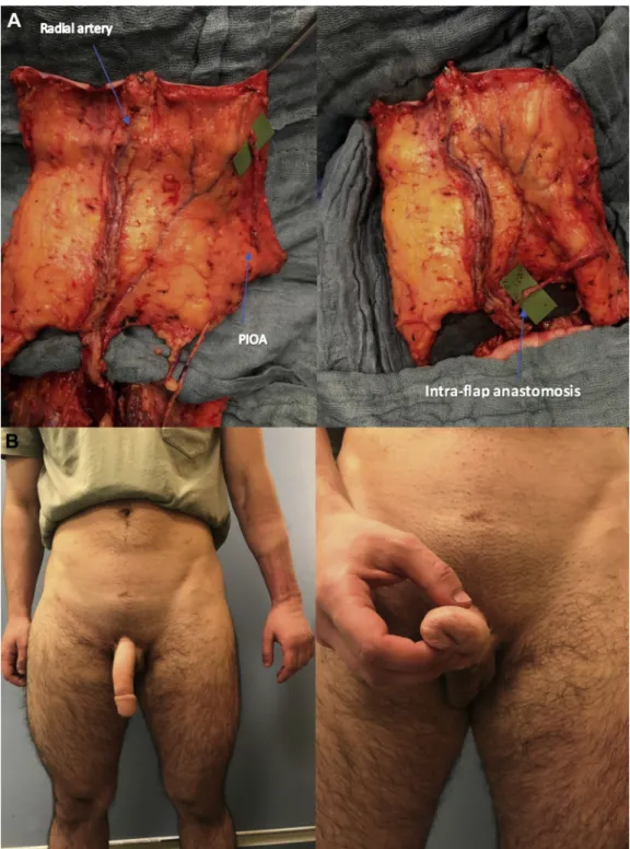
Figure 1. (Panel A) The flap at the end of the dissection seen from the superficial side. The vascular pedicle and sensory nerves can be seen. The urethral island is longer than the shaft to reach the reconstructed fixed part of the urethra more comfortably. (Panel B) The flap seen from its deep surface. A green background is placed behind the PIOA. The perforator (and pivot point) is in the middle of the flap in this case and the pedicle is proximal. Dissection shall be carried out proximally enough to let the pedicle comfortably reach the radial artery. PIOA ¼ posterior interosseous artery. Figure 1 is available in color online atwww.jsm.jsexmed.org.
INTRAOPERATIVE CONSIDERATIONS
In a typical phalloplasty procedure, 2 surgical teams operate simultaneously. The urologist performs the vaginal resection and reconstructs the fixed part of the urethra. At the same time, the plastic surgeons raise the free radial forearm flap under tourniquet. During radial dissection, the PIOA perforators are visualized along a line between the lateral epicondyle to the distal radio-ulnar joint between the extensor digiti minimi and the extensor carpi ulnaris muscles. The PIOA arises from the common interosseous artery and infrequently from the ulnar artery in the proximal forearm and runs along the intramuscular septum.6 The artery courses deep in the proximal anterior compartment of the forearm to pierce the upper aspect of the interosseous membrane and enter the posterior compartment of the forearm. In the proximal forearm, the PIOA runs deep alongside the posterior interosseous nerve (PIN). The PIOA emerges at the lower border of the supinator muscle and pierces the muscle belly of the abductor pollicis longus. In the distal forearm, the PIOA becomes superficial and travels on the extensor pollicis longus and extensor indicis muscles. The PIOA continues distally in the forearm to the proximal edge of the extensor retinaculum.
The PIOA anastomoses with the anterior interosseous artery 3 cm proximal to the distal radioulnar joint then extends to join the dorsal carpal arch. The anastomosis between the PIOA and anterior interosseous artery can reliably be found 5 cm proximal to the ulnar styloid at the proximal edge of the pronator quad-ratus muscle. Venous drainage is provided by paired venae comitantes that accompany the PIOA.
Along the intermuscular septum, the PIOA gives off 4e7 septocutaneous perforators. In the middle one-third of the forearm, the PIOA consistently gives off a cutaneous perforator accompanied by 2 venae comitantes that connect the superficial and deep venous systems.7 The biggest perforator is identified during flap dissection. Depending on its position and the size of the proximal and distal PIOA vessels, the PIOA pedicle can be either raised proximally or distally based (Figure 2).
The choice between a proximally or a distally based flap depends on the size of the perforator and of the distal PIOA. A distally based pedicle is generally preferred for 2 reasons: it allows more comfort-able reach of the radial artery for anastomosis (a proximal perforator is a better pivot point), and dissection is easier because it will not go deep in the septum and next to the PIN. When the distal PIOA is too small for anastomosis, a proximal pedicle is chosen.
If raised proximal, the pedicle is ligated distally from the main perforator and the PIOA pedicle is dissected proximally to its origin deep to the supinator muscle. If raised reverse, the pedicle is ligated proximally from the main perforator, and the PIOA is dissected as distally as possible.8,9
After suprafascial flap dissection is completed, the tourniquet is released, and the flap is left in place for 20 minutes to let the circulation settle. During this period the recipient vessels are
prepared. After 20 minutes, the flap is evaluated clinically and an ICG perfusion scan is performed.10In case of slow capillary refill and insufficient perfusion on ICG scan of the dorsoradial part of the flap, a supramicrosurgical anastomosis is performed between the FRFA pedicle and the PIOA (Figure 3).
Depending on the position of the perforator and the length and size of the PIOA pedicle, this anastomosis can be performed end-to-end to a side branch of the radial artery or to a superficial palmar branch of the radial artery (Figures 4A and 5A). End-to-side anastomosis are avoided due to the big discrepancy in wall thickness between the 2 vessels. During flap dissection and in preparation for the anastomosis, major side branches of the radial artery are ligated far from its origin to leave an adequate stump for anastomosis. This intra-flap anastomosis is artery-only. Figure 2. Dissection is centered on a line between the lateral epicondyle to the distal radioulnar joint within the mid-forearm between the radial and ulna bones. The septocutaneous perfora-tors are identified along the intermuscular septum. The biggest septocutaneous perforator is identified. The PIOA pedicle is distally based in this patient. PIOA ¼ posterior interosseous artery. Figure 2 is available in color online atwww.jsm.jsexmed.org.
Figure 3. Intraoperative indocyanine green perfusion scan after complete flap dissection. Note the well-defined zone of hypo-perfusion in the territory of the posterior interosseous artery. A supramicrosurgical anastomosis between the FRFA pedicle and the PIOA is indicated to optimize perfusion. FRFA ¼ free radial fore-arm; PIOA ¼ posterior interosseous artery.
The caliber is between 0.5e0.8 mm and is performed with an 11.0 suture. ICG scan after supercharging of the flap shows improved perfusion of the dorsoradial part of the flap.
The rest of the phalloplasty is performed as previously described. 1 Typical vascular anastomoses include end-to-side anastomoses of the radial artery and the femoral artery;
end-to-end anastomoses of the cephalic vein and the saphenous vein. In case of an inadequate connection between the superficial and deep venous system, additional venous anastomoses are performed between 1 or both venae comitantes and side branches of the venous network in the groin region. Coronaplasty is performed 1 week after the procedure.11
Figure 4. (Panel A) Intraoperative view of a FRFA, still attached to the forearm vessels, before (left) and after (right) intra-flap anas-tomosis. A distally based PIOA perforator is shown. The proximal pivot allows easy transposition to reach a proximal side branch of the radial artery. Larger side branches are intentionally left long during dissection to ease anastomosis. (Panel B) 1-year postoperative result on a frontal view that shows the donor site (left) and a close up of the urethral meatus (right). The flap has survived completely and the patient is able to void while standing through a patent urethra. FRFA ¼ free radial forearm; PIOA ¼ posterior interosseous artery. Figure 4 is available in color online atwww.jsm.jsexmed.org.
POSTOPERATIVE MANAGEMENT AND
FOLLOW-UP
The neophallus is positioned in a slightly upward position to prevent kinking and is monitored during the first 5 postoperative days. All measurements include flap color, capillary refill, tem-perature, and Doppler signal. All patients receive a suprapubic urinary diversion. The transurethral catheter is removed after
10 days and voiding starts after 11 days. Discharge of the patient is usually during the third postoperative week.
OUTCOME
In a 12-month period, all patients (n ¼ 27) having FRFA flap phalloplasty were enrolled. Based on the clinical presence of Figure 5. (Panel A) Intraoperative view of a FRFA, still attached to the forearm vessels, before (left) and after (right) intra-flap anas-tomosis. A proximally based PIOA perforator is shown. The distal pivot allows distal transposition to reach the superficial palmar branch of the radial artery for end-to-end anastomosis. (Panel B) 1-year postoperative result on a frontal (left) and three-quarter view (right). The flap has survived completely and the patient is able to void while standing through a patent urethra. FRFA ¼ free radial forearm; PIOA ¼ posterior interosseous artery. Figure 5 is available in color online atwww.jsm.jsexmed.org.
significant large perforators of the PIOA or hypoperfusion on the perioperative performed ICG scan, arterially supercharging of the flap was needed in 15 cases. No marginal necrosis was observed in these patients. Surgical time was prolonged by an average of 17 minutes. There were no cases of postoperative PIN damage. There were no significant differences in urethral complications (fistula, stenosis or necrosis) between the 2 groups (P > .5) (Figures 4B and 5B).
Various methods of vascular augmentation have been devel-oped to address partial loss of a flap; “supercharging” is 1 of those techniques. Arterial supercharging is a valuable tool to improve flap inflow. Multiple experimental studies show that increased arterial inflow results in arterial augmentation and improved flap survival. Improved flap survival is accompanied by better blood supply and increased microvascular density.12e15Arterial inflow was demonstrated more important for improved survival of distal flap areas than venous outflow.16Distal arterial supercharging is more effective than proximal arterial supercharging in increasing flap survival.17Venous supercharging is demonstrated useful to improve outflow and flap survival in cases of venous insuffi-ciency.13 Other methods of vascular augmentation include the delay phenomenon. This technique, also referred to as vascular ischemic preconditioning, describes the observation that a tissue rendered partially ischemic will undergo neovascularization and enhance its vascularity. When used, the delay phenomenon has been shown to promote flap survival, increase the length-to-breadth ratio in random pattern flaps, and ensure the reliable transfer of larger volumes in axial pattern flaps.18 The main drawback of this technique is the need for a staged intervention. The technique is a technically challenging addition to the standard FRFA phalloplasty procedure due to the small size of the PIOA vessels, the difficulty to identify corresponding acceptor side branches on the radial artery pedicle, the close relationship between the PIOA and the PIN and the vulnera-bility of the newly constructed intra-flap anastomosis during subsequent tubing of the flap.
COMPLICATIONS
No complications related to PIOA dissection were observed. Complications that might be encountered include damage of the PIN during pedicle dissection, rupture of the intra-flap anasto-mosis during transfer or tubing of the flap, or thrombosis if the PIOA results in hypoperfusion of the distal-most part of the flap.
TAKE-HOME MESSAGE
Arterial supercharging is effective in improving perfusion of large FRFA flaps used in phalloplasty when dorsoradial hypo-perfusion is detected on an ICG hypo-perfusion scan. It is a technically challenging addition to the standard technique because of the small size of the vessels, the close relationship between the PIOA and the PIN and the vulnerability of the newly constructed intra-flap anastomosis
Corresponding Author: Edward De Wolf, MD, Department of Plastic and Reconstructive Surgery, Gent University Hospital, Gent, Belgium. Tel: 3293320394; Fax: 3293323899; E-mail:
Edward.dewolf@ugent.be
Conflicts of Interest: The authors report no conflicts of interest. Funding: None.
STATEMENT OF AUTHORSHIP
Category 1
(a) Conception and Design
M. Cherubino; S. Vieni; S. D’Arpa (b) Acquisition of Data
E. De Wolf; K. Claes; C. E. Sommeling (c) Analysis and Interpretation of Data
E. De Wolf; K. Claes; D. Opsomer; S. Monstrey; S. D’Arpa Category 2
(a) Drafting the Article E. De Wolf; S. D’Arpa
(b) Revising It for Intellectual Content E. De Wolf; S. D’Arpa
Category 3
(a) Final Approval of the Completed Article
E. De Wolf; K. Claes; C. E. Sommeling; D. Opsomer; M. Cherubino; S. Vieni; S. Monstrey; S. D’Arpa
REFERENCES
1. Monstrey S, Hoebeke P, Selvaggi G, et al. Penile reconstruc-tion: Is the radial foream flap really the standard technique? Plast Reconstr Surg 2009;124:510-518.
2. Sinove Y, Kyriopoulos E, Ceulemans P, et al. Preoperative planning of a pedicled anterolateral thigh (ALT) flap for penile reconstruction with the multidetector CT scan. Handchir Mikrochir Plast Chir 2013;45:217-222.
3. Chang TS, Hwang WY. Forearm flap in one-stage recon-struction of the penis. Plast Reconstr Surg 1984;74:215-258. 4. Gilbert DA, Horton CE, Terzis JK, et al. New concept in phallic
reconstruction. Ann Plast Surg 1987;18:128-136.
5. Hage JJ, de Graaf FH. Addressing the ideal requirements by free flap phalloplasty: Some reflections on refinements of technique. Microsurgery 1993;14:592-598.
6. Appleton SE, Morris SF. Anatomy and physiology of perforator flaps of the upper limb. Hand Clin 2014;30:123-135.
7. Shibata M. Posterior interosseous artery perforator flap. In: Blondeel PM, Hallock GG, Morris SF, et al., eds. Perforator flaps. St Louis: Quality Medical Publishing; 2006. p. 270-282.
8. Agir H, Sen C, Alagöz S, et al. Distally based posterior inter-osseous flap: Primary role in soft tissue reconstruction of the hand. Ann Plast Surg 2007;59:291-296.
9. Ishiko T, Nakaima N, Suzuki S. Free posterior interosseous artery perforator flap for finger reconstruction. J Plast Reconstr Aesthet Surg 2009;62:e211-e215.
10. Li K, Zhang Z, Nicoli F, et al. Application of indocyanine green in flap surgery: A systematic review. J Reconstr Microsurg 2018;34:77-86.
11. Sommeling CE, De Wolf EJ, Salim A, et al. New technique for coronaplasty in penile reconstruction. J Sex Med 2018; 15:920-e923.
12. Nakayama Y, Soeda S, Kasai Y. The importance of arterial inflow in the distal side of a flap: An experimental investiga-tion. Plastic & Reconstructive Surgery 1982;69:61-67. 13. Zheng J, Xi S, Ding M, et al. Effects of venous superdrainage
and arterial supercharging on dorsal perforator flap in a rat model. PLoS One 2016;11:e0160942.
14. Ueda K, Harashina T, Oba S, et al. Which vessel is more important in the supercharged flap—artery, vein, or both? An experimental study. J Reconstr Microsurg 1994;10:153-155.
15. Shimpei M, Toshiharu M, Kiyonori H. Effect of recipient arterial blood inflow on free flap survival area. Plast Reconstr Surg 2008;121:505-513.
16. Zhang Y, Wang T, Wei J, et al. What’s the remedy for the distal necrosis of DIEP flap, better venous drain or more arterial supply? PLoS One 2017;12:e0171651.
17. Chang H, Nobuaki I, Minabe T, et al. Comparison of three different supercharging procedures in a rat skin flap model. Plast Reconstr Surg 2004;113:277-283.
18. Hamilton K, Wolfswinkel EM, Weathers WM, et al. The delay phenomenon: A compilation of knowledge across specialties. Craniomaxillofac Trauma Reconstr 2014; 7:112-118.