* The work was carried out within the frameworks of the fundamental research of the State Institute «V. Danilevsky Institute of Endocrine Pathology Problems of the National Academy of Medical Sciences of Ukraine»: «To investigate the role of adipocynes in development of cardiovascular complications in patients with type 2 diabetes mellitus with nonalcoholic fatty liver disease» (state registration number — 0116U007262) and in within the frame-work of the agreement on joint frame-work with the National Institute for Public Health and the Environment in Bilthoven, the Netherlands (agreement b/n RIVM dated October 18, 2008).
The research funding institution is NAMS of Ukraine.
The authors guarantee collective responsibility for everything published in article. The authors guarantee a lack of conflict of interest and their own financial interest. The manuscript was received by the editor on 19.10.2018.
CIRCUL ATING LEVELS OF TUMOR NECROSIS FACTOR-ALPHA IN TYPE 2 DIABETES MELLITUS PATIENTS
WITH NON-ALCOHOLIC FATTY LIVER DISEASE TAKING
INTO ACCOUNT THE VASCUL AR COMPLICATIONS*
T. Tyzhnenko1, N. Krasova1, M. Gorshunska2, Z. Leshchenko1, A. Cherniaieva1,2, A. Gladkih1, O. Plohotnichenko1, E. Jаnsen3,
V. Роltorak1, I. Karachentsev1,2, N. Kravchun1
1 SI «V. Danilevsky Institute of Endocrine Pathology Problems of the NAMS of Ukraine», Kharkiv, Ukraine; 2 Kharkiv Postgraduate Medical Academy, Kharkiv, Ukraine;
3 National Institute for Public Health and the Environment, Bilthoven, The Netherlands
tyzhnenko@ukr.net
Non-alcoholic fatty liver disease (NAFLD) is a clinicopathological change characterized by the accumulation of triglycerides in hepa-tocytes and has frequently been associated with obesity, type 2 diabetes mellitus (T2D), hyperlipidemia, and insulin resistance (IR) [1]. NAFLD is strongly associated with hepatic IR, inflammation and fibrosis. NAFLD is not only associated with metabolic syndrome but is also an independent risk factor for cardio-vascular disease (CVD), with CVD accounting for majority of deaths in patients with this di-sease [2, 3]. The most important focus of the NAFLD-related chronic diseases during the last 10 years has involved chronic liver disease,
CVD and T2D; e. g., a recent meta-analysis showed that NAFLD increased overall morta-lity by 57 % mainly from liver-related and CVD causes, and increased risk of incidents T2D by approximately two fold [4.]. Dyslipidemia, IR, increased production of proinflammatory cytokines, low adiponectin, high PAI-1 levels, hypertension, and hyperglycemia are main fac-tors that lead to NAFLD, further aggravate the course of NAFLD, and accelerate the progress of atherosclerosis and the development of CVD [5]. Collectively, all the above findings support the notion that there is a causal, bi-directional link between NAFLD and T2D [6]. It is well-known that the balance between pro- and
anti-inflammatory cytokines is fundamental in the control of systemic and hepatic insulin action, and as a consequence, in the development of NAFLD [7]. Adipose tissue has been recog-nized as an active endocrine organ and a main energy store of the body [8].
Tumor necrosis factor-alpha (TNF-α) is an proinflammatory cytokine and its expression is hugely increased in the fat cells of obese hu-man subjects and patients with IR. This cy-tokine acts as the link between systemic in-flammation, IR and obesity [9], which plays an important role in the development of T2D.
However, there are reports about absence of relationship between circulating TNF-α, obe-sity and T2D [10].
The question remains about the role of pro-inflammatory cytokines, in particular TNF-α, in initiating and maintaining inflammation at various stages of development of NAFLD, and the data presented in the literature are few and contradictory. Therefore, the purpose of our work was to study the circulating levels of TNF-α in patients with type 2 diabetes and to determine the nature of vascular complications in the presence and absence of NAFLD.
MATERIALS AND METHODS
The general population consisted of pa-tients with T2D with long duration of the disease against the background of metabolic syndrome, with varying degrees of glycemic control and hepatic homeostasis disorders, in the absence of renal failure, in the age range of 28 to 80 years. For the analysis, 146 people were selected: 95 of them were type 2 diabe-tics in the presence of NAFLD and 51 patients with T2D without NAFLD. The data were col-lected through a standard questionnaire. All patients were interviewed regarding a full medical history that included age, sex, occupa-tion, duration of diabetes, mode and duration of treatment, presence of any associated illness, surgical history, personal history of smoking/ alcohol/drug abuse, dietary habit and family history of diabetes. Controls (n = 21) were indi-viduals with no clinically significant abnormal physical findings. None of the controls had any personal history of diabetes at the time of blood donation, which was ascertained with a ques-tionnaire completed by each healthy volunteer. All cases and controls signed an informed con-sent for clinical and biochemical studies and
the protocols were approved by the institution-al review board of SI «V. Danilevsky Institute of Endocrine Pathology Problems of NAMS of Ukraine». Cases and controls from study were all Caucasoids and residents of Kharkiv region (Ukraine). The cases were cli nically and biochemically confirmed as T2D. The diag nosis NAFLD was verified in accordance with the recommendations of the American Gastroenterological Association (AGA) and the American Association for the Study of liver di sease based on the clinical course of the di-sease, lipid and carbohydrate metabolism, ac-tivity of alaninaminotransferase (ALT), aspar-tataminotransferase (AST), ratio ALT/AST and sonographic examination [26]. Nutritional sta-tus was assessed by measuring weight, height and abdominal circumference using well estab-lished techniques. Statistical analysis was per-formed using parametric and nonparametric methods. To compare the indices with normal distribution Student’s t-test was used and for comparison variables with abnormal distribu-tion Mann-Whitney’s U-test was used. The data are presented as mean ± SEM.
RESULTS AND DISCUSSION
Compared to control subjects, T2D pa-tients were characterized by a significant (p < 0,05 – 0,001) increase in body mass index (BMI), increased levels of free fatty acids, tri-glycerides (TG), TNF-α, insulin, HOMA-IR indexes and decreased insulin sensitivity (QUICKI). Patients with T2D and NAFLD have a higher BMI (35.10 ± 0.77 kg/m2) than type 2 diabetic patients without NAFLD
(32.13 ± 0.82 kg/m2) (p < 0.001). The waist-to-hip ratio (WHR) among patients with T2D and NAFLD (0.99 ± 0.01) is higher (p < 0.05), than among patients with T2D without NAFLD (0.96 ± 0.01). In diabetic patients, an increase in the HOMA-IR index was verified in the pre-sence of NAFLD (9.86 ± 1.44) compared to the group of patients with T2D, uncomplicated NAFLD (6.65 ± 0.62), (p < 0.05). The increase
in the indexes detected during the examination of the patients proves the presence of NAFLD-related pathogenic factors that adversely af-fects tissue sensitivity to insulin. In patients with T2D and NAFLD in comparison with non-NAFLD patients with T2D, features of dysli-pidemia with a significant increase in TG con-tent (3.96 ± 0.57 mmol/l vs. 2.22 ± 0.15 mmol/l) were established (p < 0.05). It was found that the average level of TNF-α in patients with T2D was 3-fold higher as compared to the control group (5.47 ± 3.44 pg/ml, p < 0.05). Stratification of the diabetic population on the base of the presence/absence of NAFLD shown a more pro-nounced increase in circulating levels of TNF-α in the presence of NAFLD (20.36 ± 4.81 pg/ml vs 10.05 ± 1.62 pg/ml, p < 0.05) which substan-tiates this parameter utility for further applica-tions as a diagnostic marker for the aforemen-tioned complication.
It was found that the level of TNF-α in this cohort of patients (in the presence of NAFLD) was associated with body weight. Thus, in the group with elevated BMI (26–29 kg/m2) TNF-α level was (17.98 ± 5.34) pg/ml, while in the group with BMI ≥ 30 it was (33.58 ± 5.90) pg/ml ((5.47 ± 3.44) pg/ml in the control group, p < 0.05). It is known that with the development of obe-sity there is not only an increase in the size and number of adipocytes (hypertrophic/hyper-plastic expansion of adipose tissue), but also a change in their functional activity, which pro-motes the development of clinical and metabolic changes associated with obesity. The increase in the severity of IR in patients with T2D com-plicated by NAFLD, is accompanied by an in-crease in the activity of the pro-inflammato-ry link of the cytokines presented by TNF-α, which can be considered as a trigger of the com-plication. Despite a fairly large number of stu-dies of the NAFLD development mechanisms, there is no clear idea of the inflammatory cy-tokine dependence on the NAFLD stage. Thus, the study of the TNF-α role in the pathogenesis and progression of NAFLD represents an un-doubted interest.
It is known that IR and arterial hyper-tension are two interrelated events that lead to atherosclerotic vascular damage. Hyper-insulinemia is pathogenically associated with the arterial hypertension development, because
high levels of insulin increase the activity of the sympathetic nervous system, thereby in-creasing cardiac output and peripheral vascu-lar resistance. Hyperinsulinemia stimulates the proliferation of the vascular smooth muscle cells, which leads to an increase in vascular rigidity, as well as stimulates the activity of the renin-angiotensin system, increases endo-thelin production [11]. It is believed that high-er levels of systolic and diastolic blood pres-sure are recorded in patients with NAFLD, but we did not find a statistically significant difference between groups in the presence and absence of NAFLD (systolic pressure (142.20 ± 3.84) mm Hg, diastolic pressure (92.30 ± 3.24) mm Hg for NAFLD vs. systolic pressure (142.86 ± 1.86) mm Hg, diastolic pres-sure (87.17 ± 2.86) mm Hg for non-NAFLD, p>0.05). In our study, the presence of arteri-al hypertension (AG) was found in more than half of obese patients. Hypertension was also significantly more common in the group of pa-tients with NAFLD (76.56 % vs. 64.71 % in the absence of NAFLD), but the statistical signifi-cance of this difference was not proved, per-haps due to the small sample size.
Many studies have shown the relationship between NAFLD and hyperlipidemia. We found significant differences (p < 0.01 – p < 0.001) in TG and the other dyslipidemia parameter le vels between patients with and without NAFLD. Thus, in 85.19 % of patients with T2D, in the presence of NAFLD there was an increase in the level of total cholesterol, 62.22 % — LDL cholesterol, 61.81 % — level of triglycerides, and in 100 % of patients was revealed decrease in HDL cholesterol. At the same time, only in 29.41 % of patients with T2D without NAFLD there was an increase in the level of total cho-lesterol, 17.64 % — LDL chocho-lesterol, 41.17 % — level of triglycerides, and in 47.05 % of patients was reduced HDL cholesterol. Accordingly, our data indicate a significantly enhanced risk of cardiovascular complications in the examined diabetic patients with NAFLD.
In several recent studies there was a high risk of coronary heart disease (CHD) in pa-tients with NAFLD. In our study, 67.37 % of patients with T2D were diagnosed with CHD. It was found that coronary artery disease was observed in 63.2 % of patients with NAFLD
and in 68.6 % of patients who had no concomi-tant liver disease (p > 0.05). Our data do not coincide with the results of a study conducted by Wong V. W. et al. (2011), wherein, particu-larly, the clinical implications of coronary an-giography in patients with NAFLD were ana-lyzed; the endpoint combines cardiovascular mortality, nonfatal myocardial infarction and the need for coronary intervention. There were 612 patients participated in the Wong’s study, 356 (58.2 %) patients had ultrasound signs of NAFLD, 318 (52.0 %) participants had eleva-tion of ALT, and 465 (76.0 %) patients had CHD (the CHD was observed in 84.6 % of NAFLD patients and in 64.1 % non-NAFLD patients, p < 0.001). Having made an amendment to the demographic indicators, the authors estab-lished a direct relationship between the pre-sence of NAFLD and the development of coro-nary heart disease, elevation of ALT and the formation of coronary artery disease. Having analyzed the obtained data, the authors of the study concluded that the presence of NAFLD can provoke the development of coronary ar-tery disease. However, according to the au-thors, «fatty liver» cannot «predict» CHD and morta lity in patients with established coronary artery disease [12]. On the other hand, experi-mental date with respect to the myocardium morphology of young animals with the alimen-tary obesity model indicated the development of circulatory disorders (plethora and thrombosis of vessels, diapedesis hemorrhages, myocardial hypertrophy, interstitial and perivascular ede-ma, focal cardiosclerosis), which results in the fatty myocardial dystrophy [13]. Explanation the reasons for the discrepancy between our research data and Wong’s ones may consist of the fact that a genetically different contingent of patients was examined. It is known that NAFLD can be a risk factor for the develop-ment of T2D and cardiovascular disease. It has been shown by Gastaldelli A. et al. (2009) that NAFLD increases the risk of CHD occurrence irrespective of other predictors and manifesta-tions of metabolic syndrome. [14]. Our study included patients who already had T2D in the presence and absence of NAFLD, while the work of fo reign authors was a prospective study invol ving patients with NAFLD and without liver di sease who had undergone coronary
an-giography. Consequently, differences in the fre-quency of CHD in patients are also associated with long-term T2D in our cohort of patients, but, as we know, T2D is a lesion of the macro- and microvasculature. Pathogenic mechanisms of close relationship between T2D and coronary artery disease, which predetermine the early development and aggressive course of coronary atherosclerosis, are still not completely clear and actively studied. Among them a significant role is given to oxidative stress. As it is known, hyperglycemia in uncontrolled diabetes is the cause of metabolic dysfunction, auto-oxidation of glucose, non-enzymatic glycosylation of pro-teins, and activation of glucose metabolism through the polyol pathway with an increase in the manifestation of oxidative stress [15].
Thus, the main explanation for the same incidence of coronary artery disease in pa-tients with T2D in the presence and absence of NAFLD is the independent contribution of T2D to the overall risk of CVD.
The participation of cytokines including TNF-α and endothelial dysfunction in the progression of vascular pathology has been established. At the same time, TNF-α is not sufficiently defined in the formation of cardio-vascular risk under T2D in combination with NAFLD. The obtained results indicate that the proinflammatory cytokine TNF-α can be considered as a marker of cardiovascular risk, since the level of this cytokine is increased in patients with hypertension and high body mass and abdominal obesity [16–18]. It is possible that cytokines can play a key role in the pro-gression of NAFLD, since TNF-α and other cy-tokines cause not only damage of hepatocytes, but also the development and progression of IR and endothelial dysfunction. It is known that TNF-α activates the nuclear transcription fac-tor kappa B (NF-kB) in adipocytes and hepa-tocytes, which leads to increased phosphoryla-tion of the type I insulin receptor, a violaphosphoryla-tion of insulin binding to the receptor, a decrease in the activity of glutathionetransferase 4 and phosphoinositol-3-kinase and, thus, to reduce the uptake and utilization of glucose by cells, the elevation of hyperglycemia and the deve-lopment of IR and oxidative stress. The activa-tion of NF-kB also stimulates the producactiva-tion of inducible NO-synthase, contributing to the
development of inflammatory reaction in the vascular wall, adhesion of the monocytes to the endothelium and the entire cascade of oxidative stress. Under the influence of TNF-α, smooth muscle and endothelial cells of blood vessels in-crease the production of monocyte chemotaxis protein 1, which plays an important role in the development of microcirculatory disorders. TNF-α also promotes expression and synthesis of the Bcl-2 protein that activates hepatocyte apoptosis [19–21].
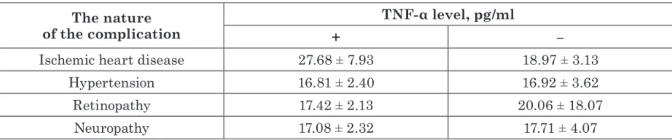
However, studies on the contribution of TNF-α to the development of cardiovascu-lar complications in patients with T2D and NAFLD are ambiguous. Stratification of the diabetic patients in the presence and absence of vascular complications shown a significant in-crease (p < 0.05) in circulatory levels of TNF-α compared to control subjects in both subgroups of patients, with greater emphasis on its se-verity in association with clinically manifested macroangiopathies-CVD and hypertension in the presence of NAFLD (see Table 1, Table 2).
An analysis of the TNF-α level in T2D pa-tients with and without NAFLD, as well as in
the presence and absence of vascular compli-cations was demonstrated significant changes. Thus, in patients with NAFLD and CVD the mean TNF-α level was grater, than in the pa-tients with T2D without NAFLD, but in the presence of CVD (p < 0.05, see Table 1, 2). It was found the difference between TNF-α le-vels in non-NAFLD diabetic patients with different types of vascular complications (p < 0.05 – p < 0.02). There were almost 2-fold higher TNF-α levels in patients with T2D and NAFLD regardless of vascular complica-tion type (p < 0.05, see Table 1). In general, the group of patients without the liver disease had lower levels of TNF-α, but the lowest levels of TNF-α were noted in patients with T2D with-out the liver or vascular complications (p < 0.05, see Table 2).
Thus, the absence of significant differences in the concentration of circulating proteins at the background of macro/microvascular com-plications with IR and NAFLD confirms the leading influence of IR and dysglycemia on the above parameters of the chronic inflammatory process in patients with T2D complicated by Table 1 Circulatory levels of TNF-α in patients with type 2 diabetes,
complicated with nonalcoholic fatty liver disease, stratified in the presence (+) and absence (-) of macro-/microvascular complications
The nature
of the complication + TNF-α level, pg/ml –
Ischemic heart disease 27.68 ± 7.93 18.97 ± 3.13
Hypertension 16.81 ± 2.40 16.92 ± 3.62
Retinopathy 17.42 ± 2.13 20.06 ± 18.07
Neuropathy 17.08 ± 2.32 17.71 ± 4.07
Table 2 Circulatory levels of TNF-α in patients with type 2 diabetes,
without nonalcoholic fatty liver disease, stratified in the presence (+) and absence (-) of macro-/microvascular complications
The nature
of the complication + TNF-α level, pg/ml –
Ischemic heart disease 10.88 ± 1.89 6.99 ± 2.92
Hypertension 10.92 ± 1.76 3.54 ± 2.54*
Retinopathy 10.93 ± 1.76 3.46 ± 2.56*
Neuropathy 10.31 ± 1.82 7.74 ± 3.39
Note:
NAFLD. Our data suggest the presence of ad-ditional factors that modulate the total TNF-α level in patients with T2D and NAFLD against the background of vascular complications. In the examined patients with T2D and NAFLD, a significant increase in TNF-α level was de-tected, regardless of the presence of concomi-tant vascular disease, which may be indicative of the predominant effect of IR and dysglyce-mia on the above-mentioned parameters of the chronic inflammatory process.
Recently published data on the develop-ment of cardiovascular disease form the view that most new biomarkers do not improve the likelihood of predicting the risk of its
develop-ment when included in a model that is based on traditional risk factors [22]. At the same time, we suggest, that the found associations of such new biomarker as TNF-α level, along with other metabolic markers (e. g., BMI), and the risk of NAFLD development not only help us to understand their role in the pathoge nesis of the disease, but also substantiate prospects of their use in the preventive therapy. We agree with statement, that diabetes and NAFLD are reciprocal risk factors and when they are occur together, an increasing body of data demonstrates that diabetes is more difficult to manage and that NAFLD is more likely to progress [23].
CONCLUSION
Non-alcoholic fatty liver disease is closely associated with hormonal and metabolic risk factors and markers of cardiovascular disease and type 2 diabetes and may increase the risk of development and progression of cardiovascu-lar complications.
Stratification of the diabetic population on the base of the presence/absence of NAFLD shown a more pronounced increase in circulat-ing levels of TNF-α in the presence of NAFLD,
which substantiates this parameter utility for further applications as a diagnostic marker.
There was no significant differences in the TNF-α levels in T2D patients with NAFLD re-gardless macro/microvascular complications, which confirms the leading influence of hepatic insulin resistance and a possible presence of additional factors, that modulate the general level of this proinflammatory cytokine.
REFERENCE
1. Berlanga A, Guiu-Jurado E, Porras JA, Auguet T. Clin Exp Gastroenterol 2014; 7: 221-239. doi: 10.2147/CEG. S62831.
2. Smith BW, Adams LA. Crit Rev Clin Lab Sci 2011; 48: 97-113. doi: 10.3109/10408363.2011.596521.
3. Targher G, Bertolini L, Rodella S, et al. Diabetes Care 2007; 30: 2119-2121. doi: 10.2337/dc07-0349.
4. Musso G, Gambino R, Cassader M, Pagano G. Ann Med 2011; 43: 617-649. doi: 10.3109/07853890.2010.518623. 5. Fotbolcu H, Zorlu E. World J Gastroenterol 2016; 22 (16):
4079-4090. doi: 10.3748/wjg.v22.i16.4079.
6. Loria P, Lonardo A, Anania F. Hepatol Res 2013;43: 51-64. doi: 10.1111/j.1872-034X.2012.01031.x.
7. Niederreiter L, Tilg H. Liver Res 2018;2: 14-20. doi: org/10.1016/j.livres.2018.03.003.
8. McGown C, Birerdinc A, Younossi ZM. Clin Liver Dis 2014; 18 (1): 41-58. doi: 10.1016/j.cld.2013.09.012. 9. Phillips CM, Perry IJ. J Clin Endocrinol Metab 2013;
98: E1610-E1619. doi: 10.1210/jc.2013-2038.
10. Rakotoarivelo V, Lacraz G, Mayhue M, et al. EBioMedi-cine 2018; 30: 237-247. doi: 10.1016/j.ebiom.2018.03.033.
11. Abasova LI, Dashdamirov RL, Bahshaliev AB. Med Novosti 2010; 11: 27-29.
12. Wai-Sun Wong V, Lai-Hung Wong G, Wai-Kwok Yip G, et al. Gut 2011; 60 (12): 1721-1727. doi: http://dx.doi. org/10.1136/gut.2011.242016.
13. Chernyavskaya EA, Volina VV, Babiichuk VG. Probl Cryobiol Cryomed 2018; 28 (2): 108-119.
14. Gastaldelli A, Kozakova M, Hojlund K, et al. Hepatology 2009; 49: 1537-1544. doi: 10.1002/hep.22845.
15. Likidlilid A, Patchanans N, Peerapatdit T, et al. J Med Assoc Thai 2010; 93: 682-693.
16. DeFronzo RA. Diabetologia 2010; 53 (7): 1270-1287. doi: 10.1007/s00125-010-1684-1.
17. Brea A, Mosquera D, Martin E, et al. Arterioscler Thromb Vasc Biol 2005;25: 1045-1050. doi: 10.1161/01. ATV.0000160613.57985.18.
18. Sansbury BE, Hill BG. Free Radical Biol Med 2014; 73: 383-399.
19. Ziol M, Handra–Luca A, Kettaneh A, et al. Hepatology 2005;41(1): 48-54. doi: 10.1002/hep.20506.
20. Torer N, Ozenirler S, Yucel A, et al. Scand J Gastro-enterol 2007; 42 (9): 1095-1101.
CIRCUL ATING LE VELS OF TUMOR NECROSIS FACTOR-ALPHA IN T YPE 2 DIABE TES MELLITUS PATIENTS
WITH NON-ALCOHOLIC FAT T Y LIVER DISE ASE TAKING INTO ACCOUNT THE VASCUL AR COMPLICATIONS
T. Tyzhnenko1, N. Krasova1, M. Gorshunska2, Z. Leshchenko1,
A. Cherniaieva1,2, A. Gladkih1, O. Plohotnichenko1, E. Jаnsen3,
V. Роltorak1, I. Karachentsev1,2, N. Kravchun1
1 SI «V. Danilevsky Institute of Endocrine Pathology Problems of the NAMS of Ukraine, Kharkiv, Ukraine; 2 Kharkiv Postgraduate Medical Academy, Kharkiv, Ukraine;
3 National Institute for Public Health and the Environment, Bilthoven, The Netherlands
tyzhnenko@ukr. net
The relationship between non-alcoholic fatty liver disease (NAFLD) and risk factors for vascular disorders and type 2 diabetes mellitus (T2D) remains the subject of discussion. Therefore, the purpose of our work was to determine the circulatory levels of tumor necrosis factor-alpha (TNF-α) in patients with T2D and to deter-mine the nature of the effect of vascular complications in the presence and absence of NAFLD on this indicator. 146 patients with T2D aged from 28 to 80 years old (68 men / 78 women) were examined, with varying degrees of glycemic control and overweight, without renal insufficiency. 95 surveyed people had NAFLD. 21 practi-cally healthy persons were studied as controls. Compared to controls, patients with T2D at the background of impaired glucose homeostasis were characterized by a marked increase in body mass index (BMI), increased levels of free fatty acids, triglycerides (TG), TNF-α, basal insulin, HOMA-IR indices, and decreased insulin sensitivity. It was found that the average level of TNF-α in patients with T2D (17.88 ± 3.42 pg/ml) was 3-fold higher than in the control group — 5.47 ± 3.44 pg/ml (p < 0.05). Patients with T2D with NAFLD have a greater BMI (35.10 ± 0.77 kg/m2) than T2D patients without NAFLD (32.13 ± 0.82 kg/m2, p < 0.001). Stratification of the diabetic population on the base of the presence/absence of NAFLD shown a more pronounced increase in circula ting levels of TNF-α in the presence of NAFLD (20.36 ± 4.81 pg/ml vs 10.05 ± 1.62 pg/ml, p < 0.05), which substantiates this parameter utility for further applications as a diagnostic marker. It was found the diffe-rence between TNF-α levels in non-NAFLD diabetic patients with different types of vascular complications (p < 0.05 – p < 0.02). There were almost 2-fold higher TNF-α levels in patients with T2D and NAFLD regardless of vascular complication type (p < 0.05). In general, the group of patients without the liver disease had lower le-vels of TNF-α, but the lowest lele-vels of TNF-α were noted in patients with T2D without the liver or vascular com-plications (p < 0.05). There was no significant differences in the TNF-α levels in T2D patients with NAFLD re-gardless macro/microvascular complications, which confirms the leading influence of hepatic insulin resistance and a possible presence of additional factors, that modulate the general level of this proinflammatory cytokine.
K e y wo r d s : type 2 diabetes mellitus, non-alcoholic fatty liver disease, tumor necrosis factor-alpha, vas-cular complications. ЦИРК УЛЯТОРН² Р²ВН² ФАК ТОРУ НЕКРОЗУ ПУ Х ЛИН-α У ХВОРИХ НА ЦУКРОВИЙ Д²АБЕ Т 2 ТИПУ ЗА НАЯВНОСТ² НЕ А ЛКОГОЛЬНО¯ ЖИРОВО¯ ХВОРОБИ ПЕЧ²НКИ З УРА Х УВАННЯМ СУДИННИХ УСК ЛА ДНЕНЬ Тижненко Т. В. 1, Красова Н. С. 1, Горшунська М. Ю. 2, Лещенко Ж. А. 1, Черняєва А. О. 1,2, ГладкихО. І. 1, Плохотніченко О. О. 1, Йенсен Е. 3, Полторак В. В. 1, Караченцев Ю. І. 1,2, Кравчун Н. О. 1 1 ДУ «Інститут проблем ендокринної патології ім. В. Я. Данилевського НАМН України»; м. Харків, Україна; 2 Харківська медична академія післядипломної освіти, м. Харків, Україна; 3 Національний інститут охорони здоров’я та довкілля, м. Білтховен, Нідерланди tyzhnenko@ukr. net Взаємозв’язок неалкогольної жирової хвороби печінки (НАЖХП) з факторами ризику судинної патології та цукрового діабету (ЦД) 2 типу залишається предметом дискусій. Тому метою нашої ро-боти було визначення циркуляторних рівнів фактору некрозу пухлин альфа (TNF-α) у хворих на ЦД 2 типу та встановлення характеру впливу судинних ускладнень за наявності та відсутності НАЖХП на цей показник. Обстежено 146 пацієнтів з ЦД 2 типу віком від 28 до 80 років (68 чоловіки / 78 жін-ки), з різним ступенем глікемічного контролю та надлишкової маси тіла, без ниркової недостатності.
21. Fon Tacer K, Kuzman D, Seliskar M, et al. Physiol Genomics 2007; 31 (2): 216-227. doi: 10.1152/physiolge-nomics.00264.2006.
22. Kim HC, Greenland P, Rossouw JE, et al. J Am Coll Car- diol 2010;55: 2080-2091. doi: 10.1016/j.jacc.2009. 12.047.
23. Hazlehurst JM, Woods C, Marjot T, et al. Metab Clin Experim 2016; 65: 1096-1108. doi: 10.1016/j.metabol. 2016.01.001.
95 обстежених мали НАЖХП. 21 практично здорову людину розглядали в якості контролю. Порівняно до контролю хворі на ЦД 2 типу за умов порушеного глюкозного гомеостазу характеризувалися ви-разним підвищенням індексу маси тіла (ІМТ), зростанням рівнів вільних жирних кислот, тригліце-ридів (ТГ), TNF-α, інсуліну натще, індексів HOMA-IR та зниження чутливості до інсуліну. Виявлено, що середній рівень TNF-α у хворих на ЦД 2 типу (17,88 ± 3,42 пг/мл) в 3 рази вищий, ніж у групі контролю — 5,47 ± 3,44 пг/мл (р < 0,05). Хворі на ЦД 2 типу за наявності НАЖХП мають більший ІМТ (35,10 ± 0,77 кг/м2, p < 0,001), ніж хворі на ЦД 2 типу без наявності НАЖХП (32,13 ± 0,82 кг/м2). Стратифікація діабетичного загалу за наявності/відсутності НАЖХП засвідчила більш виразне під-вищення циркуляторних рівнів TNF-α за НАЖХП (20,36 ± 4,81 пг/мл проти 10,05 ± 1,62 пг/мл, p < 0,05), що обґрунтовує доцільність використання цього показника для подальшого застосування в якості діагностичного параметру вищеозначеного ускладнення. Виявлено розбіжності між рівнями TNF-α у пацієнтів із цукровим діабетом, які не мали печінкового ураження, з різними типами судинних ускладнень (р < 0,05 – р < 0,02). Встановлено, що пацієнти з ЦД 2 типу за НАЖХП у порівнянні до гру-пи без печінкового ураження мали майже в 2 рази вищі рівні TNF-α незалежно від типу судинного ускладнення (p < 0,05). Найнижчі рівні TNF-α відмічені у хворих на ЦД 2 типу без супутньої патоло-гії (p < 0,05). Відсутність достовірного впливу судинних ускладнень на концентрацію циркулюючого TNF-α за умов НАЖХП засвідчує провідний вплив печінкової інсулінорезистентності та можливу на-явність додаткових факторів, які модулюють загальний рівень даного прозапального цитокіну. К л ю ч о в і с л о в а : цукровий діабет 2 типу, неалкогольна жирова хвороба печінки, фактор некрозу пухлин альфа, судинні ускладнення. ЦИРК УЛЯТОРНЫЕ УРОВНИ ФАК ТОРА НЕКРОЗА ОПУ ХОЛИ-α У БОЛЬНЫХ СА Х АРНЫМ ДИАБЕ ТОМ 2 ТИПА ПРИ НА ЛИЧИИ НЕ А ЛКОГОЛЬНОЙ ЖИРОВОЙ БОЛЕЗНИ ПЕЧЕНИ С УЧЕ ТОМ СОСУДИСТЫХ ОСЛОЖНЕНИЙ Тыжненко Т. В. 1, Красова Н. С. 1, Горшунская М. Ю. 2, Лещенко Ж. А. 1, Черняева А. А. 1,2, Гладких А. И. 1, Плохотниченко О. А. 1, Йенсен Э. 3, Полторак В. В. 1, Караченцев Ю. И. 1,2, Кравчун Н. А. 1 1 ГУ «Институт проблем эндокринной патологии им. В. Я. Данилевского НАМН Украины»; г. Харьков, Украина; 2 Харьковская медицинская академия последипломного образования, г. Харьков, Украина; 3 Национальний институт охраны здоровья и окружающей среды, г. Билтховен, Нидерланды tyzhnenko@ukr. net Взаимосвязь неалкогольной жировой болезни печени (НАЖБП) с факторами риска сосудистой патологии и сахарного диабета (СД) 2 типа остается предметом дискуссий. Поэтому целью нашей рабо-ты было определение циркуляторных уровней фактора некроза опухоли альфа (TNF-α) у больных СД 2 типа и установление характера влияния сосудистых осложнений при наличии и отсутствии НАЖБП на этот показатель. Обследовано 146 пациентов с СД 2 типа в возрасте от 28 до 80 лет (68 мужчин / 78 женщин), с разной степенью гликемического контроля и избыточной массы тела, без почечной не-достаточности. 95 обследованных имели НАЖБП. 21 практически здорового человека исследовали в качестве контроля. По сравнению с контролем больные СД 2 типа с нарушенным глюкозным гоме-остазом характеризовались выраженным повышением индекса массы тела (ИМТ), ростом уровней свободных жирных кислот, триглицеридов (ТГ), TNF-α, инсулина натощак, индексов HOMA-IR и сни-жением чувствительности к инсулину. Выявлено, что средний уровень TNF-α у больных СД 2 типа (17,88 ± 3,42 пг/мл) в 3 раза выше, чем в группе контроля — 5,47 ± 3,44 пг/мл (р < 0,05). Больные СД 2 типа при наличии НАЖБП имеют больший ИМТ (35,10 ± 0,77 кг/м2), чем больные СД 2 типа без на-личия НАЖБП (32,13 ± 0,82 кг/м2, p < 0,001). Стратификация пациентов с СД 2 типа по наличию/от-сутствию НАЖБП показала более отчетливое повышение циркуляторных уровней TNF-α при НАЖБП (20,36 ± 4,81 пг/мл против 10,05 ± 1,62 пг/мл, p < 0,05), что обосновывает целесообразность использования данного показателя в качестве диагностического параметра вышеуказанного осложнения. Было об-наружено различие между уровнями TNF-α у диабетических пациентов без НАЖБП с различными типами сосудистых осложнений (p < 0,05 – p < 0,02). Установлено, что пациенты с СД 2 типа и НАЖБП по сравнению с группой без поражения печени имели почти в 2 раза большие уровни TNF-α незави-симо от типа сосудистого осложнения (p < 0,05). Самые низкие уровни TNF-α отмечены у больных СД 2 типа без сопутствующей патологии (p < 0,05). Отсутствие достоверного влияния сосудистых ослож-нений на концентрацию циркулирующего TNF-α при наличии НАЖБП свидетельствует о ведущей роли печеночной инсулинорезистентности и возможном наличии дополнительных факторов, которые модулируют общий уровень данного провоспалительного цитокина. К л ю ч е в ы е с л о в а : сахарный диабет 2 типа, неалкогольная жировая болезнь печени, фактор некроза опухоли альфа, сосудистые осложнения.