Quantitative in vitro - in vivo
extrapolation
Analysis of 19 compounds of varying embryotoxic potency
Report 340720001/2008 W. Slob et al.
RIVM Report 340720001/2008
Quantitative in vitro - in vivo extrapolation
Analysis of 19 compounds of varying embryotoxic potency
W. Slob G. Janer J.G.M. Bessems B.C. Hakkert A.J.A.M. Sips A. Verhoef G. Wolterink A.H. Piersma Contact: A.H. Piersma
Laboratory for Health Protection Research ah.piersma@rivm.nl
This investigation has been performed by order and for the account of Ministry of Health, Welfare and Sports (VWS), within the framework of project V/340720 and of the Ministry of Housing, Spatial Planning and the Environment (VROM) within the framework of project M/601200
© RIVM 2008
Parts of this publication may be reproduced, provided acknowledgement is given to the 'National Institute for Public Health and the Environment', along with the title and year of publication.
Abstract
In vitro-in vivo extrapolation for toxicological risk assessment. Valid or not?
Animal tests can not always simply be replaced by in vitro tests (carried out in test tubes or petri dishes). This is underlined by research at the National Institute for Public Health and the Environment (RIVM). Existing research data from an in vitro test on developmental effects have been evaluated for their usefulness. This concerns disturbances in the development of rat embryos due to exposure to chemicals. This kind of alternative tests are being developed due to the public objection to animal tests (in vivo) for the purpose of quantitative risk assessment.
In the investigated ‘Whole Embryo Culture’ assay rat embryos are exposed to various concentrations of a particular chemical. Next, it is determined at which concentration (in vitro) effects occur that are similar to those in the whole animal (in vivo).
This was investigated for 19 different substances. There appears to be a clear relationship between the determined potencies in the in vitro and in vivo tests, but the scatter (uncertainty) in this relationship is very large (about a factor of hundred). This can partially be explained by the different ways in which the in vivo tests have been performed (the pregnant rats are for example exposed at different days in the gestation). In addition, differences in the uptake or excretion of a substance by the body could play a role. This study describes a first initiative to account for these differences. Even if these aspects are taken into account, the uncertainty in the outcome remains large.
Given this uncertainty, the full replacement of the in vivo animal test by the ‘Whole Embryo Culture’ assay for quantitative risk assessment is hitherto not feasible.
Key words:
Rapport in het kort
In vitro-in vivo extrapolatie voor toxicologische risicobeoordeling. Valide of niet?
Dierproeven zijn niet altijd eenvoudig te vervangen door in vitro testen (uitgevoerd in reageerbuizen of petrischaaltjes). Dat illustreert onderzoek van het RIVM. Bestaande onderzoeksgegevens uit een in
vitro test gericht op ontwikkelingsstoornissen zijn getoetst op hun bruikbaarheid. Het gaat hierbij om
stoornissen in de ontwikkeling van rattenembryo’s als gevolg van blootstelling aan chemische stoffen. Dit soort alternatieven wordt ontwikkeld onder invloed van maatschappelijke bezwaren tegen
dierproeven (in vivo) ten behoeve van kwantitatieve risicobeoordeling.
In de onderzochte ‘Whole Embryo Culture’-test worden rattenembryo’s in vitro blootgesteld aan verschillende concentraties van een bepaalde stof. Vervolgens wordt bekeken bij welke concentratie (in
vitro) de effecten overeenkomen met effecten in het intacte dier (in vivo).
Dit werd voor 19 verschillende stoffen onderzocht. Er blijkt een duidelijke relatie te zijn tussen de gemeten potentie in in vitro- en in vivo testen, maar de ruis (onzekerheid) in deze relatie is erg groot (circa factor honderd). Dit wordt voor een deel veroorzaakt doordat de beschikbare in vivo studies op verschillende manieren zijn uitgevoerd (bijvoorbeeld doordat zwangere ratten op verschillende dagen van de dracht aan stoffen zijn blootgesteld). Ook zouden verschillen waarmee een lichaam stoffen opneemt of uitscheidt een rol kunnen spelen. Deze studie beschrijft een eerste aanzet om deze verschillen te verdisconteren. Zelfs als deze aspecten worden meegewogen blijft de onzekerheid in de uitkomst groot.
Gegeven deze onzekerheid is het volledig vervangen van de in vivo dierstudie door de ‘Whole Embryo Culture’ test voor kwantitatieve risicobeoordeling vooralsnog niet haalbaar.
Trefwoorden:
Acknowledgement
Agnes Oomen and Esther Brandon are acknowledged for providing the toxicokinetic profiles of the compounds selected for PBTK modelling. Jan van Eijkeren is acknowledged for most of the PBTK modelling. Andreas Freidig and Miriam Verwei from TNO Quality of Life are acknowledged for kindly sharing their toxicokinetic models for all-trans-retinoic acid and methotrexate.
Contents
Samenvatting 11 Summary 13
1 Introduction 15
1.1 Objective of the study 16
2 Materials and methods 17
2.1 The WEC test 17
2.2 Derivation of BMCs 17
2.3 In vivo developmental toxicity studies 19
2.4 BMD derivation from in vivo data 19
2.5 Approaches of relating BMCs to BMDs 19
3 Results 21
3.1 In vitro BMCs 21
3.2 In vivo BMDs 26
3.3 Correlation in vitro and in vivo: empirical approach 28
3.4 In vitro – in vivo correlation: toxicokinetics based approach 29
3.4.1 Preliminar attempts 29
3.4.2 Toxicokinetic modelling 31
3.4.3 Relation between BMCs and in vivo internal dose parameters 40
4 Discussion 45
5 Conclusions and recommendations 49
Samenvatting
Om het testen in proefdieren te verminderen is en wordt een scala aan in vitro methoden ontwikkeld. Op dit moment worden deze alternatieven voornamelijk gebruikt voor het screenen en categoriseren (in klassen indelen) van stoffen. Om deze te kunnen gebruiken voor kwantitatieve risicobeoordeling is het noodzakelijk om een in vitro concentratie te vertalen naar een in vivo dosering. Dit rapport onderzoekt de correlatie tussen ‘dosismaten’ uit de in vitro ‘Whole Embryo Culture’ (WEC) test en ‘dosismaten’ uit in vivo ontwikkelingstoxiciteit studies. Om ‘equipotente’ in vitro concentraties en in vivo doseringen te vinden is de ‘Benchmark Dose’ benadering toegepast
De WEC-test had een hoge reproduceerbaarheid tussen laboratoria. Het was opmerkelijk dat drie eindpunten waarop geanalyseerd werd (koplengte, kop-romplengte en totale morfologische score) een sterke correlatie vertoonden. Dit biedt de mogelijkheid om de WEC test simpeler te maken door slechts op groeiparameters te scoren en de meer tijd kostende eindpunten weg te laten.
De in vitro Benchmark Concentrations (BMC’s) en in vivo Benchmark Doses (BMD’s) waren sterk gecorreleerd maar met aanzienlijke spreiding. Indien op basis van deze gevonden correlatie de BMD geschat zou worden uitgaande van de BMC, dan zou het betrouwbaarheidsinterval een aantal ordegroottes omvatten.
Verschillen in opzet van de in vivo studies en toxicokinetische eigenschappen van de diverse stoffen zouden een deel van de ruis kunnen verklaren. De correlatie verbeterde door (semi-kwantitatieve) verdiscontering van dergelijke aspecten. Bovendien werden voor een vijftal stoffen toxicokinetische computermodellen ontwikkeld om deze verschillen kwantitatief mee te nemen. Deze modellen werden gebruikt om de interne dosis van de in vivo (externe) BMD’s in te schatten. Er bleek dat op zijn minst een deel van de ruis is te verklaren door verschillen in de opzet van de in vivo studie (bijvoorbeeld verschillen in blootstellingsscenarios) en in de toxicokinetiek (absorptiesnelheid, biobeschikbaarheid, halfwaardetijd).
Het is duidelijk dat verdere validatie nodig is voordat een in vitro WEC-test een in vivo studie voor ontwikkelingstoxiciteit volledig kan vervangen. Dit is waarschijnlijk alleen mogelijk op basis van een database met gegevens van hoge kwaliteit bestaande uit studies met vergelijkbare opzet (bij voorkeur volgens de OECD Technical Guidelines). Deze is momenteel niet beschikbaar voor teratogeniteit. Bovendien zal deze methodiek een kinetisch computermodel vergen voor elke te beoordelen stof. Op dit moment is echter geen gevalideerde combinatie van in vitro testen voorhanden om de benodigde toxicokinetische parameters te bepalen.
Summary
To reduce testing in experimental animals, a variety of in vitro methods have been and are being developed. At present, these alternatives are mainly used for screening and categorizing compounds. In order to use them for quantitative risk assessment, it is necessary to translate an in vitro concentration towards an in vivo dose. This report examines the correlation between dose descriptors from the in vitro Whole Embryo Culture test (WEC) and dose descriptors from in vivo developmental toxicity tests. To that end we applied the Benchmark Dose approach to estimate equipotent in vitro concentrations and in
vivo doses.
The WEC test had a high reproducibility among laboratories. Interestingly, the three endpoints analyzed (head length, crown-rump length and total morphological score) were strongly correlated. This indicates the possibility of simplifying the WEC assay by measuring growth parameters as endpoints and omitting more time-consuming endpoints.
The in vitro Benchmark Concentrations (BMCs) and in vivo Benchmark Doses (BMDs) were clearly correlated, but with considerable scatter. Therefore, if the BMD were estimated from the BMC the confidence interval of such an estimate would span various orders of magnitude.
Differences in study-design of the in vivo studies as well as in toxicokinetic properties of the various compounds might explain part of the scatter. Taking such differences (semi-quantitatively) into account improved the correlation. In addition, for a subset of five substances, we developed mathematical toxicokinetic models to account quantitatively for the differences in toxicokinetics as well as in the study designs underlying the relevant BMDs. These models were used to estimate internal dose descriptors for the in vivo (external) BMDs. It appeared that at least part of the scatter was due to differences in the in vivo study design (e.g. differences in exposure scenario’s) and in toxicokinetics (absorption rate, bioavailability, half-life).
Clearly, further validation is needed before an in vitro WEC test could fully replace an in vivo developmental study. This will only be possible if a high quality in vivo database is available consisting of studies with similar designs (preferably conform OECD Guidelines), which is currently not the case. Further, the method probably requires a mathematical toxicokinetic model for each compound to be assessed. To date, however, a validated set of in vitro studies that could estimate the required toxicokinetic parameters required for toxicokinetic modelling does not exist.
1
Introduction
A variety of in vitro and in silico methods have been and are being developed with the aim to reduce testing in experimental animals. At present, these models are mainly used for screening and categorizing compounds, while, for most endpoints, in vivo testing is still required for deriving a dose level that is used as a Reference Point (RP), also denoted as Point of Departure (PoD), in the hazard characterization of the compound. The translation of an in vitro concentration towards an in vivo dose (RP/PoD) is not trivial. However, if this translation could be achieved, the in vitro models might be considered to be used as a stand alone for quantitative hazard characterization, without the need for an additional in vivo study (provided that the in vitro test sufficiently covers the in vivo endpoints for which it is deemed predictive).
For assessing embryotoxic effects, a promising in vitro test system that seems to realistically mimic embryogenesis in vivo is the rodent post-implantation Whole Embryo Culture (WEC) system. This method is used to assess the effects of chemicals on the development of the complete embryo within its intact visceral yolk sac outside the uterus, during a critical phase in organogenesis. Although the test uses embryos, both exposure and assessment of effects occur in vitro, and for simplicity, it will be referred to as an in vitro test. Recently, within the framework of an ECVAM validation study (Genschow et al., 2002), a heterogeneous set of 20 chemicals were tested in the WEC system in four different laboratories. This set of compounds was composed such that it contained similar amounts of compounds considered strongly teratogenic, weakly teratogenic, or not teratogenic. The latter classification was based on existing in vivo embryotoxicity and teratogenicity data and was established by consensus among the experts in the ECVAM validation study (Genschow et al., 2002).
The WEC validation study yielded a large data set that appeared very suitable for analysis by the Benchmark Dose (BMD) approach. This provided a tool for deriving equipotent concentrations from the concentration-response data, that will be denoted as (in vitro) BMCs. The resulting BMCs could be used to examine the reproducibility of the WEC system, and to examine the extent to which they correlated to equipotent in vivo BMDs.
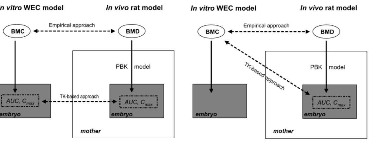
In predicting in vivo BMDs from in vitro concentrations (BMCs), two different approaches could be used: an empirical approach and an approach taking into account toxicokinetics. The empirical approach simply considers the correlation between the observed in vitro concentrations and in vivo doses. If such a correlation is strong enough, it might be used for predicting in vivo doses from in vitro concentrations. The general concept is depicted in Figure 1. The main findings of the empirical approach have been published recently (Piersma et al., 2008).
In a toxicokinetics-based approach the BMCs from in vitro tests are translated into equivalent in vivo doses based on the toxicokinetics of each substance. Ideally, all toxicokinetic information is based on
in vitro studies as well. In this approach, no in vivo testing would be needed in establishing a RP to be
used in hazard characterization. It is assumed that exposure in the in vitro system (Whole Embryo Culture) mimics exposure in the embryo in vivo and that equivalent exposures in vitro in the WEC test will result in the same effects. A physiologically-based toxicokinetic (PBTK) model is used to relate
the internal exposure of the embryo in vivo to an external dose in the dam (rat). All compound-specific parameters in the PBTK model are preferably estimated from in vitro studies.
Figure 1. Graphical representation of the two approaches used in this report.
The figure on the left illustrates the ideal situation where in a toxicokinetics (TK)-based approach the internal exposure of the embryo in the pregnant dam (AUC, Cmax) is compared to the same dose metrics in the in vitro WEC assay. However, the concentration-time profile and thus the AUC and Cmax are unknown in the WEC assay as no samples were taken during the in vitro assay to measure the concentrations. Therefore, in this report, the TK-based approach necessarily had to be the compromise as illustrated on the right (comparison of predicted AUC and Cmax for the in vivo assay to the in vitro BMC).
A PBTK model describes, by means of mathematical equations, the main physiological processes that determine the toxicokinetics of a substance. As output information, a PBTK simulation yields plasma-concentration time curves in blood and all organs/tissue that are specified in the model (Andersen 1995; Schmitt and Willmann 2005) including internal dose parameters like area under the curve (AUC) and maximum concentration (Cmax). By iterative modelling, an external dose can be assessed that would result in a plasma concentration-time profile or at least an AUC and/or a Cmax as much as comparable to the in vitro concentration-time profile.
1.1 Objective of the study
The goal of the study described in this report was to explore the feasibility of quantitative extrapolation of in vitro concentration-effect relationships to in vivo dose-effect relationships, and to consider the importance and the feasibility of accounting for toxicokinetic differences among compounds in that extrapolation.
In vitro WEC model In vivo rat model
BMD BMC Empirical approach AUC, Cmax TK-base d ap proach PBK model
In vitro WEC model In vivo rat model
BMD BMC Empirical approach AUC, Cmax TK-based approach PBK model AUC, Cmax
embryo embryo embryo
mother mother
2
Materials and methods
2.1
The WEC test
In the post-implantation whole embryo culture (WEC) assay rat embryos are removed from the dams at gestation day 10, and these embryos are then cultured and exposed for 48 hours in vitro to various concentrations of the compound of interest. During this period, major aspects of organogenesis are realized, including heart development, closure of the neural tube, development of ear and eye, brachial bars and limb buds. Disturbance during this period may lead to general retardation of growth and development or to specific malformations in one or several organ anlagen. After culture, the morphology of the embryos is carefully assessed and a series of endpoints are scored (Brown and Fabro, 1981). In this study we selected the evaluations for three of these endpoints: Head length (HEAD), Crown-Rump Length (CRL), and Total Morphological Score (TMS) calculated as the sum of scores for all organ anlagen. Comparison of control embryos with exposed embryos forms the basis for conclusions regarding the embryotoxicity of tested compounds. Further details on the method can be found in Piersma et al. (2004).
2.2
Derivation of BMCs
The ECVAM validation study (Genshow et al., 2002) generated concentration-response data for the selected compounds by selecting test concentrations based on a particular sequential scheme, where new concentrations were chosen based on findings at earlier applied concentrations. We considered concentration-response data for each compound (n=20), endpoint (n=3), and laboratory (n=4) separately, resulting in 20 x 3 x 4 = 240 datasets. We analyzed each of these datasets using the benchmark dose approach, i.e. a dose-response model was fitted to the data, and the fitted model was used to estimate the concentration associated with a particular effect size. For each of the three endpoints considered, a 5% change compared to the controls was chosen as the effect size (Benchmark Response, BMR). The associated concentration is the benchmark concentration (BMC), for which
05
.
0
)
0
(
)
0
(
)
(
=
−
f
f
BMC
f
where f denotes the fitted function to the data. By definition, the BMCs can be considered as equipotent concentrations for the whole set of compounds. The choice of a 5% change in response as the Benchmark Response (BMR) here is in accordance with the proposed default value for continuous data (e.g., Slob and Pieters, 1998), but it may be noted that for the purpose of establishing the relative potencies of the compounds this choice is not crucial.
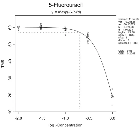
For each separate dataset a model was selected according to the procedure described in Slob (2002). Figure 2 illustrates the method of deriving a BMC for a particular data set (in this example 5-fluorouracil).
In some cases the concentration-response data were not suitable for deriving a BMC value. In these cases, only one or two concentrations had been tested: since these concentrations did not show a clear response testing of other concentrations was omitted.
The calculations were done using the PROAST software, a general software tool for dose-response modeling, which will soon be available from the RIVM website.
-2.0 -1.5 -1.0 -0.5 0.0 log10Concentration 10 20 30 4 0 5 0 60 TM S
5-Fluorouracil
y = a*exp(-(x/b)^d) version: 11.tmp3 var- 0.00526 a- 60.13774 b- 0.92658 d 1.94223 loglik 43.38 conv : T RUE sf.x : 1 dtype : 1 selected : lab R CES 0.05 CED 0.2008Figure 2. Total morphological scores (TMS) versus concentration.
Observed TMS (small circles: individual foetus, large circles: group means) plotted against concentration (in µg/ml), with a fitted dose-response function. The horizontal dashed line indicates the level where the mean TMS response is decreased by 5%, and the vertical dashed line indicates the associated concentration (BMC, here denoted as CED, value: 0.20 µg/ml).
2.3
In vivo developmental toxicity studies
For all 20 substances a literature search for embryotoxic and teratogenic effects in vivo was performed. Literature searches were performed with the substance name together with combinations of the search terms rat, teratogen, teratogenic, teratogenicity, malformation, development, embryo, foetus. Since the
in vitro WEC developmental toxicity tests were performed using rat embryos, the literature search was
focused primarily on developmental toxicity studies in the rat. For 5-bromo-2’-deoxyuridine, data from mice were used in the absence of rat data. No in vivo study was found for isobutyl-ethyl valproic acid, and this compound was excluded from further analysis.
2.4
BMD derivation from in vivo data
For the 19 remaining substances reductions in foetal weight and/or increased incidences of malformations were selected as the endpoints for estimating the (in vivo) benchmark doses (BMDs), based on visual inspection of the data (not formal dose-response analysis). The reason for choosing these endpoints is that they represent the key effects in developmental toxicity tests, and one or both of these endpoints were affected by all those compounds that induced developmental toxicity. It is important to base the BMDs on the same endpoints for all compounds, as the resulting BMDs should reflect equipotent doses. The criterion for the benchmark dose for each substance was defined as a 10% decrease in foetal weight and/or a 10% additional incidence of malformations. These values were chosen for practical reasons (observable changes), and are not crucial in the context of this study. As mentioned for BMCs, we only use these equipotent doses to establish the relative potencies of the different substances in vivo. The various studies that were often available for the same compound were evaluated as a whole, in a sort of weight of evidence approach, taking into account the quality of the study, maternal toxicity, gestation day(s) of exposure, exposure route, etc. The variation in study types and study results for the same compound could be quite large, overwhelming the potential increase in precision resulting from a formal dose-response analysis. Besides that, a formal dose-response analysis was often not possible due to limited reporting of the data, or due to the very limited number of dose groups in the study design.
2.5
Approaches of relating BMCs to BMDs
For all the 19 compounds the in vivo BMDs were plotted against in vitro BMCs. In addition, the influence of various potentially relevant factors (number of administration days in the in vivo studies, half-life of the compound) on the correlation were explored.
A toxicokinetic model was developed for five compounds representative for the three categories of teratogenicity as predefined in the ECVAM validation study on the basis of existing in vivo data. These compounds were: methotrexate, all-trans-retinoic acid and 5-fluorouracil (strongly teratogenic), salicylic acid (weakly teratogenic) and acrylamide (not teratogenic
).
The intention was to develop (preferably physiologically based) toxicokinetic models, while restricting the complexity of these models to two compartments at most. However, it soon appeared that the physicochemical properties of the compounds of interest and the paucity of data prohibited such a restrictive approach. Moreover, the intention was to develop models as much as possible based on in
vitro-in silico estimates of model parameters. However, for most compounds toxicokinetic data were
mainly based on in vivo experiments, so that intention was not feasible from the outset.
In practice, for each compound literature was searched for (toxico-) kinetic data and available models. Based on these data, models were developed or just implemented from the literature. After implementation the models were calibrated to in vivo data that were available (model parameterization), unless model parameters were already provided, as was the case where published models were found in the literature. Only data from rats were taken into account.
Model runs were performed using the estimated BMD values as the dose input and model results were reported in the form of AUC, Cmax of both the total and free plasma concentration and Tmax.
3
Results
3.1
In vitro BMCs
Table 1 shows the (geometric) mean of the BMC values resulting from the reported concentration-response data by each of the four laboratories, for each of the three endpoints. The agreement between the BMCs obtained by the four laboratories was quite good, although laboratory 3 deviated from the general pattern for various compounds (Table 1) and showed a substantially larger variation in background values (Figure 3) than the other three laboratories. The latter three laboratories showed a very consistent pattern (Table 1) in BMCs. The compounds were ranked with respect to the (geometric) means of the BMCs from the different laboratories, omitting the results from laboratory 3 because of its somewhat deviating results. As Table 1 shows, including laboratory 3 would have led to only a slightly different ranking, although for some compounds the difference would have been large (e.g. methoxyacetic acid). There is a reasonable agreement between the ranking based on the in vitro results, and the a priori assessed category of embryotoxic potency based on expert judgment using all available information (mainly in vivo results). The most deviating compounds in this comparison are diphenhydramine and acrylamide, which are in the mid range according to the WEC test (for all three endpoints: TMS, CRL, and HEAD), but were considered non-teratogenic according to the experts (Table 1). For D-(+)-camphor the agreement of the BMCs with the expert category was good for CRL and HEAD, but not so good for TMS. For dimethadione a good agreement was obtained for TMS, but not for HEAD.
Some of the cells in Table 1 are empty. In these cases no BMC value could be derived. This happened in some cases when only one concentration was tested without showing an effect, or when no dose-response was observed (see methods). If higher concentrations would have been tested for these chemicals, a BMC value might have been derived.
Figure 4 shows that a high correlation exists between the (geometric) mean BMC values for the three endpoints: CRL, HEAD and TMS.
Table 1. BMC values [µg/ml] for CRL, HEAD and TMS.
Endpoint: CRL Lab 1 Lab 2 Lab 3 Lab 4 GM GM2 Cat.
Methotrexate 0.022 0.0090 0.021 0.016 0.016 3 all-trans-Retinoic acid 0.067 0.41 0.076 0.13 0.071 3 Aminonicotinamide 0.085 0.075 1.9 0.073 0.17 0.078 3 5-Fluorouracil 0.26 0.037 0.077 0.15 0.11 0.12 3 Methylmercury chloride 3.2 2.4 0.53 3.6 1.9 3.0 3 Hydroxyurea 2.6 5.8 2.3 3.3 3.3 3 Diphenhydramine 5.9 1.4 0.43 5.5 2.1 3.5 1 5-Bromo-2’- deoxyuridine 8.8 4.0 0.087 8.0 2.2 6.6 3 Boric acid 27 16 22 21 21 2 Acrylamide 24 12 3.4 42 14 23 1 Valproic acid 98 58 76 76 2 Salicylic acid 129 65 58 136 90 105 2 Lithium 169 78 115 115 2 Pentyl-4-yn-VPA 145 122 115 127 127 2 D-(+)-camphor 300 116 64 130 187 1 Methoxyacetic acid 521 273 5.0 95 91 238 2 Isobutyl-ethyl-VPA 284 284 284 1 Dimethylphthalate 1.9 297 24 297 1 Saccharin 423 423 1 Dimethadione1 2
Endpoint: HEAD Lab 1 Lab 2 Lab 3 Lab 4 GM GM2 Cat. Methotrexate 0.013 0.013 0.013 0.013 3 Aminonicotinamide 0.11 0.049 1.2 0.034 0.12 0.056 3 5-Fluorouracil 0.12 0.034 0.057 0.087 0.067 0.071 3 all-trans-Retinoic acid 0.19 0.23 0.21 0.19 3 Hydroxyurea 1.8 5.4 1.6 2.5 2.5 3 Diphenhydramine 5.6 0.99 1.3 5.0 2.4 3.0 1 Methylmercury chloride 4.5 2.3 0.73 3.7 2.3 3.4 3 5-Bromo-2’- deoxyuridine 6.6 4.9 0.083 15.6 2.6 8.0 3 Boric acid 25 13 37 23 23 2 Acrylamide 22 31 12 50 25 32 1 Pentyl-4-yn-VPA 78 102 74 84 84 2 Valproic acid 144 78 50 66 78 91 2 Lithium 169 119 44 49 81 100 2 Methoxyacetic acid 100 191 44 72 88 111 2 D-(+)-camphor 285 118 38 109 184 1 Salicylic acid 413 172 80 159 174 224 2 Isobutyl-ethyl-VPA 227 227 227 1 Dimethylphthalate 1.2 264 18 264 1 Dimethadione 280 280 280 2 Saccharin 430 430 1
Endpoint: TMS Lab 1 Lab 2 Lab 3 Lab 4 GM GM2 Cat.
Methotrexate 0.012 0.014 0.020 0.015 0.015 3 Aminonicotinamide 0.14 0.087 38 0.027 0.34 0.069 3 all-trans-Retinoic acid 0.10 0.27 0.14 0.15 0.11 3 5-Fluorouracil 0.20 0.15 0.15 0.10 0.14 0.15 3 Methylmercury chloride 3.3 2.6 0.18 3.5 1.5 3.1 3 5-Bromo-2’- deoxyuridine 4.0 2.1 0.076 5.7 1.4 3.6 3 Hydroxyurea 9.0 4.8 1.7 4.2 4.2 3 Diphenhydramine 4.8 5.4 0.34 5.5 2.6 5.2 1 Acrylamide 36 44 13 41 30 40 1 Boric acid 30 41 33 61 40 42 2 Lithium 42 100 57 50 59 59 2 Dimethadione 63 63 63 2 D-(+)-camphor 128 39 48 62 70 1 Pentyl-4-yn-VPA 141 140 89 121 121 2 Valproic acid 157 112 78 110 111 125 2 Methoxyacetic acid 292 83 19 125 86 145 2 Salicylic acid 252 205 78 168 161 205 2 Isobutyl-ethyl-VPA 395 395 395 1 Dimethylphthalate 466 578 38 548 274 528 1 Saccharin 1002 69 263 1002 1
GM = geometric mean over all four laboratories, GM2 = geometric mean omitting laboratory 3. The GM2 is used for ranking. Categories 1, 2, 3 indicate non-, weak, and strong embryotoxic potency, respectively, according to expert judgment (Brown, 2002). 1The concentrations tested did not induce a significant effect on CRL in any of the laboratories.
1 2 3 4 Laboratory 0.46 0.51 0.56 0.61 0.66 lo g10 (b ack gro und ) CRL 1 2 3 4 Laboratory 1.70 1.74 1.78 1.82 1.86 log 10 (b ack groun d) TMS 1 2 3 4 Laboratory 0.1 0.2 0.3 0.4 lo g10 (ba ck gro und ) head
Figure 3. Estimated background values per laboratory
Estimated background values in each of the 20 substances tested plotted against laboratory. CRL: crown-rump length (mm); TMS: total morphological score (points); HEAD: head length (mm).
-2 -1 0 1 2 log BMCHEAD -2 -1 0 1 2 log B M CCRL -2 -1 0 1 2 log BMCCRL -2 -1 0 1 2 3 lo g BM CTM S -2 -1 0 1 2 log BMCHEAD -2 -1 0 1 2 3 log BMC TM S
Figure
4. Correlations of BMC values between the three endpointsCorrelations of BMC values between the three endpoints. CRL: crown-rump length (mm); TMS: total morphological score (points); HEAD: head length (mm).
3.2
In vivo BMDs
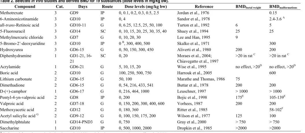
Table 2 provides details on the animal studies that played an important role in assessing the BMD for each substance. The aim was to derive an in vivo dose for each of these compounds that would result in equally strong embryotoxic effects. To that end the dose was established at which a 10% decrease in foetal weight and/or 10% additional incidence of malformations was reported, and that dose was considered a (rough) estimate of the in vivo BMD. For those studies where both endpoints were reported, these effects occurred at similar doses (Table 2).
For three substances (6-aminonicotinamide, pentyl-4n-VPA, and methoxyacetic acid) developmental effects considerably larger than 10% were observed at all dose levels tested. The BMDs for these three substances were calculated by introducing in the model the dose-response data obtained in other studies for the same substance (6-aminonicotinamide) or structurally related substances (i.e., hexyl-4n-VPA for pentyl-4n-VPA; 2-methoxyethanol and di(2-methoxy-ethyl)phthalate for methoxyacetic acid), assuming that the slope of the dose-response would be equal (see Table 2).
On the other hand, for four substances, developmental effects smaller than 10% were observed at all dose levels tested. For these substances only lower bounds for the BMD could be derived (BMD > highest dose level tested; see Table 2). For another substance, acrylamide, the developmentally toxic effects observed concurred with effects in the dams, and were hence regarded as a category 1 (non-teratogenic) compound by the ECVAM expert group in the WEC validation study. However, it cannot be excluded that (possibly at higher doses) acrylamide has, in addition, a direct embryotoxic effect. Therefore, the dose at which these developmental toxic effects occurred was considered as a lower bound for the BMD for acrylamide.
Table 2. Selected in vivo studies and derived BMD for 19 substances (dose levels in mg/kg bw).
Compound Cat. Days Route Dose levels (mg/kg bw) Reference BMDfoetal weight BMDmalformations
Methotrexate 3 GD9 IP 0, 0.1, 0.2, 0.3, 0.5, 2.5 Jordan et al., 1976 0.15
6-Aminonicotinamide 3 GD10 IP 0, 4 Sandor et al., 1978 2.4-3.6 A
all-trans-Retinoic acid 3 GD10-11 G 0, 6.25, 12.5, 25, 50, 100 Turton et al., 1992 6 5
5-Fluorouracil 3 GD14 SC 0, 10, 15, 20, 25, 30, 35, 40 Shuey et al., 1994 25 25
Methylmercuric chloride 3 GD7 G 0, 10, 20, 30 Lee and Han, 1995 9
5-Bromo-2’-deoxyuridine 3 GD10 IP 0 B, 300, 400, 500 Skalko et al., 1971 300
Hydroxyurea 3 GD6-15 G 0, 50, 150, 300, 450 Aliverti et al., 1980 200 200
Diphenhydramine 1 GD1-21,
16-21
SC 0, 20 Moraes et al., 2004;
Chiavegatto et al., 1997
>20 in rat C >20 in rat C
Acrylamide 1 GD6-21 G 5, 10, 15, 20 Wise et al., 1995 no effect, >20D no effect, >20D
Boric acid 2 GD10 G 100, 250, 500, 750 Harrouk et al., 2005 600
Lithium carbonate 2 GD6-15 G 50, 100 Marathe and Thomas, 1986 75
Dimethadione 2 GD6-15 G 0, 54, 216, 433, 541 Buttar et al., 1978 200 200
D-(+)-camphor 1 GD6-17 G 0, 216, 464, 1000 Leuschner, 1997 > 1000 > 1000
Pentyl-4-yn-valproic acid 2 GD8 IP 0, 200 Bojic et al., 1998 175E 105-139E
Valproic acid 2 GD7-18 G 0, 150, 200, 300, 400, 600 Vorhees, 1987 200 200
Methoxyacetic acid 2 GD12 G 0, 180, 360 Ritter et al., 1985 58-102F
Acetyl salicylic acid G 2 GD9-12 G 0, 100, 150, 175, 200 Wilson et al., 1977 125 100
Dimethylphtalate 1 GD14-PND3 G 0, 750 Gray et al., 2000 > 750 > 750
Saccharine 1 GD10 IP 0, 500, 1000, 2000 Dropkin et al., 1985 >2000 >2000
A Estimated from the results in Sandor et al. (1978) and the dose-response in Astroff et al. (2002) B Uninjected females were used as controls.
C Some effects on brain dopamine systems were observed in the offspring at maternally toxic doses.
D At doses of ≥ 10mg/kg bw/day marked neurotoxic effects of acrylamide in the dams, resulting in decreased body weight. The increased number of dead pups at birth at
20 mg/kg bw/day is considered to be secondary to maternal toxicity.
E Estimated from the results for Pentyl-4-yn-VPA and the dose-response for Hexyl-4-yn-VPA (Bojic et al., 1998). F Estimated from the results for MAA and the dose-response for MAA, 2-ME and DEMP.
G Acetyl salicylic acid is rapidly metabolized to salicylic acid (Wilson et al., 1977), which was the compound used in the WEC test.
3.3
In vitro - in vivo correlation: empirical approach
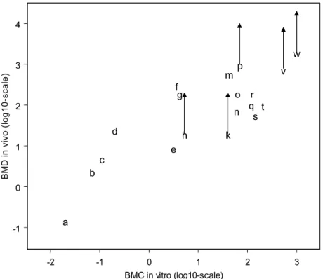
Figure 5 shows the in vitro BMC values plotted against the in vivo BMD values for the nineteen compounds. For some substances, the in vivo BMD is depicted by an arrow, to indicate that, theoretically, the in vivo BMD should be somewhere above the lower bound of the arrow (see previous paragraph).
This figure shows a correlation between the BMC and the BMD. Thus, although limited endpoints were assessed in vitro, the WEC provides information on the in vivo embryotoxicity. On a double logarithmic scale a straight line can be fitted with a slope of 0.7. This slope is lower than one, which indicates that the curve would bend on the non-transformed concentration/dose scales (i.e., its slope would progressively decrease with increasing doses).
-2 -1 0 1 2 3 BMC in vitro (log10-scale) -1 0 1 2 3 4 B M D in v ivo ( lo g 1 0 -sca le ) a b c d e f g h k m n o p qr st v w
Figure 5. In vivo BMD values plotted against in vitro BMC values for the endpoint TMS.
A positive arrow indicates that the BMD would have been higher than the highest dose tested, indicated by the base of the arrow. Compounds: a methotrexate, b 6-aminonicotinamide, c all-trans-retinoid acid, d fluorouracil, e methylmercury, f 5-bromo-2’-deoxyuridine, g hydroxyurea, h diphenydramine, k acrylamide, m boric acid, n lithium, o dimethadione, p D-(+)-camphor, q pentyl-4-yn-valproic acid, r valproic acid, s methoxyacetic acid, t salicylic acid, v dimethylphthalate, w saccharine.
3.4
In vitro – in vivo correlation: toxicokinetics-based approach
The toxicokinetics-based approach attempted to account for the differences in toxicokinetic parameters between different substances in the extrapolation between the in vitro BMC obtained in the WEC test and the in vivo BMD. The selection of the compounds to be used in the ECVAM validation study was partly based on toxicokinetic considerations (Brown, 2002). For example, the compounds used were themselves active agents (i.e., they did not need metabolic activation) and readily passed the placenta. Nevertheless, there could still be relevant differences in other toxicokinetic properties of the selected substances that might explain some of the scatter in the BMD to BMC correlation, such as absorption and half-life.
3.4.1 Preliminar attempts
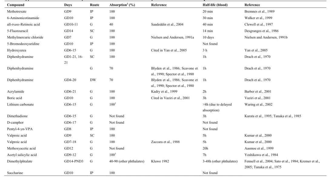
Two relevant toxicokinetic parameters, absorption and half-life, where compiled for all the compounds (Table 3). Systemic absorption was high (40-100%) for all compounds for which we found data. Half-life ranged between 14 min and 3 hours for most compounds, but some compounds had considerably longer half-life (up to 10 days). Another factor that could explain some of the scatter in the correlation between BMC and BMD is the varying dosing regimes in the in vivo studies. Thus, a single dose was administered in some studies, whereas up to 20 repeated doses were administered in others.
To cause an equally large effect a single dose might be equal to, but generally higher than the dose in a repeated dosing scheme (unless the repeated dosing scheme missed the critical window and the single dose did not). Therefore, one may imagine that a single dose study might result in a higher BMD than a repeated dose study. Similarly, one may imagine that the BMD of a compound would have been higher had the half-life of the compound been smaller.
Figure 6 shows the same plot as in Figure 5, but now with (dashed) upwards arrows for those compounds with one (or both) of these two properties (more than 2 dosing days and/or long half-life). This figure illustrates that taking these two aspects into account could potentially improve the correlation between the in vitro BMC and the in vivo BMD. Further, it can be seen that the slope of the fitted line (on double log-scale) would move closer to unity. This implies that the relationship between BMC and BMD plotted on the non-transformed concentration/dose scales would get closer to a straight line.
Table 3. Approximated1 values for absorption and half-life in blood of the compounds included in the study.
Compound Days Route Absorption2 (%) Reference Half-life (blood) Reference
Methotrexate GD9 IP 100 20 min Bremnes et al., 1989
6-Aminonicotinamide GD10 IP 100 30 min Walker et al., 1999
all-trans-Retinoic acid GD10-11 G 40 Saadeddin et al., 2004 40 min Clewell et al., 1997
5-Fluorouracil GD14 SC 100 14 min Desgranges et al., 1986
Methylmercuric chloride GD7 G 100 Nielsen and Andersen, 1991a 10 days Nielsen and Andersen, 1991b
5-Bromodeoxyuridine GD10 IP 100 Not found
Hydroxyurea GD6-15 G 100 Cited in Yan et al., 2005 3 h Yan et al., 2005
Diphenhydramine GD1-21,
16-21
SC 100 1h Drach et al., 1970
Diphenhydramine G 70 Blyden et al., 1986; Scavone et
al., 1990; Spector et al., 1980
1h Drach et al., 1970
Diphenhydramine GD4-20 DW 70 Blyden et al., 1986; Scavone et
al., 1990; Spector et al., 1980
1h Drach et al., 1970
Acrylamide GD6-21 G 100 Kadry et al., 1999 2h Barber et al., 2001
Boric acid GD10 G 100 Cited in Vaziri et al., 2001 3h Vaziri et al., 2001
Lithium carbonate GD6-15 G 1003 >8h (due to delayed
absorption)
Waring et al., 2002
Dimethadione GD6-15 G Not found 3h Kurata et al., 1995; Tanaka et al., 1985
D-camphor GD6-17 G Not found Not found
Pentyl-4-yn-VPA GD8 IP 100 Not found
Valproic acid GD9 SC 100 5h Kumar et al., 2000
Valproic acid GD7-18 G 100 Zaccara et al., 1988 5h Kumar et al., 2000
Methoxyacetic acid GD12 G Not found 20h Aasmoe et al., 1999
Acetyl salicylic acid GD9-12 G 1003 7h Yoshikawa et al., 1984
Dimethylphtalate GD14-PND3 G 40-90 (other phthalates) Kluwe 1982 3-48h (other phthalates) Fennell et al., 2004; Sato et al., 1984; Kremer et al.,
2005; Tanaka et al., 1975
Saccharine GD10 IP 100 Not found
1Absorption and half-life were obtained from the literature without considering the dose administered. In addition, when no data for rats was found, studies with other species were considered. 2Absorption is assumed to be 100% when administration route is subcutaneous or intraperitoneal.
-2 -1 0 1 2 3 BMC in vitro (log10-scale) -1 0 1 2 3 4 B M D in vi vo ( lo g1 0-s ca le ) a b c d e f g h k m n o p qr st v w
Figure 6. In vivo BMD values plotted against in vitro BMC values for the endpoint TMS.
A dashed upward arrow is added on compounds that either had a long half-life or were administered in multiple doses. See legend of Figure 5 for explanation of the other arrows and compound identification.
3.4.2 Toxicokinetic modelling
To further explore the possibility of improving the correlation between BMCs and BMDs by accounting for the toxicokinetic behaviour of the compounds, toxicokinetic models were developed for five substances. These will be briefly discussed.
3.4.2.1 Sodium salicylate
For salicylate, no PBTK model was available from literature. Therefore the model was built using the data as described below. The data for this compound both suggested and did not allow for more than the development of a one-compartment model with saturable plasma binding (see Figure 7). Although salicylate is metabolised in vivo, none of the metabolites seems to be exhibit teratogenic potency (Greenaway et al., 1984). Thus, it was not required to take an active metabolite into account in this model, i.e. only disappearance of the parent (metabolic clearance) rather than quantitative formation of metabolite(s) had to be taken into account.
Figure 7. One compartment model describing sodium salicylate disposition.
The plasma volume of distribution
V
d is dependent on the plasma concentration Cpbecause of non-saturable binding of sodium salicylate to albumin in plasma and in tissue interstitial fluid. Input: intravenous injection (
D
iv) or orally administered dose (Dpo ,Kabs); elimination: metabolism (
k
m) and renal clearance (k
u). Metabolites are eliminatedby renal clearance (
k
mu)The absorption of sodium salicylate after oral administration appears to be fairly complete, so the fraction absorbed from the intestines was set to 1.
Saturable plasma binding was modelled based on data obtained from in vitro experiments of three different references (Daston et al., 1990; Dean et al., 1989; Yoshikawa et al., 1984). Plasma-tissue binding was modelled by Yoshikawa et al., (1984). Partition between plasma and tissue interstitial fluid is governed by equilibration of the free concentration and different bound concentrations due to different albumin concentrations in plasma and fluid. Further, partition between interstitial fluid and intracellular fluid is based on the pH partition hypothesis (Rowland and Tozer 1995). Together with physiological parameters on the tissues interstitial and intracellular content and the albumin concentration in plasma and the tissues interstitial fluid, the plasma-tissue partition can be calculated. Thus, for the partition coefficients, the objective to base model parameters on in vitro-in silico methods was met.
Metabolic (from the literature it is not clear that the (vast) majority of metabolites is formed in the liver) clearance and renal clearance parameters were tuned to in vivo data of Yoshikawa et al (1984) and to data of Varma and Yue (1984) on excretion by urine of the parent compound and metabolites in rat (Figure 8). IV
D
k
m uk
(
)
d p pA V C C
=
,
po absD
K
0 5 10 15 time (hour) 0 20 40 60 80 pl as ma c onc ent ra tion ( m g/ L) Yoshikawa 0 5 10 15 20 25 time (hour) 0 10 20 30 40 50 perc ent age of dos e
Varma; cumulative renal clearance upper line, o: metabolites; lower line, *: parent compound
Figure 8. Model fit to sodium salicylate data of Yoshikawa et al. (1984) and Varma and Yue (1984).
Fitted parameter values: ku = 0.3 [h-1], km = 0.35 [h-1], kmu = 10 [h-1], Kabs = 1 [h-1]
3.4.2.2 Acrylamide
Acrylamide has a metabolite, glycidamide, which is (also) an active agent, although probably not for teratogenic activity. Therefore, quantitative formation of this metabolite was taken into account in the kinetic model. In the literature, a one compartment modelling approach was found describing the kinetics of acrylamide only. Also, a five compartment PBTK model was found describing both acrylamide and glycidamide (Kirman et al., 2003). This model was tuned by the authors on data, fitting 15 parameters ‘by eye’. Another approach found in the literature, describing the kinetics of both acrylamide and glycidamide by a classical one compartment model, was adopted as a working model (Calleman et al., 1992). The structure of the latter model is shown in Figure 9. In contrast to PBTK models, which are basically compartment models too, classical compartment models describe kinetics by transfer and elimination parameters that are not formulated in terms of the physiological system of consideration, or in terms of the physicochemical properties of the compound of consideration.
Figure 9. One compartment models for acrylamide (ACR) and its metabolite glycidamide (GLY).
The formation of glycidamide is saturated (Vmax, KM). Non-saturated metabolism of acrylamide was divided into glutathione conjugation (kGSH, ACR) and hemoglobin adduct formation (kHb, ACR). Furthermore, elimination of glycidamide was divided into glutathione conjugation (kGSH, GLY), DNA adduct formation (kDNA) and hemoglobin adduct formation (kHb GLY).
The model was tuned for non-saturated metabolism of the parent compound, glycidamide formation and total elimination on in vivo data of Calleman et al. (1992). The outcome regarding systemic exposure is presented in Figure 10.
In Figure 11, a comparison is made between the outcome for the acrylamide and glycidamide models for different routes of administration against the data published by Barber et al. (2001). The outcome of this comparison shows that the models can not be used to predict systemic exposure for these compounds for any route of exposure.
,
max MV
K
, GSH ACRk
k
Hb ACR,k
GSH GLY, DNAk
Hb GLY,k
ACR GLY0 50 100 150 Dose mg/kg) 0 2000 4000 6000 8000 Acrylamide 0 50 100 150 Dose mg/kg) 0 100 200 300 400 500 Glycidamide 0 50 100 150 Dose mg/kg) 0 0.2 0.4 0.6 AUC_GLY/AUC_ACR 0 50 100 150 Dose mg/kg) 10 20 30 40 50
60 percentage ACR converted to GLY
Figure 10. Model fit of the acrylamide (ACR) model with data of Calleman et al. (1992).
In the two top panels, on the y-axis the systemic exposure as area under the curve (AUC) is given. The two lower panels show the resulting ratio of AUCs of GLY and ACR and the percentage of ACR converted to GLY
0 2 4 6 8 10 time (hour) 0 10 20 30 40 50 concentrati on (m g/L) ACR; 50 mg/kg I.P. 0 2 4 6 8 10 time (hour) 0 1 2 3 4 conc ent ra tion ( m g/ L)
GLY; 50 mg/kg ACR I.P.
0 2 4 6 8 10 time (hour) 0 2 4 6 8 conc ent ra tion ( m g/ L)
ACR; 20 mg/kg P.O.; Fabs=0.6
0 2 4 6 8 10 time (hour) 0 0.5 1 1.5 2 concentrati on (mg/L)
GLY; 20 mg/kg ACR P.O.; Fabs=0.6
Figure 11. Model comparison of the acrylamide model with rat data of Barber et al. (2001).
3.4.2.3 5-Fluorouracil
Kanamitsu et al. (2000) described a classical kinetic model for oral administration of 5-FU in rat (200 µmol/kg) based (parameterised) on data from an intraperitoneal administration of 200 µmol/kg in rat (Desgranges et al., 1986), apparently assuming that the first order absorption rate constant to be equal for the oral and the intraperitoneal route
(ka = 0.08 min-1 = 4.8 h-1) (Figure 12). This classical model was coded (programmed) in ACSL language and verified for performance using the same intraperitoneal data from Desgranges et al. (1986). Although it is not clear from Kanamitsu et al. (2000) why the model was developed for blood kinetics as the Desgranges et al. (1986) data are from plasma, blood and plasma were assumed to be equal in our modelling experiment for 5-FU. Results are shown in Figure 13 and are regarded sufficient although overpredicting actual blood concentrations by 30-40%. Subsequently, this model was used to predict AUC and Cmax values for the developmental toxicity study on 5-FU, were 5-FU was administered s.c., assuming again first order absorption and the same absorption rate constant primarily. In order to assess the possible consequence of non-validity of this assumption, some deviating absorption rate constants were tried as well (ka = 24 h-1 and ka = 120 h-1), see Table 4.
Portal Vein Cpv, Vpv Liver Cliver, Vliver Systemic blood Csyst, Vsys 5-FU Q fb x CLint(0) x Eact/E0 CLr
Figure 12. Physiological model for the description of the time profiles of 5-FU concentrations in rats
following oral dosing (Kanamitsu et al., 2000).
0 40 80 120 160 200 0 50 100 150 200 250 Time (min) 5-F U in p las ma (µ M ) plasma_in vivo syst.blood_insilico
Figure 13. Model verification.
Model verification using the Kanamitsu et al. (2000) in silico model for blood kinetics that was coded in ACSL against the Desgranges et al. (1986) measured plasma concentrations. Predicted 5-FU concentration (µM) in systemic blood (rats) in pink and measured 5-FU concentration in plasma of rats (Desgranges et al. 1986) over time in black after intraperitoneal administration of 200 µmol/kg 5-FU.
3.4.2.4 All-trans-retinoic acid
A PBTK model for the kinetics of RA was described in a study of Clewell et al. (1997). The model structure is presented in Figure 14.
With this in silico model, plasma concentrations of RA after intravenous or oral administration were predicted. Figures 15 and 16 show the comparison between the measured systemic blood levels and the
in silico predicted systemic blood level. The plasma RA levels after an oral dose of 5 mg/kg were
predicted with the in silico model (Figure 17).
0.1 1 10 100 1000 10000 100000 0 1 2 3 4 5 6 Tim e (h) R A p lasm a co n c. ( n g /m l)
0.015_in silico 0.015_in vivo 0.25_in silico 0.25_in vivo 5_in silico 5_in vivo
Figure 15. All-trans-retinoic acid (RA) blood concentration (ng/ml).
Predicted / measured in rats upon i.v. admin. of 0.015, 0.25 and 5.0 mg/kg RA
0 1 10 100 1000 0 5 10 15 20 25 30 Time (h) R A pl as ma c onc . (ng/ ml ) in vivo in silico
Figure 16. All-trans-retinoic acid (RA) blood concentration (ng/ml).
Predicted / observed in man upon p.o. admin. of 1.1 mg/kg RA (Clewell et al., 1997).
0 1 10 100 1000 10000 0 5 10 15 20 25 30 Time (h) R A pl as m a co nc . ( n g/ m l)
Figure 17. Predicted plasma concentrations of all-trans-retinoic acid (RA).
3.4.2.5 Methotrexate
For methotrexate, only the parent compound is taken into account. For methotrexate, biphasic concentration time curves after intravenous dosing are reported in the literature. Some authors even report tri-phasic kinetics with an initial half-life of a few minutes only. However, some doubt the validity of this observation and consider it as an artefact introduced by the sampling method (micro-dialysis). From the data, representative half-life times of the initial and terminal phase were estimated and a classical kinetic model, i.e., a model consisting of the sum of exponential terms, was developed. The response to the bolus injection was considered as a fundamental solution, of which the convolution with first order uptake could model kinetics from an oral, intraperitoneal or subcutaneous administered dose and the convolution with step function kinetics from an intravenous injection. The predicted plasma concentration-time curves for methotrexate were compared with in vivo data after intravenous administration from different studies. These comparisons are shown inFigure 18.
0 2 4 6 8 10 time (hour) 0 100 200 300 400 500 conc ent rat ion (mi croM) Ekstrom 100 mg/kg 0 2 4 6 8 10 time (hour) 0 500 1000 1500 conc ent ra tion ( m ic ro M) deSouza 250 mg/kg 0 1 2 3 time (hour) 0 100 200 300 concentrati on (m ic roM ) Bremnes 50 mg/kg 0 5 10 15 time (hour) 0 2000 4000 6000 conc ent ra tion ( m ic ro M) Bremnes 1000 mg/kg
Figure 18. Model comparison of methotrexate-model.
With data of Ekstrøm et al. (1996), De Sousa Maia et al. (1996) and Bremnes et al. (1989)
0.44
initV
=
L/kg,k
init=
2.2 h
-1,V
dist=
7.5 L/kg
,k
term=
0.33 h
-13.4.3 Relation between BMCs and in vivo internal dose parameters.
Using the described kinetic models, five relevant internal dose parameters were estimated. Table 4 provides details of the in vivo studies used for determination of kinetic parameters where Table 5 shows the values for the estimated internal dose parameters.
Table 4 1. Overview of the BMDmalformations derived and the dosing design of in vivo teratogenicity studies
that the BMDmalformations was based on and that were used for determination of kinetic parameters.
Compound Frequency (# / 24 h) Period 3 Duration (# days) Route BMDmalformations (mg/kg bw/d) Acrylamide 2 1 GD6-21 16 16 16 gavage gavage gavage 20 100 1000 Methotrexate 1 GD9 1 i.p. 0.15
all-trans-Retinoic acid 1 GD10-11 2 gavage 5
Sodium salicylate 1 2, every 12 h GD9-12 GD9-12 4 4 gavage gavage 100 100 5-Fluoro uracil 1 GD14 1 s.c. ka = 4.8/h4 ka = 24/h5 ka = 120/h5 25 1 Based on Table 2
2 BMD > 20 mg/kg bw/d. Thus exact BMD is not known. Therefore an approach was chosen were 20 and additionally dose levels of 100 and 1000 mg/kg bw were chosen as arbitrary BMDs to work with
3
GD = Gestation Day 4
Absorption rate constant as taken from Kanamitsu et al. (2000)
5 Deviating absorption rate constants tried to assess the possible consequence of non-validity of the assumption ka = 4.8/h, (see text 3.4.1.3).
Table 5. Overview of modelled kinetic parameters derived for the BMDs that were based on the in vivo teratogenicity studies as given in Table 4.
Compound AUCBMD, 0-t (µg·h/ml) Cmax (µg/ml) Tmax (h) AUC BMD, 0-t, free (µg·h/ml) Cmax, free (µg/ml) Acrylamide 960 6120 70050 16.8 86.3 869 0.6 0.66 0.67 960 6120 70050 16.8 86.3 869 Methotrexate 0.39 0.39 0.26 0.26 0.3 0.3 0.04 0.24 0.05 0.16 all-trans-Retinoic acid 21.5 2.9 0.9 21.5 2.9 Sodium salicylate 13200 9800 232 160 2.0 2.2 4100 2300 98 48 5-Fluoro uracil 15.4 15.4 15.4 22.1 36.3 43.6 0.26 0.11 0.048 15.4 15.4 15.4 22.1 36.3 43.6
AUCBMD, 0-t in vivo area under the plasma concentration time curve calculated for the dosing period, e.g. GD
9-14
Cmax maximum plasma concentration
Tmax time point at which maximum plasma concentration is reached
AUC0-t, free and Cmax, free are the parameters corrected for the free (not bound to plasma proteins) fractions of the
compounds.
Two main ways of presenting the internal dose metric were used, i.e. AUC and Cmax in the systemic blood circulation (next to the descriptor of probably minor importance, i.e. Tmax). Further, another variable was introduced, i.e. expressing AUC and Cmax based on the free fraction (i.e. corrected for plasma protein bound compound), as opposed to that based on total compound. In Figure 19, these in
vivo kinetic parameters are plotted against the BMD values. BMDs were strongly and linearly (slope ~
1) correlated to Cmax values, indicating that the Cmax values for these compounds do not provide additional information to the BMDs. This could mean that for the five compounds that were further investigated by kinetic modelling, initial kinetics resulting in the Cmax is probably similar for these five componds. In contrast, the correlation between AUC values and BMDs was weaker. The reason could be that the AUC incorporates, among other factors, the differences in half-life of the substances and dosing regimes in the in vivo studies.
For acrylamide (A), three BMD values were used as the input for PBTK modelling (20, 100 and 1000 mg/kg, see Table 4), which resulted in three AUCs and three Cmax values. For 5-fluoro uracil (F), three Cmax values are presented because three ka values were used (to represent the uncertainty in the absorption for this compound). For methotrexate (M) two Cmax, free and two AUCfree values are presented because of different binding fractions reported in the literature. For salicylic acid (S), two AUC and two Cmax values are present because of the variation in dose administration (once or in two portions per day).
-1 0 1 2 3 BMD (log10-scale) -1 0 1 2 3 4 5 A U C ( log10-sc al e) S S F F F M M R A A A -1 0 1 2 3 BMD (log10-scale) -1 0 1 2 3 C m ax ( log1 0-sc al e) S S F F F M M R A A A -1 0 1 2 3 BMD (log10-scale) -2 0 2 4 A U C .f ree ( log1 0-sc al e) S S F F F M M R A A A -1 0 1 2 3 BMD (log10-scale) -1 0 1 2 3 Cm ax .f ree ( log1 0-sc al e) S S F F F M M R A A A
Figure 19. Predicted toxicokinetic parameters at the BMDs plotted against the BMDs.
M = methotrexate, F = 5-FU, A = acryl amide, S = sodium salicylate, R = all-trans-retinoid acid.
-2 -1 0 1 2 BMC (log10-scale) -2 0 2 4 A U C .f ree ( log 10-sca le ) SS F F F M M R A A A -2 -1 0 1 2 BMC (log10-scale) -1 0 1 2 3 4 5 A U C ( log1 0-scal e) S S F F F M M R A A A
Figure 20. Predicted toxicokinetic parameters at the BMDs plotted against the BMCs.
M = methotrexate, F = 5-FU, A = acrylamide, S = sodium salicylate, R = all-trans-retinoid acid.
Figure 20 shows the correlation between the internal dose parameters (AUC and Cmax as such or calculated as free/unbound) associated with the in vivo BMD (y-axis) and the in vitro BMC (x-axis). Comparing the correlations between estimated in vivo AUCs (corresponding to the BMDs) and BMCs to the correlations between the BMDs and BMCs for the same compounds (see Figure 5; compounds a, c, d, k and t) it seems that the correlation has improved.
-2 -1 0 1 2 BMC (log10-scale) -1 0 1 2 3 C m ax ( log10-sc al e) S S F F F M M R A A A -2 -1 0 1 2 BMC (log10-scale) -1 0 1 2 3 C m ax. fr ee ( log1 0-scal e) S S F F F M M R A A A
4
Discussion
The analysis of the available WEC data showed that this particular in vitro test system has a high reproducibility among laboratories. One of the laboratories showed some deviating results in comparison with the other three laboratories, possibly related to a factor not systematically kept under control in this laboratory (e.g. temperature during incubation) as indicated by relatively large variations in the background values. The three endpoints analyzed (HEAD, CRL and TMS) were strongly correlated. Comparison of the type of malformations observed in the WEC test with the type of malformations observed in vivo for all compounds tested did not reveal that the WEC test was predictive at this level of detail (data not shown). This is probably caused by the reductionistic nature of the WEC model and/or by the difference in exposure conditions as compared to the in vivo model.
The ranking of embryotoxic potency resulting from these in vitro results shows a good correlation with the three categories of embryotoxicity assessed by expert judgment. However, for some of the compounds the BMCs did not completely agree with this categorization, in particular two of them (diphenhydramine and acrylamide), while another two agreed only partly (dimethadione and D-camphor). For instance, dimethadione was correctly categorized by the parameter TMS, but not by HEAD. It should be noted that any discrepancy is not necessarily due to a limitation of the WEC test: the characterization of in vivo embryotoxicity by expert judgment in some cases had to be based on a limited database.
Ranking on the basis of BMC values implies ranking on effective concentrations reaching the embryo. However, the classification for embryotoxicity is not only based on the effective doses (concentrations) administered in vivo, but also on the relationship between embryotoxicity and maternal toxicity. The applied doses in the in vivo studies for substances like diphenhydramine and acrylamide might not have been high enough to achieve embryotoxic effects or might have induced maternal toxicity complicating the interpretation of developmentally toxic effects. For some studies, it is not trivial to conclude whether or not the developmentally toxic effects observed are a consequence of maternal toxicity. The assumption that the developmentally toxic effects are secondary to maternal toxicity when they co-occur may lead to an underestimation of the direct developmental toxic potency of a substance (see for example acrylamide). In contrast to the in vivo studies, maternal effects do not interfere in the WEC test. Thus, comparing the effective concentrations in the WEC test (extrapolated to an in vivo dose) with the doses that induce maternal toxicity in vivo, could support or reject a direct developmental effect.
Results show that for the substances for which a BMD could be derived, a clear in vitro - in vivo correlation exists. However, a considerable uncertainty would remain if the BMD were estimated from the BMC using this correlation: the confidence interval of such an estimate would span various orders of magnitude. This uncertainty is similar to that obtained in a previous assessment of the in vitro-in
vivo extrapolation based on the embryonic stem cell test (ratio predicted/observed ranged from 0.005 to
The substances for which only a lower bound level for the BMD could be assessed were not in disagreement with the overall picture, but, of course, incorporating these values does not really help in establishing the correlation. Further, differences in dosing regimens in the in vivo study seemed to account for at least part of the scatter in the BMC-BMD correlation. These notions illustrate that validating an in vitro–in vivo correlation may be strongly hampered by errors in the estimated (equipotent) BMD.
In vitro dose descriptors that we used in this study were the geometric means of the data obtained in
three of the four laboratories that participated in the ECVAM validation of the WEC test. We excluded data from one laboratory (laboratory 3) because we aimed at exploring the possibilities of extrapolating
in vitro to in vivo data by considering the highest quality data available. Of course, if a correlation as
established in Figure 5 were actually used in practice, potential errors in a single available BMC, like the ones found in laboratory 3, should be taken into account. Obviously, this would only add to the large prediction errors in extrapolating BMCs to BMDs.
To account for the variability in dosing regimes and kinetic parameters between the substances in a more quantitative way, toxicokinetic models were developed for five substances. These models were used to predict internal dose measures (Cmax and AUC) that are supposed to reflect the in vivo exposure of foetuses to the chemicals. BMD and Cmax showed a strong correlation, indicating that replacing the external dose (BMD) by Cmax would not improve the correlation with the in vitro BMC. In contrast, the correlation between BMD and AUC was weaker, and the AUC correlated better to the BMC than the BMD did. Both the differences in dosing regimes (number of dosing days), and the differences in some toxicokinetic properties of the substances (e.g. elimination rates) might have contributed to the poorer correlation. For example, four doses of sodium salicylate were administered, but only one dose of methotrexate; and the half-life for sodium salicylate in plasma (~6 h) is considerably higher than the other four chemicals (~20 min to 2 h). Cmax and Cmax-free differed only to a minor extend, similarly to AUC versus AUC-free. This indicates that no major differences in terms of binding to plasma proteins exist among these five chemicals.
The substances tested in the WEC study were purposely selected to be active themselves (not their metabolites) and they all had high systemic absorption. This limits the potential of toxicokinetic modelling to have an impact on the in vitro-in vivo correlation for this set of substances. Thus, it may be expected that, in the more general case, toxicokinetic models will more substantially improve the estimation of in vivo BMDs from the WEC test. If the WEC test were to be used as an alternative to in
vivo studies in risk assessment, the required toxicokinetic parameters should be estimated from
toxicokinetic studies. This could be a single in vivo study, or a set of in vitro toxicokinetic test systems. A validated set of such test systems is currently not available. It should be noted that if such systems would become available in the future, the costs of applying the whole set of in vitro tests to each study.
Further analysis of in vitro-in vivo relationships that could be useful in human risk assessment strongly depends on the availability of a high quality in vivo database. High quality is defined here as being based on similar study designs, especially with respect to route of administration, number of administrations, exposure window during gestation etc. In addition, general quality aspects are relevant, such as purity of the test substance and animal facility standards as prescribed in OECD Test
in
Guidelines. Admission to the numerous confidential studies that have been performed by industry under e.g. the European Pesticide Act could substantially improve the validation of in vitro-in vivo relationships.
We hypothesize that in vitro alternative tests may be particularly useful in the category and read-across approaches where data on relatively closely related compounds can be used to predict embryotoxicity of related compounds for which limited or no (in vivo) data are available. Indeed, within a category, effective exposure regimes are expected to be similar (assuming that chemicals within a category act on the same target). For example, if a critical window of exposure (e.g. certain gestational days) exists for a certain chemical this is likely to be the critical window for related chemicals. In vitro models such as the WEC may thus prove useful in screening and prioritising compounds within classes for further development.
![Table 1. BMC values [µg/ml] for CRL, HEAD and TMS.](https://thumb-eu.123doks.com/thumbv2/5doknet/3061517.8872/23.892.148.811.210.595/table-bmc-values-µg-ml-crl-head-tms.webp)


