Identification of Endocrine Disruptive Effects in the Aquatic Environment
a Partial Life Cycle Assay in Zebrafish
P.W. Wester, E.J. van den Brandhof, J.H. Vos, L.T.M. van der Ven
This investigation has been performed by order and for the account of the European
Commission (DG SANCO, project B6-7920/98/00025), and the Dutch Environment Ministry (VROM) within the framework of project M/640920, “Development and Validation of a Test Method for the Identification of Endocrine Disrupting Chemicals in the Environment”.
Contents
SAMENVATTING ... 3 SUMMARY... 7 1. INTRODUCTION... 11 2. METHOD DEVELOPMENT ... 13 2.1. VITELLOGENIN ANALYSIS... 132.2. PARTIAL LIFE CYCLE STUDY, PROTOCOL DESIGN... 21
2.3. POPULATION MODELLING... 26
2.4. HISTOPATHOLOGY ATLAS... 28
3. PARTIAL LIFE CYCLE STUDY, APPLICATION WITH REFERENCE COMPOUNDS ... 30
3.1. INTRODUCTION... 30
3.2. PLC-TEST WITH ESTROGEN: 17β-ESTRADIOL... 31
3.3. PLC-TEST WITH ANTI-ESTROGEN: TAMOXIFEN... 40
3.4. PLC-TEST WITH ANDROGEN: METHYLDIHYDROTESTOSTERONE... 50
3.5. PLC-TEST WITH ANTI-ANDROGEN: FLUTAMIDE... 58
3.6. PLC-TEST WITH ANTITHYROID AGENT: PROPYLTHIOURACIL... 64
3.7. PLC-TEST WITH A FIELD SAMPLE: THE LOES SURVEY... 74
4. DISCUSSION AND EVALUATION... 83
4.1. SPECIES... 83
4.2. ASSESSMENT OF INDIVIDUAL PARAMETERS... 84
4.3. EVALUATION OF EXPERIMENTAL SETUP... 95
4.4. ASSESSMENT OF POPULATION IMPACT... 97
5. CONCLUSIONS ... 101
ACKNOWLEDGEMENTS ... 102
REFERENCES ... 103
ANNEX 1 - TEST CONDITIONS FOR THE ZEBRAFISH SCREENING ASSAY... 107
ANNEX 2 - HISTOLOGY PROTOCOL... 110
Samenvatting
In dit rapport wordt de ontwikkeling en toepassing beschreven van een methode om effecten van hormoonverstorende stoffen in het aquatisch milieu te onderzoeken. Het project is uitgevoerd in opdracht van het Ministerie VROM (DGM / SAS) en is gesponsord door de Europese Commissie (DG SANCO).
De primaire doelstelling betrof de ontwikkeling en verdere uitwerking van een onderzoeksprotocol om in kleine laboratoriumvissen effecten te detecteren van
hormoonverstorende stoffen. In dit protocol werden zebravissen gedurende een korte maar essentiële periode van de voortplanting en ontwikkeling blootgesteld, te weten 21 dagen voor volwassen dieren en 42 dagen voor nakomelingen (Partial Life Cycle Study, PLC).
Blootstellingsconcentraties werden gekozen op basis van een voorafgaande range-finding test van 4-10 dagen. De parameters waren voortplanting (eiproductie, bevruchting, uitkomen van de eieren, ontwikkeling van juvenielen, waaronder geslachtsdifferentiatie),
vitellogeninegehaltes (VTG), en histopathologische afwijkingen van relevante
doelwitorganen. Voor de bepaling van VTG zijn histologische methoden ontwikkeld als alternatief voor de gebruikelijke ELISA. Deze methoden hebben een vergelijkbare gevoeligheid, er kunnen semi-kwantitatieve bepalingen mee worden uitgevoerd op grote aantallen monsters, en bovendien wordt door toepassing efficiënt gebruik gemaakt van de geteste dieren.
Een tweede belangrijke doelstelling betrof de ontwikkeling van een digitale atlas van
histopathologische veranderingen die werden waargenomen als gevolg van blootstelling aan hormoonactieve stoffen in gevoelige organen, in het bijzonder de geslachtsorganen, van zebra- en andere kleine laboratoriumvissen. De gegevens voor de atlas zijn verkregen uit de experimenten die in dit project zijn uitgevoerd. Deze atlas is vrij beschikbaar op Internet (http://www.rivm.nl/fishtoxpat/) ten behoeve van research, testen en training.
Het onderzoeksprotocol werd toegepast met een reeks bekende hormoonactieve stoffen, voornamelijk zoals voorgesteld door de Validation and Management Group eco (VMG eco), onderdeel van de Organisatie voor Economische Samenwerking en Ontwikkeling (OESO, OECD). Deze waren het oestrogeen 17β-oestradiol (E2), het anti-oestrogeen tamoxifen, het androgeen methyldihydrotestosteron (MDHT), het anti-androgeen flutamide en de
schildklierremmer propylthiouracil (PTU). Ook is een veldmonster getest, te weten een effluent van een rioolwaterzuiveringsinstallatie en een synthetisch analoog hiervan, in het kader van LOES, het Landelijk Onderzoek naar oEstrogene Stoffen in het aquatisch milieu. De bevindingen bij de individuele dieren werden voorts getoetst in een populatiemodel om een schatting te maken van de effecten op populatieniveau.
Het oestrogeen werd getest tot een concentratie van 1 nM omdat 10 nM E2 in een voorstudie volledige blokkade van ovarium activiteit te zien gaf. Deze concentratie van 1 nM gaf afname in het aantal legsels (maar wel gecompenseerd door een toename van de legselgrootte) en bij nakomelingen een groeibevordering en feminisatie (verschuiving in de geslachtsverhouding), waarbij soms zelfs mannelijke dieren ontbraken. Bij ouderdieren werd verhoging van VTG-gehaltes gezien en de testis vertoonde remming van de spermatogenese. Deze effecten werden gezien vanaf 0,32 nM, niettemin bleek de voortplanting niet beïnvloed.
Het anti-oestrogeen tamoxifen gaf een afname in het aantal eilegsels, bevruchting, uitkomen van eieren, overleving en groei van juvenielen te zien. De histologische bevindingen waren karakteristiek, namelijk plooivorming in de eicelmembraan, eidegeneratie, en VTG-verlaging bij vrouwtjes. Bij mannetjes werd verstoring in de synchronisatie van de spermatogenese gezien en stimulatie van de Leydigcellen. Daarnaast werd met verhoging van de dosis een toename in ontstekingsprocessen gezien in buikorganen wat zou kunnen wijzen op verstoring van immunologische afweer. De nakomelingen ontwikkelden zich vrijwel allemaal tot mannetjes. Effecten werden gezien vanaf 10 µg/L.
Als androgeen is gekozen voor de niet-aromatiseerbare vorm MDHT omdat
methyltestosteron in een voorstudie overwegend oestrogene effecten te zien gaf. Bij 10 µg/L werd al spoedig geen eileg meer gevonden en histologisch bleek ovulatie geremd te zijn, wat aangeeft dat bij deze concentratie geen voortplanting mogelijk is. Bij ouderdieren en
nakomelingen (afkomstig van niet behandelde ouders) werd bij hoge concentraties VTG-inductie gezien. Deze oestrogene effecten zijn mogelijk het gevolg van directe activatie van de oestrogeenreceptor bij deze hoge concentraties. De testis vertoonde verstoring van
spermatogenese en afwijkingen van de Sertoli- en Leydigcellen. Nakomelingen bleven achter in de groei. Bij lagere concentraties werden lagere eiproductie en achterblijvende groei van nakomelingen gezien. Vanaf de laagste concentratie (0.1 µg/L) trad volledige masculinisatie op bij het nageslacht, hetgeen een kritisch effect is voor het voortbestaan van de populatie.,
Bevruchting, uitkomen van eieren en overleving en conditie van nakomelingen waren niet beïnvloed.
Het anti-androgeen flutamide veroorzaakte een afname in het aantal eilegsels en in de overleving van nakomelingen. Histologisch werden in de testis veranderingen gezien als toename van Leydigcellen, stimulering van spermatogoniën en Sertolicellen en remming van de vroege spermatogenese. Er waren geen aanwijzingen voor effecten op VTG of op de vrouwelijke dieren. Na blootstelling van alleen de ouderdieren werd masculinisatie gevonden.
Met de schildklierremmer PTU werden tot 100 mg/L geen effecten op reproductie gezien. Vanaf 1 mg/L werd bij ouderdieren en juvenielen struma waargenomen, afname van schildklierhormoon (adulten) en glycogeengehalte in de lever. In het nageslacht werd remming van de groei en ontwikkeling (metamorfose) gezien.
Uit een veldstudie is een veldmonster van een verdachte locatie getest, samen met een analoog synthetisch mengsel en E2 als controles. Het monster en het synthetisch mengsel gaven alleen bij vrouwelijke adulten verhoging van VTG te zien en een verschuiving naar vrouwelijke ontwikkeling bij het nageslacht. De effecten waren minder dan bij E2, waar ook VTG-inductie bij mannelijke dieren werd gevonden. Blootstelling aan het synthetisch mengsel veroorzaakte veranderingen in geslachtsorganen van adulten die overeenkwamen met die bij anti-oestrogeen tamoxifen. Geconcludeerd werd dat het effluent enige oestrogene activiteit vertoont, en dat het nettoresultaat van een mengsel kan verschillen van wat men op grond van de individuele oestrogene componenten zou verwachten.
In het PLC- protocol werd een semi-statische blootstelling aan een referentiestof toegepast met tweemaal per week een verversing. Behalve bij PTU werd bij chemische analyse een – soms snelle – afname in de testverbinding gezien; hierdoor kunnen de uitkomsten een onderschatting zijn in relatie tot nominale concentraties.
Met het populatiemodel werd vastgesteld dat, met inachtneming van de testopzet (keuze van de concentratiereeks, spreiding in de uitkomsten, beperkte testduur, soortspecifieke
voortplantingsstrategie, etcetera), overlevingskansen van de populatie van zebravissen verminderen bij blootstelling aan MDHT en tamoxifen, ten gevolge van veranderde
geslachtsverhoudingen. Geslachtsdifferentiatie bleek bepalend voor overlevingskansen van de populatie.
De conclusie luidt dat het zebravis-PLC-protocol een bruikbare methode is om de verschillende effecten van hormoonactieve stoffen te identificeren. Histopathologische evaluatie is hierbij cruciaal omddat het een hoge specificiteit en gevoeligheid heeft, en tevens aanwijzingen kan geven voor een werkingsmechanisme. Bovendien zijn voor de
histopathologie minder dieren nodig dan voor evaluatie van reproductieparameters. De inductie van VTG is met name een bruikbare methode voor het detecteren van risico’s van verbindingen met een hoge oestrogene activiteit, omdat duidelijk waarneembare VTG-veranderingen daar gepaard gaan met schadelijke andere hormonale effecten.
Summary
Here is described the development and application of a detection method for pollutants with endocrine activity in the aquatic environment. This project was sponsored by the European Commission (DG SANCO) and the Dutch Ministry of the Environment (VROM).
The principal objective was the development and further validation of a detection method of endocrine effects by means of a test protocol in small laboratory fish. This protocol was designed to expose zebrafish during a limited but critical window in the reproductive and developmental stages; for the selected species, this was 21 days for reproductive adults, followed by 42 days for progeny (Partial Life Cycle Study, PLC). Exposure concentrations were determined on the basis of a pilot range-finding test of 4-10 days. Parameters were reproductive endpoints (egg production, fertilisation, hatching, juvenile development and sexual differentiation), vitellogenin (VTG) levels, and histopathology of (endocrine) target organs. For VTG analysis, histological methods were developed and tested, as an alternative for ELISA. These methods allow identification and semiquantitative determination of VTG almost equally sensitive as ELISA, with a high throughput, and maximising the informative output of a minimised number of animals.
Another major objective was the development of a digital atlas of histopathological changes in small laboratory fish, zebrafish in particular, induced by endocrine active substances, notably changes in endocrine target organs / tissues. Data were obtained from the experiments conducted in this project. The atlas, publicly available on the Internet
(http://www.rivm.nl/fishtoxpat/), .is intended for use as a reference in research and testing and for educational purposes.
The designed test protocol was applied to a spectrum of reference endocrine active
compounds, in line with the proposal by the Validation and Management Group eco (VMG eco) functioning under the Organisation for Economic Co-operation and Development (OECD). These were 17β-estradiol (E2) as estrogen, tamoxifen as anti-estrogen, methyldihydrotestosterone (MDHT) as androgen, flutamide as anti-androgen and
propylthiouracil (PTU) as anti-thyroid agent. Also a field sample (sewage treatment works effluent and its synthetic analogue) was tested as part of a national field trial (LOES). The
data from the experiments were used in a mathematical fish population model, to estimate the effect of the detected endocrine disruption in individuals at the population level.
The results for estrogen, where exposure levels were tested up to 1 nM E2 (10 nM causing histologically complete ovarian inactivation in a pilot test), showed a reduction in number of clutches, which was however compensated by increased clutch size, and enhancement of juvenile growth in exposed groups. There was also feminisation in the offspring, sometimes leading to complete absence of males. In adult males and females, a clear induction of VTG was seen, and testis morphology indicated inhibition of spermatogenesis. Effects were noted from 0.32 nM onwards; nevertheless, reproduction was largely uneventful up to 1 nM
Results from the anti-estrogen (tamoxifen) study showed the number of egg clutches, fertilisation, hatching, survival and length / weight to decrease. Histologically, typical effects on the gonads were seen, such as wrinkling of the oocyte membrane; other degenerative changes took place, and VTG was decreased in females. In the testis, asynchrony of spermatogenesis was seen, together with activation / proliferation of Leydig cells. Remarkably, enhancement of abdominal inflammatory processes was observed with increasing dose, which may point towards an (in)direct effect on the animals’ immune competence. In juveniles, tamoxifen induced sex reversal, indicated by a nearly 100% male population. The overall lowest effect concentration was 10 µg tamoxifen /L.
In the androgen study, methyltestosterone was initially tested, but the clear, induced,
estrogenic effects, attributed to aromatisation, of the preliminary study led to investigation of the non-aromatisable MDHT. In the 10 µg/L group, spawning was inhibited within a few days, associated with histologically observed inhibited ovulation; for this reason, it is anticipated that this concentration is incompatible with reproduction. VTG was induced in adults and juveniles. These estrogenic effects were possibly due to direct interaction with the estrogen receptor at high concentrations. Testis morphology indicated disturbance of
spermatogenesis and effects on Sertoli / Leydig cells. In juveniles, growth (body weight and length) was reduced. At 1 µg/L, egg production was reduced (concentration related); there was also a gain in body weight in juveniles. At low concentrations (0.1 µg/L and higher) complete masculinisation was induced in developing juveniles. This is considered as the critical effect with respect to extrapolated survival chances of the population. At higher
concentrations gonad development was inhibited. No effects were seen on survival, fertilisation rate, hatching and the condition factor (juveniles) in any of the groups.
Concerning anti-androgen, flutamide at 1 mg/L caused a reduction in egg clutches and in the condition of juveniles, and caused a concentration-dependent reduced juvenile survival. Histological changes in the testis included an increase in interstitial cells, hypertrophy of spermatogonia and Sertoli cells, and the inhibition of early spermatogenesis. No effects were seen in VTG, and there were no histological changes in females. In juveniles, paradoxally enough, masculinisation (partial) was seen after parental exposure.
In the study with the anti-thyroid drug PTU, it was shown that even at 100 mg/L no adverse effects on reproductive parameters were evident. In both adult and juvenile zebra fish, struma was observed for 1 mg/L and above. In plasma of adults, a dose-dependent decrease in thyroid hormones was indeed measured. Liver glycogen was reduced as well, this being attributed to the known glyconeogenetic activity of thyroid hormones. Developmental effects were limited to reduced growth and metamorphosis.
A field sample (a sewage treatment plant effluent) was tested from a so-called hot spot and a synthetic analogue was examined, E2 being used a positive control. The field sample and the synthetic analogue induced VTG in females but not in males; in juveniles there was a shift towards the female phenotype. The effects were less prominent than in the E2-exposed fish, where males too exhibited VTG induction. By contrast, histological effects of the synthetic analogue in adult gonads of both sexes were identical to those from the tamoxifen study (anti-estrogen). From the study it can be concluded that the effluent had an endocrine disruptive potency (shift in sex differentiation). Results also showed that the net effect of a mixture could differ from expected effects of the individual estrogenic constituents.
The PLC-protocol included a semi-static exposure regime for the reference compound, with biweekly renewal of the exposure medium. With the exception of PTU, chemical analysis showed a decline in test compound concentrations, which, in some cases, was fairly rapid. The detected effects may therefore be an underestimation of the actual hazard of the compounds at the given nominal concentrations.
Population modelling showed that under the conditions of the test method (selection of concentrations, variation in results, limited duration, reproduction strategy of zebra fish, etc.) an increase in the risk of extinction of the zebrafish population took place after exposure to MDHT and tamoxifen due to skewed sex ratios. Sex differentiation was critical for chances of survival of the population .
The zebra fish PLC protocol is concluded to be a useful method for identifying various effects of endocrine disrupting chemicals. Histopathological evaluation, with its high specificity and sensitivity, is essential; it can also contribute in identifying the mode of action. Fewer animals are needed for histopathological evaluation than for evaluation of reproduction parameters. VTG induction appears to be useful in specific identification of risk from compounds with a high estrogenic potential, since appreciable VTG increase is
1. Introduction
Environmental effects of endocrine active substances have raised many concerns world-wide since the early nineties (Colborn et al., 1993; Vos et al., 2000). This concern was raised after abnormalities in sexual differentiation in certain wildlife populations, and it has triggered research, hypotheses and speculation towards insidious effects in wildlife and man, such as population decline in wildlife, reduction in sperm counts, gonad abnormalities and endocrine related tumours in humans.
Most of the concerns and research was focussed towards the aquatic environment, and the need was felt for more specific or dedicated toxicity test protocols. Indeed, current
ecotoxicity testing guidelines (OECD TG 201 Fish early life stage and OECD draft TG 212 Egg and sac fry) are not able to identify mechanistic pathways, including the endocrine system, necessary for the characterisation of EDC effects. The emphasis in development of predictive tests for endocrine disruption currently was predominantly on in vitro bioassays, while the in vivo effects on the (mainly reproductive) functioning of organisms and
populations remained relatively unattended. Therefore, the qualitative and quantitative significance of the data from such in vitro bioassays is not well known and, thus, for proper risk assessment (and consequently risk management) in vivo studies are indispensable. Various international scientific, regulatory and industrial groups have identified the lack of adequate in vivo models and testing protocols for endocrine disrupting chemicals (EDCs) in the aquatic environment (EU, OECD, EMWAT, EDSTAC, CEFIC). Since in human risk assessment histopathological screening of various organs and tissues of laboratory rodents is the cornerstone in hazard identification, it is proposed to extend the current test protocols using fish with histopathology; this will not only cover the detection of EDCs but also other categories of toxic compounds.
Thus the aim of the present project is twofold:
1. development and testing of a reproduction study in zebrafish, with the principle aim to identify effects indicating endocrine disruption (in case of an estrogenic action, increased levels of circulating vitellogenin), and relate these to reproductive performance. Both parent and offspring animals are monitored for relevant parameters such as reproduction indices, sex distribution, development and histopathology. This will enable the
thyroid abnormalities), in regard to reproductive hazard and will help to estimate consequences for population dynamics and ecology.
2. development and validation of an in vivo total body histopathological screening of small fish by exposure to a variety of known EDC’s. Such a screening has been developed in the past by our group, using guppy (Poecilia reticulata) and medaka (Oryzias latipes) as test species; the small size of the test fish allows group-wise whole body
histopathological examination with the organs still in situ. This cost-effective
methodology enabled screening of structural effects, induced by various environmental contaminants in a variety of relevant organs including endocrine organs and endocrine responsive tissues. Not only a range of target organs can be identified by this protocol, adequate knowledge of pathophysiology and toxicological pathology may also give indications for organ interactions, mechanism of action and consequently functional impact on the organism (Wester and Vos, 1994; Wester et al., 2002; Van der Ven et al., 2003b). Histopathology has been mentioned by several bodies (OECD, CEFIC) as an important need in further development and validation of test methods to detect EDCs. In the present project the aim is to introduce this histopathological screening protocol for the oviparous zebrafish Danio rerio, a widely used laboratory species for which more
information on reproductive physiology is available and which is more suitable for reproduction studies than e.g. the live bearing guppy. From a variety of established EDCs a number is selected to be used as reference compounds, and the attention is focused on (but not limited to), effects on endocrine responsive tissues. Such a protocol with fish is intended to be incorporated in future ecotoxicity testing guidelines. The results of this investigative work on histopathology will be issued as a digital histopathology atlas available through internet to aid researchers and students in training and harmonisation of terminology and interpretation.
The project was proposed on a call for tender(DGXXIV/98/B2/008) from the European Commission, and was granted in 1998, contract no. B6-7920/98/00025. Furthermore, this project was supported by the Dutch Environment Ministry (VROM- SAS), project
M/640920, Development and Validation of a Test Method for the Identification of Endocrine Active Substances. The start of the project was April 1999.
2. Method development
2.1.
Vitellogenin analysis
This chapter is excerpted from a paper which will be published in Aquatic Toxicology:
Vitellogenin expression in zebrafish Danio rerio: evaluation by histochemistry,
immunohistochemistry, and in situ mRNA hybridisation (LTM Van der Ven, H Holbech, M
Fenske, EJ Van den Brandhof, FK Gielis-Proper, PW Wester).
Introduction
Vitellogenin (VTG) is an important biomarker for assessing endocrine disruption, in particular estrogenic stimulation in aquatic vertebrates. This yolk precursor protein is produced in the liver after stimulation of hepatic estrogen receptors, secreted to the blood, and incorporated in the developing oocytes. Hence, under physiological conditions, VTG is mainly present in sexually active females, since males do not produce appreciable levels of estrogen (Kime, 1998). The presence of pollutants with estrogenic activity in the field may cause elevated VTG levels in aquatic vertebrates, and similarly, field samples can be tested for estrogenic activity by laboratory models employing VTG expression as an endpoint (Sumpter and Jobling, 1995; Kime, 1998).
The most widely used detection method is the VTG ELISA with antisera specific to or cross-reactive with the species used in the model (Kime et al., 1999). This method enables
quantitative analysis of VTG contents in blood plasma or whole body or organ homogenates. Alternatively, changes in expression and levels of VTG can be detected with sophisticated histological techniques, which may offer substantial advantages over ELISA and other extraction methods:
• more information may be retrieved from the same animal, thereby reducing the number of animals needed for analysis,
• increased quality of information, since it integrates VTG expression with other histological endpoints,
• cost-effectiveness since most of these techniques can be completed on routine sections within a short time,
We have explored the employability of immunohistochemistry, histochemical stainings, and
in situ mRNA hybridisation for analysis of VTG expression as an indicator of estrogenic
activity in our zebrafish by comparing them with traditional ELISA.
Materials and methods
Animals, exposures, and histological pre-processing
The zebrafish were exposed to 17β-estradiol (E2; Fluka, Buchs, Switzerland), for 10-21 consecutive days in a semi-static system (see Chapter 2.2). Experiments were performed with serial dilutions in concentration ranges of 0 - 10 nM and 0-1 nM. Standard chemical analysis showed actual exposure levels of 40-80% of nominal values. Male fish were exposed to all concentrations, females only to the control and the highest concentration.
After the exposure period, four fish of each group were bled from the tail vein as described in Chapter 2.2: Partial Life Cycle Study, protocol design. In the narrow range experiments, blood of several fish was pooled, in the wide range experiments blood samples were stored individually. A typical blood yield of normal sized adult fish was 5 µL in males, 7 µL in females. An equal volume of a 6 µg/mL aprotinin (protease inhibitor, Sigma) in phosphate buffered solution was added to the blood. All fish were submitted to routine histological processing, including fixation in Bouin's fixative, embedding in paraffin, and preparation of horizontal sections (5 µm), the latter on amino-acyl silane (AAS) coated glass slides for special histological techniques (see below).
Histology, histochemistry, immunohistochemistry, and in situ hybridisation
Tissue sections were submitted to the following techniques: • standard H&E staining,
• histochemical staining of VTG, making use of the typical high concentration of phosphate groups in VTG (see Annex 2, histology procedures),
• immunohistochemistry with a zebrafish-specific rabbit anti-lipovitellin polyclonal antiserum (generous gift of Dr. Holbech, see also the ELISA section),
• in situ mRNA hybridisation using a 275-bp probe, which is a digoxin labelled PCR transcript of a vector construct containing a VTG PCR product from female zebrafish liver (Dr. Juliette Legler and Dr. Bart van der Burg, the Netherlands Institute for Developmental Biology (NIOB), Utrecht, the Netherlands (Legler et al., 2002).
For objective measurement, staining intensities of representative areas of the tissue structure of interest were quantified on digital images (20x magnification) with standard image analysis software.
ELISA
The blood/aprotinin-diluted samples were centrifuged in a micro-hematocrit centrifuge (600 rpm,/ 5 min), and stored at -20°C until analysis in an ELISA using a polyclonal rabbit IgG raised against lipovitellin (yolk protein) purified from zebrafish ovaries (Holbech et al., 2001), or with a polyclonal rabbit antiserum raised against plasma VTG purified from female zebrafish stimulated with ethynylestradiol (Fenske et al., 2001).
Statistics
Differences between control and exposed animals were tested for statistical significance with an ANOVA or T-test. Linear relationships between data sets (mean exposure group values) were calculated using the Pearson product moment correlation coefficient (r).
Results
Histological techniques
With routine H&E histology, the cytoplasm of hepatocytes in control males was pale eosinophilic; after stimulation with E2, liver cell cytoplasm became clearly basophilic, as a result of increased mRNA levels (Fig. 2.1.1a). The increased staining intensity was
reproducibly measurable at a level of exposure of 1 nM E2/L, and overall basophilic staining intensity increased significantly with concentration of exposure (Fig. 2.1.2).
Phosphoprotein staining, analysed in plasma compartments on the sections, showed a low intensity in non-exposed males, which progressively and statistically significantly increased with increasing E2 exposure levels (Fig. 2.1.1b, Fig. 2.1.2). Again, the lowest level of detection was at exposure to 1 nM E2.
VTG mRNA was detected in males only exposed to E2 and in all females. The signal of the hybridisation was limited to hepatocytes, visible as a diffuse or punctuated pattern throughout the cytoplasm. Notwithstanding morphological drawbacks, the measured signal intensity increased significantly with the level of exposure of E2, with a lower signal detection limit at the exposure level of 1 nM/L (Fig. 2.1.2). Furthermore, there was a linear correlation between VTG mRNA in situ signal and the other method indicative of mRNA (liver basophilia;
r=0.94, Table 2.1.1), and also between the mRNA in situ staining and the methods detecting VTG protein in the plasma (ELISA, immunohistochemistry, and phosphoprotein staining, r=0.79, r=0.80, and r=0.86, respectively; Table 2.1.1).
Immunohistochemistry for VTG showed well localised intracellular compartments in the hepatocytes, but only in females and estrogen exposed males (1 nM and higher), not in control males (Fig. 2.1.3). These localised areas, suggestive of RER/Golgi regions, were too small to yield a significant overall increase in measurable staining intensity at any level of exposure to E2 (no dose response, Fig. 2.1.2), suggesting that the protein does not accumulate in the cells. This is further supported by additional immunopositivity in extracellular,
perivascular spaces in the liver, which most likely are spaces of Disse (Fig. 2.1.3).
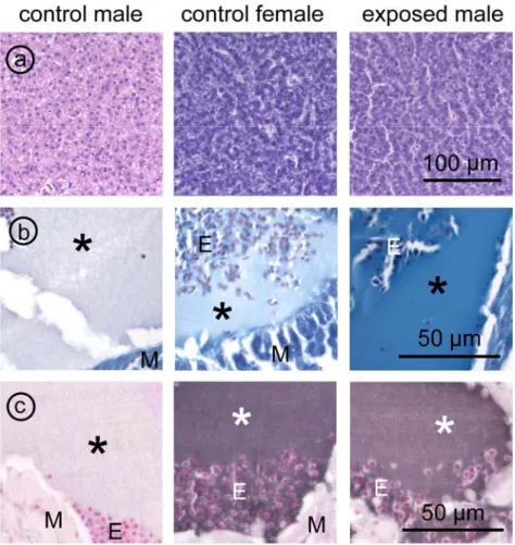
Fig. 2.1.1 Microphotographs of (immuno)histochemical detection of VTG
a Liver sections, representing pale acidophilia in control male zebrafish (left), and dark basophilia in
control female (middle) and male exposed to 1 nM E2 (right).
b,c Blood plasma in the heart,
b histochemical staining for phosphoproteins. High intensity staining is found in the male exposed to E2
(1 nM), faint staining in the control female, and virtual absent staining in the control male. Measurements of colour or staining intensity of these three parameters are given in Fig. 2.1.2.
c strong immunostaining intensity for vitellogenin in control female (middle) and male exposed to 1 nM
In the histological sections, the circulatory system showed areas of cell free plasma,
particularly in large vessels and in the heart. In these areas, there was also immunoreactivity with the anti-VTG antiserum in females and estrogen exposed males, from a concentration of 1 nM E2 (Fig. 2.1.1c). The intensity of immunostaining was significantly related to the dose of exposure to E2 (Fig. 2.1.2), confirming accumulation of VTG in the circulation.
Measurement in an exposure range between 0.1 and 1.0 nM confirmed 1 nM as the lower limit of detection (not shown).
ELISA
The ELISA for VTG used in a wide exposure range detected an increase of VTG in male plasma at an exposure level of 0.1 nM E2; further increase was dose dependent (Fig. 2.1.2). The lower detection limit in the ELISA used in the narrow exposure range was at an exposure level of 0.32 nM E2 (not shown). An ANOVA could not be performed on this latter series, since the plasma samples were pooled in this experiment. Both ELISAs detected VTG in female plasma at high levels.
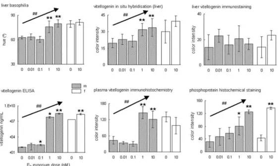
Fig. 2.1.2 - Comparison of semiquantitative representations of various histological detection methods of vitellogenin mRNA (liver basophilia, in situ mRNA hybridisation) or peptide
(immunohistochemistry, phosphoprotein staining) with the quantitative vitellogenin ELISA on plasma.
Each parameter was measured in males (m), exposed to a logarithmic dilution range of E2 (0-10 nM), and in females (f), control as well as exposed to a high concentration of E2 (10 nM). Each
concentration group contained six animals. Statistical significance of differences between exposure and control groups was calculated in a Student's T-test (* p<0.05; ** p<0.001); statistical significance dose-dependent effects was calculated in a single factor ANOVA (## p<0.001).
Correlations for methods to detect VTG protein
The correlations between the tested VTG parameters, liver H&E, plasma ELISA, plasma immunohistochemistry, and plasma phosphoprotein staining, were calculated on pooled average group data of both sexes. Table 2.1.1 shows that there were consistent high
correlation coefficients between all these parameters, in both test ranges of exposure to E2.
Discussion
Several histological tools are available to identify and quantify VTG, which, to date mainly has been detected by immunochemical methods in plasma samples. Advantages of these histological methods were mentioned in the introduction of this chapter, and they have satisfactory specificity, sensitivity and validity.
Fig. 2.1.3 - Immunohistochemical staining for vitellogenin in the liver of a male zebrafish exposed to 1 nM E2 (right), as well as a control male (not exposed, left).
Vitellogenin is present in the cytoplasm of hepatocytes in a perinuclear area reminiscent of RER/Golgi (arrows). Apparently excreted vitellogenin is also present in the space of Disse (arrowhead), between hepatocytes and the sinusoid lining.
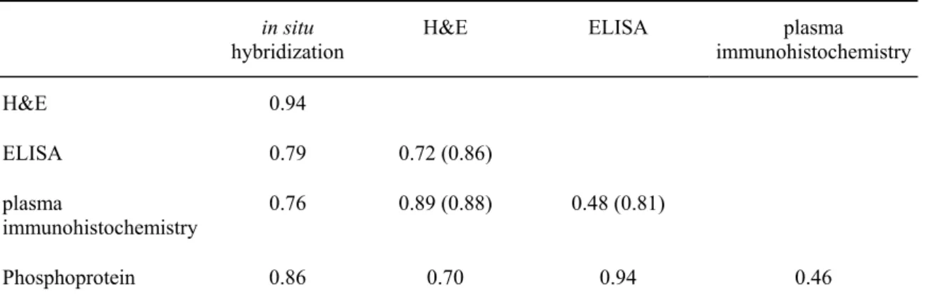
Table 2.1.1 - Correlation coefficients (R) in situ
hybridization H&E ELISA immunohistochemistryplasma
H&E 0.94
ELISA 0.79 0.72 (0.86)
plasma
immunohistochemistry 0.76 0.89 (0.88) 0.48 (0.81)
Phosphoprotein 0.86 0.70 0.94 0.46
Results of application of the various detection techniques for vitellogenin mRNA or protein on male zebrafish exposed to a wide range of concentrations of E2 were compared. The values in brackets represent the data from the narrow range series.
The specificity of the employed methods is indicated by staining patterns and locations, as well as correlations of staining intensities with exposure to (endogenous) E2 and correlations between methods. With respect to specificity, the histochemical staining of phosphoproteins (phosvitin, which is part of the VTG complex), is independent of the fish species, since this method will stain all phosvitins, and was equally successfully applied on histological sections of guppy (Wester et al., 1985).
The sensitivity of the histological methods was comparable. VTG mRNA and peptide were both reproducibly detected in male zebrafish after exposure to 1 nM E2. Digitised
measurements excluded subjective bias which may play a role in visual inspections.
The sensitivity of the ELISA was higher than of the histological methods, although not more than one dilution factor of of E2, compared to the histological detection of VTG. For
quantitative analysis of VTG expression, all methods have their specific (dis)advantages.
applications
These experiments were conducted to analyse whether histological evaluation of VTG
expression, either at the level of mRNA or of the peptide, can be useful in the histopathogical analysis of biological effects of exposure of (xeno-)hormones in the aquatic environment. From these results, routine H&E, focusing on liver basophilia, appears to be an acceptable method to screen for estrogen-like stimulation, at least in males. For purpose of validation, H&E could be supplemented with the relatively simple immunohistochemical detection of VTG in an area in a large vessel or the heart, devoid of erythrocytes. An alternative validation method, when no VTG antiserum for the species under study is available, is provided by the phosphoprotein staining method. The in situ mRNA hybridisation is a more laborious
method, yielding inferior results from the morphological viewpoint, without additional value compared to the other histological methods. Immunohistochemical detection of VTG in the liver could be useful for mechanistic studies, i.e. it indicates qualitative responses after estrogen agonist or antagonist stimulation.
ELISA and immunohistochemistry both have a high throughput, i.e. many samples can be processed within a relatively short time. An important advantage of immunohistochemistry is that the slides remain available for review, and embedded fish for other histological
determinations. The limit of exposure to E2 yielding detectable VTG expression by histological methods is below or at the level at which histopathological effects are found (VTG accumulations, alterations in the gonads). These histological methods for the detection of VTG can therefore be considered as valid markers for the induction of clinical
(histopathological) effects, and thus as valuable for hazard identification. In perspective of employing correlates for clinical relevance, there is only limited value in higher sensitivity of the biochemical analysis of plasma by ELISA compared to the other described methods, since ELISA detects VTG induction below an E2 exposure level that yields clinically relevant effects.
2.2.
Partial Life Cycle Study, protocol design
The partial life cycle (PLC) protocol as designed was aimed to detect endocrine effects with emphasis on the reproductive system, in both parents and offspring of laboratory fish. Such a protocol is more time and cost effective compared to a multi-generation or full life cycle assay, although the latter has the potential of generating more information, particularly on reproductive parameters in next generations. The central parameters measured in the PLC were reproductive success, juvenile development, histopathology of target organs and vitellogenin (VTG) levels. The test was further developed based on experience gathered during the various tests carried out in the course of the project. To validate the protocol, various reference EDCs were tested.
Selection of the species
The species used is Danio rerio (zebrafish). This species was selected as an easy-to-breed laboratory fish for which extensive knowledge from toxicology (Meinelt and Staaks, 1994; Kime, 1995; OECD, 1993) and developmental biology is available (Laale, 1977). These small fishes (approximately 3-4 cm, 0.55-1.0 g for males and females, respectively) are particularly suitable for whole body histology, while a sufficient volume of blood can be sampled for biochemical analysis (4-10 µL). The fish has a short life cycle, is sexually mature after approximately 3-4 months, and there is some sexual dimorphism. They are continuous (non-seasonal) breeders, and eggs are normally produced every 3-5 days under laboratory conditions (Niimi and LaHam, 1974; Laale, 1977; Westerfield, 2000).
Our stock was initially (1998) obtained from a commercial supplier, and after an initial antibiotic treatment subsequently bred in our facility and kept successfully under apparently disease-free conditions. Details on husbandry are described in the zebrafish atlas
(http://www.rivm.nl/fishtoxpat).
Test conditions
During the tests, adult fish were kept at a density of 2 L medium per fish in full glass containers covered with glass plates. Juveniles were kept at 150 mL per 5 fish for 21 days, thereafter at 300 mL per 5 fish. Animals were fed ad libitum for 5 minutes twice a day with defrosted artemias (commercially obtained).
Environmental conditions Before and after renewal of the media, pH and oxygen were
measured in all containers. pH values were considered acceptable between 6.5 and 8.5, and dissolved oxygen concentration in the test solution during the test was considered acceptable at a minimum of 60% saturation. Oxygen supply was by aeration through glass tubes.
Hardness was measured once in the stock control medium (upper limit 14 dH°), before use in the PLC. The temperature was monitored daily and maintained at 27 ± 2 ºC by keeping the containers in a water bath (Fig. 2.2.1). Nitrite was measured when increased respiration was observed in the fish. Light / dark regimen was kept at 14-10 hours (see Annex 1).
Experimental media A single stock of test compound was prepared for each PLC test and
kept at 4 °C. From these stocks, pre-dilutions were prepared each week, and final test solutions were prepared at room temperature from these pre-delutions at the day of use. If necessary a solvent was used, usually 0.01% DMSO. Ethanol, which was used initially, was abandoned since this facilitated microbial growth in the tanks. Solvent concentration was equalised in all test and control groups. The carrier medium was Dutch Standard Water (DSW, see Annex 1). Contact of the test system with synthetic materials was kept to a minimum to avoid the introduction of endocrine active contaminants, such as plasticisers. Test compound concentration was maintained in a semi-static way, i.e. with medium changes twice a week (3-4 day intervals). Duplicate water samples were taken daily for test compound analysis in such an interval. The concentration of stock solutions was monitored, and in some cases also of the highest test concentration with or without aeration, and with or without test organisms. Medium samples were kept frozen at –70°C until analysis.
Used medium was discarded after charcoal filtration, and containers were cleaned at each medium change (with 96% alcohol, then thoroughly rinsed).
Range finding assays
Initially, a concentration range finding experiment was conducted for each compound, in which adult and fry were exposed for a short period (usually fry 4 days, adults 9 days) to a dilution series with the highest concentration at water solubility (including solvent) or at sublethal concentrations derived from literature data. The concentration exposure range consisted of logarithmic dilutions of the top concentration. Decisive parameters in the range finding assay indicating toxic effects were: reproduction success, mortality, clinical
pathology and histopathology.
PLC
Protocol for adults
Spawning units consisting of two males and one female with an age range of 8 –14 months were selected from the batch, on the basis of successful reproduction, as indicated by the number of clutches (at least two clutches in eight days), by fertilisation rate (at least 100 fertilised eggs per clutch), and by hatchability (at least 50% per brood), all under reference conditions. Three spawning units were used per treatment (see Annex 1). The adults were exposed to a range of three concentrations of test compound with a dilution factor of preferably 3.2, or 10 at most; carrier medium (DSW) served as control. The highest test concentration was based on absence of toxic effects and successful reproduction as
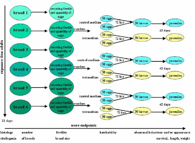
anticipated from the range finding test. The total exposure period for adults was 21 days (Fig. 2.2.2).
Breeding protocol was as follows: immediately after each medium renewal, spawning units were placed in a breeding trap with a mesh sieve without spawning substrate in a 6L
container. The next day eggs were collected and sexes separated until the next spawning episode.
Protocol for eggs
Immediately after separation of the sexes the produced eggs were collected by siphoning from the bottom of the tank. Fertilisation ratio was expressed as the percentage of fertilised eggs (non-fertilised eggs appear opaque). Fertilised eggs were rinsed with temperature controlled DSW to remove debris (remaining feed and excreta). From the spawning brood numbers 2, 4 and 6 produced during the exposure period, four groups of 50 fertilised eggs were used for further incubation. Two of these groups were placed in control medium (with
solvent if applicable), and the two other groups in the same test concentration as the parents had (Fig. 2.2.2). Two groups of each of the broods 2, 4 and 6 from control parents were also incubated in the highest test concentration. Incubation was performed in 10 cm diameter petridishes with 50 mL test medium. This design allows duplicate observations and
distinction between parental and postnatal effects. Eggs were maintained at 28.5 ± 2 ºC and mortality and hatching were scored after 24, 48 and 72 hours.
Protocol for larvae and juveniles
If available, a total of 50 hatchlings obtained from pooled duplicate groups of eggs was transmitted to a 1.5 L glass tank for continued exposure for another 42 days (see Annex 1). During each of the biweekly medium changes, the juveniles were photographed in a small volume of medium to facilitate counting and data storage. The volume of medium was adjusted to the actual number of fish.
Parameters
Both adult and juvenile fish were inspected daily for mortality, abnormal appearance and behaviour. After 21 days of exposure, adult fish are euthanised in an aqueous solution of 100 mg/L tricaine methanesulphonate (MS-222, CAS RN 886-86-2, Sigma-Aldrich), neutralised with sodium bicarbonate (2/1 MS-222, w/w). From two females and four males per
concentration group the tail was cut with a pair of scissors and blood obtained from the incision with a heparinised glass capillary. The blood was diluted with an equal volume of aprotinin (6 µg/mL), stored on ice and centrifuged at 600 g for 5 min. to separate and collect plasma, which was stored at –20 ºC for future analysis of e.g. VTG. Specimen were fixed in Bouin’s fixative for 24 hours, thereafter kept in 70% ethanol until further histological processing; for details see Annex 2.
Juveniles were euthanised in MS-222 after 42 days, blotted dry, length measured on calibrated paper, weighed and fixed as above. Moribund animals were also sampled for histology, if indicated. Condition factor (K) was calculated for individual fish by the
equation: K= weight (g) x 100/(length cm)3. One day prior to euthanasia, fish were not fed to reduce intestinal content that might interfere with histology.
Data treatment and interpretation
For statistical analyses, the experimental unit of the PLC-tests is the spawning unit
(two males, one female) in a single tank. Egg clutches deposited in one tank were defined as repeated measures because these egg clutches, which originate from the same experimental unit, are not independent from each other. The same accounts for the juveniles hatched from the egg clutches originating from the same experimental unit; these are also defined as repeated measures. ANOVAs, t-tests and regressions were executed with means of repeated measures per aquarium. All life history parameters were analysed with One way ANOVA followed by Dunnett’s Multiple Comparison to compare treatments with the controls (GraphPad Prism 2.01). Exceptions were hatching of eggs and juvenile parameters coming from the same parents but subjected to different exposure concentrations. These data were analysed by paired t-tests with the adult couples defining the pairs. Linear regression was applied to number of egg clutches, to total number of eggs per aquarium, to fertilisation rate and also to hatching and juvenile parameters from the treatments in which the juveniles were exposed to the same concentration as their parents.
2.3.
Population modelling
The ecological relevance of endocrine disrupting effects in individual fish is in the impact at the population level. As a tool to extrapolate effects measured in the PLC-test to the
population level, the zebrafish model of Oertel (1992) was applied. The model is described in detail by Schäfers et al. (1993). In summary, the Individual Based Model (IBModel) is based on long-term experiments in large aquaria with zebrafish or guppy, representing opposite reproduction strategies. Zebrafish are typical r-strategists, with a life history directed to maximize reproductive rate (r); r-strategists typically produce large numbers of offspring and show no brood care behaviour (Nagel, 2002; Halliday, 1993). Populations of r-strategists can recover relatively easy from environmental disturbances.
K-strategists such as the guppy have a life history adapted to maximize competitiveness and survival. They typically produce less offspring than r-strategist, but invest in some form of brood care (e.g. viviparity in guppies). K-strategists mostly live in relative stable
environments and the population is relatively sensitive to environmental disturbances. In the laboratory setting of Oertel, population dynamics were monitored and some of the population parameters were estimated with additional experiments.
Variables which were entered in the model were as measured in the PLC-tests: number of clutches, clutch size, fertilisation, hatching, developmental variables (survival, length,
weight), and sex differentiation. The Von Bertalanffy growth curve was used to estimate time to develop from hatching to adult animals. It was assumed that maturity was reached when the animals reached 24 mm of length (24.9 mm for females and 23.1 mm for males; Laale, 1977). Sex differentiation was incorporated as proportion of the juveniles that developed into females. The model accounts for predation of progeny by adult zebrafish, varying with life stage; this characteristic was maintained. Mortality of adults was set at 0 % in all cases, also when mortality had occurred among adults. Exposure concentration levels used during the present study were set at non-lethal levels for adults and it was assumed that possible mortality among adults was incidental. Mortality due to exposure within the population is already represented by juvenile mortality.
The modelled system was set at 800 L water and 200 L of refugium, in contrast to 200 L water and 50 L refugium in the laboratory setup of Schäfers et al. (1993); pilot calculations showed that such a larger system better accounts for changes in life history parameters which may have an impact on population size or survival. With the smaller system, population
extinction occurred in a relatively high proportion of simulations, thereby possibly veiling effects of PLC test variables.
Output parameters were extinction risk (survival changes of the population), day of
extinction, and average population density. The calculation period was 2004 days (equals 4-5 generations); pilot calculations showed that there were no additional effects after 3000 days (equaling 6-9 generations). To understand the results of the calculations, and to validate the specificity of the outcomes on zebrafish populations (r-strategists), the IBModel was also employed to estimate effects on
K-strategists, using the guppy model defaults and the results from the PLC-tests with zebrafish. For this purpose, results from the other PLC variables (adult reproduction parameters and growth and survival of juveniles) were assumed to affect guppies and zebrafish similarly.
2.4.
Histopathology atlas
Introduction
One of the endpoints of the project was an inventory of the normal histology and effects of endocrine disruptors in small laboratory fish. To facilitate dissemination it was decided to present this in the format of a digital Toxicological Pathology Atlas of Small Laboratory Fish. This atlas is intended as a reference guide, to help investigators and other professionals interested to use histology and pathology of small fish. Although the data are focused on the model species Danio rerio, it must be acknowledged that the information will be applicable to other species to a large extent. Material from other sources than the current project is also included; references are included in the concerning sections. Acknowledgement is made to those scientists that have contributed as peer reviewers in their respective expert fields.
Outline of the atlas
The atlas is a html-based product (hypertext markup language), optimised for Microsoft Internet Explorer 5.0+. This format enables easy browsing and the inclusion of advanced techniques to improve understanding, such as image animation or sophisticated detail identification. The possibility of instant updating is also considered as a major advantage.
The atlas contains five main sections, which are available from a top menu (Fig. 2.4.1). These include:
• normal histology, aiming to show overview and detail sections of all organ systems; • histopathological effects of exposures to endocrine active compounds, including estrogen
agonists and antagonist, androgen agonist and antagonist, and thyroid antagonist; this module contains major results from the present project;
• MRI animations for better understanding of the (zebra)fish anatomy; • a text search module;
• a general information module.
Each section has an index menu on the left-hand side, and a contents area. For the
histological modules, this contents area consists of a central image part and a descriptive text, which contains interactive links to the image.
The atlas was presented at the annual meetings of the Society of Environmental Toxicology and Chemistry (SETAC) Europe and America in Brighton and Nashville, TN, respectively, both in 2000. It was announced to expert organisations, and hyperlinks are now available at websites of e.g. Society of Toxicological Pathology. A mailing list with approximately 200 addresses of colleagues in this area is used to announce major updates.
The atlas has been used in practice as a basis for an OECD workshop held in September 2002 at the RIVM. Also it is often cited and used in OECD guidelines under development, and it will be included in a workshop CD ROM to be organised in October 9-10, 2003 (Fraunhofer Institut, Hannover).
The atlas is available on the institute’s website http://www.rivm.nl/fishtoxpat/; for dedicated users a CD ROM version can be provided.
Fig. 2.4.1 – Screenshot of the Toxicological Pathology Atlas of Small Laboratory Fish
(http://www.rivm.nl/fishtoxpat). The top menu indicates the main sections of the atlas and is available throughout the application. The left-hand menu is activated by selection of specific items and contains links to pages with detailed information. These contents pages have a title, a single or a composite image, or a
(animated) sequence of related images, and a descriptive text, which contains links which activate indicators to areas of interest on the image.
3. Partial Life Cycle study, application
with reference compounds
3.1.
Introduction
In order to test the practical applicability of the prototype test protocol, a number of reference compounds was tested. The compounds selected were 17β-estradiol (E2), a natural estrogen. Initially, ZM 189,154, a preclinical drug from a pharmaceutical industry R&D program, was tested as anti-estrogen, in accordance with the proposed compound from the OECD panel, but analytical difficulties and potential problems with future supply made us to choose tamoxifen, a therapeutic anticancer drug. For androgen initially methyltestosterone was selected, but appeared to have significant estrogenic properties and thus the non-aromatisable methyldihydrotestosterone was chosen. As anti-androgen the therapeutic drug flutamide was selected and for antithyroid the therapeutic drug propylthiouracil. Finally, a field sample from a suspected hot spot for estrogenic effects was investigated.
3.2.
PLC-test with estrogen: 17
β-estradiol
Introduction
As reference estrogen, 17β−estradiol (E2) was chosen. E2 is one of the natural estrogens in vertebrates. It is often used a reference compound, although many researchers prefer ethynylestradiol (EE2), a synthetic pharmaceutical and the active ingredient in oral
contraceptive. Both E2 and EE2 are found in environmental surface water samples mainly through sewage treatment effluent, but EE2 is known to be a more potent estrogen.
Materials and methods
E2 (CAS 50-28-2, Fluka) was dissolved in stock medium ethanol 96% and stored at 4 0C. End solutions were prepared from this stock and contained maximal 0.01% solvent.
The test was performed as described in “General Protocol”. Briefly, range finding tests were conducted up to 21 days using concentrations from 1-100 nM with adults and 1-1000 nM with fry. These tests revealed significant effects in histology in both sexes such as
accumulation of VTG in circulation, body cavities and interstitial tissues, with dilation of these compartments. In excessive cases this resulted in hydrops, ascites and abduction of scales (see Atlas). This increased vitellogenesis was also observed in 4 dph larvae at 10 nM E2 and higher. In addition, hepatocellular basophilia was seen in conjunction with
vitellogenesis, and collapse of the ovaries (extensive atresia and absence of vitellogenic oocytes). The absence of vitellogenic oocytes in the 10 nM-exposed females indicated cessation of reproduction at this concentration. Therefore, 1 nM was chosen as the top concentration for the PLC test, with 0.32 and 0.1 nM as mid- and low concentrations. Analysis of the exposure medium for actual concentrations of E2 revealed a gradual decline of 102.0 - 28.4 - 12.5 - 3.8 (percentage of nominal value of 1 nM) at days 1-4, respectively. A similar decline was found in the 1000 nM medium at days 1-2.
Adults in triplicate spawning units per concentration were exposed for 21 days, and eggs were collected, incubated and juveniles were sampled after 42 days of exposure to the same or complementary medium compared to their parents. To examine effects of high E2
concentrations on histopathology of the gonads in more detail, data from the preceding range-finding assay (range 1 - 10 - 100 nM) were included.
Animals were monitored daily for general health and clinical effects such as mortality, abnormal behaviour and appearance. Eggs were monitored for fertility and hatching. At termination of the experiment, animals were euthanised, length and weight were measured (juveniles), and blood was collected from adults for VTG determination. Animals were fixed
in toto for histopathology of target organs (gonads, plasma, liver, etcetera), or for further
development of VTG immunohistochemistry. The results reported below are from the PLC, unless specified otherwise.
The experiments were approved by the Institute’s Animal Experiment Committee (AAP 199900019, 199900608, 199800376 and 200100203).
Results and discussion
In life observations - adultsIn life observations during adults exposure revealed no effect on clinical appearance and behaviour. In the control and 0.1 nM groups only two out of three units were reproductive, and the data in Table 3.2.1 are based on the reproductive units only. There was a
concentration-dependent decrease of the number of clutches, whereas the clutch size increased in a concentration-dependent way. These effects apparently compensated each other, as the resulting total number of eggs showed no change. The fertilisation rate was also unaffected.
Table 3.2.1 - Reproduction parameters of P generation after exposure with E2
concentration
(nM) number ofclutches 1 clutch size total number ofeggs fertilisation rate(%)
control 7.0 ± 0 a 183 ± 8 a 1280 ± 59 61.3 ± 16.8
0.1 6.5 ± 2.1a 243 ± 68 a 1649 ± 958 72.8 ± 1.1
0.32 4.3 ± 0.6 a 234 ± 56 a 1009 ± 226 67.4 ± 24.9
1 3.3 ± 2.5 a 448 ± 199 a 1159 ± 465 70.3 ± 28.5
All values are average ± sd of three spawning units (two in control and 0.1 nM due to non spawning).
1 maximum number of clutches is 8.
a significant (p<0.05), linear regression; non spawners in control and 0.1 nM are not taken into
In life observations - juveniles
It appeared that parental nor juvenile exposure had any effect on hatching percentage (Table 3.2.2). No abnormal appearance or behaviour was observed in any of the treatment groups. Increased mortality was recorded with the 1 nM exposed juveniles (Table 3.2.3).
Table 3.2.2 - Hatching after exposure to E2
treatment P - F1 (nM) n1 hatching (%) control - control 2 90.3 ± 2.6 0.1 - control 2 93.5 ± 3.5 0.32 - control 2 87.0 ± 18.4 1 - control 2 85.5 ± 4.9 0.1 - 0.1 2 89.5 ± 7.8 0.32 - 0.32 3 93.0 ± 4.0 1 - 1 2 89.3 ± 2.5
Values represent the average ± sd of all replicates.
1 number of spawning units
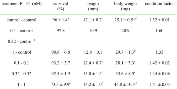
Table 3.2.3 - In life observations of F1 zebrafish exposed to E2 for 42 days
treatment P - F1 (nM) survival
(%) length(mm) body weight(mg) condition factor control - control 96 ± 1.4a 12.1 ± 0.2b 25.1 ± 0.5c,d 1.32 ± 0.01 0.1 - control 97.8 10.9 20.9 1.60 0.32 - control 1 1 - control 90.8 ± 6.8 12.8 ± 0.1 29.7 ± 1.3d 1.35 0.1 - 0.1 93.2 ± 3.7 12.4 ± 0.7b 28.1 ± 5.3c 1.42 ± 0.02 0.32 - 0.32 92.4 ± 1.9 13.0 ± 1.4b 33.6 ± 8.5c 1.44 ± 0.08 1 - 1 73.3 ± 9.9a 14.2 ± 1.0b 45.8 ± 10.5 c 1.41 ± 0.03
Values are mean ± sd of two replicates, except for 0.1 control (single observation) and 0.32 -0.32 (triplicate observation).
1no data for the 0.32 nM-control group due to insufficient offspring in the 0.32 nM a p<0.05, Dunnett’s test
b,c p<0.05, p<0.01, linear regression for exposed juveniles d p<0.05, T-test
Importantly, there was a significant concentration-related increase in length and body weight in the exposed juveniles (with similarly exposed parents; linear regression), which is possibly related to the anabolic properties of estrogens. There was also an increased juvenile body weigth after exposure of parents only to 1 nM E2.
Histopathology - adults
In the PLC, only moderate effects were observed in the highest exposure group (1 nM): moderate to strong basophilia in male hepatocytes (Fig. 3.2.1), and occasionally eosinophilic (proteinaceous, vitellogenic) intra-/extravascular plasma. Histology in the lowest exposure group (0.1 nM) was comparable to control: no aberrant VTG expression (eosinophilic hepatocytes in males, no colloidal plasma accumulations), no gonadal pathology. There was some variation of the intensity of these effects between this and other studies where E2 was used as a test compound (see Chapters 4.1, VTG analysis; and 3.7, test with field sample). No obvious changes in females (possibly increased atresia in the ovaries) were detected.
Fig. 3.2.1 - Routine H&E staining of zebrafish liver; control male liver stains acidophilic (eosin), control female liver stains basophilic (haematoxylin) due to high contents of mRNA. Liver of
males exposed to E2 stains as control female, due to induction of vitellogenin mRNA
expression.
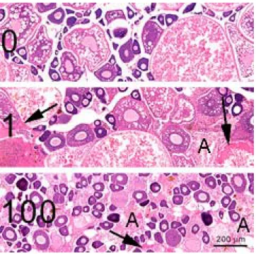
Fig. 3.2.2 - Zebrafish ovaries showing a concentration dependent decrease of vitellogenic oocytes and an increase of atretic follicles (A), compared to control (0). Arrows indicate
accumulations of vitellogenin. E2 concentrations (1-100) in nM (range finding test). H&E staining
At high concentrations (10 nM and up, range finding test), severe intravascular and interstitial VTG accumulations were detected in both males and females (Fig. 3.2.2). No vitellogenic oocytes were present in the ovaries, and a high incidence of atretic follicles, compared to control ovaries, was observed (Fig. 3.2.2).
In the testis, microscopic observation revealed an increase of spermatogonia (Fig. 3.2.3); morphometrically, this increase proved to be relative, since there was a decrease in size of progressed classes of spermatogenic cysts (mainly spermatocytes) in size (Fig. 3.2.4); the
Fig. 3.2.3 - Zebrafish testis showing a decrease of progressed stages of
spermatogenesis (SC , spermatocytes and ST, spermatides) and a subsequent relative increase of early stage spermatogonia (SG), after exposure to E2 (bottom), compare to control (top). Note
eosinophilic vitellogenin accumulations in the interstitial tissue in the E2-exposed specimen. H&E staining.
average cyst size (µm2)
0 2000 4000 sg sc st
#
*
#
*
ratio cysts per phase (%)
0 35 70 sg sc st 0 1 10 100
Fig. 3.2.4 -Morphometrical analysis of spermatogenic cysts after exposure to 0-1-10-100 nM E2. There
is a concentration dependent decrease in cyst size of spermatocytes (sc) and spermatids (st), linear regression , p< 0.05 (#); when compared to the control, sc and st are smaller in the highest concentration (p<0.05, t-test). sg, spermatogonia.
suggested decreased proportion of spermatocytes was not statistically significant. This indicates a decreased proliferation of spermatogonia and a decreased progression of
differentiation. These conclusions were further supported by labelling experiments with BrdU and immunostaining of PCNA (not shown). There were no aberrations of Leydig or Sertoli cells.
Other observations included occasional chronic inflammation and fibrosis in pancreas or bile ducts, and were considered as background pathology.
Vitellogenin Pooled plasma of 2-3 females or 3-5 males was analysed for VTG contents with
ELISA (M. Fenske, UFZ Leipzig; Fig. 3.2.5). These values revealed a concentration dependent increase in males after exposure to E2, up to a similar value as found in females after exposure to 1 nM E2. Although these represent mean values, since they were obtained from pooled plasmas, the increase could not be verified statistically on these singular entries. There was no change in females. Details on methodology and interpretation of VTG analysis are further discussed in the chapter on VTG analysis (see Chapter 2.1).
Histopathology - juveniles
Vitellogenesis in juveniles was not convincing (with 1 nM as highest concentration). On the other hand, there was a marked statistically significant shift in sex ratio, after each of the exposure concentrations. This shift is mainly due to decreased percentage of males and increase of undifferentiated individuals (Fig. 3.2.6 left). This indicates either a mere delay of differentiation, or a real shift in sex ratio, depending on final phenotype of the yet
undifferentiated specimen. The results from experiment with the field sample (Chapter 3.7), using only 1.0 nM support the latter option, since the significant shift in sex ratio observed in this case was mainly due to the absence of males and an increase of females after the E2 exposure (Fig. 3.2.6 right panel). This experiment also showed that the induced shift is due to juvenile exposure, since there was no effect of parental exposure only. The different outcome between these two experiments may result from a generalised delayed development in
vitellogenin after E2 (range)
1 10 100 1000 10000 100000 1000000 0 0.1 0.32 1 0 0.1 0.32 1.0 µ female male nM
Fig. 3.2.5 – VTG-ELISA of zebrafish plasmas after exposure to a range of E2 concentrations, given as nM. Each bar
represents a mixed sample of either 2-3 females or 3-5 males.
juveniles in the first experiment, indicated by the lower overall average length (13.0 ± 1.6 mm) compared to the overall average length in the second experiment (16.5 ± 0.4 mm). In this second experiment, ovary maturation was determined on the presence of the most matured stage of oocytes, defined by size and progression of vitellogenesis (Fig. 3.2.7). This analysis showed that exposure to 1 nM E2 inhibited maturation of oocytes significantly, compared to control.
Population modelling
By means of the zebrafish model no significant effects of the E2 treatments in the PLC-test were found on population survival and population size of zebrafish (Fig. 3.2.8), in spite of skewed sex ratio and decreased juvenile survival. The large CVs of extinction chances and population size of the model simulations have probably prevented the appearance of more
Fig. 3.2.7 - Staging of ovaries according to the most advanced oocytes present in the gonad, in control animals and after exposure to 1 nM E2. u, undifferentiated, further numbers on the horizontal axis indicate arbitrary classes (defined in the atlas). Exposure to E2 induced a
statistically significant shift to the left (***p<0.0001 in a Chi-squared test), indicated a delayed development in females. 0 25 50 75 u 0 0-1 1 1-2 2 2-3 3 3-4 4 4-5
ovary maturation stage
percentage of animals per class
control estradiol
***
Fig. 3.2.6 - Sex ratios in two separate assays after 21 (P) - 42 (F1) days of exposure. Both graphs show
the relative presence of female (f), male (m), and undifferentiated (u) specimen. The left graph shows the effects of a concentration range of E2 (0.1 - 0.32 - 1.0 nM; c, control), adults and offspring exposed, in the left graph, there is only exposure to 1.0 nM, both in adults and offspring (middle set), or in adults only (right set). *, **, p< 0.05, 0.01 respectively, T- test.
F1 sex ratio at 42 dph - E2 exposed
0 25 50 75 100 c - c 0.1 - 0.1 0.32 - 0.32 1.0 - 1.0 sex (p ercen tag e ) f m u
*
*
*
0 20 40 60 80 100 c - c E2 - E2 E2 - c sexc (percent age) f m u**
obvious effects of E2.
Again, the 10 nM concentration was not tested because no eggs were found to develop at this concentration (data from range finding). Therefore, this concentration was not included in the PLC-test, but it can be anticipated that at 10 nM E2 the population’s reproduction and
survival are severely compromised.
Conclusions
• At 10 nM E2 ovaries were completely blocked within a few days. Therefore, this
concentration must be considered as incompatible with normal reproduction and survival of the population.
• At 1 nM there was a lower number of egg clutches but an increased number of eggs per clutch. Total number of eggs was unaffected. Survival was decreased in offspring. • There was a dose dependent increase in length and weight of the exposed juveniles; this
could be related to the anabolic properties of estrogenic hormones.
• At 0.32 nM and higher, vitellogenesis was induced in males, and the sex ratio was skewed in offspring with a preference for females to develop. Therefore, this histologically
detectable VTG increase could serve as an indicator for adverse other effects. Overall, this was the lowest observed adverse effect level.
• Spermatogenesis was inhibited in adult males (1 nM, concentration dependent), as well as ovary development in juvenile females (1 nM) .
Fig. 3.2.8 - Population model for E2. No effects of treatment on population extinction. Total
extinction in one of the control and 0.1 populations due to non-spawning. Treatments indicate P and F1 exposure, respectively. DSW, control medium; other groups are indicated with nominal values of E2 exposure in nM.
effect of E2 on population extinction after 2004 days
DSW-DSW 0.1 -0.1 0.32 -0.32 1 -1 0 25 50 75 100 ex tin ct ion ( % )
• Up to 1 nM, combined effects had no influence on survival of the population or population size.
• In view of the decline in E2 concentrations during the exposure period, the results are most likely an underestimate when expressed as nominal values.