ESTABLISHING
ZEBRAFISH
EMBRYONIC MODELS TO INVESTIGATE
PANCREATIC
CANCER
TREATMENT
AND PARP INHIBITOR EFFICACY
Word count: 17,716Lore Dobbelaere
Student number: 01507858
Supervisor(s): Prof. Dr. Kathleen Claes
Mentor: Jeroen Vierstraete
A dissertation submitted to Ghent University in partial fulfilment of the requirements for the degree of Master of Science in the Biomedical Sciences
Preface
In the next fifty pages you will find the work I conducted in the last two years, my master thesis. To make all of this possible, I had a tremendous amount of help coming from a diversity of people whom I would like to thank first.
To start, I would like to thank Ghent University in general and more specific its Faculty of Medicine and Health Sciences to allow me to follow this education and to get a Master’s degree of Sciences in Biomedical Sciences. These five years have been an incredible and educational experience for me.
I wish to show my gratitude to my promotor Prof. Dr. Kathleen Claes. She has given me the opportunity to work in her research group on a very interesting subject. She also gave me the chance to learn about the academic world. Additionally, I would like to thank every member of the Center of Medical Genetics in Ghent (CMGG) for teaching me. I had the opportunity to attend great lectures from professors and PhD students and I have learned interesting laboratory techniques from lab technicians. Some CMGG members just friendly showed me the way around the lab when I was lost.
Above all, I would like to thank my mentor, Jeroen Vierstraete, for his guidance. He showed me how to correctly produce scientific data and how to write a scientific research. He has spent an endless amount of time teaching me brilliant techniques and had unlimited patience despite my tremor. He trusted me to do experiments individually, gave me responsibilities and in particular he believed in my abilities.
I would like to show my gratefulness to my parents for giving me the opportunity to complete this education and I would like to thank especially my mother for calming me during many stressful times. Finally, my friends have always been an enormous part of my life and they have formed me into the person that I am today. I will be forever grateful to them.
Corona crisis
The Corona crisis influenced scientific research around the world. Due to measures taken by the Belgian government I was unable to continue laboratory work. Experiments were discontinued from the 16th of March on and this thesis was written with the obtained results supplemented with literature studies. Some experiments were completed, however many were started but not finished. The original goal was twofold.
To start, the optimization of the dissociation protocol of PANC-1 and Mia-Paca-2 cells was planned. Three methods were used to dissociate PANC-1 cells. In the first method, EDTA was used as dissociation compound. This resulted in low yield of cells with moderate viability. To improve the viability trypsin and TrypLE were investigated to be used as dissociation compounds. However, during optimization steps, the Corona crisis started and these experiments were prematurely terminated.
Engraftment of human cells in zebrafish larvae requires countless hours of practice. To practice, injection of HTC116 cells were used because these cells are easy to engraft. Injection with PANC-1 cells was tried a few times and conducted successfully in some larvae. The next step would have been optimization of the functional read-out and incubation of successfully injected larvae with anticancer compounds (gemcitabine and FOLFORINOX) to verify tumor response to compounds.
Culturing of the Mia-Paca-2 cell line was initialized one week before lockdown. Our goal was to engraft Mia-Paca-2 cells in zebrafish larvae and treat the engrafted larvae with gemcitabine to verify the higher sensitivity to gemcitabine in the zebrafish model. This cell line was freshly purchased and thus only had one vial that was in culture. Upon notice of the immediate lockdown measures, it was decided to freeze all cultures for long-term storage. Therefore, no experiments on this cell line could be conducted to optimize the dissociation protocol and no engraftment was performed using this cell line.
The second goal of this thesis was to create a model to establish novel PARP inhibitors (PARPi)
in vivo. To do this, a Rad51 foci assay, an acridine orange assay and an eye size test were
planned. The Rad51 foci assay was mostly completed. All five clinically available PARPi were investigated at a 400µM concentration and additionally olaparib and talazoparib were investigated at a 5µM concentration. Furthermore, a dose response was planned for all five PARPi. This, however was not completed. The dose response of rucaparib and veliparib were conducted. Due to a mistake in the staining protocol, the rucaparib slides had to be redone. The dose response of veliparib was successful but more data points are needed. For olaparib, an acridine orange staining and eye size test were done to prove cytotoxicity of the PARPi. The acridine orange staining obtained satisfactory results and the eye size test was concluded.
I regret the lab time that was taken away causing unfinished experiments. However, safety comes first at all cost.
List of abbreviations
µL microliter
µM micro molar
3D 3 Dimensional
ADP Adenosinediphosphate AMPK AMP-activated protein kinase AO acridine orange
APC Adenomatous polyposis coli ATM serine/threonine kinase
ATR ataxia telangiectasia and Rad3-related protein BER Base Excision Repair
BRCA1 breast cancer 1 BRCA2 breast cancer 2 CA19-9 Cancer Antigen 19-9
CDKN2A cyclin-dependent kinase inhibitor 2A CI Confidence interval
cm Centimetre
CT Computed Tomography
CtIP retinoblastoma binding protein 8 DAPI 4′,6-diamidino-2-phenylindole
DDR DNA damage response
DMEM Dulbecco's Modified Eagle Medium DNA desoxyribonucleicacid
dpf days post fertilization dpi days post injection DSB Double Strand Breaks ECM Extra Cellular Matrix
EDTA Ethylenediaminetetraacetic acid
FAMMM Familial Atypical Multiple Mole and Melanoma FAP Familial Adenomatous Polyposis
FBS Fetal Bovine Serum
FDA Food and Drugs Administration
FOLFIRINOX Folonic Acid, 5-Fluorouracil, Irinotecan, Oxaliplatin GDP Guanosinediphosphate
GEF Guanine nucleotide Exchange Factor GTP Guanosinetriphosphate
H&E Haematoxylin and Eosin
HBOC Hereditary Breast and Ovarian Cancer hpf hours post fertilization
HR Homologous Recombination
IPMN Intraductal Papillary Mucinous Neoplasm KRAS Kirsten Rat Sarcoma
LOF Loss Of Function
mFOLFIRINOX modified FOLFIRINOX mL Millilitre MLH1 MutL homolog 1 mm Millimetre MSH2 MutS homolog 2 MSH6 MutS homolog 6
mTOR mechanistic Target Of Rapamycin
Nacre melanocyte inducing transcription factor A NAD+ Nicotinamide-adenine-dinucleotide
NBS1 Nibrin
NHEJ Non-Homologous End Joining PALB2 Partner And Localizer of BRCA2 PanIN Pancreatic Intraepithelial Neoplasia PARP poly (ADP-ribose) polymerase PARPi PARP inhibitor
PBS Phosphate Buffered Saline PCR Polymerase Chain Reaction
PDAC Pancreatic Ductal Adenocarcinoma PDX Patient Derived Xenograft
Penstrep penicillin and streptomycin
PFA paraformaldehyde
PMS2 PMS1 Homolog 2
PP Pancreatic Polypeptide PVS Perivitelline space
Rad51 DNA repair protein RAD51 RNA ribonucleic acid
RPA DNA Replication Protein A seDNA single end DNA
SMAD4 Mothers against decapentaplegic homolog 4 SSB Single Strand Break
ssDNA single stranded DNA STK11 serine/threonine kinase 11 TGF-β Transforming Growth Factor-β TGS Tumor Suppressor Gene TP53 Tumor Protein 53
Table of contents
1. Summary ... 8 2. Introduction ... 9 2.1 The pancreas ... 9 2.2 Pancreatic cancer ... 10 2.2.1 Incidence ... 10 2.2.2 Survival rates ... 10 2.2.3 Risk factors ... 102.2.3.1 Non-modifiable risk factors ... 10
2.2.3.2 Modifiable risk factors ... 11
2.2.4 Pathogenesis ... 12 2.2.4.1 Molecular Profile ... 12 2.2.4.2 Screening of PDAC ... 14 2.2.5 Treatment ... 15 2.2.5.1 Surgery ... 15 2.2.5.1 Chemotherapy ... 16 2.2.5.3 PARP inhibitors ... 17
2.2.6 Using zebrafish to model pancreatic Xenografts ... 22
2.2.6.1 Patient derived xenografts ... 22
2.2.6.2 Zebrafish xenografts ... 22
3. Objectives ... 24
4. Materials and methods ... 25
4.1 Zebrafish embryonic xenografts ... 25
4.1.1 Optimization on cell lines ... 25
4.1.1.1 Maintenance of cell lines ... 25
4.1.1.2 Labeling cells ... 25
4.1.2 Injecting larvae ... 26
4.1.2.1 Maintenance of zebrafish strain ... 26
4.1.2.2 Injection ... 27
4.1.2.3 Hematoxylin and eosin staining ... 28
4.2 PARPi ... 28
4.2.1 Maintenance of the zebrafish strains ... 28
4.2.2.1 DMSO toxicity ... 28
4.2.2.2 Rad51 foci ... 28
4.2.2.3 Acridine orange assay ... 29
4.2.2.4 Eye size test ... 30
5. Results ... 31
5.1 Optimization xenograft procedure ... 31
5.1.1 Cell dissociation ... 31 5.1.2 Engraftment ... 32 5.2 PARPi ... 34 5.2.1 DMSO toxicity ... 34 5.2.2 Rad51 foci ... 34 5.2.2.1 PARPi comparison ... 35
5.2.2.2 Assessing potency of talazoparib ... 36
5.2.2.3 Dose response ... 37
5.2.3 Acridine orange assay ... 37
5.2.4 Eye size test ... 39
6. Discussion ... 41
6.1 Zebrafish embryonic xenografts ... 41
6.1.1 Cell dissociation ... 41 6.1.2 Engraftment ... 42 6.2 PARPi ... 43 7. General conclusion ... 46 8. Poster ... 47 9. Reference list ... 48 10. Addendum ... 53 1. H&E protocol ... 53 2. Ford protocol ... 53
3. KAPA2G Robust HotStart ReadyMix Ford ... 54
1. Summary
Pancreatic cancer is a devastating disease with a five-year survival rate below 9%. This poor prognosis is mainly due to late diagnosis, rapid progression, early metastasis, refractoriness to therapy and lack of therapeutic approaches.
To improve prognosis, personalized therapies are warranted. Such personalized therapy is usually performed by developing and testing patient derived mouse xenografts (mPDXs). However, several disadvantages do not allow the clinical use of mPDXs. We investigated if zebrafish embryonic xenografts (zPDX) could be a suitable alternative. To engraft human pancreatic cells in zebrafish embryos, we aimed to optimize the engraftment protocol. Three dissociation methods were investigated. The first method was mechanical but results were unsatisfying. The next method was enzymatic dissociation with trypsin or TrypLE. Some successful injections of PANC-1 cells into zebrafish were accomplished. We aimed to optimize functional read-outs of those xenografts. However, experiments were not finished due to the measures imposed by the Belgian government as response to tackle back the Covid-19 outbreak.
As approximately 10% of all pancreatic cancer patients have mutations in HR genes, these patients may benefit from therapy with PARP inhibitors (PARPi). New PARPi are being developed in search for better therapy with less adverse effects. Therefore, we tested if zebrafish larvae could serve as in vivo model. Three assays were used to evaluate this being the Rad51 foci, the acridine orange assay and an eye size test. Not all experiments were completed but results showed that indeed zebrafish can be used as in vivo models to establish novel PARPi.
2. Introduction
2.1 The pancreas
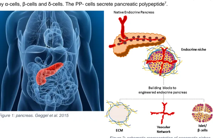
The pancreas is a lobulated organ of the digestive system. The pancreas is located on the posterior wall of the abdominal cavity1 as shown in figure 1. Macroscopically, four parts can be distinguished: a head, a neck, a body and a tail. The pancreas consists of an endocrine and an exocrine part. The exocrine gland secretes pancreatic fluid into the duodenum. This pancreatic fluid contains enzymes for digestion like trypsin, lipase, protease and amylase2. These enzymes are inactive inside the pancreas and later activated in the duodenum to prevent damaging of the pancreas by the enzymes. The pancreas also secretes natriumbicarbonate to neutralize the gastric acid3.
The endocrine part of the pancreas secretes hormones and is structured in units that are independently vascularized4. These units are called niches and consist of extracellular matrix (ECM) and islets (figure 2). The pancreatic islets represent only about 1% of the pancreatic tissue though it receives over 15% of all the blood flow to the pancreas5. The islet itself is an independent organ that consists of single cells like parenchymal cells, α-, β-, and δ-cells and pancreatic polypeptide (PP) cells6. These cells form a 3D structure that are connected to each other by vascular endothelial cells (VECs)7. Glucagon, insulin and somatostatin are secreted respectively by α-cells, β-cells and δ-cells. The PP- cells secrete pancreatic polypeptide7.
Figure 1: pancreas. Geggel et al. 2015
Figure 2: schematic representation of pancreatic niches. Citro et al. 2018
2.2 Pancreatic cancer
2.2.1 Incidence
Globally, pancreatic cancer is the 11th most common cancer8. However, it is the 7th highest cause of cancer related deaths. The incidence of pancreatic ductal adenocarcinoma (PDAC) is highest in developed countries. Europe has the highest incidence, followed by Australia, New-Zeeland and then Asia and Central America9. The incidence is still rising which is a major concern10. Pancreatic tumors are found in both the endo- and exocrine parts of the pancreas. The most common pancreatic tumor (85% of all cases) is the pancreatic ductal adenocarcinoma (PDAC) which arises in the exocrine glands. Pancreatic neuroendocrine tumors are the second most frequent tumors, but make up only about 5% of all pancreatic cancer cases11. Most PDAC arise in the head of the pancreas (60-70%). The rest of the tumors are found in the body (15%) and the tail (15%). However, most carcinomas have already spread to other parts of the human body upon diagnosis12.
2.2.2 Survival rates
The 5-year survival rate for PDAC is only about 8% and most patient die within six months after diagnosis. As said before it is the seventh leading cause of cancer related deaths8. This poor prognosis is mainly due to late diagnosis, rapid progression, refractoriness to therapy and lack of therapeutic approaches13.
PDAC is often diagnosed at a late stage because of non-specific symptoms. Many patients are falsely diagnosed with other diseases due to the vague symptoms14. Because of the relative rarity of the disease, many doctors will only see one case every few years, so awareness among doctors is of great importance to facilitate diagnosis. Metastasis is common due to the close proximity of major blood vessels15. These two elements (late diagnosis and early metastasis) lead to unresectable tumors in 80% of all cases16. Up to date, surgical removal the tumor is the only fully curative option.
2.2.3 Risk factors
There are two main subgroups of risk factors: non-modifiable and modifiable risk factors. 2.2.3.1 Non-modifiable risk factors
Non-modifiable risk factors cannot be altered by changing the life style of a person. Age is an important non-modifiable risk factor. Pancreatic cancer is most common in elderly people. 90% of all diagnoses are made in patients above the age of 55, with most patients being 70 years or older13. Pancreatic cancer is a multistep process. Several mutations in pancreatic cells need to occur in order for the cells to transform into malignant pancreatic cells. The older a person gets, the more chance the cells mutates in all the necessary genes13.
Another non-modifiable risk factor is the sex13. More males are affected. The gap between the sexes is larger in the most developed countries. An explanation of this male predominance can be different exposure to environmental factors or alternating genetic factors between the sexes. Ethnicity also seems to play a role. In the United States the incidence of pancreatic cancer among the African-American population is much higher than the Caucasian population17. The pacific
Islanders and the Asian-Americans are population groups with lower incidence rates. It is thought that the higher incidence in the African-American population is rather due to other, modifiable, risk factors like smoking, alcohol consumption and overweight, although genetic differences are also suggested18.
Blood group is also a non-modifiable risk factor. Compared with patients that have blood group O, patients with blood group A, B or AB have significantly more risk at pancreatic cancer19. Gut microbiota have most likely an influence on the development of pancreatic cancer20.
The next non-modifiable risk factor is diabetes. It is well known that patients with diabetes type I have two times more chance at developing pancreatic cancer21. However type II diabetes also gives an increased risk at pancreatic cancer 22.
Chronic pancreatitis is the ongoing inflammation of the pancreas. It is mainly caused by alcohol abuse and gallstones23. According to the results of 7 studies, patients with pancreatitis have 13 times more chance at developing pancreatic cancer than the general population13. The risk for pancreatitis is very divergent in different countries but overall the risk is relatively low.
Pancreatic cancer can be inheritable. A person can have germline mutations in predisposition genes of pancreatic cancer. Most of these genes are identified within multi-cancer familial syndromes24. Peutz-Jeghers syndrome is one such syndrome. It is an autosomal dominant disorder in which the tumors suppressor gene STK11 is mutated. Intact STK11 modulates many processes like energy metabolism, cell growth and apoptosis mainly through controlling the AMPK/mTOR pathway24. FAMMM or Familial Atypical Multiple Mole and Melanoma syndrome is known for its inactivating mutations of CDKN2A24. The normal function of CDKN2A is explained
later on. Lynch syndrome is characterized by mutations in mismatch repair genes24. Often mutated genes are MLH1, MSH2, MSH6 and PMS2. Loss of these genes creates genomic instability causing mainly colorectal tumors. Familial Adenomatous Polyposis (FAP) syndrome is also linked to an increased risk at colorectal cancer due to mutations in APC gene24. Germline mutations in
BRCA1 and BRCA2 characterize Hereditary Breast and Ovarian Cancer (HBOC)24. These genes play important roles in DNA damage repair. But also PALB2 and ATM have been frequently found in HBOC. PALB2 is a localizer of BRCA2 and ATM plays a role in DNA repair of DNA double strand breaks. All of the aforementioned syndromes induce an increased risk at pancreatic cancer. Cancer can also be familial. The term familial is used when two or more first degree relatives have been diagnosed with pancreatic cancer, but a clear genetic component has not been discovered25. People that have familial risk factors have nine times more chance at the development of pancreatic cancer in comparison with people that have no familial history. The more first degree relatives a person has, the higher the likelihood of development of pancreatic cancer26.
2.2.3.2 Modifiable risk factors
Modifiable risk factors are elements associated with a person’s lifestyle that can be adjusted in order to decrease the risk for pancreatic cancer. Important examples are cigarette smoking and alcohol abuse. Smoking of cigarettes is considered the most crucial modifiable risk factor. A study showed that there is a 74% increased risk in smokers in comparison with non-smokers27. Even former smokers have 20% more chance at developing pancreatic cancer. The association
between alcohol misuse and pancreatic cancer is up to date unsure28. There are suspicions that moderate alcohol abuse is not linked with pancreatic cancer but heavy misuse is. In addition, heavy drinking can lead to chronic pancreatitis which is a known risk factor for pancreatic cancer29. Obesity is a big problem in the modern world. People with obesity have an increased risk at pancreatic cancer in a way that every increase in BMI by 5 points increases the risk with 10%. It is expected that the rising incidence of pancreatic cancer is linked to the rising obesity numbers30.
2.2.4 Pathogenesis
PDAC is a multistep process. It starts with mutations from normal mucosa to precursor lesions. The precursor lesions lead to invasive malignancy. There are three types of precursors recognized. The first one is pancreatic intraepithelial neoplasia (PanIN). This is a microscopic lesion that occurs in the small pancreatic ducts and is non-invasive. It is thought that PanIN plays a role in localized pancreatitis development. This results in an epithelial injury which may propagate into a neoplastic process31. It is estimated that it takes up to 11.3 years for men and 12.3 years for women to develop from PanIN to pancreatic adenocarcinoma32.
A second precursor type is intraductal papillary mucinous neoplasm (IPMN). This is a heterogeneous group of cystic lesions33. The risk at malignancies is highly dependent on the ducts the lesions arose from. Malignant cells were found in 70% of the biopsies taken from the main duct in comparison with 25% in biopsies from side branches13.
The last group is mucinous cystic neoplasm (MCN). This group accounts for a quarter of the resected pancreatic cysts33. They are less common than IPMN and do not involve the pancreatic duct system. They are also more common in women. 1% of all abdominal CT scans will identify a cystic lesion of the pancreas. Guidelines are needed to make sure these lesions are dealt with correctly13.
2.2.4.1 Molecular Profile
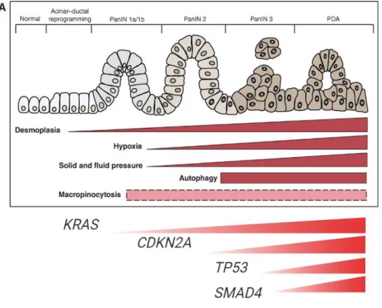
The molecular heterogeneity of pancreatic cancer has been studied in whole exome-sequencing studies. These studies identified four common alterations: the oncogenic KRAS mutations and mutations of the tumor suppressor genes CDKN2A, TP53 and SMAD4.
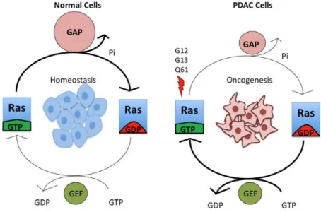
KRAS mutations are the first genetic alterations in the development of PDAC (figure 4). Activating
KRAS mutations are found in 92% of all PDAC cases34. This makes it the determining factor of PDAC progression. KRAS is a member of the RAS superfamily and encodes for a GTPase that is involved in regulating processes of the cell like proliferation, differentiation, survival and migration. When the gene is intact, the KRAS protein cycles between an GTP-bound active state and a GDP-bound inactive state. The conversion from the active to the inactive state is modulated by guanine nucleotide exchange factors (GEF) and GTPase-activating proteins. GEFs catalyze exchange of GDP for GTP and GTPase-activating proteins enhance the ability of KRAS to hydrolyze GTP. This process is showed in figure 3.
Figure 3: KRAS cycle. Zeitouni D. et al.
In a non-dividing cell KRAS is bound to GDP and thus is inactive. Growth factor stimulation causes the activation of the exchange of GDP for GTP making KRAS active. Mutations in the KRAS gene reduce the ability of KRAS to hydrolyze GTP making it constitutively active, hence driving tumor development. PanIN lesions are categorized into three groups of progression (I-III). PanIN lesions develop easily as an effect of KRAS mutations but these KRAS mutations are not sufficient to cause PDAC34.
Besides the KRAS oncogene, mutations in several tumor suppressor genes (TSGs) are required to develop PDAC34. TSGs play a role in the regulation of the cell by inhibiting proliferation. TSGs can induce apoptosis, cell cycle arrest and senescence. In PDAC, CDKN2A, TP53 and SMAD4 are key TSGs that are frequently mutated34. Interestingly, the mutations occur mostly in a specific order (see figure 4).
CDKN2A encodes for a cyclin-dependent kinase inhibitor that inhibits the entry to the S-phase of
the cell cycle. Most often, CDKN2A is the earliest TSG mutation that is seen in PDAC34. CDKN2A inactivation is often seen in slightly advanced PanIN stages prior to PDAC development. Loss of
CDKN2A occurs directly after KRAS activation to inhibit the cell from going into the senescence
state (see figure 4).
The second frequently mutated TSG is TP53. It encodes for the transcription factor p53. Loss of function (LOF) mutations are observed in a quarter of all tumors34. Point mutations undermine the ability of p53 to bind DNA, hence making it unable for p53 to activate factors to initiate apoptosis. Mutations in TP53 are commonly observed in advanced PanINs after the loss of CDKN2A (see figure 4) although not in every pancreatic cancer case a TP53 mutation is observed35. It is also thought that TP53 mutations play an important role in the metastasis of PDAC. The function of p53 as both tumor suppressor and enhancer of metastasis shows the complexity of the molecular pathogenesis of PDAC. Inactivation of other TSGs are also seen but in lower frequency.
The role of the transforming growth factor-β (TGF-β) pathway is crucial in the development of PDAC as it can both induce apoptosis in some cases and promote invasion and metastasis in
other cases34. SMAD4 is a transcriptional co-activator that regulates the anti-proliferative effect of TGF-β signaling. SMAD4 deletions accelerate tumor development. Mutations in SMAD4 are frequently found in advanced PanINs following CDKN2A mutations making SMAD4 the last step in tumor induction (see figure 4). Loss of SMAD4 is linked to a poor prognosis in comparison with wild type SMAD4 expression. Recent studies are conflicting in the role of the TGF-β pathway through saying that it does not play a role in metastasis34.
Figure 4: schematic representation of transformation from pancreatic cell to malignant cell. Rushika, M. et al.
2.2.4.2 Screening of PDAC
Population screenings are not recommended due to a low lifetime risk13. However, screening is recommended in people that match the definition of familial pancreatic cancer or patients of hereditary cancer syndromes. The low risk at developing pancreatitis together with the high risk of developing pancreatic cancer in a pancreatitis patient makes the pancreatitis population a good group for pancreatic cancer screening13. Intensive research including liquid biopsy is done to find potential biomarkers. Currently, serum cancer antigen 19-9 (CA19-9) is the only marker approved by the FDA in routine management of pancreatic cancer. However, this marker cannot be used as a screening biomarker due to the low positive predictive value. Patients with pancreatitis also have elevated levels of CA19-9. Its role is mainly to establish prognosis and to follow treatment response36. Treatment response translates in decrease of CA19-9 levels. Cell free DNA, tumor cells in circulation, volatile organic compounds in exhaled air and DNA mutations in pancreatic juice are all subjects of investigation but none of them identified a validated and specific biomarker13. This remains a major challenge.
2.2.5 Treatment
2.2.5.1 Surgery
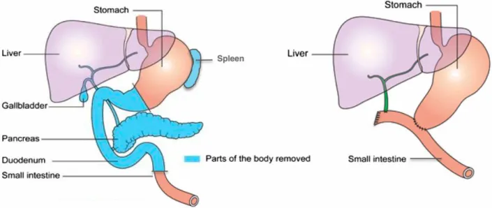
As stated before, surgical resection is the only potentially fully curative option. A resectable tumor is one that has no entanglement of the superior mesenteric artery, coeliac axis, portal vein or superior mesenteric vein. A tumor is borderline resectable based on the degree of entanglement of the above mentioned structures37. The surgical options are distal or total pancreatectomy or pancreatico-duodenectomy (also called Whipple’s surgery). The choice of the type of surgeries depends on the anatomical location of the tumor(s)13.
Total pancreatectomy or the removal of the pancreas is difficult due to the anatomy of the pancreas. The pancreas is in contact with almost all abdominal organs. In addition, the pancreas is densely connected to the duodenum and encloses the distal common bile duct. This makes the surgery extremely complicated38. The surgery is represented in figure 5. The 5-year survival rate of a total pancreatectomy in PDAC patients is 18,9% according to a study by Reddy et al.39
Figure 5: total pancreatectomy. Lund et al.
During a Whipple’s surgery, also called pancreatico-duodenectomy, the head of the pancreas, parts of the duodenum, the bile duct, the gallbladder and part of the stomach are removed. This requires outstanding surgical expertise. Figure 6 shows the modifications made during the surgery. The 5-year survival rate after a Whipple’s surgery is 15-20%40.
Figure 6: Whipple’s surgery41
Interests in minimally invasive techniques in all areas are rising because this is correlated to less pain, shorter hospitalization time, fewer blood loss, quicker recovery, less costs and better physical appearance (cosmesis)42. The first minimally invasive technique is the laparoscopic distal pancreatectomy43. Robotic techniques to improve Whipple’s surgery are investigated in several studies44. Results are a lower complication rate and less margin involvement. However, robotic surgery is very expensive and cost-effectiveness evaluations have yet to be made45.
2.2.5.1 Chemotherapy
Chemoradiotherapy is cancer therapy that uses anticancer-drugs (chemotherapeutic agents) or radiation to cure cancer patients. Chemotherapy can be administered in two settings: Adjuvant and neoadjuvant therapy. Adjuvant therapy involves surgical resection of the tumor, followed by chemotherapy administration. The advised chemotherapy option has shifted over the years. Chemoradiotherapy has long been used in locally advanced pancreatic cancer until the LAP07 study showed that there was no significant difference in overall survival with chemoradiotherapy compared to chemotherapy alone46. The CONKO-001 study47 proved that administration of gemcitabine after surgery significantly increased disease free survival. Later, the ESPAC-4 study showed that dual therapy of capecitabine and gemcitabine caused better overall survival compared to gemcitabine alone48. Recently, the PRODIGE-24 study showed that mFOLFIRINOX treatment resulted in an improved median disease free survival compared to gemcitabine. mFOLFIRINOX is a cocktail drug that consists of modified folinic acid, 5-fluorouracil, irinotecan and oxaliplatin. However, mFOLFIRINOX was also associated with more risk at complications49. Because of these findings, the standard treatment choice is based on the fitness of the patient after surgery. Patients in the University Hospital in Ghent that are very fit receive adjuvant mFOLFIRINOX and the less fit patients receive gemcitabine50.
In addition to adjuvant therapy, there is also neo-adjuvant therapy. In this kind of therapy chemotherapy is administered before surgery. Neo-adjuvant therapy attempts to eliminate micro-metastases in combination with shrinking of the primary tumor. Neo-adjuvant therapy is further
often used to treat borderline resectable tumors. The shrinking of the tumor and the elimination of micro-metastases lead to a decreased incidence of tumor recurrence13. In the University Hospital in Ghent patients with borderline resectable tumors receive FOLFIRINOX as neo-adjuvant therapy. There is an indication that gemcitabine plus nab-paclitaxel treatment has the same response as FOLFIRINOX but this therapy is not reimbursed in Belgium46.
Due to late diagnosis, many patients already have metastases upon diagnosis. Control of these metastases are mainly just symptom control, management of jaundice and palliative chemotherapy51. Administration of FOLFIRINOX was compared to gemcitabine in palliative patients in which FOLFIRINOX showed to induce longer overall survival but again FOLFIRINOX generates more adverse effects52. Palliative patients in Ghent University Hopsital receive either the gemcitabine plus nab-paclitaxel treatment or FOLFIRINOX as first line chemotherapy. The second line chemotherapy is based on the first line. If the patient received gemcitabine or gemcitabine plus nab-paclitaxel as first line therapy, the second line chemotherapy will either be nonaliposomal irinotecan53 or FOLFOX. If the patient received FOLFIRINOX as first line chemotherapy, the second line therapy will be gemcitabine.
In order to improve prognosis, more targeted therapy is under investigation. Approximately 10% of all PDAC patients have mutations in HR genes54. Those genes are involved in repair of DNA double strand breaks (DBS) through homologous recombination (HR). These patients may benefit from therapy with PARP inhibitors. Recently, the POLO trial showed that pancreatic cancer patients with BRCA1/2 germline mutations have longer progression-free survival if treated with PARPi55. The PARPi treatment is less toxic and induces less adverse effects than the classic chemotherapies discussed above.
2.2.5.3 PARP inhibitors
Double Strand Break repair
DNA damage is a common phenomenon in human cells. DNA double strand breaks (DSBs) are especially toxic for cells, and can lead to genomic instability and ultimately tumorigenesis56. There are two major pathways to repair those double strand breaks: non-homologous end joining (NHEJ) and homologous recombination (HR). NHEJ is the most frequent outcome, where the DNA ends are simply re-ligated. However, this is an error-prone process. In contrast, HR uses the undamaged sister chromatid to repair the break57. This generates an error-free repair, but is only available in the late S-/G2- phase of the cell cycle.
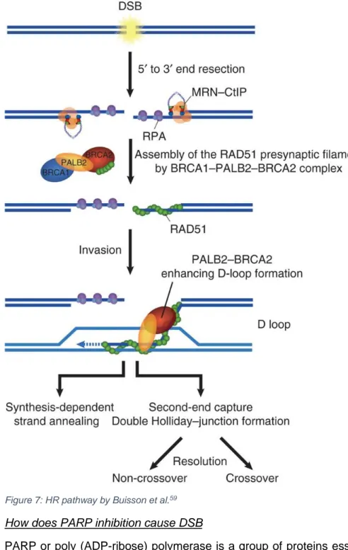
Homologous recombination works as follows (figure 7). Upon a DSB, the MRE11-RAD50-NBS1 complex is recruited and bound to the break and activates the ATM kinase which initiates the DNA repair response of the cell58. CtIP-mediated nuclease activity leads to the formation of single stranded DNA (ssDNA). This ssDNA can be coated with DNA replication protein A (RPA) which activates ATR and facilitates the HR response. RAD51 is then localized to the ssDNA by the BRCA1-PALP2-BRCA2 complex59, replaces the RPA complex and searches for the homologous sequence on the sister chromatid.
Figure 7: HR pathway by Buisson et al.59
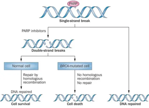
How does PARP inhibition cause DSB
PARP or poly (ADP-ribose) polymerase is a group of proteins essential in DNA damage repair. PARP1 is of importance in the repair of single strand breaks (SSB) through base excision repair (BER). PARP inhibitors (PARPi) will inhibit the function of PARP by trapping and/or hinder the catalytic functionality. The unrepaired SSB causes replication fork stalling which induces a DSB. Normally, a DSB has two DNA ends but when a DSB is formed through replication fork stalling, there is only one DNA end. This is called single end DNA (seDNA). Normally, this break is repaired through the HR pathway but when the HR pathways is impaired this break is repaired by NHEJ which is error-prone and causes cell death60. This prevents the single strand break from being repaired correctly.
Synthetic lethality and PARPi
Synthetic lethality is the phenomenon where a deficiency in two genes leads to cell death, but deficiency in only one of those genes does not61. This approach is interesting in cancers with patients carrying germline mutations in DDR genes. As tumors deriving from these patients are often resulting from a second hit mutation, therapies that utilize the synthetic lethality approach could selectively target the tumor, while leaving normal tissue unharmed.
For PARPi, this is the case in tumors with a BRCA1/2 mutation causing disfunction of the HR pathway. PARPi treatment causes cell death in BRCA1/2 mutated cells due to excessive DNA damage. This phenomenon is showed schematically in figure 8. Cells with one or two functional
BRCA1/2 allele(s) will survive62, however, when both alleles are mutated, the cell goes into apoptosis due to excessive DNA damage.
Figure 8: synthetic lethality caused by PARPi by A. Sonnenblick. et al.62
Different PARPi
Up to date there are five clinically available PARPi, being olaparib, niraparib, rucaparib, veliparib and talazoparib. PARPi work by trapping PARP onto damaged DNA and inhibiting the catalytic cavity of PARP. Some PARPi are more potent to trap and others are more potent to inhibit.
Olaparib is the best known PARPi and is reimbursed for the treatment of recurrent ovarian cancer. Olaparib is developed to inhibit the NAD+ activity in the catalytic cavity of PARP1 and PARP263,64. It is clinically proven that olaparib also benefits patients with metastatic breast or prostate cancer with germline BRCA mutations. A clinical trial of phase III was conducted in metastatic pancreatic cancer patients with germline mutations in BRCA1 or BRCA255. The overall survival of the patients
receiving olaparib is longer than those in the placebo group, nonetheless, more adverse effects were observed in the olaparib group.
Niraparib is a known maintenance treatment in recurrent ovarian cancer. Niraparib is targeted against PARP1 and PARP2. It inhibits the enzymatic activity which causes to form more PARP-DNA complexes inducing more PARP-DNA damage leading to cell death and apoptosis65. Clinical approval was granted after the phase III NOVA trial which proved that it was stated that the niraparib group had longer progression free-survival65. However these effects were regardless of the presence of BRCA mutations. Up to date, niraparib is not used in the clinic to treat pancreatic cancer. There are three clinical trials currently recruiting pancreatic patients in the United States that will test niraparib in pancreatic cancer patients as maintenance treatment (NCT03553004, NCT03601923, NCT03404960).
Rucaparib inhibits PARP-1, 2 and 3 by trapping these PARPs64. Rucaparib is approved as a treatment for ovarian cancer with BRCA-mutations and as maintenance therapy for recurrent ovarian cancer66. The phase III ARIEL3 trial showed longer progression-free-survival in the rucaparib group compared to the placebo group. Currently, there are no phase III trials ongoing for maintenance rucaparib treatment in pancreatic cancer patients.
Veliparib inhibits the catalytic activity of PARP64. The trapping efficacy of veliparib is much lower in comparison with olaparib, rucaparib and niraparib67. Veliparib was shown to have a moderate effect as anticancer therapy in prostate cancer. It is clinically used in combination treatment68. Currently there are no phase III trials ongoing of veliparib treatment in ovarian cancer patients but phase II trials are promising67.
The fifth clinically available PARPi is talazoparib. Talazoparib is thought to be the most potent PARP trapper up to date67. The efficacy of talazoparib in ovarian cancer is still under investigation. Talazoparib has a larger structure and is more rigid than the other PARPi. The rigidity is thought to be the cause of the higher potency67. This PARPi also showed better progression free survival in comparison with a standard single agent therapy like capecitabine, eribulin, gemcitabine, or vinorelbine69.
Adverse effect observed in PARPi treatment
Despite the less severe toxic effects of the PARPi in comparison with the classic chemotherapy, there are still adverse effects observed. The most common adverse events are nausea, anemia, thrombocytopenia, hypertension, sepsis and fatigue64,65,67. The anemia and thrombocytopenia are caused by inhibition of the HR pathway in rapidly divining cells that result in more mutations and more apoptosis of these cells. Because of these adverse effects, there is a constant search for new PARPi with fewer adverse effects.
Developing novel PARPi
New PARPi are being developed in the search of better therapy with less adverse effects. PARPi development is mainly performed using several in vitro assays70. Only in late stages of development, in vivo testing of PARPi is done with mouse xenografts. However these mouse xenografts are expensive and are highly time consuming. Thus, failure at this stage of development would be very unfortunate. Therefore, there is a high interest in developing alternatives of in vivo models that are less expensive, take less time and can be used earlier in development of a PARPi.
Zebrafish
Zebrafish or Danio rerio is a tropical fish that is member of the teleostei infraclass and is estimated to be arisen 340 million years ago from its common ancestor71. This ancestor underwent an additional genome duplication in comparison with other vertebrates causing zebrafish to have 4 copies of each chromosome. These duplications are called ohnologues71. Zebrafish have their origin in Southeast Asia. An adult fish is about two and a half centimeters to four centimeters long. Zebrafish models are often used in genetic studies, but also in toxicological studies, environmental health studies, metabolic diseases and so on. Zebrafish are widespread used for studying the function of vertebrate genes71. The Welcome Trust Sanger Institute initiated the Zebrafish genome-sequencing project in 2001. This led to insights in orthologues between the human and zebrafish genome. 71,4% of all human genes have at least one orthologue in zebrafish71. Reciprocally, 69% of all zebrafish genes have an orthologue in the human genome. 47% of these human orthologues have a one-to-one relationship with the zebrafish orthologue.
Advantages of zebrafish are high fecundity, rapid development, feasible manipulation due to ex
utero development, small size and easy manipulation. Zebrafish are aquatic models that can easily
take up small molecules if added to the medium72 making compound administration effortless. Zebrafish as a solution to early in vivo PARPi testing
A zebrafish model could be an answer to the problems of the PARPi development. Zebrafish are much cheaper than mice. It is relatively easy to manipulate the zebrafish genome by for example making an overexpression or a gene knockdown. It is already known that several repair pathways are conserved between zebrafish and human73. The HR pathway is one of them. There are several techniques available to study gene pathways in zebrafish74.
Assays to study HR and cell death in zebrafish
Visualization of the HR pathway can be done as shown by Vierstraete et al.74 Rad51 binds to Brca2 when the HR pathway is activated as discussed above. Rad51 can be visualized with immunohistochemical staining74. In this assay two antibodies are used to visualize the Rad51 foci. The primary antibody binds Rad51 and the second antibody is fluorescently labeled and binds the primary antibody making the Rad51 foci visible. This assay is visualized in figure 9.
The acridine orange (AO) staining is a technique that can be used to identify cells in apoptosis. Acridine orange is a permeable nucleic acid dye that is closed off in different compartments like lysosomes. These compartments cannot enter the nucleus of the cell. However, when a cell is going into apoptosis, the pH in the cell changes and the acridine orange is released from the lysosomes in the cytoplasm. Once in the cytoplasm the acridine orange can localize to the nucleus and intercalate with partially uncoiled DNA. This process makes the acridine orange highly fluorescent which can be visualized and quantified easily75.
2.2.6 Using zebrafish to model pancreatic Xenografts
Due to the low survival rates and especially the short time of survival after diagnosis, the prognosis of PDAC is very poor. Nowadays, the choice of treatment is mainly based on the condition of the patient. In order to improve the prognosis, more personal therapy approaches are needed. Up to date it is unpredictable to assess which patient will respond to which treatment. With PARPi being more and more interesting, it would be favorable to predict the response to a PARPi and other chemotherapy.
2.2.6.1 Patient derived xenografts
An option for predicting the response to the therapy of a PDAC patient is by transplanting a piece of the primary tumor of the patient into an immune compromised mouse76. This technique is called a patient derived xenograft (PDX). PDXs are used for drug development but also for predicting the outcome of a therapy for a patient.
However, this approach has multiple downsides. For starters, the time upon engraftment in a mouse can take up to several months, this is time the patient does not have. Additionally, the immune system of the mice would reject the human tumor tissue, therefore an immunocompromised strain is required for mice xenografts increasing the costs of the study76. Because of these increased costs, fewer therapies can be tested.
2.2.6.2 Zebrafish xenografts
A solution to these problems can be found in using zebrafish xenografts (zPDX). Firstly, xenografts engraft much faster in zebrafish in comparison with the time of engraftment needed in mice (days instead of months)77. Secondly, less tumor material is needed for engraftment, which makes that, with the same amount of material harvested from a patient, more zebrafish can be engrafted causing the ability to implement more biological replicates favoring the statistics78. Only about 50 to 100 cells per larvae need to be injected for successful engraftment. It has already been proven that human cells can be engrafted in zebrafish embryos78. Finally, zebrafish have no adaptive immune system in the embryonic stage making the use of an immunocompromised strain unnecessary which reduces the cost. The reduced costs enable to inject more embryos and thus gaining more xenografts to test multiple therapies. Other advantages are stated above (high fecundity, rapid development, feasible manipulation due to ex utero development, small size and easy manipulation and effortless compound administration). The Casper strain is a strain of zebrafish that lacks pigment and thus is transparent throughout their whole life79. This makes visualization of the xenotransplanted tissue easier.
Fior et al. proved that diverse behaviors of cancer cells could be measured in vivo only 4 days after developing a PDX model of colorectal cancer cells in zebrafish larvae80. When they compared
zebrafish xenografts to mouse xenografts, they observed similar sensitivities to chemotherapy. They also used the zebrafish xenografts to test the response to treatment in patients and found correlations between patients relapse time and zebrafish xenograft response to treatment. Because of the promising results of this research group, we want to test if this model can also be used for predicting treatment outcome in PDAC patients.
3. Objectives
The goal of this master thesis is twofold. The first goal is to establish and optimize a zebrafish xenograft protocol to engraft pancreatic cancer cells and assess their sensitivity to different compounds. For this optimization work, we will attempt to engraft two pancreatic cancer cell lines, PANC-1 and Mia-Paca-2. Upon establishing proper engraftment, we will treat these xenografts with the chemotherapeutic compound gemcitabine, to which Mia-Paca-2 is exceptionally sensitive. Our aim is to use our functional-readouts to observe an increased tumor response of the Mia-Paca-2 cell line to the treatment, compared to less sensitive PANC-1 cells. These optimizations will eventually lead to a zebrafish platform where we aim to predict the response to treatment in pancreatic cancer patients. In the long run, we want to engraft primary tumor material into zebrafish larvae. Subsequent to successful engraftment, compounds (gemcitabine and FOLFIRINOX) will be added to the medium of the larvae. The response to therapy in zebrafish should be similar to the response to treatment in the patient. This will ultimately lead to a platform in which a prediction can be made about which chemotherapy the patient will respond to.
The second aim is development of a zebrafish model for in vivo establishment of new PARPi. Mutations in the HR pathway confer an increased risk of breast, ovarian, prostate and pancreatic cancer. PARPi target tumor cells with deficiencies in the HR pathway. It is well-known that ~10% of all pancreatic cancer patients have mutations in HR genes and thus could benefit from a PARPi treatment. There is a constant search of novel PARPi with higher potency and less adverse effects. Establishment of those PARPi nowadays is mainly done in vitro followed by mouse xenografts late in development. To improve and fasten development of new compounds there is need for an assay that foresees in vivo data early in development process, provides fast results and all of that at an affordable cost. A zebrafish could accomplish the needs. Using several techniques, we will check the efficacy of all five clinically available PARPi.
4. Materials and methods
4.1 Zebrafish embryonic xenografts
4.1.1 Optimization on cell lines
4.1.1.1 Maintenance of cell lines
We aimed to inject two cell lines to engraft in zebrafish larvae. The PANC-1 cell line and the Mia-Paca-2 cell line. All cells were cultured in a humidified incubator at 37°C and 5% CO2. The PANC-1 cell line is a human pancreatic cancer cell line. The medium used is Dulbecco’s Modified Eagle Medium (DMEM) enriched with 10% fetal bovine serum (FBS) and antibiotics being 1% penicillin and streptomycin (penstrep). The Mia-Paca-2 cell line is another human pancreatic cancer cell line that should be more sensitive to gemcitabine. These cells were maintained in DMEM enriched with 10% FBS and 2,5% horse serum and 1% penstrep. The use of these cell lines was approved by the local Ethical Committee (code EC 107-2018/mf).
4.1.1.2 Labeling cells
The human pancreatic cancer cells need to be labeled with a fluorescent dye (DiI) for later visualization. DiI is a fluorescent lipophilic cationic dye from Thermo Fisher Scientific (catalogue number: V22888). Upon labeling, cells need to be dissociated in order to collect them. In this thesis, three approaches were conducted in order to find the most optimal dissociation method for pancreatic cancer cells.
EDTA
The protocol that was followed is manufactured by Fior et al.80 Cells should be at 70% confluence for the right balance between optimal cell density and dividing cells. Medium was removed and cells were washed with 3 mL PBS 1x. After that PBS 1x was removed. DiI was diluted 1:1000 in PBS and 2mL was added to the cells. Cells were incubated for 10 minutes at 37°C followed by an incubation on ice for 15 minutes. During this procedure, cells are covered with aluminum foil to avoid fading of fluorescent signal. Afterwards, DiI is removed and cells were washed again with 3 mL PBS 1x. Subsequently, cells were harvested using EDTA. EDTA is a Ca2+ chelator that will remove the Ca2+ ions. Integrins needs these Ca2+ ions to maintain cell adhesion81. 2mL of EDTA was added to the cells and this was incubated for 3 minutes at 37°C. A cell scraper was used to detach the cells from the surface of the flask. The suspension containing the cells was transferred into 1,5mL Eppendorfs. In order to remove dead cells and debris, the Eppendorfs were centrifuged for 4 minutes at 300rcf at 4°C. Two pellets were formed during the centrifugation of the Eppendorfs. The pellet on the side of the Eppendorf contained dead cells and debris and needed to be removed. The lower pellet contained the vital cells. The excess fluid together with the upper pellet was removed and the lower pellet was resuspended in 60µL medium. The cells were counted and the viability is measured.
In order to quantify cell density and calculate viability, samples of cell suspension were diluted 1:10 in trypan blue. Viable cells have intact cell membrane and do not stain with trypan blue. Dead cells however have permeable membranes which allows the dye to enter and stain the cells. The cells were counted using a Burker count chamber. This chamber consist of 9 large squares (1mm2). Each square is subdivided into 16 smaller squares with double lines (0,05 mm apart). These double lines form 0,0025mm2 squares. The depth of the square is 0,1mm2 making the volume in the chamber 1µL. The larger square have triple lines. When counting cells, only cells
inside the 16 squares and those that touch the upper or left triple lines were counted as showed in Figure 10. Cells outside of the larger square or those touching the bottom or right triple lines were not counted. Using this dilution, every counted cell resembles 1 million cells per mL. Three of those large squares are counted and average of those three results will be taken as end result.
Figure 10: Method of counting cells in Burker count chamber
Trypsin
The previous protocol was adjusted to obtain higher viability of the cells. Washing and incubating with DiI was done as described above. Subsequently, cells were incubated for 3-7 minutes with 2mL trypsin (Life Technologies Europe, Thermo Fisher Scientific catalogue number: 25300-054). Trypsin is an enzyme which degrades proteins by cutting peptide bindings between lysine and arginine. This dissociates the cells from each other and from the surface so scraping of the cells was unnecessary. The cells are harvested and centrifuged as described above. An extra step was added to inhibit the trypsin. After removing the excess fluid cells were resuspended in DMEM to inhibit trypsin and prevent it from being toxic to the cells because incubation of the cells with trypsin for too long will damage the membrane and kill the cells. After a second centrifugation the cells were counted as described above.
TrypLE
In order to obtain even higher viability and retain more cell surface proteins, a third condition was investigated to dissociate cells. Again, the cells are washed and stained as described above. For harvesting the cells, TrypLE (Thermo Fisher Scientific, catalogue number 12605010) was used. This is a mixture of three enzymes and is thought to be less aggressive then trypsin. Washing the cells was done with PBS 1x as described above. 2mL of TrypLE was added to the cells and incubated for 3-7 minutes at room temperature. Without scraping, the cells are transferred to 1,5mL Eppendorfs and centrifuged as described above. After removal of the remnant, 60µL of cold PBS 1x was added. The cells were counted and the viability was measured as described above.
4.1.2 Injecting larvae
4.1.2.1 Maintenance of zebrafish strain
All zebrafish lines were housed in a Zebtec semi-closed recirculation housing system at a constant temperature (27°C-28°C), pH (~7,5), conductivity (~550µS) and light/day cycle (14/10). Fish were fed twice a day with dry food (Gemma Micro, Skretting) and once with artemia (Ocean Nutrition). The Casper strain is used for engrafting the cells. This strain has mutations in two genes (rov and
engraftment easier82. All experiments were approved by the local animal ethics committee, application number ECD 18/75.
4.1.2.2 Injection
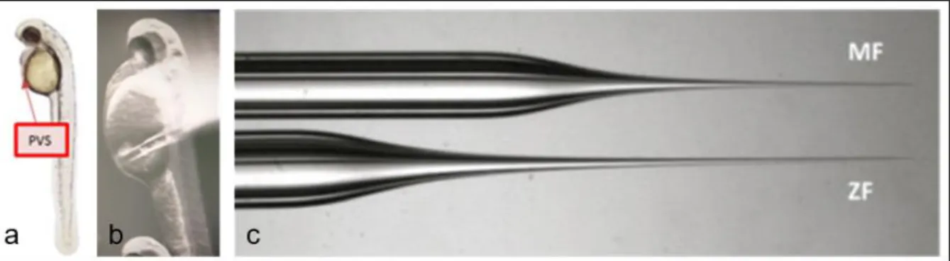
48 hours post fertilization (hpf) larvae were injected with labeled human pancreatic cells. In this stage, most of the larvae were still in their chorion. The chorion was removed using the pronase, a mixture of enzymes that digest proteins to single amino acids83. The larvae were sedated with tricaine after which they could be positioned on an agar plate with lanes. The head was positioned upwards and the back of the larvae was positioned against an agar lane. An example of the positioning is showed in figure 11.
After positioning, the larvae could be injected with the labeled cells. For injection, needles were made by pulling a borosilicate into two pieces with a TW100-4 World precision instruments with heat at 400-600, fil: 4, Vel: 46-60, Del: 200 and pull: 100. The remaining halves have a tip of approximately 1cm. This allows for shortening of the needle if obstructions occur. The optimal location for injection was in the perivitelline space (PVS) indicated in figure 12. The ideal amount of injection is described as approximately the size of an eye.
Figure 11: positioning larvae on agar plates. The head
is positioned upwards and the back is positioned against the agar lane. Black lines are indicate lanes.
Figure 12: injection side and needle; a shows the PVS schematically in a zebrafish larvae; b shows an injection into
After injection, the larvae were kept sedated another ten minutes to allow them to recover and prevent them from twitching and pushing out freshly injected cells. Then, larvae were transferred back into E3 medium and held in an incubator at 34°C. This temperature is a compromise between the optimal temperature for a zebrafish and the optimal temperature for human cells being 28°C and 37°C respectively84. Screening was done 24 hours post injection (hpi). In this screening, the viability of the zebrafish was checked together with the viability of the injected tumor cells. Larvae that were wrongly injected, had too few injected cells or appeared unacceptable were discarded. 4.1.2.3 Hematoxylin and eosin staining
After successful injection, the zebrafish could be stained with a hematoxylin and eosin (H&E) staining. This is a standard technique that is often used by pathologists to screen for cancer cells in a biopsy. Hematoxylin stains the basophilic structures purple, such as DNA and RNA. Eosin, on the other hand, stains the acidophilic structures in several shades of red. This will mostly be the cytoplasm and its structures. To do a H&E staining, the fish are euthanized with tricaine and fixed overnight in paraformaldehyde (PFA) 4%.
The next day, the fish can be dehydrated by incubating in a series of alcohol solution going from 30% ethanol, to 50%, 70%, 96% and iso-propanol (considered as 100% ethanol). The next incubation is in an iso-propanol/toluene mixture before incubation in toluene. The fish are then dehydrated and can be embedded in paraffin. After incubation at 55°C in paraffin overnight, the fish can be positioned and cut into 5µm sections. The H&E staining itself is performed in the Microm HMS 740 (protocol in addendum 1).
4.2 PARPi
4.2.1 Maintenance of the zebrafish strains
For the experiments with the PARPi the brca2cmg35 zebrafish strain and the Tg(EF1a:
mCherry-zGem)oki011 zebrafish line were used. The housing of the strains is outlined above.
4.2.2 Treatment of PARPi
The five PARPi that were investigated were olaparib (Selleckhem: S1060), niraparib (Absource Diagnostics: S2741), rucaparib (Selleckhem: S1098), veliparib (Enzo Life Sciences: ALX-270-444-M005) and talazoparib (Bioconnect: S7048).
4.2.2.1 DMSO toxicity
As PARPi are often dissolved in DMSO, DMSO was used as negative control in the Rad51 assay, acridine orange assay and in the eye size test. However DMSO or dimethylsulfoxide itself has a certain toxicity. To check the toxicity of DMSO, an experiment was set up in which wild type larvae at 72hpf were exposed for seven hours to a concentration of DMSO ranging from 1% to 5%. For each concentration six larvae were incubated. After incubation, larvae were checked for possible malformations and viability.
4.2.2.2 Rad51 foci
Breeding
To measure the amount of Rad51 foci (marker for HR), an immune fluorescent staining was performed. For the Rad51 foci experiment, the Tg(EF1a: mCherry-zGem)oki011 strain was used.
This is a transgenic line that expresses geminin with a fluorescent tag at the amino-terminal region to visualize the cell cycle. Geminin is a DNA replication inhibitor that is present in high levels in S- and G2- phase cells. At 24hpf, larvae were screened for the presence of a geminin signal. All larvae without the fluorescent tag were discarded.
Incubation
At 3 days post fertilization (dpf), the larvae were incubated with PARPi. Fish were kept in 2mL E3 medium enriched with the desired amount of PARPi for 7 hours. For all five PARPi we incubated fish with a concentration range of PARPi between 5µM and 800µM.
Embedding
After incubation, the zebrafish were euthanized using tricaine followed by fixation for 1 hour using PFA 4%. The next day, fish were dehydrated and embedded in paraffin as described above. Sections of 5µm of the region that encloses the intestinal tract were made. In this region many dividing cells are present thus many cells in the S-/G2- phase of the cell cycle74.
Immunofluorescent staining
To stain the larvae, mCherry (visualizing Geminin; 1:1000; Abcam; ab125096) and anti-Rad51 (1:2000; Santa Cruz; H-92) were used. The secondary antibodies involved Goat-anti-rabbit Dylight 488 antibody (1:1000 Sigma-Aldrich) and Goat-anti-mouse Dylight 594 antibody (1:1000 Sigma-Aldrich). The slides were counterstained with DAPI+fluoromount (Sigma-Aldrich).
Counting foci
After staining, the slides were scanned on a Zeiss Axio Observer.Z1 inverted microscope using the Zen pro 2012 software. A 100x magnification was used and Z-stacks (0.22 µm thickness) were made for each section. The number of Rad51 foci was measured in geminin positive cells only, as only these cells are capable of performing HR.
4.2.2.3 Acridine orange assay
The larvae used for the acridine orange assay were offspring of brca2cmg35/+ heterozygotes. Larvae 6hpf were assembled and treated with olaparib for 24 hours at a concentration of 5µM. At 32hpf, larvae were dechorionated and incubated for 15 minutes in acridine orange (AO) solution. Afterwards, the embryos were washed three times with E3 medium and sedated with tricaine. For imaging, the embryos were positioned laterally on methylcellulose droplets. Images were taken with a Nikon SMZ18 stereoscope (FGP; 3.2x magnification, 800ms integration). ImageJ was used to select areas of the body and by using “process – find maximum” the number of apoptotic cells could be quantified (noise tolerance being 5). A normalization was preformed to correct for the variations in the selected body size using the Region of interest manager tool. The average of each group was taken and the number of apoptotic cells for each embryo was normalized over the average area of the group.
DNA of each embryo was collected to perform genotyping. Embryos were first euthanized with tricaine after which DNA extraction was performed. Next, PCR was performed on this DNA using the ford protocol (addendum 2), Cmg35_F and Cmg35_R primers and the KAPA2G Robust HotStart ReadyMix Ford master mix (see addendum 3). PCR products could be placed on a
capillary gel electrophoresis that can detect the 13 base pair deletion in case of mutated brca2 (see addendum 4).
4.2.2.4 Eye size test
For the eye size test the same strain was used as the AO assay (offspring of brca2cmg35/+ heterozygotes). At 6hpf, embryos were collected and incubated with 5µM olaparib. The solution with olaparib was renewed at 52hpf. At 72hpf the larvae were sedated using tricaine and placed laterally on methylcellulose droplets. Visualization of the larvae was done with a Leica M165FC stereomicroscope at a 4x magnification. Again, ImageJ was used, this time to measure the area of the eye as a quantitative marker for growth malformations. Genotyping of the brca2 gene was done as described above.
5. Results
5.1 Optimization xenograft procedure
5.1.1 Cell dissociation
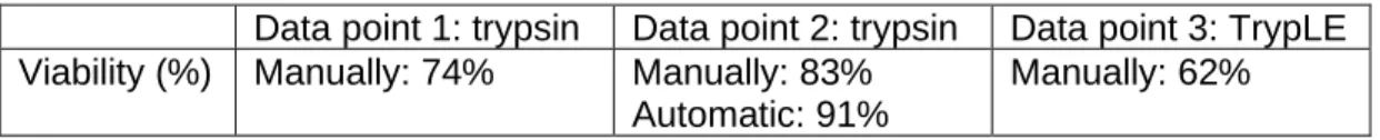
An essential step in the xenograft procedure involves cell dissociation upon labeling. Initially, the protocol as described by Fior et al. using EDTA mediated dissociation of cells was conducted. However, we experienced low viability after dissociating cells (figure 13). In addition, injection of these cells proved troublesome, as cells clotted causing clogging up of the needle to be a continuous issue. After adding EDTA, cells were scraped from the surface and collected. Toluidine blue staining showed that scraping these cells was quite harmful, as many cells were dead after collection. During cell death, DNA is set free from cells and DNA is a highly viscous substance. Thus, low viability leads to a more viscous cell substance which causes increased clogging up of the injection needle. Furthermore, the relatively large size of the PANC-1 cells might further accelerate clogging up of the injection needle in these conditions. Lastly, we noticed that EDTA dissociated cells were still clustered in relatively large groups, which can also cause needle clogging. In order to improve cell viability, we used two additional methods to dissociate the cells with as goal optimization of the protocol in order to obtain both more single cells and a higher viability. This would ultimately lead to a decreased clogging up of the needle. The viability obtained through the different methods are displayed in figure 13.
Figure 13: Viability of cells upon different dissociation methods displayed in percentages. For the EDTA and TrypLE
method, one data point was included. For the trypsin method two data points were included. The error bar indicates 95%CI.
As mechanical dissociation proved too aggressive and ineffective, we next tried enzymatic dissociation protocols. In the second method we used trypsin. Trypsin is an enzyme that cuts peptides between lysine and arginine. Trypsin dissociation is a known technique to split and harvest cells. It was no longer necessary to scrape the cells, causing less debris and dead cells. A downside of using trypsin for too long is that it can become toxic for the cells as it degrades the membranes of these cells. To avoid this toxicity, we added DMEM after dissociation in order to
0 20 40 60 80 100 120 p erce n ta ge v iab le ce lls (10 0%) dissociation compound
Viability of cells upon different dissociation methods
EDTA trypsin TrypLE
inhibit the trypsin. Because trypsin impairs the membrane we investigated a third substance to harvest cells. However, the goal was to create a tumor in the zebrafish larvae so once the cells are injected they have to be able to recognize other human cells to form a tumor.
The last method also involves enzymatic digestion. Here, trypsin was replaced with TrypLE. This is another enzyme that is known to be more gentle to cells, is easy to use and room-temperature stable. Like trypsin, the cells did not need to be scraped and the additional step for inhibiting the enzyme function was not necessary.
We wanted to compare these three methods in order to find the most suitable protocol. Unfortunately, the Corona crisis did not allow further experiments leaving us with only three data points for the PANC-1 cell line. Table 1 displays the viability obtained in the three datapoints. The first attempt of conducting the trypsin method the cells were counted manually and some mistakes were made resulting in a low viability (74%) displayed in table 1. The second data point was obtained when conducting the trypsin method a second time. Cells were counted both manually and automatically and results are displayed in table 1. When counting manually, a lower viability was seen (83%) compared to the automatic count (91%). The last data point was obtained through performing the TrypLE method. Cells were only counted manually and viability was very low (62%).
Table 1: viabilities obtained after dissociation using the trypsin and TrypLE method
Data point 1: trypsin Data point 2: trypsin Data point 3: TrypLE Viability (%) Manually: 74% Manually: 83%
Automatic: 91%
Manually: 62%
After dissociation and collection, cells were quantified. To count the cells, two methods were compared. The first one was counting with a Burker count chamber (manually) and the second one is an automatic count with Luna-II. Using the automatic cell counter would be advantageous as this is much faster than manual counting. After multiple attempts and comparisons between the manual and automatic method, we concluded that the latter was too inaccurate. The Luna-II typically underestimated the density of the cells by a factor of 10. However, in this device you have the ability to change parameters such as cell size and cluster analysis. We therefore planned to perform several experiments with the PANC-1 and Mia-Paca-2 cell line in order to get the automatic counting method more reliable. The experiments were unfortunately not completed due to the Corona crisis. In general, there was little variation in the cell density between the different dissociation methods.
5.1.2 Engraftment
We performed some injections of PANC-1 cells in zebrafish larvae that were harvested using the EDTA and trypsin method. We aimed to inject labeled tumor cells in the PVS of the larvae. This required a lot of skills, and therefore I had to perform multiple injections before being proficient enough to inject cells with high accuracy. Figure 14a displays a H&E staining of a fish with tumor cells injected into the yolk sac. Cells are concentrated in the tumour but do not develop a compact structure as expected in a real tumour.For this reason, injection in the PVS was desired like shown in figure 14b.