Detection of infectious Cryptosporidium oocysts by cell culture: applicability to environmental samples
F. M. Schets, G.B. Engels, M. During, A.M. de Roda Husman
This investigation has been performed by order and for the account of General Directorate for Environmental Protection, directorate of Drinking Water, Water and Agriculture, within the framework of project M/289202/IC, Infectivity of Cryptosporidium and Giardia in recreation water.
Abstract
Cryptosporidium is one of the important causative agents of gastrointestinal illness in
humans. Cryptosporidium infections are often waterborne and can be transmitted through drinking water or recreational water. Estimation of the risk of infection with Cryptosporidium after exposure to drinking water requires information on oocyst infectivity. According to the Dutch drinking water regulations, the annual risk of infection should not exceed one infection per 10,000 persons. We evaluated the applicability of in vitro cell culture assays on HCT-8 and Caco-2 cells for determination of oocyst infectivity in naturally contaminated water samples. Experiments with Cryptosporidium oocysts showed considerable variability in infectivity. Dilution and survival experiments indicated that only relatively large numbers of fresh or aged oocysts produced infection in cell cultures. Naturally contaminated Dutch surface waters usually contain low numbers of Cryptosporidium oocysts. The cell culture assays are not sensitive enough to detect infectivity of such low numbers of oocysts. The assays can however successfully be used to study the effect of disinfection processes on oocyst infectivity. Surrogate methods, such as vital dye exclusion assays, used to determine oocyst viability as opposed to infectivity were found to overestimate the number of infectious oocysts. Using oocyst viability in risk assessment will therefore overestimate the risk of infection with Cryptosporidium. For accurate risk assessment, further improvement of the detection method for Cryptosporidium in water is required. PCR detection of infection in cell lines may improve sensitivity whereas enhanced recovery of the detection method may result in the detection of higher numbers of oocysts.
Contents
Samenvatting 5
Summary 7
1. Introduction 9
2. Materials and methods 13
2.1 Cryptosporidium oocysts 13
2.2 Routine cell culture 13
2.3 Cell culture for infectivity studies 13
2.4 Induction of excystation 14
2.5 Infection of cell monolayers 14
2.6 Detection of infection 14
2.7 Comparison of different Cryptosporidium oocyst lots 15 2.8 Comparison of infections in cell culture and mice 16
2.9 Environmental samples 17
2.10 Ageing of Cryptosporidium oocysts in surface water 18
3. Results 19
3.1 Optimisation of the infection procedure 19 3.2 Comparison of Cryptosporidium oocyst lots 20 3.3 Comparison of cell culture and animal infectivity 25
3.4 Environmental samples 26
3.5 Ageing: comparison of viability methods 27
4. Discussion 31
4.1 Comparison of Cryptosporidium oocyst lots 31 4.2 Infectivity in cell culture vs. infectivity in mice 32 4.3 Infectivity in cell culture vs. surrogate viability methods 32 4.4 Cell culture infectivity assays in disinfection studies 33 4.5 Cell culture infectivity assay for environmental samples 33
5. Conclusions 35
Acknowledgements 36
References 37
Appendix 1 Mailing list 43
Appendix 2 Cell culture protocol (in Dutch) 44
Samenvatting
Cryptosporidium is één van de belangrijke veroorzakers van gastro-enteritis bij de mens. Cryptosporidium infecties worden vaak via water overgedragen, dit kan zowel via drinkwater
als recreatiewater zijn. Mensen met een verstoord immuunsysteem kunnen bij infectie met
Cryptosporidium levensbedreigend ziek worden. Mede hierdoor is Cryptosporidium een
belangrijk probleem geworden voor gezondheidsdiensten en waterleidingbedrijven.
Cryptosporidium oöcysten komen voor in het Nederlandse oppervlaktewater wat gebruikt
wordt voor drinkwaterproductie. Ze zijn bovendien buitengewoon resistent tegen chloor in concentraties die gebruikt worden bij desinfectie van drinkwater. Het aantal oöcysten in het leidingwater is meestal laag en ligt onder de detectielimiet van de methode. Dit betekent echter niet dat er geen oöcysten in het leidingwater kunnen voorkomen. Daarom moeten Nederlandse waterleidingbedrijven volgens het Waterleidingbesluit een risicoanalyse voor
Cryptosporidium uitvoeren. De risicoschatting wordt gebaseerd op de concentratie Cryptosporidium in het ruwe water en de verwijderingcapaciteit van de
drinkwaterzuiveringsprocessen. Het jaarlijkse infectierisico bij consumptie van drinkwater moet lager zijn dan één infectie per 10.000 personen. Alleen infectieuze Cryptosporidium oöcysten vormen een mogelijk risico voor de volksgezondheid. Daarom is informatie over de infectieusiteit van oöcysten nodig om, bij blootstelling aan drinkwater, betrouwbaar de kans op infectie met Cryptosporidium te kunnen berekenen. Wij evalueerden de toepasbaarheid van in vitro celkweekmethoden op de humane intestinale cellijnen HCT-8 en Caco-2 om de infectieusiteit van Cryptosporidium oöcysten afkomstig uit natuurlijk besmet water te bepalen. In deze monsters water zitten vaak weinig oöcysten die bovendien aan ongunstige milieufactoren zijn blootgesteld. Detectiemethoden dienen dan ook een hoge mate van gevoeligheid te bezitten. Uit onze experimenten is gebleken dat de huidige
celkweek-methoden niet gevoelig genoeg zijn om infectieuze Cryptosporidium oöcysten in Nederlandse oppervlaktewateren te detecteren. De concentratie oöcysten in deze monsters ligt meestal onder de concentratie die gebleken is nodig te zijn om in vitro infectie van cellijnen te verkrijgen. Daarom is het op dit moment niet mogelijk om bij risicoschattingen gebruik te maken van gegevens over de infectieusiteit van Cryptosporidium oöcysten. Het is
desalniettemin mogelijk om de levensvatbaarheid van Cryptosporidium oöcysten in monsters oppervlaktewater met lage concentraties oöcysten te bepalen met behulp van zogenaamde vital dye in- of exclusie methoden. Deze methoden overschatten echter de infectieusiteit van oöcysten in vergelijking tot muis- en celkweekmethoden. Celkweekmethoden kunnen informatie geven over het effect van zuiveringsprocessen op de aantallen infectieuze oöcysten. Wanneer echter levensvatbaarheid van oöcysten in plaats van infectieusiteit van oöcysten wordt gebruikt in de risicoanalyse, zal het aantal infectieuze oöcysten in het ruwe water waarschijnlijk worden overschat. Dit kan een overschrijding van de grenswaarde voor het infectierisico (één infectie per 10.000 personen per jaar) tot gevolg hebben. Overschatting van de kans op infectie met Cryptosporidium kan voor waterleidingbedrijven leiden tot onnodige kosten voor aanvullend onderzoek of aanpassingen in de zuivering. Voor een goede
risicoanalyse is informatie over de infectieusiteit van Cryptosporidium oöcysten daarom onontbeerlijk. De celkweekmethoden zijn mogelijk in de toekomst beter toepasbaar voor milieumonsters wanneer het rendement van de detectie methode voor Cryptosporidium in water verbeterd wordt en de celkweekmethoden zelf gevoeliger zijn. Detectie van de infectie in cellijnen met behulp van PCR in plaats van met behulp van microscopie zou een
verhoogde gevoeligheid van de celkweekmethoden kunnen bewerkstelligen en dient nader onderzocht te worden.
Summary
Cryptosporidium is one of the important causative agents of gastrointestinal illness in
humans. Cryptosporidium infections are often transmitted through contaminated drinking water or recreational water. Infected immunocompromised humans may develop severe and potentially fatal illness and therefore Cryptosporidium has become a major concern for public health and water utilities. Cryptosporidium oocysts are ubiquitous in surface water and are extremely resistant to chlorination in concentrations commonly used for drinking water disinfection. Dutch surface waters that are used for drinking water production contain (low numbers of) Cryptosporidium oocysts. It is unknown whether these oocysts are infectious and belong to human pathogenic species. The number of oocysts in finished water is usually low and lies beneath the detection limit of the method. Cryptosporidium oocysts may however be present in finished water. Therefore, the Dutch drinking water regulation requires drinking water companies to perform a quantitative risk assessment for Cryptosporidium. The risk assessment is based on the concentration of oocysts in the source water and the efficiency of drinking water treatment processes. The annual risk of infection for consumers of drinking water should not exceed one infection per 10,000 persons. Methods to determine infectivity of detected oocysts are needed, because only infectious oocysts form a potential health risk.
In vivo methods, such as human volunteer studies, provide the most reliable information on
oocyst infectivity, but these assays are not practical for routine use. Mouse assays are the most commonly used animal models for assessing Cryptosporidium infectivity, but they are expensive, time consuming, not widely applicable and may be considered unethical. In vitro cell culture assays using human intestinal cells are useful tools for determination of oocyst infectivity.
We evaluated the applicability of these infectivity assays for determination of the infectivity of oocysts from naturally contaminated water samples. In these water samples oocyst counts are often low and the oocysts have been exposed to unfavourable environmental conditions. Detection of infectious Cryptosporidium oocysts in surface water thereby requires an assay with a high level of sensitivity. We assessed the sensitivity of cell culture assays on HCT-8 and Caco-2 cells with Cryptosporidium oocysts from different sources. We compared infectivity in cell culture with infectivity in mice and with methods that determine oocyst viability, like vital dye staining. The cell culture assays were also used to determine
infectivity of Cryptosporidium oocysts seeded to river water samples and of Cryptosporidium oocysts naturally occurring in river water. We calculated ID50 values by using the logit
response model in order to compare our results mutually and with those reported in international literature. Regression analysis resulted in correlations that indicated the reliability of the calculated ID50 values.
Experiments with various Cryptosporidium oocyst suspensions resulted in variable ID50
values, indicating considerable variety in infectivity. The variety between C. parvum isolates from different sources, between lots from the same source, on different cell lines and in different experiments confirm previously published results. Several studies indicate that cell
culture assays are at least as sensitive to infection with Cryptosporidium oocysts as mouse assays. Due to a limited data set we cannot confirm this from our experiments.
Dilution and survival experiments indicated that only relatively large numbers of fresh and aged oocysts were able to produce infection in cell cultures. Naturally contaminated Dutch surface water samples contain low numbers of both fresh and aged oocysts. In vitro cell culture assays are not sensitive enough to detect infectious oocysts in such low numbers. We observed that even seeded water samples did not produce the expected infectivity on HCT-8 cells. This may either be explained by lack of sensitivity of the cell culture assay or may be inherent to the variability of this method.
In conclusion, the cell culture assays presently available are not sensitive enough for
detection of infectious Cryptosporidium oocysts in Dutch surface water samples. The assays can however successfully be used to study the effect of disinfection processes on oocyst infectivity. Surrogate methods like vital dye in- or exclusion assays can be used to determine oocyst viability in samples with low oocyst concentrations. These methods however
overestimate oocyst infectivity in comparison to mouse and cell culture infectivity. Using oocyst viability instead of oocyst infectivity in risk assessment will probably overestimate the risk of infection with Cryptosporidium. As a result, the limit of one infection in 10,000 persons per year may be exceeded and drinking water companies may face unnecessary costs for additional research or adjusted drinking water treatment. For accurate risk assessment information on oocyst infectivity is indispensable. Further improvement of the detection method for Cryptosporidium in water is therefore required. Enhanced recovery of the detection method and PCR detection of infection of cell lines may improve sensitivity and should be studied.
1.
Introduction
Cryptosporidium is a coccidian parasite that infects intestinal epithelial cells of many animal
species, including humans. It is one of the important causative agents of gastrointestinal illness in humans (De Wit et al., 2001). In immunocompetent individuals cryptosporidial diarrhoea is usually self-limiting and the infection tends to be located mainly in the small intestine (McDonald et al., 1990). Besides diarrhoea, symptoms usually include nausea, vomiting, abdominal pain and mild fever (Arrowood, 1997). In immunocompromised individuals, e.g. those with AIDS, persons undergoing cancer chemotherapy, transplant patients and malnourished children (Griffiths et al., 1994), diarrhoea may be severe,
persistent and life-threatening (You et al., 1996). The infection can spread extra intestinally, particularly to the respiratory tract (McDonald et al., 1990). Until now, no consistently effective, approved therapeutic agent with anticryptosporidial activity is available.
Cryptosporidium infections are often waterborne and can be transmitted through
contaminated drinking water or recreational water (Rose et al., 1997). Over 50 reported outbreaks of cryptosporidiosis associated with either contaminated drinking water or recreational water have occurred in the USA, UK, Canada, Australia and Japan since 1983 (Rochelle et al., 2002). In the Netherlands, drinking water associated outbreaks of
cryptosporidiosis have not been reported to date. Cryptosporidium does however circulate in the Dutch population (de Wit et al., 2001) and has been frequently detected in surface water used for recreational purposes or drinking water production (Medema et al., 2001; Medema
et al., 2002). In the summer of 1995 an elevated prevalence of cryptosporidiosis has been
observed among patients with gastrointestinal complaints. A primary source for this outbreak was not identified, but one of the risk factors appeared to be swimming in a swimming pool (Van Asperen et al., 1996). A study that showed the presence of Cryptosporidium oocysts in the backwash water of pool filters and the pool water of learner pools confirmed that
swimming pools in the Netherlands can be a source of cryptosporidiosis (Schets et al., 2003). Recently, a small outbreak, likely to be related to swimming in a swimming pool, has been reported (Schets and Van Lierop, 2004).
Infected immunocompromised humans may develop severe and potentially fatal illness and therefore Cryptosporidium has become a major concern for public health and water utilities.
Cryptosporidium oocysts are ubiquitous in surface water (Rose et al., 1997) and are
extremely resistant to chlorination in concentrations commonly used for drinking water disinfection (Korich et al., 1990). In case of defects in drinking water treatment, e.g. break-through of filters, the drinking water distribution system may become contaminated. The number of oocysts in finished water is usually low (but relevant for public health) and lies beneath the detection limit of the method. Therefore, the Dutch drinking water regulation requires drinking water companies to perform a quantitative risk assessment for
Cryptosporidium. The risk assessment is based on the concentration of oocysts in the source
for consumers of drinking water should not exceed one infection per 10,000 persons. Methods to determine infectivity of detected oocysts are urgently needed because only infectious oocysts form a potential health risk.
In vivo methods, such as human volunteer studies, provide the most reliable information on
oocyst infectivity, but these assays are not practical for routine use and considered unethical. Mouse assays are the most commonly used animal models that are considered as the ‘gold standard’ for assessing Cryptosporidium infectivity. They are however expensive, time consuming, not widely applicable and may be considered unethical. Moreover, data generated with mouse infectivity models vary considerably, both in different experiments (Rochelle et
al., 2002) and between different mouse assays (Slifko et al., 2002). Isolates of C. parvum
genotype I (human genotype) do not efficiently infect standard mouse infectivity models (Rochelle et al., 2002; Peng et al., 2000).
At this moment, the widespread standard method for detection of Cryptosporidium oocysts in water is USEPA method 1623 (Anonymous, 2001). This method includes concentration by Envirochek filtration and purification by immunomagnetic separation. No discrimination can be made between infectious and non-infectious oocysts. Information on oocyst viability can be obtained when vital dye staining (Campbell et al., 1992) or in vitro excystation (Robertson
et al., 1993) is performed. However, viability determined with these surrogate methods does
not always correlate with in vivo (Neumann et al., 2000) and in vitro (Bukhari et al., 2000) infectivity assays and therefore cannot be used to accurately assess oocyst infectivity. For decades, in vitro cell culture assays have been used to study the life cycle and infection mechanism of Cryptosporidium and to test the efficay of therapeutical agents against cryptosporidiosis. More recently, the assays have been developed into useful tools for determination of oocyst infectivity (Upton et al., 1994a). Over 20 different cell lines have been tested and various growth conditions and assay formats were applied (Schets et al., 1998). A comparison of 11 continous cell lines showed that the human ileocecal
adenocarcinoma cell line HCT-8 supported in vitro infection with Cryptosporidium oocysts better than the other cell lines in this study (Upton et al., 1994b). However, other researchers reported that optimal results were obtained with Caco-2 cells (Rochelle et al., 1997). The human colon adenocarcinoma cell line Caco-2 differentiates in cell culture, which may make it a beter model of the human intestine. Moreover, Caco-2 cells support infection by a range of water related pathogens, which is a practical advantage over cell lines that do not, like HCT-8.
Cell culture assays are based on in vitro release of sporozoites from the oocyst after induction of excystation. After release from the oocyst, the sporozoites invade the cultured cells and start the Cryptosporidium reproductive cycle. Clusters of foci of infection (the various stages of the reproductive cycle) are formed. They can be detected microscopically after labeling with specific fluorescent antibodies (Slifko et al., 1997a; Slifko et al., 1997b). When cell culture is combined with PCR techniques and specific primers and probes are used, specific detection of C. parvum oocyst infectivity is possible (Di Giovanni et al., 1999). C. parvum is the only Cryptosporidium species that is recognized as a human pathogen (Casemore et al., 1997) although other species, like C. felis, C. canis, and C. meleagridis have occasionally
been isolated from immunocompromised hosts (Fayer et al., 2000). Transmission studies have shown that C. parvum genotype II (cattle genotype) has a wide host range. C. parvum genotype I (human genotype) on the other hand is not infectious for mice and cattle and seems to be specifically host related to humans. Morgan et al. (2002) have proposed to consider the human genotype (genotype I) as a separate species named C. hominis n. sp. After implementation and optimization of the Focus Detection Method on the HCT-8 cell line (Slifko et al., 1997) and the Caco-2 cell line we evaluated the applicability of these infectivity assays for determination of the infectivity of oocysts from surface water samples. In these water samples oocyst counts are often low. Moreover, the oocysts have been exposed to unfavourable environmental conditions and therefore only few oocysts may be capable of infecting human intestinal cells in vitro. Detection of infectious Cryptosporidium oocysts in water thereby requires an assay with a high level of sensitivity. We assessed the sensitivity of the cell culture assays by using Cryptosporidium oocyst suspensions from different sources. The cell culture infectivity assays were also used to determine infectivity of Cryptosporidium oocysts ageing in surface water under controlled conditions in the laboratory. Here, oocyst infectivity in cell culture was compared to oocyst viability after vital dye staining. The infectivity assays were used to determine the infectivity of Cryptosporidium oocysts seeded in river water samples and of Cryptosporidium oocysts naturally occurring in river water as well.
2.
Materials and methods
2.1
Cryptosporidium oocysts
Cryptosporidium parvum type II oocysts were purchased from the National Institute of
Veterinary Research (Brussels, Belgium), Waterborne Inc. (New Orleans LA, USA) and Moredun Animal Health (Moredun Scientific Limited, Penicuik, Scotland, UK). C. parvum oocysts provided by the University of Alberta (Edmonton, Canada) were also tested for their ability to infect Caco-2 and HCT-8 cells. Oocyst suspensions with an excystation percentage of at least 70 %, determined according to Robertson et al. (1993), were used.
2.2
Routine cell culture
Cell cultures were acquired from the American Type Culture Collection (ATCC, Manassas, VA, USA). For routine culturing of Caco-2 (ATCC HTB-37) and HCT-8 (ATCC CCL-244) cells, Dulbecco’s Modified Eagle Medium (DMEM) with 25 mM HEPES and 4500 mg/l glucose, supplemented with 10 % heat inactivated foetal bovine serum, 0.1 % Modified Eagle Medium (MEM) non-essential amino acids, 0.1 % L-glutamine and 50 µg/ml gentamicin (DMEM and all supplements: GibcoBRL, Life Technologies, Paisley, Scotland, UK) was used. Cells were grown in 75 cm2 culture flasks (Corning Costar, Corning, NY, USA), which were incubated in a 5 % CO2 atmosphere at 37 °C. Caco-2 cells were passaged once a week
and HCT-8 cells every three to four days. The procedures followed for routine culturing, passage and storage of the human intestinal cell lines Caco-2 and HCT-8 are described in detail in a protocol (in Dutch) in Appendix 2.
2.3
Cell culture for infectivity studies
For infectivity studies Caco-2 and HCT-8 cells were grown in supplemented DMEM on Lab-Tek™ II multi chamber slides (8 wells per slide; Nunc, Life Technologies, Paisley, Scotland, UK). Prior to seeding with cells, chambers were filled with 350 ml pre-warmed (37 °C) supplemented DMEM and slides were placed in a CO2-incubator at 37 °C for 30-60 min to
enhance attachment and growth of the cells. Chambers were seeded with approximately 1.105 HCT-8 cells or approximately 5.104 Caco-2 cells. Slides were incubated in a 5 % CO2
atmosphere at 37 °C and grown to suitable confluence within 24-36 h for HCT-8 and seven days for Caco-2. In case a colour change of the medium was observed in Caco-2 cultures, the culture medium was refreshed after three days.
2.4
Induction of excystation
Three different treatment procedures were used to induce excystation of the C. parvum oocysts.
1) Oocysts were incubated in 10 % cold (4 °C) bleach for 10 min (Slifko et al., 1997b). 2) Oocyst were incubated in acidified Hanks’ Balanced Salts Solution (aHBSS, pH 2.75)
containing 1 % trypsin (porcine; GibcoBRL, Life Technologies, Paisley, Scotland, UK) for 1 h (Di Giovanni et al., 1999).
3) Oocysts were incubated in aHBSS for 1 h at 37 °C, followed by one wash with HBSS (GibcoBRL, Life Technologies, Paisley, Scotland, UK) and subsequent incubation in freshly prepared 1 % bovine bile (Sigma-Aldrich Chemie, Steinheim, Germany) solution and 0.44 % sodium bicarbonate (Merck, Darmstadt, Germany) solution at 37 °C for 30, 60 or 120 min (Campbell et al., 1992; Robertson et al., 1993).
Before adding the pre-treated oocysts to the cell monolayers, they were washed with HBSS and resuspended in a final volume of 100 µl HBSS.
2.5
Infection of cell monolayers
Sixty to seventy percent confluent monolayers of HCT-8 or Caco-2 cells were used for infectivity studies and inoculated with pre-treated oocysts. Cells were grown on multi chamber slides as described in section 2.3. Shortly before inoculation, the cell culture medium was removed, monolayers were carefully rinsed with Dulbecco’s Phosphate Buffered Saline (DPBS; GibcoBRL, Life Technologies, Paisley, Scotland, UK) at room temperature. Subsequently, 350 ml fresh, pre-warmed (37 °C) culture medium was added. Slides inoculated with oocysts pre-treated with bovine bile and sodium bicarbonate after acid treatment were incubated at 37 °C in a CO2-incubator. Infection was stopped at 3, 16, 24, 48,
72, 96 or 120 h post inoculation. The culture medium was decanted and the infected cells were rinsed with DPBS once. In each experiment a blank slide, containing uninfected cells, and a positive control slide, containing C. parvum oocysts from the National Institute of Veterinary Research (Brussels, Belgium), were included.
2.6
Detection of infection
Slides with infected monolayers (see 2.5) were fixed with 100 % methanol for 10 min at room temperature and treated for 30 min at room temperature in the dark with 400 ml Blocking Reagent (Boehringer Mannheim, Germany) per chamber. After removal of the Blocking Reagent by decanting, chambers were covered with 100 ml primary antibody
(anti-Cryptosporidium-sporozoite IgM (Capricorn Products, Scarborough ME, USA) 1:5 diluted in
DPBS or 100 ml polyclonal anti-C. parvum rabbit antiserum (kindly provided by Dr. D. de Graaf, National Institute of Veterinary Research, Brussels, Belgium) 1:10 diluted in DPBS. Slides were incubated at 37 °C for 30 min, after which they were rinsed with DPBS four times before adding 100 µl secondary antibody per chamber (FITC-anti-mouse IgM 1:5 diluted in DPBS or 100 µl FITC-labelled polyclonal anti-rabbit Ig 1:10 diluted in DPBS, respectively; both antibodies from PharMingen Europe). Slides were incubated at 37 °C in the dark for another 30 min, followed by four washings with DPBS.
Alternatively, a direct staining method was used. After fixation and treatment with Blocking Reagent, chambers were flooded with 75 µl SporoGlo rat polyclonal anti-sporozoite antibody (Waterborne Inc., New Orleans LA, USA) 1:20 diluted in Phosphate Buffered Saline (PBS, 0.01 M, pH 7.2, SVM Z. 3000). Slides were incubated at 37 °C for 1 h and subsequently washed twice with DPBS.
To allow detection of oocysts, 75 µl of a fluorescein-isothiocyanate (FITC) labelled monoclonal antibody (Cryptosporidium and Giardia staining reagent without Evans Blue, Cellabs Diagnostics, Brookvale, Australia) 1:5 diluted in PBS was simultaneously incubated with the secondary antibody or SporoGlo.
After indirect or direct staining of oocysts, slides were dried with a medium warm hairdryer. Slide chambers were removed, slides were mounted with DABCO-glycerol mounting
medium and sealed with colourless nail-polish. Stained slides were screened for the presence of foci of infection at 250x magnification using epifluorescence microscopy (Zeiss Axioskop, Carl Zeiss, Jena, Germany). Foci were studied in detail at 1000x magnification and with Nomarski Differential Interference Contrast (DIC) microscopy. A well was recorded positive when both sporozoite invasion and clustering of foci of infection were observed (Slifko et al., 1999).
2.7
Comparison of different Cryptosporidium oocyst lots
2.7.1 Immunofluorescence (IF) stain of oocysts on membrane filters
Cryptosporidium oocyst suspensions from various sources (see 2.1) were diluted in PBS to
obtain concentration series of 500, 400, 300, 200, 100, 50, 25, 10 or 5 oocysts per 100 µl. The number of oocysts in each dilution was determined in triplicate by filtration of a 100 µl portion of the dilution through a 1.2 µm pore size 13 mm diameter membrane filter (Isopore, Millipore, Billerica, MA, USA). Each filter was incubated with 70 µl of a 1:5 dilution of a fluorescein-isothiocyanate (FITC) labelled monoclonal antibody (Cellabs Cryptosporidium and Giardia staining reagent without Evans Blue, Brookvale, Australia) in PBS at 37 °C for 30-45 min, in the dark. Subsequently, the filter was rinsed with circa 1 ml PBS, mounted onto a slide with DABCO-glycerol mounting medium, covered with a cover slip and examined by epifluorescence microscopy at 250x magnification.
2.7.2 Infection of cells with different oocyst lots
Four to eight replicates of each dilution of different oocyst lots were pre-treated with aHBSS, bovine bile and sodium bicarbonate as described in section 2.4. Cell monolayers were
prepared as described in sections 2.3 and 2.5. Pre-treated oocysts were inoculated onto HCT-8 or Caco-2 monolayers (4-8 wells per dilution) and incubated in a CO2 incubator at
37 °C for 72 h. After termination of the infection, the slides were treated as described in section 2.6, using the direct staining method. The presence of foci of infection and the number of clusters of foci of infection were recorded for each well.
2.8
Comparison of infections in cell culture and mice
2.8.1 Infectivity in mice
Five-day-old neonatal CD-1 mice were infected with C. parvum oocysts provided by the University of Alberta (Edmonton, Canada) by oral administration. The oocysts were directly brought into the stomachs by using sterile syringes without needles. Four cohorts of ten neonatal mice were infected with 50, 100, 200 or 400 oocysts. The oocyst dose was determined by IF counts (see 2.7.1) of the dilutions of the oocyst suspension prior to administration. One week after infection the mice were sacrificed. The intestines were
excised and homogenised in 5 ml sterile saline by using an Ultra Turrax blender (IKA Werke GmbH & Co, Staufen, Germany) at maximum speed for 30 sec. Ten µl of each homogenate was applied to a well on a 4-well slide (heavy teflon super-cured coating, Nutacon,
Leimuiden, the Netherlands), dried at 37 °C, fixed with 100 % methanol and subsequently air dried. Fixed slides were stained with a 1:5 in PBS diluted FITC-labelled monoclonal antibody (Cellabs Cryptosporidium and Giardia staining reagent without Evans Blue, Brookvale, Australia), 12 µl per well. Slides were incubated in the dark at 37 °C for 30-45 min, after which they were rinsed with PBS, air dried, mounted with DABCO-glycerol mounting medium, covered with a cover slip and examined by epifluorescence microscopy at
250x magnification. The presence of oocysts indicated infection of the intestines of the mice.
2.8.2 Infectivity in cell culture
Monolayers of HCT-8 and Caco-2 cells were infected with portions of the same dilutions that were administered to the mice (see 2.8.1). Monolayers were prepared as described in sections 2.3 and 2.5, whereas oocysts were pre-treated with bovine bile and sodium bicarbonate as described in section 2.4. The infection of cell cultures was terminated 72 h post inoculation (see 2.5 and 2.6). Infected slides were stained by using the direct staining method and examined as described in section 2.6.
2.9
Environmental samples
2.9.1 Sample filtration and concentration
Water samples were taken from the rivers Rhine at Lobith and Meuse at Roosteren within the framework of RIWA project ‘Viable and pathogenic Cryptosporidium and Giardia in source water’ (Medema et al., 2002). The samples were concentrated by using Envirochek filtration capsules (Pall Gelman Laboratory, Ann Arbor MI, USA) according to the manufactures’ instructions and SOP MGB/M003 (Anonymous, 2003a). In brief: samples were filtered at a filtration speed of 2 L/min. Filtered samples were eluted from Envirochek filters with approximately 130 ml elution buffer (1 g Laureth-12, 10 ml 1M Tris pH 7.4, 2 ml 0.5 M EDTA pH 8.0, 150 ml Antifoam A, 1 L distilled water) by agitation in a wrist-action laboratory shaker for 5 min at 600 rpm. The eluate was decanted into a 250 ml conical centrifuge tube and the elution procedure was repeated with another 130 ml elution buffer; this eluate was decanted into the same centrifuge tube. After centrifugation (10 min, 1080 x G, without centrifuge brake) of the final eluate and aspiration of the supernatant the volume of the pellet was determined.
2.9.2 Purification of concentrates
Concentrated samples were purified by immunomagnetic separation (IMS) using the Dynal GC-Combo system (Dynal, Oslo, Norway) according to the manufactures’ instructions and SOP MGB/M004 (Anonymous, 2003b). In brief: 1 ml of 10 x SL buffer A and 1 ml of 10 x SL buffer B (both supplied by Dynal) were added to a volume of 0.5 ml of the concentrated water sample. The final volume was adjusted to 10 ml with distilled water, followed by adding 100 ml of resuspended anti-Cryptosporidium Dynabeads and 100 ml of resuspended anti-Giardia Dynabeads. The mixture was incubated on a rotating mixer (25 rpm) for 1 h at room temperature. The Dynabeads-(oo)cyst-complexes were collected by using a magnet (Dynal MPC-1), the supernatant was aspirated and the complexes were resuspended in 1 ml 1 x SL buffer A. The (oo)cyst-Dynabeads-complexes were captured with another magnet (Dynal MPC-M), the supernatant was carefully removed . The (oo)cyst-bead complexes in the partly purified concentrate were not dissociated as described by the manufacturer, but pre-treated with aHBSS, followed by incubation in bovine bile and sodium bicarbonate, as
described above in section 2.4.
2.9.3 Infection of cell monolayers with concentrated water samples
Before applying the samples to the cell monolayers, the samples were washed with HBSS and resuspended in 100 µl HBSS. The pre-treated concentrates (the equivalence of 12-26 L river water) were equally split and inoculated into 3-6 chambers with 60-70 % confluent monolayers of Caco-2 or HCT-8 cells, which were prepared as described above. Inoculated slides were further incubated in a 5 % CO2 atmosphere at 37 °C. Infection was stopped by
Veterinary Research (Brussels, Belgium) were used as a positive control. Uninfected cell monolayers were blank controls. Fixed slides were stained by using the direct staining method and examined as described in section 2.6.
2.10
Ageing of Cryptosporidium oocysts in surface water
Cryptosporidium oocysts (Moredun Animal Health) were inoculated into a 250 ml
Erlenmeyer flask with 100-200 ml sterile (15 min 121 °C) river water from the Lekkanaal to a final concentration of 1000 oocysts per 100 µl. The flask was stored at 15 °C in the dark, rotating at 100 rpm. A sample was taken from the flask immediately after inoculation and mixing (t = 0), every six to seven days during the next six weeks, and once again after approx. four weeks until a final storage time of approx. two to three months.
An IF count of the oocysts in each sample was performed as described in section 2.7.1. The oocysts were additionally stained with propidium iodide (PI, 1 mg/ml in PBS, 0.01 M, pH 7.2) for 2 min at room temperature. A vital dye exclusion assay with DAPI (di-amine-phenylindole) and PI was performed as described by Campbell et al. (1992). Oocysts were pre-treated with aHBSS for 1 h at 37 °C, washed twice with HBSS and resuspended in a volume of 100 µl. Ten µl DAPI solution (2 mg/ml in methanol) and 10 µl PI solution were added and the suspension was incubated for 2 h at 37 °C. Subsequently, the suspension was washed with HBSS and the oocysts were resuspended in 100 µl HBSS. Ten µl wet-slides were prepared and examined. Oocysts were screened for sporozoite contents with Nomarski DIC microscopy. Dilution series (1000, 500, 300, 100, 50 or 10 oocysts/100 µl) of the samples were made, the concentration of oocysts in each dilution was counted. Eight 100 µl replicates of each dilution were pre-treated with aHBSS, bovine bile and sodium bicarbonate (see 2.4) and inoculated onto monolayers of Caco-2 cells (see 2.3, 2.5). The infection was terminated after 72 h and the obtained slides were stained with the direct method and prepared for microscopy (see 2.6). The presence of foci of infection and the number of clusters of foci of infection were recorded for each well.
3.
Results
3.1
Optimisation of the infection procedure
Several publications in international literature have suggested procedures for infection of intestinal cell lines with Cryptosporidium oocysts (Rochelle et al., 1997; Slifko et al., 1997a; Slifko et al., 1997b; Di Giovanni et al., 1999). We optimised a protocol for the infection of HCT-8 and Caco-2 cell lines to be used in our laboratory (Appendix 3). The results of the optimisation experiments are described in the paragraphs below.
3.1.1 Oocyst pre-treatment to induce excystation
Oocyst pre-treatment with 10 % cold bleach or acidified HBSS with 1 % trypsin did not result in sufficient excystation of the oocysts. After 2 h of incubation, only 1 to 3 % of the oocysts had excysted. Acid treatment for 1 h followed by incubation in 1 % bovine bile and 0.44 % sodium bicarbonate for 30 min, resulted in excystation of approximately 25 % of the oocysts. This increased to approx. 60 % and approx. 70 % after respectively 60 min and 2 h incubation in bovine bile and sodium bicarbonate. With increased incubation time in bovine bile and sodium bicarbonate, the number of excysted oocysts increased. However,
sporozoites released from excysted oocysts rapidly disintegrate in suspension and will not be able to produce infection on cell monolayers unless handled very carefully. Therefore oocysts will henceforth be treated with acidified HBSS for 1 h at 37 °C, followed by an incubation in bovine bile and sodium bicarbonate that was restricted to 30 min. Treated like this, the majority of the oocysts will be sufficiently triggered to excyst when incubated on cell monolayers, but will not already have done this in suspension.
3.1.2 Detection of infection
Indirect staining of the infection sites with the combination anti-Cryptosporidium-sporozoite IgM and FITC-anti-mouse IgM resulted in weak fluorescence signals. The combination of polyclonal anti-C. parvum rabbit primary antibody and FITC-labelled polyclonal anti-rabbit Ig secondary antibody gave a much stronger fluorescence signal which was equal to the fluorescence signal obtained with the direct stain SporoGlo. It was observed that antibody solutions containing Evans Blue caused a considerable amount of disturbing red background fluorescence. Henceforth slides were stained with SporoGlo direct stain in combination with the Cellabs antibody directed to the oocyst wall, without Evans Blue.
At any moment during the experiments both empty oocyst shells and intact oocysts were detected. Counting of the number of foci of infection on cell monolayers was
labour-intensive. Non-synchronous excystation and infection, and secondary infection resulted in the production of large clusters of various reproductive stages. It was therefore impossible to relate the number of foci present in a well to the number of infectious oocysts present in the
sample originally applied to that well. The most-probable-number method (Slifko et al., 1999) in which dilutions were inoculated in replicate, and presence or absence of infection was scored, was far more convenient and therefore used in further experiments.
3.1.3 Optimal exposure of cells to oocysts
Foci of infection were seen on both HCT-8 and Caco-2 cells as green fluorescing circular spots. The foci predominantly appeared in large clusters indicating that secondary infection took place. Studying the foci with DIC microscopy revealed various stages of the
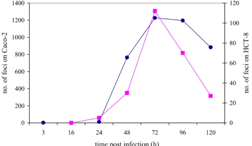
reproductive cycle of Cryptosporidium: meronts and macrogametocytes were clearly distinguishable. When inoculated with 1000-7000 pre-treated oocysts, the first foci of infection on Caco-2 cells were observed 3 h post infection. On HCT-8 cells the first foci of infection were seen 24 h post infection. On both cell lines the number of foci increased with increasing incubation time (Figure 1) and reached its maximum 48 –72 h post infection. Prolonged incubation (96 h or longer) resulted in deterioration of the cell monolayers and poor readability of results. Further results were read at 72 h post infection.
0 200 400 600 800 1000 1200 1400 3 16 24 48 72 96 120
time post infection (h)
no. of foc i on C ac o-2 0 20 40 60 80 100 120 no. of foc i on HC T -8
Figure 1 The number of foci of infection on Caco-2 (●) and HCT-8 (■) cells in a time series
3.2
Comparison of Cryptosporidium oocyst lots
Dilution experiments, using the most-probable-number method for detection of infection, indicated that infectivity of various C. parvum isolates varied. Variation was observed both between oocysts from different sources and among oocysts from different lots of one isolate from one source. Different experiments with identical lots of oocysts also resulted in different minimal numbers of oocysts required for a positive result, different numbers of positive wells (Tables 1-3) and different ID50 values (Table 4). ID50 values, indicating the number of
logit response model (Finch et al., 1993). The parameters for the logit model were calculated by regression analysis using the method of the least squares. Dilutions were recorded positive when foci of infection were detected in at least one well, however replicates appeared to produce a variable level of infection which was shown by the variable number of clusters of foci in a positive well (data not shown).
Both cell lines seemed almost equally sensitive to infection with Cryptosporidium oocysts, however a larger number of clusters of foci was observed in positive wells on HCT-8 than on Caco-2 when infected with equal numbers of oocysts (data not shown).
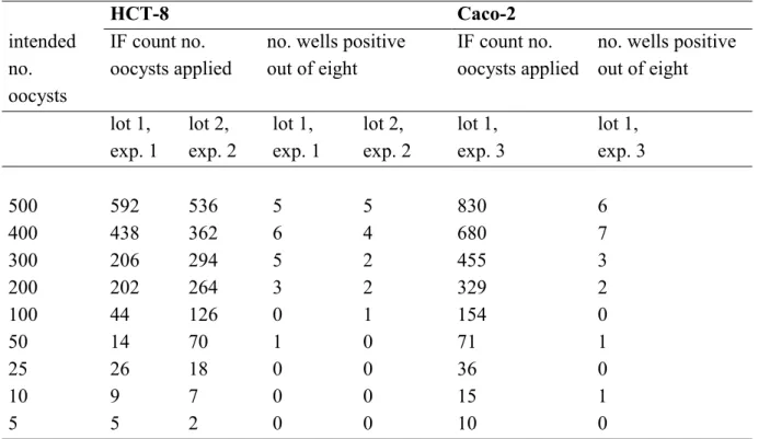
Table 1 Infectivity of Cryptosporidium parvum oocysts, purchased from Moredun Animal Health, on HCT-8 and Caco-2 cell lines. The immunofluorescence (IF) counts of the number of oocysts applied to each of eight replicate wells per dilution and the corresponding number of positive wells are displayed for two lots of oocysts used in three different experiments.
HCT-8 Caco-2 intended no. oocysts IF count no. oocysts applied
no. wells positive out of eight
IF count no. oocysts applied
no. wells positive out of eight lot 1, exp. 1 lot 2, exp. 2 lot 1, exp. 1 lot 2, exp. 2 lot 1, exp. 3 lot 1, exp. 3 500 592 536 5 5 830 6 400 438 362 6 4 680 7 300 206 294 5 2 455 3 200 202 264 3 2 329 2 100 44 126 0 1 154 0 50 14 70 1 0 71 1 25 26 18 0 0 36 0 10 9 7 0 0 15 1 5 5 2 0 0 10 0
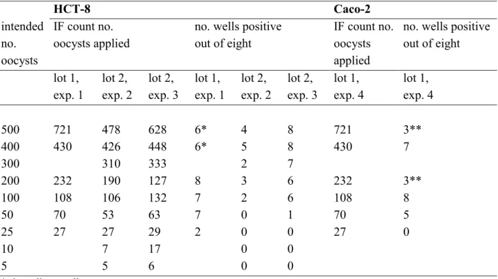
Table 2 Infectivity of Cryptosporidium parvum oocysts, purchased from Waterborne Inc., on HCT-8 and Caco-2 cell lines. The immunofluorescence (IF) counts of the number of oocysts applied to each of eight replicate wells per dilution and the corresponding number of positive wells are displayed for two lots of oocysts used in four different experiments.
HCT-8 Caco-2 intended no. oocysts IF count no. oocysts applied
no. wells positive out of eight
IF count no. oocysts applied
no. wells positive out of eight lot 1, exp. 1 lot 2, exp. 2 lot 2, exp. 3 lot 1, exp. 1 lot 2, exp. 2 lot 2, exp. 3 lot 1, exp. 4 lot 1, exp. 4 500 721 478 628 6* 4 8 721 3** 400 430 426 448 6* 5 8 430 7 300 310 333 2 7 200 232 190 127 8 3 6 232 3** 100 108 106 132 7 2 6 108 8 50 70 53 63 7 0 1 70 5 25 27 27 29 2 0 0 27 0 10 7 17 0 0 5 5 6 0 0
* six replicate wells ** three replicate wells
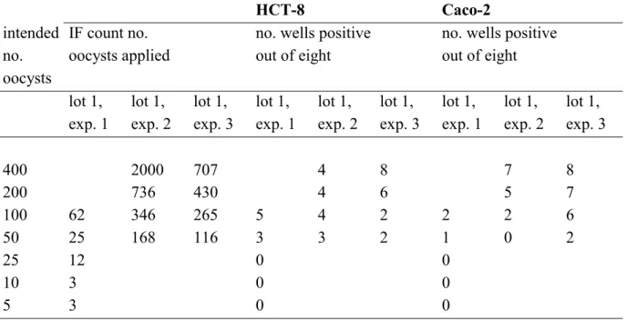
Table 3 Infectivity of Cryptosporidium parvum oocysts, provided by the University of Alberta, Edmonton, Canada, on HCT-8 and Caco-2 cell lines. The
immunofluorescence (IF) counts of the number of oocysts applied to each of eight replicate wells per dilution and the corresponding number of positive wells are displayed for one lot of oocysts used in three different experiments.
HCT-8 Caco-2 intended no. oocysts IF count no. oocysts applied
no. wells positive out of eight
no. wells positive out of eight lot 1, exp. 1 lot 1, exp. 2 lot 1, exp. 3 lot 1, exp. 1 lot 1, exp. 2 lot 1, exp. 3 lot 1, exp. 1 lot 1, exp. 2 lot 1, exp. 3 400 2000 707 4 8 7 8 200 736 430 4 6 5 7 100 62 346 265 5 4 2 2 2 6 50 25 168 116 3 3 2 1 0 2 25 12 0 0 10 3 0 0 5 3 0 0
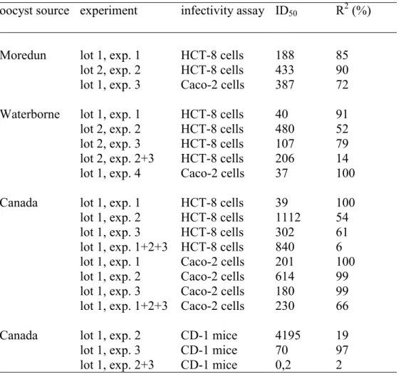
Table 4 ID50 values for C. parvum type II oocysts from various sources (Moredun Animal
Health; Waterborne Inc.; University of Alberta, Canada) in cell culture and CD-1 mouse infectivity assays. ID50 values were calculated by using the logit response
model; regression analysis resulted in R2 values, indicating the reliability of each calculation.
oocyst source experiment infectivity assay ID50 R2 (%)
Moredun lot 1, exp. 1 HCT-8 cells 188 85
lot 2, exp. 2 HCT-8 cells 433 90
lot 1, exp. 3 Caco-2 cells 387 72
Waterborne lot 1, exp. 1 HCT-8 cells 40 91
lot 2, exp. 2 HCT-8 cells 480 52
lot 2, exp. 3 HCT-8 cells 107 79
lot 2, exp. 2+3 HCT-8 cells 206 14
lot 1, exp. 4 Caco-2 cells 37 100
Canada lot 1, exp. 1 HCT-8 cells 39 100
lot 1, exp. 2 HCT-8 cells 1112 54
lot 1, exp. 3 HCT-8 cells 302 61
lot 1, exp. 1+2+3 HCT-8 cells 840 6 lot 1, exp. 1 Caco-2 cells 201 100
lot 1, exp. 2 Caco-2 cells 614 99
lot 1, exp. 3 Caco-2 cells 180 99
lot 1, exp. 1+2+3 Caco-2 cells 230 66
Canada lot 1, exp. 2 CD-1 mice 4195 19
lot 1, exp. 3 CD-1 mice 70 97
lot 1, exp. 2+3 CD-1 mice 0,2 2
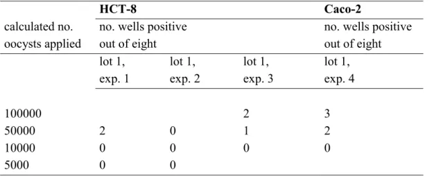
To test whether the cell culture assays were specific for detection of infectivity of C. parvum oocysts only, one isolate of C. muris was used. C. muris oocysts appeared to produce positive test results on both HCT-8 and Caco-2 cells at extremely high oocyst doses (Table 5).
However, the supplier of the oocyst suspension (Waterborne Inc., New Orleans LA, USA) could not guarantee 100 % purity of this suspension. Therefore we cannot be certain that the infection of the cells was produced by C. muris oocysts. On microscopic examination of the suspension we incidentally observed oocysts that differed from C. muris in shape and size, but the C. muris oocysts were abundant. We could not use molecular methods to confirm what other Cryptosporidium species was present in the suspension due to the very low concentration of suspect C. parvum oocysts.
Table 5 Infectivity of Cryptosporidium muris oocysts, purchased from Waterborne Inc., on HCT-8 and Caco-2 cell lines. The calculated number of oocysts applied to each of eight replicate wells per dilution and the corresponding number of positive wells are displayed for one lot of oocysts used in four different experiments.
HCT-8 Caco-2
calculated no. oocysts applied
no. wells positive out of eight
no. wells positive out of eight lot 1, exp. 1 lot 1, exp. 2 lot 1, exp. 3 lot 1, exp. 4 100000 2 3 50000 2 0 1 2 10000 0 0 0 0 5000 0 0
3.3
Comparison of cell culture and animal infectivity
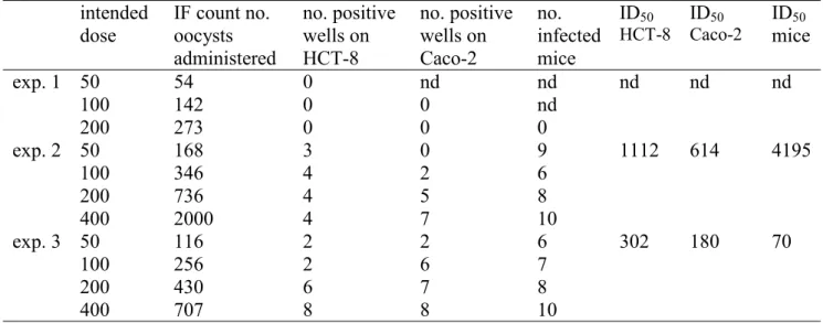
In these experiments, C. parvum oocysts from the University of Alberta, Canada, were used to compare cell culture infectivity on HCT-8 and Caco-2 cells with infectivity in CD-1 mice. Although the experimental settings were identical in all experiments (except from the mice, which were different in each experiment) variable results were obtained (Table 6).
Table 6 Cell culture (on HCT-8 and Caco-2 cells) infectivity and animal (on CD-1 mice) infectivity of Cryptosporidium parvum oocysts from the University of Alberta, Canada in three subsequent experiments. Each dose was inoculated onto eight replicate wells with HCT-8 or Caco-2 cells and was administered to ten mice.
intended dose IF count no. oocysts administered no. positive wells on HCT-8 no. positive wells on Caco-2 no. infected mice ID50 HCT-8 ID50 Caco-2 ID50 mice exp. 1 50 54 0 nd nd nd nd nd 100 142 0 0 nd 200 273 0 0 0 exp. 2 50 168 3 0 9 1112 614 4195 100 346 4 2 6 200 736 4 5 8 400 2000 4 7 10 exp. 3 50 116 2 2 6 302 180 70 100 256 2 6 7 200 430 6 7 8 400 707 8 8 10 nd= not done
3.4
Environmental samples
Surface water concentrates may have a toxic effect on cell monolayers. In the sample concentration procedure, not only target organisms are concentrated, but also a large variety of other biological and chemical substances. These may not be efficiently removed from the concentrates by the applied washing procedures and can have an adverse effect on in vitro cultured cell lines. The concentrates from the river Rhine (n=7) and Meuse (n=8) samples analysed here, did not have any toxic effect on the cell monolayers. Disturbing infection of the cell cultures with bacteria or moulds, possibly present in the river water concentrates, did not occur either.
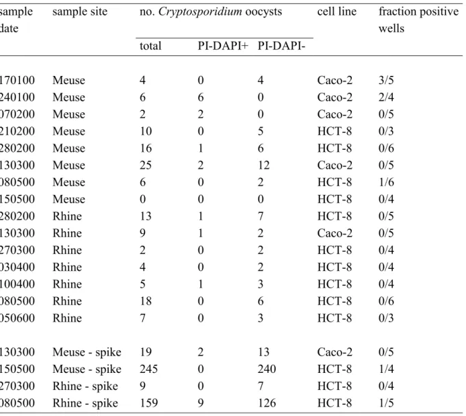
Sporozoite invasion producing foci of infection was observed in three of eight river Meuse samples, but in none of the seven river Rhine samples (Table 7). In the three positive samples, secondary infection resulting in large clusters of foci was not seen. This indicates that the oocysts were only capable of initial invasion of the cell cultures, but could not proliferate to further life cycle stages. Oocysts that only display initial invasion of cells in
vitro, are believed not to be able to reproduce and infect humans (Slifko et al., 1999). The
count results, which were performed on an equal portion of the same concentrated water sample, representing an equal volume of the original sample, showed that only few potentially infectious oocysts were present in the samples. Additionally, only two of four river water samples that were seeded with C. parvum oocysts infected the cell monolayers suggesting poor sensitivity of the cell culture assay.
Table 7 Infection of HCT-8 and Caco-2 cells with Cryptosporidium oocysts from concentrated water samples from the rivers Rhine and Meuse. The number of oocysts enumerated in the samples with an immunofluorescence stain (total) and the number of these oocysts which was potentially viable (DAPI+ and PI-DAPI-) is also displayed.
sample date
sample site no. Cryptosporidium oocysts cell line fraction positive wells
total PI-DAPI+
PI-DAPI-170100 Meuse 4 0 4 Caco-2 3/5 240100 Meuse 6 6 0 Caco-2 2/4 070200 Meuse 2 2 0 Caco-2 0/5 210200 Meuse 10 0 5 HCT-8 0/3 280200 Meuse 16 1 6 HCT-8 0/6 130300 Meuse 25 2 12 Caco-2 0/5 080500 Meuse 6 0 2 HCT-8 1/6 150500 Meuse 0 0 0 HCT-8 0/4 280200 Rhine 13 1 7 HCT-8 0/5 130300 Rhine 9 1 2 Caco-2 0/5 270300 Rhine 2 0 2 HCT-8 0/4 030400 Rhine 4 0 2 HCT-8 0/4 100400 Rhine 5 1 3 HCT-8 0/4 080500 Rhine 18 0 6 HCT-8 0/6 050600 Rhine 7 0 3 HCT-8 0/3
130300 Meuse - spike 19 2 13 Caco-2 0/5
150500 Meuse - spike 245 0 240 HCT-8 1/4
270300 Rhine - spike 9 0 7 HCT-8 0/4
080500 Rhine - spike 159 9 126 HCT-8 1/5
3.5
Ageing: comparison of viability methods
To compare oocyst infectivity and viability of ageing oocysts determined by different methods, oocyst survival in sterile river water was studied. Two Erlenmeyer flasks with sterile river water seeded with oocysts were studied for respectively 82 and 55 days. The DIC image of most oocyst remained unchanged. At the moment of inoculation into the flask, 99-100 % of the oocysts contained sporozoites and still did so after respectively 82 and 55 days. Viability as determined by vital dye in- or exclusion after acid treatment showed a
sharp decline between 26 and 33 days in flask I and between 7 and 14 days in flask II, whereas PI exclusion after IF stain (without acid treatment) declined more gradually
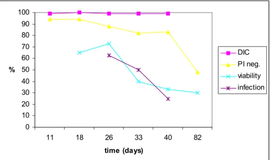
(Figure 2-3). Flask I contained approximately 1000 oocysts/100 µl. IF counts showed that the concentration remained stable for up to 82 days (Table 8). Oocyst morphology remained unchanged during this period. When applied to HCT-8 cell monolayers, this number of oocysts infected a gradually declining percentage of wells (Figure 2).
0 10 20 30 40 50 60 70 80 90 100 11 18 26 33 40 82 time (days) % DIC PI neg. viability infection
Figure 2 Ageing Cryptosporidium parvum oocysts in sterile surface water at 15 °C, flask I. Differential Interference Contrast (DIC) microscopy, propidium iodide exclusion without acid treatment (PI neg.) and the percentage of oocysts that both excluded PI after acid treatment and contained sporozoites (viability) indicated oocyst viability. Oocyst infectivity is indicated by the percent of infected HCT-8 cell monolayers (infection).
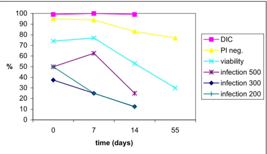
Flask II was seeded with a comparable number of oocysts per 100 µl, but here IF counts showed some inconsistency (Table 8). The number of intact oocysts detectable by IF seemed to have rapidly declined during the first week. As a result of this decline, fewer oocysts have been administered to HCT-8 cells than originally intended, but the trend of a gradual
0 10 20 30 40 50 60 70 80 90 100 0 7 14 55 time (days) % DIC PI neg. viability infection 500 infection 300 infection 200
Figure 3 Ageing Cryptosporidium parvum oocysts in sterile surface water at 15 °C, flask II. Differential Interference Contrast (DIC) microscopy, propidium iodide exclusion without acid treatment (PI neg.) and the percentage of oocysts that both excluded PI after acid treatment and contained sporozoites (viability) indicate oocyst viability. Oocyst infectivity is indicated by the percent of infected HCT-8 cell monolayers after applying approx. 500, 300 or 200 oocysts per well
(infection 500, 300, 200).
Table 8 The number of Cryptosporidium oocysts present in flasks with sterile river water on the displayed days after seeding, determined by immunofluorescence (IF).
time post-inoculation (days) IF count flask I flask II 0 1092 1148 5 1044 7 238 11 1118 14 639 18 1056 26 1197 33 1236 40 1096 55 488 82 1102
4.
Discussion
Previously, we searched the literature for possible cell culture systems to determine the infectivity of Cryptosporidium oocysts (Schets et al., 1998). Comparison of these cell culture assays resulted in an optimal protocol for infection of the human intestinal cell lines HCT-8 and Caco-2 with Cryptosporidium oocysts, which was implemented and further optimised. The protocol has been used in a large number of infection experiments with C. parvum oocysts. The infection experiments were aimed at evaluating the applicability of these cell culture infectivity assays to naturally contaminated environmental samples. Furthermore, their relation to other viability and infectivity methods such as vital dye exclusion and animal infectivity was evaluated.
4.1
Comparison of Cryptosporidium oocyst lots
Experiments with dilution series of Cryptosporidium oocysts from various vendors and with various lots from one vendor revealed considerable variety in infectivity. We calculated ID50
values for each experiment by using the logit response model (Finch et al., 1993) in order to be able to compare our results mutually and with those reported in international literature on
Cryptosporidium cell culture infectivity. Regression analysis with the method of the least
squares revealed very low correlations (R2 values) in quite a few cases, indicating that the calculated ID50 values were not reliable. The low correlations also suggested that the logit
response model might not have been the right choice to fit that particular data set. We have not tried to fit other models to our data. For comparison of infectivity of oocysts from different origin, on different cell lines or in different experiments, we used ID50 values with
correlations of 70 % or above, assuming that these were reliable ID50 values. Experiments
that generated 100 % R2 values were disregarded, because these experiments yielded only two data points, automatically resulting in 100 % correlation when performing the method of the least squares. ID50 values for Moredun oocysts ranged from 188 to 433 oocysts on
HCT-8 cells in two different experiments and appeared to be 387 on Caco-2 cells.
Waterborne Inc. oocysts had lower ID50 values (40-107 oocysts) on HCT-8, but here data
from two experiments on HCT-8 and one experiment on Caco-2 had to be disregarded because of poor correlation. C. parvum oocysts from the University of Alberta seemed to perform differently on HCT-8 cells and Caco-2 cells. No reliable ID50 could be calculated
from three HCT-8 experiments, whereas highly reliable estimates of the ID50 (R2 99%) on
Caco-2 could be obtained, which however differed considerably in the two Caco-2
experiments (180 and 614 oocysts). The variety observed between C. parvum isolates from different sources, between lots from the same source, on different cell lines and in different experiments have also been described by Slifko et al. (2002) and Rochelle et al. (2002). Human feeding trials showed similar differences with different C. parvum isolates displaying different infectivities (Okhuysen et al., 1999). It must be noted that differences are sometimes
small and the number of data is sometimes limited. The differences may be true differences between isolates or lots, but they may also be due to the inaccuracy inherent to cell culture assays. In conclusion, one of the drawbacks of the mouse infectivity assays, the variability in the obtained results, seems to apply to cell culture infectivity assays as well.
4.2
Infectivity in cell culture vs. infectivity in mice
Three experiments with neonatal CD-1 mice yielded only one reliable estimate of ID50 for
C. parvum oocysts from the University of Alberta, which was 70 oocysts. These results
suggest a higher sensitivity for the neonatal mouse assay than for the cell culture assay on Caco-2 cells, but these are the data of one experiment only. Our complete data set on mouse infectivity is limited and regression analyses with the least squares method showed that the logit response model did not fit the data in two of three experiments. Slifko et al. (2002) noted that a cell culture infectivity assay on HCT-8 cells was more sensitive to infection with
C. parvum oocysts than a mouse infectivity assay using 4-day-old BALB/c mice or neonatal
CD-1 mice. They reported average logit ID50 values of 8, 64 and 119, for these three assays
respectively. Using different oocyst lots, the observed differences were not statistically significant, resulting in the choice to report average ID50 values. Rochelle et al. (2002) found
cell culture infectivity equivalent to a standard mouse assay using CD-1 mice for the C.
parvum oocysts they used. They also compared cell culture infectivity on HCT-8 and Caco-2
cells and observed lower ID50 values on HCT-8, indicating a higher sensitivity for HCT-8
cells. We cannot draw the same conclusions from our experiments, but a larger number of clusters of foci of infection were however observed in positive wells on HCT-8 than on Caco-2 cells when infected with equal numbers of oocysts. This suggests that HCT-8 cells support the proliferation to further life cycle stages more than Caco-2 cells.
4.3
Infectivity in cell culture vs. surrogate viability methods
Although cell culture assays are easier to perform than animal infectivity assays, they still require special laboratory facilities and are time consuming. In vitro excystation and vital dye in- or exclusion assays are simpler and more widely applicable. Bukhari et al. (2000)
compared animal infectivity with in vitro excystation and in- or exclusion of the vital dyes SYTO-9, SYTO-59 and DAPI/PI after treatment of fresh and environmentally aged oocysts with low concentrations of ozone. They found that both in vitro excystation and the vital dyes overestimated oocyst infectivity in comparison to CD-1 mice. These results confirmed earlier studies that also showed the disparity between surrogate methods for oocyst infectivity and neonatal mouse assays (Korich et al., 1990; Finch et al., 1993; Black et al., 1996). In our survival experiments we observed that cell culture infectivity declined far more rapidly than
the surrogate methods we used, including vital dyes, indicating that the surrogate methods indeed overestimated oocyst infectivity compared to the cell culture assay.
4.4
Cell culture infectivity assays in disinfection studies
As opposed to analyses of naturally contaminated water samples for the presence of
Cryptosporidium oocysts, in disinfection experiments high numbers of oocysts can be used to
study the effect of disinfectants. Cell culture infectivity can be successfully used to determine oocyst infectivity in vitro after disinfection treatment. Rochelle et al. (2002) compared cell culture infectivity on HCT-8 cells (including RT-PCR detection of infection) with infectivity in CD-1 mice. Both assays were found to produce equivalent results in predicting oocyst infection after ozone or UV treatment. Slikfo et al. (2002) reported no significant differences between cell culture on HCT-8 cells (microscopic detection of infection) and mouse
infectivity after UV or chlorine dioxide treatment. They however noted that cell culture predicted slightly higher inactivation than mice did after UV treatment and slightly lower inactivation after chlorine dioxide treatment. Joachim et al. (2003) concluded that both HCT-8 cell culture and 3-day-old BALB/c mice could be successfully used to assess
Cryptosporidium infectivity after treatment with chemical disinfectants, although no results
of statistical comparison of both methods were reported. Cell culture assays provide optimal results when used to discern relatively large differences in the levels of infection or
inactivation since variability plays a less profound role (Rochelle et al., 2002).
4.5
Cell culture infectivity assay for environmental samples
Our dilution experiments indicated that relatively large numbers of oocysts were needed for positive results (i.e. infection) in cell culture. Analyses of natural samples over the years have learned that the concentration of oocysts in these samples is usually below these minimally required numbers. The surface water samples analysed in this study were extracted from the largest rivers in the Netherlands with the highest average Cryptosporidium load in Dutch surface water samples, ranging from 0.1-1.3 oocysts/L (Medema et al., 2002).
Considering the results of the dilution experiments, the absence of detection of infectious oocysts in the 15 surface water samples we examined in this study, does not necessarily mean that there were no infectious oocysts present in the river water. Relatively small samples of 12-25 L were examined. IF counts showed that the total number of oocysts in these samples ranged from 1 to 32. The fraction of these oocysts that was viable and potentially infectious was way below any of the ID50 values of the C. parvum suspensions used to evaluate the cell
culture assay. Surface water samples seeded with Waterborne Inc. oocysts, which had ID50
although oocysts numbers in the seeded samples were above the ID50’s. This may either be
due to lack of sensitivity of the cell culture method or may be inherent to the variation in the results obtained with this method.
In our survival experiments we were able to detect infectivity of aged oocysts with cell culture after 82 days in sterile river water. However, about 1,000 oocysts were required for obtaining a positive result, once again indicating that naturally contaminated water samples, containing both fresh and aged oocysts, presumably do not contain enough infectious oocysts to produce positive results in cell culture. Jenkins et al. (2003) found that C. parvum oocysts remained infectious for HCT-8 cells and neonatal BALB/c mice when stored for seven months in sterile deionized water at 15 °C. Both cell cultures and mice were inoculated with 104 oocysts. Quintero-Betancourt et al. (2003) reported that they were able to obtain positive results on HCT-8 cells when analysing reclaimed effluent samples that contained more than 100 oocysts per 100 L.
5.
Conclusions
Cell culture assays used in this study were not sensitive enough to detect infectious
Cryptosporidium oocysts in Dutch surface waters used for recreation or drinking water
production. Oocyst concentrations in those samples are usually below those required to produce infection in cell culture assays. Mouse assays have been reported to be equivalent to cell culture assays in predicting oocyst infectivity. Assessment of the risk of infection with
Cryptosporidium can not be based on oocyst infectivity. It is nevertheless possible to
determine oocyst viability in environmental samples with low oocyst concentrations by using vital dye in- or exclusion assays. These assays however overestimate oocyst infectivity compared to mouse and cell culture infectivity assays. For recreational waters this may lead to an overestimation of the chance of getting infected or ill after exposure to Cryptosporidium oocysts. In drinking water production, risk assessment is based on the number of infectious oocysts in the source water and the effectiveness of drinking water treatment processes on the number of infectious oocysts. Cell culture infectivity assays can provide information on the effect of disinfection processes on oocyst infectivity. When oocyst viability data are used in risk assessment, the number of infectious oocysts in the source water, and thus the risk of infection with Cryptosporidium, is probably overestimated. The limit of one infection in 10,000 persons per year may be exceeded, unjustly indicating that adjustment in drinking water treatment or additional research is required. For accurate risk assessment information on oocyst infectivity is indispensable. Enhanced recovery of the method for detection and enumeration of Cryptosporidium oocysts in water and improved sensitivity of the cell culture assays may result in better applicability of cell culture infectivity assays to environmental water samples in the future. PCR detection of the infection of the cells in stead of
Acknowledgements
The authors thank Erwin Duizer (RIVM-LIS) for his help with the implementation of Caco-2 and HCT-8 cell culture in our laboratory. Jack Schijven is acknowledged for his assistance in calculating ID50 values with the logit response model. We thank Willemijn Lodder, Ria de
Bruin and Harold van den Berg for their assistance with the cell culture infectivity experiments.
References
Anonymous
Method 1623: Cryptosporidium and Giardia in water by filtration/IMS/FA United Stated Environmental Protection Agency, Office of Water 4603 2001; EPA-821-R-01-025
Anonymous
Voorschrift voor concentratie van water m.b.v. Envirochek capsules t.b.v. detectie van
Cryptosporidium oöcysten en Giardia cysten
SOP MGB/M003 rev. 0, 1/9/2003 (in Dutch) Anonymous
Voorschrift voor zuivering van waterconcentraten m.b.v immonomagnetische separatie t.b.v. detectie van Cryptosporidium en Giardia
SOP MGB/M004 rev. 0, 1/9/2003 (in Dutch) Arrowood MJ
Diagnosis, in: Cryptosporidium and Cryptosporidiosis Fayer R Ed., CRC Press, Boca Raton 1997: 43-64
Asperen IA van, Stijnen C, Mank T, Boer A de, Groot JF, Medema GJ, Ham P ten, Sluiters JF, Borgdorff MW
An outbreak investigation of cryptosporidiosis in the Netherlands RIVM report 215700 001 1996; Bilthoven, the Netherlands Black EK, Finch GR, Taghi-Kilani R, Belosevic M
Comparison of assays for Cryptosporidium parvum oocysts viability after chemical disinfection
FEMS Microbiol Lett 1996; 135: 187-189
Bukhari Z, Marshall MM, Korich DG, Fricker CR, Smith HV, Rosen J, Clancy JL
Comparison of Cryptosporidium parvum viability and infectivity assays following ozone treatment of oocysts
Appl Environm Microbiol 2000; 66 (7): 2972-2980 Campbell AT, Robertson LJ, Smith HV
Viability of Cryptosporidium parvum oocysts: correlation of in vitro excystation with inclusion or exclusion of fluorogenic vital dyes
Casemore DP, Wright SE, Coop RL
Cryptosporidiosis: human and animal epidemiology in: Cryptosporidium and Cryptosporidiosis
Fayer R Ed., CRC Press, Boca Raton 1997: 65-92
Di Giovanni GD, Hashemi FH, Shaw NJ, Abrams FA, LeChevallier MW, Abbaszadegan M Detection of infectious Cryptosporidium parvum oocysts in surface and filter backwash water samples by immunomagnetic separation and integrated cell culture-PCR
Appl Environm Microbiol 1999; 65 (8): 3427-3432 Fayer R, Morgan U, Upton SJ
Epidemiology of Cryptosporidium: transmission, detection and identification Int J Parasit 2000; 30: 1305-1322
Finch GR, Daniels CW, Black EK, Schaefer III FW, Belosevic M
Dose response of Cryptosporidium parvum in outbred neonatal CD-1 mice Appl Environm Microbiol 1993; 59 (11): 3661-3665
Griffiths JK, Moore R, Dooley S, Keusch GT, Tzipori S
Cryptosporidium parvum infection of Caco-2 cell monolayers induces an apical
monolayer defect, selectively increases transmonolayer permeability, and causes epithelial cell death
Infect Immun 1994; 62: 4506-4514
Jenkins M, Trout JM, Higgins J, Dorsch M, Veal D, Fayer R
Comparison of tests for viable and infectious Cryptosporidium parvum oocysts Parasitol Res 2003; 89: 1-5
Joachim A, Eckert E, Petry F, Bialek R, Daugschies A
Comparison of viability assays for Cryptosporidium parvum oocysts after disinfection Vet Parasitol 2003; 111: 47-57
Korich DG, Mead JR, Madore MS, Sinclair NA, Sterling CR
Effects of ozone, chlorine dioxide, chlorine, and monochloramine on Cryptosporidium
parvum oocyst viability
Appl Environm Microbiol 1990; 56 (5): 1423-1428
McDonald V, Stables R, Warhurst DC, Barer MR, Blewett DA, Chapman HD, Conolly GM, Chiodini PL, McAdam KPWJ
In vitro cultivation of Cryptosporidium parvum and screening for anticryptosporidial drugs