Report 360060002/20010 C.A. Herberts et al.
Stem Cell Therapy
Evaluation of risk factors
Letter Report 360060002/2010
Stem Cell Therapy
Evaluation of risk factors
January 2010
C.A. Herberts, RIVM / Centre for Biological Medicines and Medical technology M.S.G. Kwa, Netherlands Medicines Evaluation Board
H.P.H. Hermsen, RIVM / Centre for Biological Medicines and Medical technology
Contact: C.A. Herberts
Centre for biological medicines and medical technology carla.herberts@rivm.nl
This investigation has been performed by order and for the account of Netherlands Health Care Inspectorate, within the framework of Risicosignalering van Medische Producten
© RIVM 2010
Parts of this publication may be reproduced, provided acknowledgement is given to the 'National Institute for Public Health and the Environment', along with the title and year of publication.
Abstract
Stem cell therapy holds the promise to treat degenerative diseases, cancer and repair of damaged tissues for which there are currently no or limited therapeutic options. Despite that promise there are still many obstacles in the safe use of stem cell based regenerative medicine. A thorough evaluation of potential risks will be a prerequisite step before more wide clinical application and/or registration of stem cell medicinal products will become a reality. The most important risks associated with the use of different stem cell types are evaluated in this report. The risks depend on many factors such as the origin of the cells, their differentiation status and ability to proliferate, and the route/location of administration. The risk on tumourigenicity is a critical point in the safety concerns associated with the use of stem cells. This risk is potentially higher for pluripotent stem cells than for multipotent somatic stem cells. Besides tumourigenicity also other risks like immune modulation should be evaluated and reduced before clinical application.
The risk profile of a stem cell medicinal product not only depends on the choice the stem cells to be used, but also by issues such as the differentiation status of the cells, the need for in vitro culture and/or manipulation, the reversibility of treatment, the need/possibility for concurrent tissue regeneration in case of irreversible tissue loss, and the long-term survival of engrafted cells. Currently there is no clinical experience with pluripotent stem cells probably because the perceived risks are unacceptable. However, in the last decade the use of multipotent somatic stem cells in regenerative medicine has been studied in multiple small-sized clinical trials. From these studies it appears that the application of multipotent stem cells is relatively safe. Nevertheless in several clinical trials adverse events have been reported, which emphasizes the need for additional knowledge, particularly on long term safety of stem cell therapy.
Key words:
stem cell therapy, advanced therapy, risk, medicinal product, stem cells, tumor, tumour, immunology, clinical, regulatory, regulation
Trefwoorden:
stamcellen, geavanceerde therapie, risico, geneesmiddel, stamceltherapie, tumor, immuunsysteem, klinisch, regelgeving
Table of Content
Abstract 3 Table of Content 5 Abbreviations 6 Introduction 7 Stem cells 8Pluripotent stem cells 9
Somatic stem cells 10
Risks 12
Tumourigenicity 12 Pluripotent stem cells 12 Multipotent stem cells 13 In vitro stem cell culture 14 Bystander tumour formation 14 Immune responses 15 Adventitious agents 15 Other risks 16
Clinical therapeutic experience 18 Regulatory status 20 Discussion/conclusion 21 References 23
Abbreviations
ATMP advanced therapy medicinal product
BM bone marrow
CB cord blood
DC dendritic cell
ECFC endothelial colony forming cells
ESC embryonic stem cell
FBS fetal bovine serum
G-CSF granulocyte-colony-stimulating factor
GVHD graft versus host disease
HLA human leukocyte antigen
HSC Haematopoietic stem cell
HSCT Haematopoietic stem cell transplantation
iPSC induced pluripotent stem cell
LVEF left ventricular ejection fraction
MHC major histocompatibility complex
MI myocardial infarction
MSC mesenchymal stem/stromal cells
PB peripheral blood
SCNT somatic cell nuclear transfer
SSC somatic stem cell
Introduction
The field of stem cell therapy is rapidly developing, and many clinical trials have been initiated exploring the use of stem/progenitor cells in the treatment of degenerative diseases and cancer and for the repair of damaged or lost tissues. A stem cell can be defined as a cell that can renew itself indefinitely, while producing progeny that have a multi-lineage differentiation capacity. Due to these properties, stem cells therapy holds the promise in the treatment of many degenerative and/or age-related diseases and conditions for which there are currently no or limited therapeutic options. Despite the great promise, there are still many obstacles in the safe use of stem cell-based regenerative medicine. In this report, different types of stem cells will be described and the risks associated with their use will be evaluated. As haematopoietic stem cell transplantation (HSCT) has become a standard therapy for reconstitution of the immune system in severe compromised patients, risks involved with this treatment will not be described in this review. However, it will focus on the recent developments in stem cell therapy involving the substantial manipulation of cells with stem cell-like characteristics. This will include both the theoretic/perceived risk as well as examples of adverse observations described in public literature. In addition, a concise review on the current clinical applications will be provided and the regulatory aspects of stem cell therapy will be discussed.
Stem cells
Stem cells are undifferentiated cells that have the capacity to proliferate as stem cell in undifferentiated form both in vitro and in vivo (self-renewal) and to differentiation into mature specialized cells.
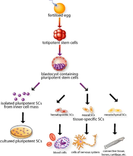
Stem cells can be subdivided based on their potency to produce different types of cells (Figure 1).
Figure 1: Origin, isolation and specialisation of stem cells1
A totipotent stem cell is a stem cell that can produce an entire organism, because it can produce all the cell types of an organism and the extra-embryonic tissues. In the mouse, only a zygote and a blastomere from a 2-cell stage embryo would be considered totipotent. Pluripotent stem cells do not have the self-organising capability to form the whole organism but have the ability to differentiate into derivatives of all three germ layers (multilineage differentiation). Pluripotent embryonic stem cell (ESC) lines can be derived from the inner cell mass of blastocyst stage embryo. Recently, pluripotent stem cells with ESC-like characteristics have been engineered from somatic/adult cells2. These cells are termed
induced pluripotent stem cells (iPSC). Multipotent stem cells have a more restricted differentiation capacity compared to pluripotent stem cells3. Multipotent adult or somatic
stem cells (SSC) are present in many, possibly all, postnatal tissues of an organism and are presumed to play a role in tissue homeostasis. They are considered to be responsible for the maintenance and regeneration of aged or damaged tissue by replacing lost cells3.
Pluri/totipotent stem cells have been generated by somatic cell nuclear transfer (SCNT). In this technique the nucleus of a somatic (donor) cell is inserted into an enucleated egg cell. This will lead to reprogramming of the donor nucleus by the host cell. Following chemical activation the cell may start to divide and develop into a blastocyst. This technique was used for cloning animals (e.g. the sheep Dolly). Currently, no human stem cell lines have been derived by SCNT research. Although straightforward, this technique is technically demanding4 (not automated) with a high failure rate. Because of the development of
induced pluripotent stem cells, this technique now seems redundant and it is unlikely that SCNT will be used for stem cell therapy in the near future. Therefore SCNT will not be discussed in this review.
Pluripotent stem cells
Research in the field of pluripotent stem cells began with the study of teratocarcinoma’s in the 1950(s). Mouse embryonic stem cells were isolated almost 30 years ago5, while successful culture of human embryonic stem cells has been described just over a decade ago6. Recently it has been discovered that (normal) somatic cells can be reprogrammed to an embryonic stem cell-like pluripotent state (iPSC) 2 (Figure 2).
The most common assay for demonstrating pluripotency is teratoma formation. However, pluripotent stem cell lines must be able to fulfil several other specific features4;7. Such
lines should have the ability to grow indefinitely and express ESC surface markers and marker genes and show ESC-like morphology. In addition, the cell line should be able to form embryonic bodies (in vitro) and/or teratoma’s (in vivo) containing all 3 germlayers (endoderm, mesoderm, and ectoderm). In addition, pluripotent stem cells must have the ability to form chimeras upon injection into early blastocysts. This latter property however can only be tested with animal (mouse) ESC as this assay cannot be performed in humans. Notably, this latter criteria is often not met while the other two are fulfilled7.
Pluripotent ESCs can be derived from the inner cell mass of blastocyst stage embryo, and can be maintained in in vitro culture conditions as established cell line. Human ESC (hESC) are very different from mouse ESC (mESC)8, they express different cell surface markers, have other (active) signalling pathways and other factors are needed in culture medium for the isolation and maintenance of the ESC. The use of ESC in clinical practice raises ethical concerns (as an embryo was needed for the generation). This is in contrast to iPSC where pluripotent stem cells are generated from the patients own cells by inducing a forced expression of certain genes (Figure 2). For iPSC generation mostly fibroblasts are used, but iPSCs have also been derived from liver, pancreas β cells and mature B cells8. There are
many similarities between iPSC and ESC for both human and mouse. They have highly similar growth characteristics, gene expression profiles, epigenetic modifications and developmental potential8;9. However, some differences in gene expression have been reported suggesting that reprogramming in iPSC is incomplete9. In addition, the generation of chimeras from iPSC appears more difficult than for ESCs and has been associated with a high rate of tumour formation10.
Figure 2: Schematic diagram of Induced Pluripotent Stem Cells11
Somatic stem cells
Multipotent somatic stem cells (SSC) are undifferentiated cells found in many organs and differentiated tissues9. SSCs vary in their differentiation capacity. They can give rise to a number of cell types which is generally limited to the cell types present in the tissue of origin. SSCs are immature cells that express stem cell markers (c-kit, MDR1, Sca-1). Cord blood and bone marrow are sources which are often used as source for SSC, more recently adipose tissue is also used.
Cord blood (CB) may comprise different stem cell populations, haematopoietic stem cells, endothelial progenitor cells, and non-haematopoietic stem cells have been identified but also stem cells for neurons and hepatocytes and cells expressing markers of pancreatic transitional cells have been reported to be present in CB3. The bone marrow contains haematopoietic stem cells (HSC) and non-haematopoietic mesenchymal stem/stromal cells (MSC), and possibly also endothelial progenitor cells.
Endothelial progenitor cells (EPS) can also be isolated from peripheral blood, especially following mobilisation with granulocyte-colony-stimulating factor (G-CSF). EPCs are identified according to the expression of both haematopoietic stem cell markers and endothelial cell markers12, however they lack a specific marker. EPC can be isolated from a different sources (e.g. BM, CB, PB), and different isolation procedures have been employed. Surface antigen markers CD133, CD34 and VEGFR2 are often used to identify EPC, however they correspond to a heterogeneous population13. Recently it has become evident that the endothelial colony forming cells (ECFC) or ‘late outgrowth EPC’ are the real progenitor or stem cells that have the ability to form new endothelium in blood vessels in vivo12;14. EPC
have the ability to differentiate into several cell types, depending on the culture conditions15
MSCs were initially isolated from bone marrow aspirates16, however it is now clear that MSC
can be isolated from a large number of tissues, including tissues harvested from pathological sites such as rheumatoid arthritic joint17. The morphology, immunophenotype and growth properties of these MSCs are similar to bone marrow-derived MSCs. However some variations in immunophenotype and osteogenic or adipogenic differentiation potential variations have been reported which appeared to be related to the site of origin18
suggesting that the differentiation potential of MSC is related to the tissue of origin. It is therefore still unclear if MSC derived from different tissues are identical or tissue specific 18-20.
MSC exhibit enormous in vitro expansion capacity. MSC can support haematopoiesis, appear non-immunogenic and have immunomodulatory/ suppressive effects. Phenotypically MSC express a number of markers (including CD44, VCAM-1, ICAM-1 and CD2917 21). However a unique and specific MSC marker, allowing their exclusive identification, has not yet been found17;20-22. MSC are capable of producing cell types of mesodermal lineage (bone,
cartilage, fat, tendon, muscle, marrow stroma). Whether MSC can differentiate into cells of other germ layers (transdifferentiation) is still a matter of debate4;19;23-27. Some groups have
observed differentiation of MSC into cell types of such as neuronal cells or hepatocytes, while others did not find such transdifferentiation. These differences may be (partly) due to differences between laboratories in MSC source used, extraction procedures, and cultivation methods. These variables are responsible for the phenotype and function of resulting cell populations. Small differences in the method of procurement and culture may be biologically significant28;29. Whether differences in culture conditions selectively
promote the expansion of slightly different populations of MSCs or cause similar cell populations to acquire different phenotypes is not clear.
Risks
The risks associated with stem cell therapy depend on many factors such as the origin of the cells, their differentiation status and ability to proliferate, and the route/location of administration. Several risks are evaluated here, starting with the risk on tumourigenicity as this is perceived as a key obstacle to the safe use of stem-cell based regenerative medicine. However, also other risks associated with the use of stem cells should be considered, these include the effect on the immune responses, in vivo differentiation and potential viral or microbial contamination.
Because there is more experience with somatic stem cells, especially MSC, compared to pluripotent stem cells (ESC/iPSC), more is known about their characteristics and the risks associated with the use of these types of cells. The risks described here for MSC may however also apply to other types of stem cells. More general risks that are not stem cell specific, which are associated with cell therapy, e.g. traceability of materials from donor to patient, irreversibility of treatment, difficulty of GMP manufacture, errors in labelling/storage/administration, non-efficacious dosing are not discussed here.
Tumourigenicity
Stem cells possess several important features making them attractive candidates for malignant degeneration. These include long life span, relative apoptosis resistance and ability to replicate for extended periods of time. In addition, similar growth regulators and control mechanisms are involved in both cancer and stem cell maintenance. The risk on tumour formation or the tumourigenic potential of stem cells is assumed to depend on their pluripotent or multipotent characteristics. In addition, also the environmental (local) context of the stem cell and the duration of in vitro culture may influence its tumourigenic potential.
Pluripotent stem cells
One of the hallmarks of pluripotency is teratoma formation. Thus, administration of (undifferentiated) pluripotent stem cells to humans may cause teratoma formation. In addition to teratomas, which are considered to be benign, also malignant tumour formation (e.g. teratocarcinoma) has been observed when administering ESCs or iPSCs in animal models30;31. ESC have been claimed to have a more tumourigenic nature when compared with their originator cells (the inner mass of early blastocysts)32, probably due to removal of the cells from the context of a developing embryo and enforcing in vitro culture. So tumourigenic risk of pluripotent cells is real, but difficult to estimate because no human trials with ESC have been conducted yet. Notably, transformation of early foetal cells which are presumably pluripotent into pre-invasive testicular carcinoma cells has been suggested to occur in humans33. Tumourigenesis may be dependent on the host/species to which the
cells are administrated34.
In addition to the risk associated with the pluripotent state, specific factors may contribute to the tumourigenic potential of iPSCs. The method for generating iPSCs from fibroblast is very similar to the focus formation assay which is an assay for testing the tumourigenicity of specific genes in fibroblasts. For the generation of iPSCs somatic cells are reprogrammed by a forced expression of several genes, some of which are oncogenes (Myc and KLF4) or
genes linked to tumourigenesis (Sox2 and Nanog, and Oct3). The use of these genes may increase the tumourigenic potential of iPSC. It has been described that up to 20% of iPSC chimeric mice develop tumours due to reactivation of c-myc9;32. The iPSC transforming
genes were initially inserted by retroviral transduction. This introduced the risk of retroviral-mediated carcinogenesis. Stable integration at multiple sites (random) can disrupt genes that are important for normal cellular homeostasis, and can thereby cause aberrant expression of an oncogene thus inducing/promoting carcinogenesis7;9. However, already new methods for the generation of iPSC have been described that do not require genomic incorporation but transient expression of iPSC genes or delivery of the relevant proteins9;35;36. As the iPSC field is moving away from genetic changes and the use of c-myc,
their safety may improve and the risk of tumourigenicity may be reduced10 and become
more similar to the tumourigenic risk associated with ESC. It should be noted that iPSC induction is associated with profound and progressive changes in the epigenetic state of the chromatin7. Epigenetic changes can cause changes in the expression of onogenes or tumour suppressor genes thereby changing the tumourigenic potential of the cell.
Taken together, knowledge on tumourigenicity of iPSC is still very limited and more knowledge is needed. Due to the teratoma and potential tumour forming capacity of ESCs and iPSCs, it is unlikely that they will be used directly in regenerative medicine. Therefore differentiation of ESC/iPSC into specific cell types is preferred as these are devoid of pluripotent capacity which may reduce the risk of tumour formation.
Multipotent stem cells
Adult somatic stem cells have been used directly in the clinic. HSC are widely used for decades for reconstitution of immune function3. Also bone marrow derived MSCs have been
used as supportive treatment in HSC transplantation. However, injection of undifferentiated stem cells in specific tissue may cause unwanted cell proliferation. Bone marrow derived stem cells have been identified as the cell of origin of Helicobacter-induced gastric cancer in a mouse model37 and sarcomas were observed after in vivo infusion of MSC3. Therefore also for SSC tumourigenicity is still a major concern when these cells are used in the clinic for other purposes than reconstitution or to perform their essential function. Notably, it has been postulated that adult somatic stem cells play a role in the aetiology of cancer where these cells may perhaps become tumourigenic32.
Autologous MSCs have been implicated in childhood leukemia38, gastric cancer39, and
osteogenic sarcoma40. Theoretically, EPC could promote tumour growth by increasing tumour microvasculature41. It is thought that a small fraction of cells within a tumour are capable of independent growth, and fulfil the criteria described for cancer stem cells37. These cells have metastatic potential, form tumours in secondary hosts and are believed to be responsible for continual renewal of cells within the tumour mass. Despite the similarities between somatic stem cells and cancer stem cells a direct link between somatic stem cells and cancer stem cells remains to be shown.
Notably in humans the incidence of solid tumours is significantly increased following bone marrow transplantation42. However it still needs to be established that this is caused by the administered stem cells and not due to other aspects of the treatment (e.g. immune suppression or chemotherapy). Recipients of solid organ transplants also appear to have a higher incidence of secondary malignancy37. Haematopoietic cells of donor origin are often found in the circulation indicating that haematopoietic stem cells are transferred with the transplanted organ43. These transferred stem cells have been shown to give rise to Kaposi
In vitro stem cell culture
The risk of uncontrolled cell proliferation is reduced with differentiated (stem) cells. Thus differentiation of stem cells prior to administration to a patient seems desirable. In vitro cultured stem cells can be guided to differentiate into a desired cell type by lineage-specific culture conditions. However, during in vitro expansion and differentiation stem cells are subject to damage from intracellular and extracellular influences. Every cell division has a small chance of introducing deleterious mutations, most of which cannot be detected by karyotyping. In vitro ESC lines have been reported to show a certain degree of dysregulation of the so-called imprinted genes, also after differentiation3.
In principle, (epi)genetic changes may cause transformation of a cell into a tumourigenic phenotype and may contribute to increased tumour formation. Several groups analysed whether MSC could undergo spontaneous transformation, and the results are conflicting. Some reported spontaneous transformation during long term culture for both mouse and human MSC3;27;46-48 while others found no evidence for transformation after extensive
genetic characterisation49;50. In France, several clinical trials with MSC have been put on
hold due to the occurrence of recurrent abnormal karyotypes in cell culture.51. However,
transformation of the karyotypic altered MSC was not observed, and the abnormal karyotype did not persist upon prolonged culturing52. Following long term culture of human MSC, both escape of senescence resulting in crises followed by transformation, and no escape of senescence has been reported. It has been suggested these ambiguous result can partly be explained by a species difference stating that murine MSC are more prone to cellular transformation than human MSC23. However, spontaneous transformation human
adipose tissue–derived MSCs after long-term in vitro culture has been described, and these transformed cells were able to produce tumours in vivo in immunodeficient mice46. Taken
together, in vitro transformation of human MSC is still a matter of debate, and if it exists it appears to be an exceptional event.
The tumourigenicity of stem cells has been predicted to increase proportionally with the length of in vitro culturing32. Spontaneous transformation of mice neural precursor/stem cells has been reported already after ~10 passages of cell culture, and these cells also able to form tumours upon administration into rodent brains48. Therefore, to minimise the risk
on transformation and/or epigenic changes short term culture of the stem cell product is advisable. This will also reduce the risk of selecting fast growing cells.
Bystander tumour formation
In addition to be tumour forming cells themselves, stem cells might affect growth/proliferation of tumour cells22. This has been studied for MSC only. In vitro and in
vivo studies have reported inhibition, enhancement and no effect of administration of MSC
on tumour growth21;25;53-55. Most likely the observed effect depends on the nature of the
cancer cells, the characteristics of the used MSC, on the integrity of the immune system and on the site of injection. Two possible mechanisms have been postulated for the stimulation of tumour growth25, MSC may provide supportive stroma creating a permissive environment for tumour growth or MSC may reduce immune rejection of the tumour cells thus allowing continued tumour growth. No mechanism for the sometimes observed decreased tumour growth has been described. Since al these studies have been performed
in vitro or in animal models the relevance of these observations for humans is unknown.
Notably, an opposite effect on tumour growth, between in vitro and in vivo situation, has also been reported21 and has complicated the assessment of the potential effect of MSC on
tumour by MSC must be considered when administering these cells to a patient, however nature of the risk is difficult to assess.
Immune responses
Administration of stem cells may have consequences for the immune system. First, the administered cells may induce an immune response themselves56. Immune recognition of
stem cells is particularly important when the cells are not autologous. Allogenic (and xenogeneic) cells may be recognized by the immune system of the patient leading to rejection and loss of function of the administered cell. The use of immune suppressants may limit this risk. Notably, ESC and especially MSC have been reported to be immune-privileged and have a low immunogenic potential17;21;57 and administration may require reduced or even no immune suppression. Upon differentiation these cells may become more immunogenic due to e.g. upregulation of a normal set of MHC molecules.
In addition to direct recognition of the stem cell, they may also have a modulating effect on immune responses. The immune modulatory effect of MSC has been described in multiple reports, mostly describing in vitro experiments. MSC have been described to suppress T cell proliferation, inhibit differentiation of monocyte and cord blood CD34+ cells into immature myeloid DC, affect DC function (skewing mature DC towards immature state57, inhibit TNF production, increase IL-10 production), and inhibit proliferation and cytotoxicity of resting NK cells and their cytokine production. A direct effect of MSC on B cells is still matter of debate (different results), however most studies indicate that MSC can inhibit B cell proliferation and/or differentiation in vitro.
In vivo control or limitation of graft-versus-host disease (GVHD) by MSC has been reported
both in humans and animal models21;53, Moreover also an immune suppressive effect was seen in an animal model of rheumatoid arthritis57. In addition, MSCs have been shown to suppress lymphocyte proliferation to allogenic or xenogenic antigens21;25;54 leading to acceptation of allo/xenotransplants in animal models57.
Taken together the in vitro and some in vivo data suggest that MSC can interact with cells of both the innate and adaptive immune system and can modulate their effector functions leading to potent immunosuppressive and anti-inflammatory effects. The secretion of various soluble factors by MSC 54 may enhance this effect. It has been described that MSC
express TLRs that after interaction induce proliferation, migration and differentiation of the MSCs and secretion of cytokines21. MSC may thus exert protective effects resulting in e.g. effective stimulation or regeneration of cells in situ or in a local immunosuppressive microenvironment. The mechanisms by which MSC exerts its immune suppressive effect are only partially known21. It is not the scope of this risk evaluation to describe this in further detail.
Thus, modulation of immune responses may occur following administration of MSC17;21;54. No
data on immune modulation of other somatic stem cells and pluripotent stem cells have been found therefore it is still unclear if these cells could have similar effects.
Adventitious agents
Viral and microbial safety is also an issue associated with the use of non-autologous and/or cultured cells, including stem cells. Patients may contract bacterial or viral infections
fatal allergic reactions. Only limited information is available on disease transmission via adult somatic stem cells other than those routinely used HCS. It has been shown that MSC are susceptible to both CMV and HSV-1 infection in vitro. However, using sensitive PCR techniques no CMV DNA could be detected in ex vivo expanded MSC derived from healthy seropositive individuals carrying CMV58. No information on the susceptibility of pluripotent stem cells has been found.
Another aspect of viral safety is the patient’s vulnerability to the contraction or reactivation of (latent) viruses due to immune suppression necessary for some types of stem cell therapy. In the case of allogenic stem cell therapy the use of immune suppressive agents may be required leading to a (severe) compromised host immune system. In HSC transplantation, allogeneic stem cell transplantation is often complicated by reactivation of herpesviruses indicating that viral activation is not only a theoretical risk.
Serum is generally needed for the in vitro culture of (pluripotent and somatic) stem cells17, and cell feeder layers may be required for isolation and propagation of stem cells. The use of animal products in tissue culture (e.g. foetal bovine serum (FBS), or non-human feeder cells) also introduces a risk of transmission of disease (e.g. prion) as well as activation of host immune system by biomolecules23. Expansion of stem cells in medium supplemented
with FBS has a potential risk of transmitting viral and prion diseases and causing immunological rejection. Autologous or donor-derived plasma may be a safer substitute for FBS and may still allow proper cell proliferation and differentiation. However, the age of the patient may influence the proliferation, and changing FBS to human platelet lysate has been described to result in accelerated/enhanced proliferation, but without genetic abnormalities23. In addition when possible, cell feeder free isolation and culturing or the use of a membrane between feeder cell and stem cell culture will enhance the viral safety of the stem cell product. It should be noted that most of the ESC lines used today have not been generated for the application in humans, but for basic research. These cell lines have thus not been isolated under FBS- and feeder cell free conditions. Now clinical application for some of these ESC lines may be dawning, and potential contaminations with adventitious agents becomes a safety issue that should be thoroughly addressed. However, because each ESC line should be considered as unique, ‘simple’ regeneration of ESC line with safer culturering techniques is not an option.
Other risks
There are several other risk factors involved with the clinical application of (stem) cells, of which only limited information is available. For example the (bio)distribution of the stem cells. MSC are known to home to specific tissues e.g. the bone marrow, muscle, or spleen, particularly when the tissues are damaged or under pathological conditions such as ischemia or cancer 19;21;25;54;55. The mechanism underlying the migration of MSC remains to
be clarified. Data suggests that both chemokines and their receptors and adhesion molecules are involved. However, it has been reported that when used to treat myocardial infarction (MI) only a few cells homed to the site of injury following iv administration. Also when injected at site of injury (intramyocardial or intracoronary injection) engraftment rate appears to be extremely low9;59. It is unclear where the non-engrafted (stem) cells go to, and more importantly what their effect is at other sites. Given the limited data, assessment of the risk is difficult.
Another risk factor associated with the use of stem cells may be the potential high number of cells needed for the beneficial effect. It is unknown how many cells are needed,
however, given the (very) low rate of retention, large number of cells may be required. Injection of concentrated cells into tissue may have unwanted effects; cells may form aggregates, particularly if sheared by passage through small needles under pressure22, and
these aggregates may cause pulmonary emboli or infarctions after infusion. Injection in e.g. the portal vain may partially circumvent this problem; however for this specialised (surgical) procedures are needed. And such specific procedure may introduce other risks. Similarly, local application of the cells may be desirable, e.g. intracardial, or at site of spinal cord injury, but also for this specific procedures and/or surgery may be required. As mentioned before, it is unlikely that undifferentiated iPSC or ESC will be used in the clinic, and that differentiation into a desired phenotype will be necessary prior to administration. However, it is unknown that dedifferentiation of stem cells can occur in
vitro. Dedifferentiation of somatic cells or redifferentiation into another cell type has been
described3. In addition, for MSC differentiation into unwanted mesenchymal cell types such as osteocytes and adipocytes has been described27. Encapsulated structures containing calcification and/or ossifications in the heart have been seen in animals treated with BM-derived MSC for (induced) myocardial infarction. Unwanted differentiation is therefore not only a theoretical risk.
The current knowledge on the mechanism of action of stem cell therapy is very limited and the cellular requirements necessary for a successful product are largely unknown14;60. For
example, when used in an autologous setting, the underlying disease, or medication may have an impact on the number and functionality function of the stem cells13;60, which can induce unwanted side effect of stem cell therapy. In addition, stem cells may constitutively excrete trophic factors and/or a variety of growth factors of which the safety is not appropriately has been assessed19.
There may also be risks associated with specific stem cell therapies. An example is the use of stem cell therapy in the treatment of myocardial infarction. The creation and/or exacerbation of arrhythmias are a major concern in clinical trials studying the beneficial effect of stem cell therapy for MI. These arrhythmias may be caused by poor cell-cell coupling, incomplete differentiation (seen in vitro with MSC), an inexcitable state of the MSC, or a heterogeneous distribution of action potential24;59. Principally, cell therapy in the heart can be predicted to have a multitude of electrical effects some potential destabilizing and others clearly beneficial.
Naturally, if allogenic stem cells are used there is a risk of stem cell-tissue rejection which may be (partially) overcome by donor-patient matching, by immunological sequestration or by the use of immune suppressants, which all have their own drawbacks.
Clinical therapeutic experience
Preclinical stem cell therapy studies have provided information on the safety and potential efficacy to warrant human trials. The first clinical trials with adult stem/progenitor cells to repair non-haematopoietic tissues were carried out with MSCs22. The initial clinical trials with MSCs were in patients with severe osteogenesis imperfecta61 and then in patients with mucopolysaccharidoses62. Other indications for which clinical trials using SSC have been initiated are suppression of GVHD severe autoimmune diseases, repair of skeletal tissue, amyotrophic lateral sclerosis, chronic spinal cord injury, non-healing chronic wounds, vascular disease, coronary artery disease and myocardial infarction (MI). Currently, the largest number of clinical trials is in patients with heart disease22. In all clinical trials
patients are monitored for side effects, toxicity and changes in clinical haematological, biochemical and cardiac parameters. These early, small-scale human trials exploring various cell types and delivery modes have shown that patients generally tolerate the treatment well without any complications or side effects14. As mentioned above, one of the main safety concerns is the occurrence of arrhytmias24;59. These were seen in several, but not all trials studying cell-based therapy in treatment of heart failure or myocardial infarction59. Because a control group was often lacking in these studies conclusions cannot
be drawn. Bone marrow-derived mononuclear cells (unselected or not specified which cells) have an excellent safety record with thus far no apparent excessive adverse arrhythmic events or sudden deaths in hundreds of treated patients treated. The used cell type and route of administration may influence the risk on arrhytmias59.
Overall, in most cases, irrespective of the treated condition or mode of administration, MSC therapy appears safe20;59;63;64, with sometimes a modest efficacy or some promise of benefit. However, as the small scale, and limited time to follow up seem a common feature of these trials, these trials are generally too insensitive to detect clear clinical benefit. For example, in MI trials, the primary end point is often Left Ventricular Ejection Fraction (LVEF) instead of more clinical relevant end points such as mortality and/or selected cardiovascular morbidity. Meta-analysis of these studies is impossible due to differences in study protocol, patient populations, as well as the heterogeneous cell populations (due to differences in procedures of cell isolation and purification) and in timing of injection. Most clinical trials studying stem cell therapy have used MSC which were often derived from bone marrow19;64. This large interest in MSC applicability for clinical approaches relies on the ease of their isolation from several human tissues, such as bone marrow, adipose tissue, placenta, and amniotic fluid, on their extensive capacity for in vitro expansion (as many as 50 population doublings in about 10 weeks) and on their multipotential differentiation capacity (osteoblasts, chondrocytes and adipocytes)19;20;60. A subsequent advantage of MSC
in the case of allotransplantation is the relatively lack of immunogenic of MSC, thereby limiting the need for immunosuppressives. In some studies MSC are chosen because of their immune-modulatory properties (e.g. enhance skin graft survival54;57). However, this last property could display adverse effects in certain circumstances such as the promotion of tumour growth. MSC have been used to facilitate the engraftment of HSC and decrease GVHD21. However, in a small study, MSC cotransplantation with HSC of HLA-identical
siblings in treatment of haematological malignancy a decreased frequency in GVHD (acute and chronic) was accompanied by an increased frequency of relapse was seen in MSC group. It may be concluded that MSC could be a double edged sword, which can prevent GVHD due to immunomodulatory properties53, but at the same time may have the potential to abrogate or weaken graft versus leukaemia activity. Another potential application for MSC is as vehicle for gene therapy; however this has not been studied in the clinic yet.
Diseases that could be treated with MSC include non-healing bone defects and fractures, inflammatory arthritis, ligament tears, enhance HSC engraftment (differentiate into hematopoetic supportive stromal cells), correct inhereditary disorders of bone and cartilage and ameliorate tissue damage after MI, and treat graft versus host disease (immunomodulatory action).
In addition to reconstitution, autologous HSC have also been used in more than 1000 patients with severe autoimmune diseases worldwide, including patients with rheumatoid arthritis, juvenile idiopathic arthritis, systemic lupus erythematosus, and systemic sclerosis. Long-term improvements of disease activity have been documented, albeit at the expense of treatment-related toxicity and mortality due to the myo-ablative therapy.
Despite substantial uncertainty of the proper identification of EPC, these cells have already been used in a relatively large number of small size clinical trials14;65, dealing with different conditions, such as peripheral arterial disease or myocardial infarction65;66. However, these studies have shown at best only modestly encouraging results, and sometimes not so beneficial effects such as a potential increase in re-stenosis14.
There is currently no clinical experience with pluripotent stem cells (ESC or iPSC). There is one trial with differentiated ESC for the treatment of spinal cord injury that was approved by the FDA in 2009. However this trial was put on hold before the first patient was included due to non-clinical findings of microscopic cysts in the regenerating injury site67. Initial
non-clinical studies in vitro and in vivo with iPSC have shown that these cells do have a therapeutic potential, however there are still too much uncertainties to apply iPSC in a clinical setting.
Regulatory status
In the European Union stem cell products that contain or consist of (stem) cells that have been subject to substantial manipulation so that biological characteristics, physiological functions or structural properties relevant for the intended clinical use have been altered or that are not intended to be used for the same essential function(s) in the recipient and the donor are regarded as Advanced Therapy Medicinal Products (ATMP) (see DIRECTIVE 2009/120/EC). Such stem cell products fall under the European Regulation 1394/2007 on Advanced Therapy Medicinal Products which came into force in December 2008. This ATMP Regulation provides a regulatory framework built on directives laid down for medicinal products. This implies that stem cell products that are used for the treatment of degenerative diseases and cancer and for the repair of damaged or lost tissues are regarded as (cell therapy) medicinal products. For the clinical application of ATMPS three options are possible. The product can be registered following a centralised European assessment procedure (Directive 2001/83/EC), it can be used as part of a clinical trial, or it can be applied under article 28 of the regulation, also known as the hospital exemption.
Currently, stem cell therapy is still in clinical trial phase, and there are no registered stem cell based medicinal products in the Netherlands or anywhere else in Europe. In the Netherlands the clinical use of stem cells is restricted to a limited number of hospitals, and stem cell therapy can only be performed in the context of scientific research in academic hospitals (see ministerial resolution CZ/IZ-2721951, Planningsregeling stamceltransplantatie).
Discussion/conclusion
Initial clinical experience with stem cell therapy may appear promising. However, many questions have not yet been answered. Due to the multiple techniques used it is difficult to extrapolate results from one study to another. The amount of data available for specific types of stem cells depends on several factors, such as the time of identification, the accessibility of the stem cell (ease of isolation) and the risk associated with the use of the stem cell. Currently, the most clinical experience has been obtained with somatic stem cells, in particular haematopoietic stem cells and mesenchymal stem/stromal cells. The clinical experience with endothelial progenitor cells is also growing. In contrast to SSC, there is currently no clinical experience with pluripotent stem cells. This is in particular due to the assumption that the application of these cells is associated with a higher risk in particular of tumourigenicity. Recent developments indicate that a clinical trial with embryonic stem cells may start in the near future. Clinical application of iPSC is still relatively far away as the technique to generate these cells is still quite new and the method to generate these cells is still rapidly developing. The perceived risk associated with iPSC is higher than for ESC as discussed above.
The clinical use of MSC appears safe, however given the limited time of follow up and the low number of patients, further study on the safety of MSC is still needed, especially on long term effects such as tumourigenicity. Autologous stem cell transplantation is perceived as non-harmful; however this only applies for non-substantially manipulated stem cells. The safety of autologous stem cells does need further evaluation if stem cells are manipulated by e.g. tissue culture and have become advanced therapy medicinal products.
As stated earlier, the risk on tumourigenicity is a critical point in the safety concerns associated with the use of stem cells, this risk is potentially higher for pluripotent stem cells (ESC, iPSC) than for somatic stem cells. Some information on the tumourigenic risk of ESC will be obtained in a planned ESC trial in the US. For iPSC, even the non-clinical information on the tumourigenicity in a context relevant to regenerative medicine (focal injection or iv administration) is still very limited (mouse iPSC) or essentially lacking (human iPSC) apart from their teratoma inducing capabilities32.
For each stem cell product the tumourigenic issue needs to be adequately addressed and should take into account the specific characteristics of the stem cell and the data already obtained with similar type of products. Possible options to mitigate the risk on tumourigenicity is the induction of differentiation, possibly accompanied by cell sorting to minimize the number of pluri/multipotent stem cells in the cell preparation56. Another
approach would be to selectively kill unwanted/stem cells by e.g. the introduction of a suicide gene, the generation of killer antibodies specific for stem cell surface antigens, chemotherapeutic treatment (hESC and iPSC are fast growing cells), or to separate tumourigenic stem cells from non-tumourigenic stem cells (by e.g. cell sorting on specific ‘tumourigenic’ surface antigens, if this is possible). Relative recently a male patient was diagnosed with a multifocal brain tumour several years after receiving neural stem cell therapy. Analysis showed that the tumour is derived from at least two donors68. It is
unknown what type of cells was used, but this incidence shows that the risk of tumourigenicity of stem cell is real.
It should be clear that while tumourigenicity is an important risks factor associated with stem cell therapy, it is not the only risk, and other risks (e.g. immune modulation) should be evaluated and if present reduced as well (if possible).
Overall stem cell therapy may represent great hope for multiple diseases and degenerative conditions, but a thorough evaluation of their potential risk will be a prerequisite step before more wide clinical application and/or registration of a stem cell product. In addition, more information on the biological mechanism of stem cell therapy is needed as well as sufficient characterisation of the cells and reproducible production of stem cell batches14. Other issues such as choice of stem cells to be used, the need/ possibility for concurrent tissue regeneration in case of irreversible tissue loss, the differentiation degree and specific identity of the transplanted cells, and the long-term survival of engrafted cells in the absence of a normal supportive tissue environment should be considered as well.
References
1. Figure 1. http://juanv.wordpress.com/2008/01/28/cure-found-for-hearts-borken-by-nature-hearts-broken-by-love-are-still-a-probleme/.
2. Takahashi,K., K.Tanabe, M.Ohnuki, M.Narita, T.Ichisaka, K.Tomoda, and S.Yamanaka. 2006. Induction of Pluripotent Stem Cells from Adult Human Fibroblasts by Defined Factors. Cell 131:861-872.
3. Pessina,A. and L.Gribaldo. 2006. The key role of adult stem cells: Therapeutic perspectives. Current Medical Research and Opinion 22:2287-2300.
4. Solter,D. 2006. From teratocarcinomas to embryonic stem cells and beyond: A history of embryonic stem cell research. Nature Reviews Genetics 7:319-327. 5. Evans,M.J. and M.H.Kaufman. 1981. Establishment in culture of pluripotential cells
from mouse embryos. Nature 292:154-156.
6. Thomson,J.A. 1998. Embryonic stem cell lines derived from human blastocysts.
Science 282:1145-1147.
7. Esteban,M.A., Y.Gan, D.Qin, and D.Pei. 2009. Induced pluripotent stem cell (iPS) technology: Promises and challenges. Chinese Science Bulletin 54:2-8.
8. Yu,J. and J.A.Thomson. 2008. Pluripotent stem cell lines. Genes and Development 22:1987-1997.
9. Saric,T. and J.Hescheler. 2008. Stem cells and nuclear reprogramming. Minimally
Invasive Therapy and Allied Technologies 17:64-78.
10. Geoghegan,E. and L.Byrnes. 2008. Mouse induced pluripotent stem cells.
International Journal of Developmental Biology 52:1015-1022.
11. Figure 2. http://www.sigmaaldrich.com/life-science/stem-cell-biology/ipsc.html. 12. Mund,J.A., D.A.Ingram, M.C.Yoder, and J.Case. 2009. Endothelial progenitor cells
and cardiovascular cell-based therapies. Cytotherapy 11:103-113.
13. Germani,A., C.Di Campli, G.Pompilio, P.Biglioli, and M.C.Capogrossi. 2009. Regenerative therapy in peripheral artery disease. Cardiovascular Therapeutics 27:289-304.
14. Pearson,J.D. 2009. Endothelial progenitor cells - Hype or hope? Journal of
Thrombosis and Haemostasis 7:255-262.
15. Sales,K.M., H.J.Salacinski, N.Alobaid, M.Mikhail, V.Balakrishnan, and A.M.Seifalian. 2005. Advancing vascular tissue engineering: The role of stem cell technology.
Trends in Biotechnology 23:461-467.
16. Friedenstein,A.J., R.K.Chailakhyan, and N.V.Latsinik. 1974. Stromal cells responsible for transferring the microenvironment of the hemopoietic tissues. Cloning in vitro and retransplantation in vivo. Transplantation 17:331-340. 17. Chamberlain,G., J.Fox, B.Ashton, and J.Middleton. 2007. Concise review:
Mesenchymal stem cells: Their phenotype, differentiation capacity, immunological features, and potential for homing. Stem Cells 25:2739-2749.
18. da Silva Meirelles,L., P.C.Chagastelles, and N.B.Nardi. 2006. Mesenchymal stem cells reside in virtually all post-natal organs and tissues. Journal of Cell Science 119:2204-2213.
19. Bieback,K. and H.Kluter. 2007. Mesenchymal stromal cells from umbilical cord blood. Current Stem Cell Research and Therapy 2:310-323.
20. Giordano,A., U.Galderisi, and I.R.Marino. 2007. From the laboratory bench to the patient's bedside: An update on clinical trials with Mesenchymal Stem Cells. Journal
21. Uccelli,A., L.Moretta, and V.Pistoia. 2008. Mesenchymal stem cells in health and disease. Nature Reviews Immunology 8:726-736.
22. Prockop,D.J. and S.D.Olson. 2007. Clinical trials with adult stem/progenitor cells for tissue repair: Let's not overlook some essential precautions. Blood 109:3147-3151.
23. Lepperdinger,G., R.Brunauer, A.Jamnig, G.Laschober, and M.Kassem. 2008. Controversial issue: Is it safe to employ mesenchymal stem cells in cell-based therapies? Experimental Gerontology 43:1018-1023.
24. Ruckdeschel Smith,R., L.Barile, E.Messina, and E.Marban. 2008. Stem cells in the heart: What's the buzz all about? Part 2: Arrhythmic risks and clinical studies. Heart
Rhythm 5:880-887.
25. Lazennec,G. and C.Jorgensen. 2008. Concise review: Adult multipotent stromal cells and cancer: Risk or benefit? Stem Cells 26:1387-1394.
26. Pittenger,M.F., A.M.Mackay, S.C.Beck, R.K.Jaiswal, R.Douglas, J.D.Mosca, M.A.Moorman, D.W.Simonetti, S.Craig, and D.R.Marshak. 1999. Multilineage potential of adult human mesenchymal stem cells. Science 284:143-147. 27. Breitbach,M., T.Bostani, W.Roell, Y.Xia, O.Dewald, J.M.Nygren, J.W.U.Fries,
K.Tiemann, H.Bohlen, J.Hescheler, A.Welz, W.Bloch, S.E.W.Jacobsen, and B.K.Fleischmann. 2007. Potential risks of bone marrow cell transplantation into infarcted hearts. Blood 110:1362-1369.
28. Luttun,A. and C.M.Verfaillie. 2007. Will the real EPC please stand up? Blood 109:1795-1796.
29. Yoder,M.C., L.E.Mead, D.Prater, T.R.Krier, K.N.Mroueh, F.Li, R.Krasich, C.J.Temm, J.T.Prchal, and D.A.Ingram. 2007. Redefining endothelial progenitor cells via clonal analysis and hematopoietic stem/progenitor cell principals. Blood 109:1801-1809. 30. Okita,K., T.Ichisaka, and S.Yamanaka. 2007. Generation of germline-competent
induced pluripotent stem cells. Nature 448:313-317.
31. Shih,C.C., S.J.Forman, P.Chu, and M.Slovak. 2007. Human embryonic stem cells are prone to generate primitive, undifferentiated tumors in engrafted human fetal tissues in severe combined immunodeficient mice. Stem Cells and Development 16:893-902.
32. Knoepfler,P.S. 2009. Deconstructing stem cell tumorigenicity: A roadmap to safe regenerative medicine. Stem Cells 27:1050-1056.
33. Almstrup,K., S.B.Sonne, C.E.Hoei-Hansen, A.M.Ottesen, J.E.Nielsen,
N.E.Skakkeb+ªk, H.Leffers, E.R.D.Meyts, K.Loveland, and E.Rajpert-De Meyts. 2006. From embryonic stem cells to testicular germ cell cancer - Should we be concerned?
International Journal of Andrology 29:211-218.
34. Erdo,F., C.Buhrle, J.Blunk, M.Hoehn, Y.Xia, B.Fleischmann, M.Focking,
E.K++stermann, E.Kolossov, J.Hescheler, K.A.Hossmann, and T.Trapp. 2003. Host-dependent tumorigenesis of embryonic stem cell transplantation in experimental stroke. Journal of Cerebral Blood Flow and Metabolism 23:780-785.
35. Yusa,K., R.Rad, J.Takeda, and A.Bradley. 2009. Generation of transgene-free induced pluripotent mouse stem cells by the piggyBac transposon. Nature Methods 6:363-369.
36. Okita,K., M.Nakagawa, H.Hyenjong, T.Ichisaka, and S.Yamanaka. 2008. Generation of mouse induced pluripotent stem cells without viral vectors. Science 322:949-953. 37. Li,H.C., C.Soticov, A.B.Rogers, and J.M.Houghton. 2006. Stem cells and cancer:
Evidence for bone marrow stem cells in epithelial cancers. World Journal of
Gastroenterology 12:363-371.
38. Greaves,M. 1999. Molecular genetics, natural history and the demise of childhood leukaemia. European Journal of Cancer 35:173-185.
39. Houghton,J., C.Stoicov, S.Nomura, A.B.Rogers, J.Carlson, H.Li, X.Cai, J.G.Fox, J.R.Goldenring, and T.C.Wang. 2004. Gastric cancer originating from bone marrow-derived cells. Science 306:1568-1571.
40. Stark,A., T.Aparisi, and J.L.E.Ericsson. 1983. Human osteogenic sarcoma: Fine structure of the osteoblastic type. Ultrastructural Pathology 4:311-329. 41. Roncalli,J.G., J.Tongers, M.A.Renault, and D.W.Losordo. 2008. Endothelial
progenitor cells in regenerative medicine and cancer: a decade of research. Trends
in Biotechnology 26:276-283.
42. Ades,L., P.Guardiola, and G.Socie. 2002. Second malignancies after allogeneic hematopoietic stem cell transplantation: New insight and current problems. Blood
Reviews 16:135-146.
43. Suberbielle,C., S.Caillat-Zucman, C.Legendre, C.Bodemer, L.H.Noel, H.Kreis, and J.F.Bach. 1994. Peripheral microchimerism in long-term cadaveric-kidney allograft recipients. Lancet 343:1468-1469.
44. Barozzi,P., M.Luppi, F.Faccheti, C.Mecucci, M.Al+¦, R.Sarid, V.Rasini, L.Ravazzini, E.Rossi, S.Festa, B.Crescenzi, D.G.Wolf, T.F.Schulz, and G.Torelli. 2003. Post-transplant Kaposi sarcoma originates from the seeding of donor-derived progenitors.
Nature Medicine 9:554-561.
45. Aractingi,S., J.Kanitakis, S.Euvrard, C.Le Danff, I.Peguillet, K.Khosrotehrani, O.Lantz, and E.D.Carosella. 2005. Skin carcinoma arising from donor cells in a kidney transplant recipient. Cancer Research 65:1755-1760.
46. Rubio,D., J.Garcia-Castro, M.C.Martin, R.De La Fuente, J.C.Cigudosa, A.C.Lloyd, and A.Bernad. 2005. Spontaneous human adult stem cell transformation. Cancer
Research 65:3035-3039.
47. Tolar,J., A.J.Nauta, M.J.Osborn, A.P.Mortari, R.T.McElmurry, S.Bell, L.Xia, N.Zhou, M.Riddle, T.M.Schroeder, J.J.Westendorf, R.S.McIvor, P.C.W.Hogendoorn, K.Szuhai, L.Oseth, B.Hirsch, S.R.Yant, M.A.Kay, A.Peister, D.J.Prockop, W.E.Fibbe, and B.R.Blazar. 2007. Sarcoma derived from cultured mesenchymal stem cells. Stem
Cells 25:371-379.
48. Siebzehnrubl,F.A., I.Jeske, D.Muller, R.Buslei, R.Coras, E.Hahnen, H.B.Huttner, D.Corbeil, J.Kaesbauer, T.Appl, S.Von H+¦rsten, and I.Blumcke. 2009. Spontaneous in vitro transformation of adult neural precursors into stem-like cancer cells. Brain
Pathology 19:399-408.
49. Kassem,M., J.S.Burns, J.G.Castro, and D.R.Munoz. 2005. Adult stem cells and cancer (multiple letters). Cancer Research 65:9601.
50. Bernardo,M.E., N.Zaffaroni, F.Novara, A.M.Cometa, M.A.Avanzini, A.Moretta, D.Montagna, R.Maccario, R.Villa, M.G.Daidone, O.Zuffardi, and F.Locatelli. 2007. Human bone marrow-derived mesenchymal stem cells do not undergo
transformation after long-term in vitro culture and do not exhibit telomere maintenance mechanisms. Cancer Research 67:9142-9149.
51. Lucas, S. Chromosomal instability and mesenchymal stem cells. Human Gene Therapy 20, 657-664. 2009.
Ref Type: Generic
52. Sensebe, L., Tarte, K., Lataillade, J. J., Fouillard, L., Rouard, H., Tirchkov, A., Vernant, J. P., and Gorin, N. C. Clinical-grade mesenchymal stem/stromal cells: Aneuploidy is not transformation. Human Gene Therapy 20, 657-664. 2009. Ref Type: Generic
53. Ning,H., F.Yang, M.Jiang, L.Hu, K.Feng, J.Zhang, Z.Yu, B.Li, C.Xu, Y.Li, J.Wang, J.Hu, X.Lou, and H.Chen. 2008. The correlation between cotransplantation of mesenchymal stem cells and higher recurrence rate in hematologic malignancy patients: Outcome of a pilot clinical study. Leukemia 22:593-599.
54. Djouad,F., P.Plence, C.Bony, P.Tropel, F.Apparailly, J.Sany, D.Noel, and C.Jorgensen. 2003. Immunosuppressive effect of mesenchymal stem cells favors tumor growth in allogeneic animals. Blood 102:3837-3844.
55. Karnoub,A.E., A.B.Dash, A.P.Vo, A.Sullivan, M.W.Brooks, G.W.Bell, A.L.Richardson, K.Polyak, R.Tubo, and R.A.Weinberg. 2007. Mesenchymal stem cells within tumour stroma promote breast cancer metastasis. Nature 449:557-563.
56. Nussbaum,J., E.Minami, M.A.Laflamme, J.A.I.Virag, C.B.Ware, A.Masino, V.Muskheli, L.Pabon, H.Reinecke, and C.E.Murry. 2007. Transplantation of
undifferentiated murine embryonic stem cells in the heart: Teratoma formation and immune response. FASEB Journal 21:1345-1357.
57. Nasef,A., N.Ashammakhi, and L.Fouillard. 2008. Immunomodulatory effect of
mesenchymal stromal cells: Possible mechanisms. Regenerative Medicine 3:531-546. 58. Sundin,M., C.Orvell, I.Rasmusson, B.Sundberg, O.Ringden, and K.Le Blanc. 2006.
Mesenchymal stem cells are susceptible to human herpesviruses, but viral DNA cannot be detected in the healthy seropositive individual. Bone Marrow
Transplantation 37:1051-1059.
59. Menasche,P. 2009. Stem cell therapy for heart failure: Are arrhythmias a real safety concern? Circulation 119:2735-2740.
60. Aranguren,X.L., C.M.Verfaillie, and A.Luttun. 2009. Emerging hurdles in stem cell therapy for peripheral vascular disease. Journal of Molecular Medicine 87:3-16. 61. Horwitz,E.M., D.J.Prockop, P.L.Gordon, W.W.K.Koo, L.A.Fitzpatrick, M.D.Neel,
M.E.McCarville, P.J.Orchard, R.E.Pyeritz, and M.K.Brenner. 2001. Clinical responses to bone marrow transplantation in children with severe osteogenesis imperfecta.
Blood 97:1227-1231.
62. Koc,O.N., J.Day, M.Nieder, S.L.Gerson, H.M.Lazarus, and W.Krivit. 2002. Allogeneic mesenchymal stem cell infusion for treatment of metachromatic leukodystrophy (MLD) and Hurler syndrome (MPS-IH). Bone Marrow Transplantation 30:215-222. 63. Tendera,M. and W.Wojakowski. 2009. Cell therapy - Success does not come easy.
European Heart Journal 30:640-641.
64. Lasala,G.P. and J.J.Minguell. 2009. Bone Marrow-derived Stem/Progenitor Cells: Their Use in Clinical Studies for the Treatment of Myocardial Infarction. Heart Lung
and Circulation 18:171-180.
65. Mobius-Winkler,S., R.Hollriegel, G.Schuler, and V.Adams. 2009. Endothelial
progenitor cells: Implications for cardiovascular disease. Cytometry Part A 75:25-37. 66. Sneider,E.B., P.T.Nowicki, and L.M.Messina. 2009. Regenerative medicine in the
treatment of peripheral arterial disease. Journal of Cellular Biochemistry 108:753-761.
67. Geron. www.fiercebiotech.com/story/geron-offers-reassurance-its-esc-safety-record/2009-08-27.
68. Amariglio,N., A.Hirshberg, B.W.Scheithauer, Y.Cohen, R.Loewenthal,
L.Trakhtenbrot, N.Paz, M.Koren-Michowitz, D.Waldman, L.Leider-Trejo, A.Toren, S.Constantini, and G.Rechavi. 2009. Donor-derived brain tumor following neural stem cell transplantation in an ataxia telangiectasia patient. PLoS Medicine 6:0221-0231.
RIVM
National Institute for Public Health and the Environment