Furan-modified peptides
for crosslinking to GPCRs
Emiel Ongenae
Promotor:
Prof. Dr. Annemieke Madder
Supervisor: Dr. Laia Miret Casals
Examiners: Prof. Dr. Marleen Van Troys
Prof. Dr. Sandra Van Vlierberghe
A dissertation submitted to Ghent University in partial fulfilment of the requirements for the degree of Master of Science in Chemistry
Acknowledgments
I’ve always been fascinated by the chemistry that fuels the wonders of the world, life itself in particular. Throughout my 5-year educative journey, the mysteries surrounding the world at a molecular level have largely been unraveled, yet its magic remains. Ghent University treated me with fantastic courses taught by passionate professors. I will always look back at my student years with a nostalgic feeling of pure joy and excitement.
A single professor did stand out, partially because the course material carried my biggest interest, but more importantly, because of the unequalled enthusiasm for her own field of study. It was a true honor to perform both my bachelor thesis and my master thesis at her research group, I felt at home. Professor Madder, thank you so much for all the opportunities I received to perform scientific research at a top level in the branch of chemistry that interests me the most.
Every final year student is guided throughout their master thesis project by a thesis supervisor, which was Laia Miret Casals in my case. If this thesis were a water skiing competition, Laia would be the speedboat driver. Laia was always thinking two steps ahead, pulling me along on her fast-paced trajectory, powered by scientific dedication and Catalan fury instead of diesel. Without the speedboat driver, there is simply no way I could have covered the same distance. Laia, I learned so much from you, working together in the lab was a delight and my greatest gratitude goes to you. I could not have completed this thesis without you.
Obviously it wasn’t just Laia and me in the lab, the whole corridor was always filled with people from the OBCR group who were more than willing to help out with whatever issue I was encountering. A special thanks to: Nathan, always ready to jam together to some song on the below-average sounding lab radio, Dorien and Jan for the cheerful chats, Jos for the technical support and everyone else for just being in an overall good mood all the time. Apart from people affiliated to the OBCR group, I want to thank people from the VIB, especially Professor Marleen Van Troys and Professor Christophe Ampe for allowing me to perform the cell experiments in their lab.
I would also like to thank all of my fellow last year students, parents, brother, family, friends and girlfriend for the fun times and support when needed, not only during my final year but
also during the four wonderful years in advance. Each and every one of you contributed to shaping me into the person I am proud to be today. In addition, the COVID-19 quarantine weighed rather heavily on me and I am very happy that I could rely on any of you for cheering me up, especially my girlfriend Laura.
Finally, I would like to thank the members of the reading commission for spending some of their valuable time on reading and grading my master thesis dissertation. I hope the reading process will be as enjoyable as possible.
Enjoy reading!
Table of Contents
1 Introduction ... 1 1.1 Peptide Therapeutics ... 1 1.2 Covalent drugs ... 2 1.3 Peptide-protein crosslinking ... 3 1.4 Furan-based crosslinking ... 4 1.5 G protein-coupled receptors (GPCRs) ... 62 Aims and Objectives ... 8
3 Relevance of the chosen model systems ... 10
3.1 Model system 1: apelin receptor ... 10
3.2 Model system 2: µ-opioid receptor ... 10
3.3 Scientific relevance of the model systems ... 11
4 Synthesis of furan-modified peptides ... 12
4.1 Peptide sequence design ... 12
4.1.1 Apelin receptor ligands ... 12
4.1.2 µ-opioid receptor ligands ... 18
4.2 Solid phase peptide synthesis ... 21
4.2.1 General principle ... 21
4.2.2 Polymer resin and linker ... 22
4.2.3 Fmoc strategy and protecting groups ... 23
Coupling reagents (HBTU + DIPEA) ... 24
4.2.4 TNBS test ... 25
4.2.5 Cleavage cocktail optimization ... 26
4.2.6 Work-up and purification ... 30
4.2.7 SPPS applied to furan-modified apelin peptides... 31
4.3 Results & discussion ... 33
5 Crosslinking experiments ... 34
5.1 Crosslinking in living human cells ... 35
5.2 Cell lysis and SDS-PAGE ... 36
5.3 Blotting and visualization ... 37
5.4 Results & conclusion ... 39
5.4.1 Apelin receptor system ... 39
5.4.2 µ-opioid receptor system ... 41
5.4.3 Conclusion ... 43
6 Visualization and simulation of ligand-receptor complexes ... 44
6.1 Visualization of apelin receptor with AP1(Nle), AP2(Nle) and AP3 ... 44
6.2 Molecular simulation of apelin 13 interacting with apelin receptor ... 48
7 COVID-19 restrictions and consequences ... 50
8 Conclusions ... 51
9 Future perspectives ... 53
9.1 Cancelled experiments due to COVID-19 ... 53
9.2 Perspectives beyond this thesis ... 54
10 Procedures and experimental data ... 56
10.1 Synthesis of furan-modified peptides ... 56
10.1.1 General and reagents ... 56
10.1.2 Procedures and instrumentation ... 56
10.1.3 Experimental data ... 63
10.2 Crosslinking experiments ... 82
10.2.1 General and reagents ... 82
10.2.2 Procedures and instrumentation ... 82
10.3 Visualization and simulation of ligand-receptor complexes ... 89
10.4 Molecular simulation of apelin 13 interacting with apelin receptor ... 90
10.4.1 CHARMM-GUI-solution builder workflow ... 90
10.4.2 OpenMM simulation source code (Python) ... 92
11 Appendix A: peptide sequences ... 97
11.1 Apelin receptor ligands: ... 97
11.2 µ-opioid receptor ligands: ... 97
1
1 Introduction
1.1 Peptide Therapeutics
As from the 1920s, peptide therapeutics have been playing part in the quickly evolving field of medical practice. The well-known first example of a peptide therapeutic is insulin, which treats high glucose concentrations in the blood of diabetic patients. A century later, insulin is still a true necessity to the health of about 150-200 million individuals worldwide[1]. Apart from
insulin, over 140 peptide therapeutics are currently available for use in general medicine, of which 11 got approved by the United States food and drug administration (FDA) during the period 2016-2019[2]. Although featuring a low toxicity in general, peptide therapeutics have
some drawbacks compared to alternatives, which translates into their currently rather modest share in the pharmaceutical industry[3]. Nevertheless, they play a key role in the treatment of
a wide variety of diseases, as shown in Figure 1.
The most fundamental hurdle for peptide therapeutics to overcome, is their rapid degradation inside the human body, most specifically under oral administration. The digestive system contains plenty of enzymes (peptidases) specialized in cleaving the amide bonds of proteins and thus the peptide drugs. Scientists have been performing extensive research to overcome this challenge, as well as other challenges like improving absorption, distribution, metabolism, excretion and enhancing activity in general. This has yielded promising results by making use of modified amino acids[4], peptide cyclization[5], conjugation[6], stapling[7] and more[8].
However, despite these solutions to overcome the challenges peptide drugs typically face, their market share has simply been overshadowed by rapid evolutions in the field of small
2 molecule drugs thanks to combinatorial chemistry and high-throughput screening technologies, benefiting from higher interest from the industry’s big corporations[9].
Although being more prevalent, the small molecule approach has some drawbacks of its own. Compared to peptides that typically contain a specific complementary 3D-structure to the targeted receptor, small molecules often lack the necessary chemical complexity required for such high specificity interaction with the receptor[9]. In this case, a receptor might only be
druggable by more complex chemical entities. On top of that, it is known that small molecules tend to act as antagonists more frequently as compared to fulfilling an agonist role and given their intrinsic less specific and complex structure, they can give rise to undesired side effects[10].
In order to increase the size of the druggable genome and thus maximizing the possibilities of treating current and future diseases, research on peptide therapeutics has received increased interest from the pharmaceutical industry over the past recent years and novel ways of tackling peptides’ intrinsic chemical and pharmacological challenges has been the topic of numerous detailed publications.
1.2 Covalent drugs
Drugs that form a covalent bond with their target are not the first thing that comes to mind in the process of new drug development in the pharmaceutical industry. This category of therapeutics has obtained a rather bad reputation due to the fear for off-target binding, something that was studied for example for bromobenzene in the 1970s[11]. It was observed
that reactive metabolites of bromobenzene generated in the liver, covalently bound to liver proteins resulting in hepatotoxic phenomena. Apart from side effects observed in the liver, direct tissue damage as well as immune reactions were reported, resulting in wide-spread aversion to therapeutics with a covalent mode of action[12].
Despite off-target binding being a major concern, it should not be forgotten that many covalent drugs play a daily role in the health of numerous individuals. Aspirin, the most broadly used medication worldwide, has a covalent mode of action. Other FDA-approved examples are found in a wide variety of applications, ranging from oncology to anti-infectives, cancer treatments and treatments for gastrointestinal and cardiovascular disorders[13]. The
3 (Figure 2) are known to target receptors. In this case, the target is a G
protein-coupled receptor (GPCR), more specifically P2Y12[14]. Examples like
clopidogrel prove the applicability and potential of a covalent mode of action for targeting receptors and more specifically GPCRs.
Apart from their mode of action, covalent drugs share another feature. They have mostly been discovered serendipitously through screening experiments of some sort. Their covalent mechanism only became apparent at some point in time after their pharmaceutical role had long been established. This is something that could change in the near future, with drug discovery research increasingly focusing on a structure-based approach, with a lesser role for serendipity. By employing a covalent mode of action, drugs obtain a higher potency and prolonged activity. This is caused by the irreversible covalent bond, which allows for the drug interaction to persist longer, even beyond drug clearance. This on its turn leads to less frequent dosing and less exposure to the drugs. On top of that, covalent drugs could prove their value in targeting known shallow binding sites [15][16].
1.3 Peptide-protein crosslinking
While peptide-protein interactions are very dynamic by nature, chemically crosslinking peptides to proteins by means of forming a covalent bond allows to freeze these interactions in place (Figure 3). Chemical crosslinking has been heavily explored over the past decade in the field of structural biology and proteomics[17]–[19]. Typically, new information on the
structure of the formed complex, binding partner identity, binding site topology and more is gathered by performing a crosslink experiment followed by enzymatic digestion and advanced mass spectrometric analysis. This strategy has successfully led to numerous new insights in the complex worlds of structural and chemical biology in the recent past and will continue to elucidate nature’s biochemical cellular processes in the coming decades[20][21].
Figure 2: Clopidogrel
4 The two most widely used classes of crosslinking reagents for the strategy described above are amine- or thiol-reactive crosslinkers (e.g. carbodiimides, reactive esters, isocyanates…) and photoreactive crosslinkers (e.g. azides, benzophenones…)[22]. While the first class relies
on specific reactivity from certain amino acids (e.g. lysine, cysteine, …) present in the target protein, the latter can react with a larger variety of amino acids to form the covalent crosslink-bond upon activation of the photoreactive chemical moiety. The two can also be combined, where the introduced bifunctional molecule possesses for example an amide-sensitive group on one side of the molecule in combination with a photoreactive moiety on the other side[23].
Apart from different classes of chemical reagents, several different implementations of these reactive groups exist in the crosslinking field. Reactive esters for example can be used in the form of a homobifunctional molecules, serving as glue to covalently bind many interacting peptide-protein pairs at once (often called ‘shotgun approach’)[24]. Photoreactive crosslinkers
on the other hand can be incorporated by means of an unnatural amino acid in the sequence of peptides or proteins in order to enable a more selective crosslinking experiment. This unnatural amino acid can be introduced either during chemical peptide synthesis, or through genetic incorporation.
In order to extract accurate information from crosslinking experiments, it is of high importance that the crosslinking event occurs in a natural cellular environment, as altered conditions like addition of certain chemical reagents or UV-light can have detrimental effects on cells and can even yield false positive results[25].
1.4 Furan-based crosslinking
Over the past ten years, the Organic and Biomimetic Chemistry Research group (OBCR) at the University of Ghent has built up considerable expertise in furan-based modification of biomolecules and subsequent interstrand crosslinking of DNA[26]–[30], RNA[31] and PNA (peptide
nucleic acid)[32][33], peptide-peptide ligations[34][35] or ligand-receptor crosslinking[36].
Incorporation of a furan residue in sequences of nature’s most relevant molecules, followed by oxidation and subsequent crosslinking to the biological binding partners has led to many noteworthy publications.
5 The reactivity of the initially stable furan moiety is based upon its oxidation to the corresponding keto-enal, which is receptive for nucleophilic attack by proximal amine or sulfhydryl groups present in the binding partner (Figure 5). The furan oxidation can be performed in several ways: in vitro by selective N-bromosuccinimide (NBS) oxidation[26] or by
singlet oxygen produced by photosensitizers when irradiated with a visible light source[29].
Alternatively, oxidation can even occur spontaneously in living cells by endogenously produced reactive oxygen species (ROS)[36].
Figure 5: Furan oxidation to keto-enal, arrows indicate possible nucleophilic attack sites.
The potential for endogenous oxidation of furan-containing molecules when applied on living cells, was serendipitously discovered through a control experiment during previous research. It was observed that furan-crosslinking unexpectedly occurred without the addition of the NBS oxidizing reagent to the cell culture when crosslink experiments on the KISS-receptor were performed with a biotin-furan-modified kisspeptin-10 (BFK) ligand. It was hypothesized that ROS produced by NADPH oxidases (NOX) contained in the cell membrane were responsible for this oxidation. This hypothesis was supported by the fact that crosslinking occurred to lesser extent when NOX inhibitors were added to the cell culture[37]. In addition, when a
6 scrambled kisspeptin-10 peptide containing a furan at the same position was added, no crosslinking was observed, highlighting the very high selectivity of the crosslinking process. In the field of structural biology and proteomics, this endogenous oxidation in cells has substantial benefits over the widely used alternatives. Experiments can be performed with complete omission of the formerly required addition of chemicals or administration of UV-light, which are known to have an influence on the cellular environment and thus interfere with the results of experiments[25].
In addition, this discovery even offers the possibility to further expand the applicability of furan-based crosslinking beyond fundamental research in structural biology and proteomics. The furan moiety is intrinsically stable prior to oxidation and is easily incorporable in peptide sequences through the commercially available Fmoc protected 3-(2-furyl)alanine, which is suitable for solid phase peptide synthesis (SPPS). These two attractive characteristics of furan-based crosslinking could potentially pave the road for novel pharmaceutical applications.
1.5 G protein-coupled receptors (GPCRs)
One of the necessities for the existence of multicellular living organisms is communication between cells and subsequent reaction to signals. This cellular communication goes under the name of cell signaling and plays a crucial role in homeostasis, metabolism, tissue function and repair, immunity and much more. The signal carriers in the world of cellular biology are called hormones and can be chemically categorized as ranging from amino acid derivatives to full peptides and more lipid-like molecules (e.g. steroids).
Signal reception of these hormones is achieved by so-called receptors, of which the G protein-coupled receptors are a major class, with over 750 known GPCRs found in human cells. The role of these receptors is to sense the hormone’s presence at the outer side of the cell membrane and to initiate a cellular response at the inner side of the cell membrane. Receptors of this class are able to sense a wide variety of signals: peptides/proteins, lipids, sugars and even energy in the form of light.
7 A signal is transduced over the cell membrane by means of a conformational change in the receptor. A GPCR consists of a single, long peptide chain, but possesses a distinct three-dimensional folded structure, just like many proteins. This folded structure consists of a series of loops that cross the cell membrane seven times, giving rise to three distinct zones in the receptor: the extracellular part responsible for hormone sensing (ligand binding), the seven transmembrane helices and the intracellular part triggering a response inside the cell. The N-terminus is located on the outside of the cell, the C-N-terminus on the inside (Figure 6).
The N-terminus on the outside of the cell together with the extracellular loops are responsible for ligand specificity and are known to vary a lot between different types of GPCRs. The N-terminus plays a particular key role when the ligands are larger molecules like peptides[38], as
small molecules are known to interact more often with the transmembrane part[39].
As GPCRs are known to regulate many cellular processes in the human body, they are a popular drug target. A third of all FDA-approved drugs are focused on this type of receptors, targeting 108 different members of this receptor family[40]. Nevertheless, there are a
considerable number of GPCRs that currently still have undiscovered ligands. These so-called orphan GPCRs are a great source of future drug targets and are being heavily investigated.
Figure 6: Schematic representation of Apelin receptor (GPCR) ECL = extra-cellular loop, ICL - intra-cellular loop.
8
2 Aims and Objectives
The OBCR group has developed a novel crosslinking strategy that involves endogenous, in situ oxidation of a furan moiety incorporated into a peptide ligand, in an attempt to bridge the gap between peptide therapeutics and covalent drugs. Peptide therapeutics as well as covalent drugs present intrinsic challenges, which could be mitigated by combining the two in one in the design of peptides with the potential to form covalent bonds towards their targets. The challenging pharmacokinetic properties of peptide therapeutics on one side can be countered by a very high potency resulting from a covalent mode of action. On the other side, off-target covalent binding should be prevented by the furan moiety only being reactive after in situ oxidation on living cells. Peptide therapeutics also possess a very specific molecular structure which grants them their typical high selectivity towards a specific target. This combination could contribute to a possible solution for the increasing problem of drug resistance that society is facing today, and tomorrow[41][42].
This thesis is focused on the synthesis and study of a variety of furan-modified peptide ligands for the apelin and µ-opioid receptor. To introduce the furan moiety into the peptide ligand, the commercially available Fmoc protected 3-(2-furyl)alanine, an unnatural amino acid containing a furan moiety (Figure 7), is utilized. The peptide ligands will be synthesized using solid phase peptide synthesis (SPPS), a procedure
that has nowadays become the go-to approach for synthesizing peptides of moderate size (less than 50 amino acids). The Fmoc protected 3-(2-furyl)alanine (Fua) is compatible with standard SPPS, yet a cleavage cocktail optimization will be performed to maximize the overall synthetic yield of the furan-modified peptides.
Once this goal is achieved, the aim of the thesis is to evaluate the general applicability and robustness of the furan crosslinking strategy based on endogenous oxidation, a method which has previously been demonstrated on the KISS-receptor - kisspeptin-10 system in a proof of concept study[37]. For this, two therapeutically relevant receptors, both being GPCRs, will be
targeted: the apelin receptor and the µ-opioid receptor. The applicability of the novel crosslinking strategy will be evaluated by cell incubation tests using the in-house synthesized furan-modified peptides, followed by Western blot experiments. Several variables in the cell
Figure 7: Fmoc-3-(2-furyl)alanine.
9 incubation experiments will be investigated in order to obtain a more detailed understanding of the factors influencing the crosslinking process and efficiency, which are currently not yet entirely understood. A schematic overview of the aims and objectives of this thesis can be found in Figure 8.
In addition to performing peptide synthesis and crosslinking experiments, in-silico visualization and simulation of the apelin ligand-receptor complex will be attempted, based on a confirmed molecular crystal structure[43]. Inside the binding pocket, distances between
the furan moiety (incorporated in the ligand) and possible nucleophiles in the receptor in close proximity to the furan, will be calculated. The specific position of the furan in the ligand sequence might result in this distance being shortened or lengthened, which could possibly be reflected in the crosslinking efficiency of the furan-modified apelin peptide with the receptor. Combining results from the crosslinking experiments with structural insights obtained from this in-silico visualization and simulation could lead to a deeper understanding of the actual covalent bond being formed. This part of the thesis was performed as a supplementary assignment to compensate for the loss of lab time caused by the COVID-19 outbreak.
10
3 Relevance of the chosen model systems
3.1 Model system 1: apelin receptor
The apelin receptor, also known as the APJR receptor, is a G protein-coupled receptor (GPCR) belonging to class A, also known as the rhodopsin-like GPCRs. The apelin receptor is consistently expressed in human cells, most notably in the central nervous system, lungs and mammary gland[44]. The apelin receptor has several known natural apelin peptide ligands:
apelin 36, apelin 17 and apelin 13 being the shortest (the number index indicates the number of amino acids in the peptide ligand, for complete structures vide infra section 4.1). On top of this, Elabela has recently been proven as a second type of endogenous peptide ligand of the apelin receptor[45]. The apelin receptor has been associated with cardiovascular regulations
and fluid homeostasis. It also plays a role in the inhibition of HIV infection and has an influence on the glucose and energy metabolism, making it an interesting drug target. The different apelin peptide ligands have non-identical potencies in regulating the functions above, the longest ligand (apelin 36) plays a more effective role in the HIV infection inhibition, while the shortest peptide (apelin 13) has a more pronounced effect on the cardiovascular system[46][47].
Although some mysteries regarding the apelin receptor and its ligands remain unsolved, furan-modified apelin ligands could prove to be very useful covalent drugs for cardiovascular and metabolic diseases.
3.2 Model system 2: µ-opioid receptor
Opioid receptors, just like the apelin receptor, can be categorized as GPCRs belonging to class A. Opioid receptors can be categorized in three main categories: µ-opioid receptors (MOR), δ-opioid receptor (DOR) and the κ-δ-opioid receptor (KOR)[48]. The opioid receptors play their part
in the mechanism of the most effective analgesics, which is the scientific term for compounds that relief pain[49][50]. As opioid receptors are some of the oldest drug targets, responsible for
the physiological effects of e.g. opium, intensive research has resulted in a wide variety of known ligands, ranging from small molecules to endogenous or synthetic peptides[51]. These
different ligands all bind to the different opioid receptors with varying affinity and specificity. Opioid pain relief therapy is an established practice but can come with harmful side effects (nausea, dizziness, vomiting) and possible addiction to the administered substances. On top of that, a typical phenomenon is desensitization of the receptor, something which is a known
11 issue for GPCRs[52]. Given the wealth of information concerning these GPCR systems and the
existence of relevant peptide ligands, it was decided to also investigate furan-modified versions thereof as a third potential proof of concept system.
3.3 Scientific relevance of the model systems
Both systems are therapeutically interesting G protein-coupled receptors belonging to class A. The choice for these systems was based on previous collaborations with Confo Therapeutics (for the apelin receptor system) and with Professor Steven Ballet from the University of Brussels (for the µ-opioid receptor system). Some digging inside Web of Science showed that the µ-opioid receptor has been studied extensively for many years, while the apelin receptor is a rather novel receptor, first discovered in 1993[53], where a lot remains to be studied (Figure
9). 0 100 200 300 400 500 600 700 1980 1985 1990 1995 2000 2005 2010 2015 2020 Apelin receptor publications (WoS)
0 500 1000 1500 2000 2500 3000 3500 4000 4500 1980 1985 1990 1995 2000 2005 2010 2015 2020 Mu opioid receptor publications (WoS)
12
4 Synthesis of furan-modified peptides
4.1 Peptide sequence design
In the following part, the sequence design of the synthesized furan-modified peptides will be addressed. All synthesized peptides include the 3-(2-furyl)alanine amino acid (except for the “native(Nle)” apelin analogue) and a biotin moiety. The furan-moiety will be responsible for forming the covalent bond in the ligand-receptor crosslinking event. A biotin moiety is attached to the peptides to allow detection of the crosslinked ligand-receptor complex through Western blotting experiments. The biotin moiety is attached via a PEG-linker to avoid as much as possible any steric interference in the interaction between the peptide ligand and the receptor. In addition, the PEG-linker increases accessibility of the biotin moiety after crosslinking, facilitating its recognition by streptavidin for visualization purposes. More information on this can be found in section 5.3. In total 9 different apelin peptides and 6 different opioid peptides were synthesized, all sequences can be found in appendix A.
4.1.1 Apelin receptor ligands
The synthesized furan-modified apelin sequences are based upon insights originating from the crystal structure of the apelin receptor obtained by Ma et al.[43] and earlier preliminary work performed by Dr.
Khoubaib Ben Haj Salah within the OBCR group. The crystal structure reported in the paper by Ma et al. describes a modified apelin 17 peptide ligand interacting with the apelin receptor. In addition, the paper reports work performed using molecular dynamics focused on the interaction between natural apelin 13 and the apelin receptor. The combined results of the work performed by Ma et al. can be found in Figure 10, where an overlay is presented of the obtained crystal structure (yellow and blue) and the molecular dynamics results (pink and light blue).
Figure 10: Overlay of the crystal structure of the modified apelin 17 - apelin receptor complex (yellow - blue) and molecular dynamics results of
the apelin 13 - apelin receptor complex (pink - light blue)[43].
13 When examining Figure 10 in detail, it is remarkable how the C-terminal parts of both peptide ligands occupy almost the exact same available space. However, the position of the N-terminal part differs a lot, which indicates a less stringent interaction of these parts with the receptor. It was hypothesized that the N-terminus of the peptide ligands play a lesser role in the ligand-receptor interaction and positioning of residues in that part is more difficult to predict or rationalize. Therefore it was decided to use the shorter natural apelin 13 as a template for ligand design. As illustrated in Table 1, synthesizing apelin 13 peptides instead of apelin 17 peptides only results in a loss of N-terminal amino acids that seem to play a less important role in binding to the receptor, no C-terminal amino acids are lost.
Apelin peptide Sequence (N-terminus ➔ C-terminus)
1 2 3 4 5 6 7 8 9 10 11 12 13 14 15 16 17
Apelin-17 Lys Phe Arg Arg Gln Arg Pro Arg Leu Ser His Lys Gly Pro Met Pro Phe
Modified-17 Lys Phe Arg Arg Gln Arg Pro hArg Cha Glu His Lys Lys Oic Nle Pro 4-Cl-Phe
Apelin-13 Gln Arg Pro Arg Leu Ser His Lys Gly Pro Met Pro Phe 1 2 3 4 5 6 7 8 9 10 11 12 13
Table 1: Apelin peptide sequences, modified-17 is the peptide synthesized by Ma et al.[43] for the crystal structure determination, blue residues are unnatural amino acids, red residues represent mutations used to form a lactam ring. The
yellow and pink represent the colors of the peptide ligands depicted in Figure 10.
In previous work performed by Dr. Khoubaib Ben Haj Salah (at the OBCR group) and Confo Therapeutics, it was studied at which positions the 3-(2-furyl)alanine could be inserted in the apelin 13 peptide, without losing the interaction with the apelin receptor. In addition, Dr. Khoubaib Ben Haj Salah studied if having a carboxylic functionality or an amide functionality at the C-terminus of the peptides made a difference towards receptor interaction (both functionalities are synthetically accessible by simply varying the linker in SPPS, see section 4.2.2). Lastly, the influence of N-terminal cyclization of the glutamine to pyroglutamic acid was also examined (Pyr(1) apelin 13), as this is a stabilized isoform largely present in the human cardiovascular system[54].
These studies were carried out with furan-modified apelin 13 peptides with amino acids 1, 5, 7, 11, 12 or 13 counting from the N-terminus being replaced by 3-(2-furyl)alanine in the peptide sequence. All sequences can be found in Table 2. As a next step, indirect radioligand binding assays (RBA) were performed to determine the binding affinity of the various
14 synthesized furan-modified apelin peptide ligands. RBA is the most sensitive quantitative approach to measuring receptor binding parameters in vitro.
Code Sequence (N-terminus ➔ C-terminus)
Fur(13) Apelin 13: H-GlnArgProArgLeuSerHisLysGlyProMetProFua-OH Fur(13) Apelin 13-NH2 H-GlnArgProArgLeuSerHisLysGlyProMetProFua-NH2
Fur(12) Apelin 13 H-GlnArgProArgLeuSerHisLysGlyProMetFuaPhe-OH Fur(12) Apelin 13-NH2 H-GlnArgProArgLeuSerHisLysGlyProMetFuaPhe-NH2
Pyr(1) Apelin 13 H-PyrArgProArgLeuSerHisLysGlyProMetProPhe-OH Apelin 13 H-GlnArgProArgLeuSerHisLysGlyProMetProPhe-OH Apelin 13-NH2 H-GlnArgProArgLeuSerHisLysGlyProMetProPhe- NH2
Fur(11) Apelin 13 H-GlnArgProArgLeuSerHisLysGlyProFuaProPhe-OH Fur(7) Apelin 13 H-GlnArgProArgLeuSerFuaLysGlyProMetProPhe -OH Fur(5) Apelin 13 H-GlnArgProArgFuaSerHisLysGlyProMetProPhe-OH Fur(1) Apelin 13 H-FuaArgProArgLeuSerHisLysGlyProMetProPhe-OH
Table 2: (Furan)-modified apelin 13 peptide sequences used in RBA.
There are three main ways of performing radioligand binding assays based on saturation binding, indirect binding, or kinetic binding[55]. The furan-modified apelin analogues were
tested with an indirect binding assay, which is based on binding competition. A fixed concentration of apelin receptor in membrane extracts was incubated with a fixed concentration of radioactively labeled apelin 13 ligand (125-I apelin 13, an apelin peptide with radioactive 125I bound to the lysine using iodinated Bolton & Hunter reagent[56]) and varying
concentrations of the unlabeled ligand of interest (furan-modified apelin peptides in this case). The radioligand binding in the absence of unlabeled ligand typically delivers the maximal binding in the assay (B0) and increasing concentrations of unlabeled ligand progressively
inhibit radioligand binding through competition. In most cases, a semilogarithmic plot of bound radioligand against the 10log of the concentration of unlabeled ligand gives an inverse
sigmoid curve with a maximum equal to the B0 and a minimum equal to nonspecific binding
(NSB), while the unlabeled ligand concentration that inhibits binding by 50% (IC50) may be used to estimate the binding affinity of the unlabeled ligand of interest (Figure 11).
In the assays performed by Confo Therapeutics, unlabeled apelin 13 (without a furan) was also included as one of the ‘unlabeled ligands of interest’ as a positive control, allowing quantitative comparisons with the furan-modified apelin analogues. In case the inverse
15 sigmoid curve (signal output) for a certain unlabeled furan-modified apelin ligand is displaced to the left compared to the inverse sigmoid curve of the unlabeled non-furan-modified apelin 13 (positive control), this means that the unlabeled furan-modified apelin has a more pronounced interaction with the receptor (stronger competition at lower concentrations directly relates to higher binding affinity). The opposite is also true, when the inverse sigmoid curve of a certain unlabeled furan-modified apelin peptide is located to the right compared to the unlabeled non-furan-modified apelin 13 (positive control), the furan-modified ligand has less binding affinity. The results of the radioligand binding assays can be found in Figure 11.
Although some of the inverse sigmoid curves of furan-modified apelin analogues are clearly located to the right compared to the non-furan-modified apelin 13 ligand (meaning lower binding affinity compared to the non-furan-modified apelin 13 ligand), these results still demonstrate interaction with the receptor at higher concentrations. The experiments prove that incorporation of the 3-(2-furyl)alanine in apelin 13 on positions 1, 5, 7, 11, 12 or 13 is allowed with conservation of biological activity. In addition, it can be observed in Figure 11A that non-furan-modified apelin 13 and two different furan-modified apelin peptides (furan at position 12 or 13) containing a carboxylic acid at the C-terminus have higher binding affinities
Figure 11: Results of indirect radioligand binding assay on (furan-) modified apelin 13 peptides, performed by Confo Therapeutics.
16 compared to their C-terminal amide equivalents. From these results it was decided to only synthesize the remaining furan-modified apelin peptides with a C-terminal carboxylic acid, which explains the lack of data for other C-terminal amide furan-modified apelin peptides in Figure 11B. In addition, it can be observed that the binding affinity of the Pyr(1) apelin 13 is very similar to the non-furan-modified apelin 13.
After completion of the experiments performed by Confo Therapeutics, furan-modified apelin peptides with the 3-(2-furyl)alanine at position 11, 12 or 13, containing a carboxylic acid at the C-terminus, were selected for further studies. Based upon the analysis of the crystal structure reported by Ma et al.[43] (vide infra, section 6.1) it can be noticed that these three
modification sites are in close proximity to nucleophilic amino acids (Lys, Cys and His) and to amino acids that can engage in electrophilic aromatic substitutions (Tyr and Trp) present in the targeted apelin receptor. Furan-modified apelin peptides with the 3-(2-furyl)alanine at positions 1, 5 or 7 were discarded as it can be observed in the crystal structure that these positions at the N-terminal part of the apelin 13 peptide have a less stringent interaction with the receptor. It was hypothesized that it would be more difficult to form a crosslinked product if the furan moiety was placed in these positions.
The next step in the sequence design was to include a biotin moiety in the selected furan-modified apelin 13 peptides for easy visualization by a fluorescent-streptavidin tag during Western blotting. It was chosen to couple the biotin via a PEG4 linker to the N-terminus of the furan-modified apelin 13 peptides, as this side of the peptide is located in a more solvent exposed area of the receptor upon interaction, as can be seen in the crystal structure. Attachment of the biotin and linker was achieved by performing a regular peptide coupling step between biotin-PEG4-propionic acid and the free N-terminal amine of the peptide, using the same coupling reagents as during the synthesis of the peptide, which will be described in more detail later in this chapter.
An additional element in the sequence design of the apelin peptides was introduced after synthesis of the first two apelin analogues, as troublesome methionine (Met) oxidation was observed during cleavage of the peptides. To solve this issue, all remaining apelin peptides were synthesized with norleucine (Nle) as a methionine replacement (Figure 12). This is a fairly common modification that has been proven to retain binding affinity[57]–[59]. This replacement
17 apelin 13 ligand (containing no furan). It is worth noting that this modification was also introduced by Ma et al. in their modified apelin 17 ligand to obtain the crystal structure[43].
Figure 12: Methionine (left), Norleucine (right), note the sterical similarity.
In total, 9 different furan-modified apelin 13 peptides were synthesized. All sequences can be found in Table 3. Looking at the codes in Table 3, AP stands for apelin peptide, the number 1,2 or 3 indicates the position of the furan moiety counting from the C-terminus, Met and Nle indicate which amino acid was included at the third position starting from the C-terminus. Figure 13 shows the detailed chemical structure of the first synthesized apelin 13 analogue: AP1(Met), which contains the 3-(2-furyl)alanine at the first position from the C-terminus, a methionine at the third position from the C-terminus and has a biotin-PEG4 at the N-terminus (every synthesized apelin analogue has a biotin-PEG4 attached to the N-terminus). An apelin peptide label starting with the letter S (e.g. SAP2(Nle)) means the apelin 13 peptide sequence was scrambled for later control experiments, as this should nullify the ligand-receptor interaction. The ‘Native’ apelin 13 peptide does not contain the 3-(2-furyl)alanine.
Code Sequence (N-terminus ➔ C-terminus)
AP1(Met): Biotin-PEG4-GlnArgProArgLeuSerHisLysGlyProMetProFua-OH
AP2(Met): Biotin-PEG4-GlnArgProArgLeuSerHisLysGlyProMetFuaPhe-OH
AP1(Nle): Biotin-PEG4-GlnArgProArgLeuSerHisLysGlyProNleProFua-OH
AP2(Nle): Biotin-PEG4-GlnArgProArgLeuSerHisLysGlyProNleFuaPhe-OH
AP3: Biotin-PEG4-GlnArgProArgLeuSerHisLysGlyProFuaProPhe-OH
SAP1(Nle): Biotin-PEG4-GlyProLysLeuNleArgProGlnHisArgProSerFua-OH
SAP2(Nle): Biotin-PEG4-GlyProLysLeuNleArgProGlnHisArgProFuaSer-OH
SAP3(Nle): Biotin-PEG4-GlyProLysLeuNleArgProGlnHisArgFuaProSer-OH
Native AP(Nle): Biotin-PEG4-GlnArgProArgLeuSerHisLysGlyProNleProPhe-OH Table 3: Furan-modified apelin 13 peptide sequences.
18 Figure 13: Chemical structure of AP1(Met).
4.1.2 µ-opioid receptor ligands
Currently, two crystal structures for the µ-opioid receptor interacting with a ligand have been reported[51][60]. However, both crystal structures contain a non-peptide ligand, making design
of the envisaged furan-modified opioid peptides a bit more challenging. As a consequence, design of the µ-opioid peptides relies on published research concerning structure-activity relationships and additional expertise in this area of Professor Steven Ballet from the University of Brussels[61], [62]. As a starting point for the sequence design of opioid peptides for
interaction with the µ-opioid receptor, the tetrapeptides endomorphin-1 and -2 (Figure 14) and the heptapeptide dermorphin (Figure 15) were used.
Endomorphin-1 is, just like endomorphin-2, considered a natural opioid tetrapeptide neurotransmitter involved in pain relief. Their C-terminal amide peptide sequences give rise to a folded tertiary structure which grants them their very strong and exclusive binding properties towards the µ-opioid receptor. Endomorphin-1 can be found in the brain and the upper brainstem, whereas endomorphin-2 is more concentrated in the lower brainstem and the spinal cord[63].
19 Dermorphin is a natural opioid agonist first isolated from frogs (Phyllomedusa)[64]. This
C-terminal amide peptide is known to bind with high potency and selectivity to the µ-opioid receptor, being 30-40 times more potent than morphine[65]. This peptide cannot be found in
the human body or other mammals, as it contains a D-amino acid (D-alanine), which is not included in the human genetic code. This means that the peptide cannot be synthesized naturally. However, bacteria, amphibians and mollusks are known to produce D-amino acid containing peptides through a posttranslational modification by amino acid isomerases.
Figure 15: Chemical structure of Dermorphin (H-Tyr-D-Ala-Phe-Gly-Tyr-Pro-Ser-NH2).
Extensive research over many years has led to the development of small synthetic opioid peptides with high bioactivity. Replacing the N-terminal tyrosine by a 2’, 6’ methylated analogue (2’, 6’-dimethyl-L-tyrosine, Dmt) proved to be a successful approach in enhancing the biological activity[66] and will therefore be incorporated in the sequence design of the
opioid peptides for this thesis. This knowledge was combined with the state of the art and expertise from Professor Steven Ballet from the University of Brussels, which has led to the six sequences of the furan-modified opioid peptides for this thesis (Table 4).
The furan-modified opioid peptides synthesized in this thesis are five to seven amino acids long and have an amide function at the C-terminus in accordance with the dermorphin and endomorphin-1 and endomorphin-2 structures. All sequences contain a Dmt-residue at the N-terminus to increase biological activity. D-alanine was introduced at position two to prevent protease cleavage (in accordance with the dermorphin sequence). A furan moiety was introduced as 3-(2-furyl)alanine at position three or four counting from the N-terminus to establish the covalent crosslink bond with the receptor. Phenylalanine was introduced at position three or four (except for OP5) as it is known that an aromatic amino acid at these positions increase the biological activity (in accordance with both the dermorphin and the endomorphin-1 sequence). A ß-alanine was included at position 5 to act as a spacer to prevent
20 steric hindrance between the pharmacophore part (positions 1-4) and the biotin-PEG3 moiety at the C-terminus.
As the N-terminal Dmt is known to be highly important for the opioid peptide bioactivity, attaching the biotin at the N-terminus in a similar way as performed with the apelin peptides was not an option.
Attaching the biotin at the N-terminal side of the peptide would most likely cause steric interference and thus lower the ligand-receptor interaction. The solution for this was to couple the commercially available Fmoc-Glu(biotinyl-PEG)-OH at the C-terminus instead (Figure 16). Incorporation of this unnatural biotin-containing amino acid in the regular solid phase peptide synthesis described later in this chapter, was realized without problems. The biotin-PEG3 moiety at the C-terminus will be used for easy visualization of the crosslinked receptor-ligand complex in Western blot assays. The opioid peptides were arbitrary labeled as opioid peptide (OP) 1-6, the sequences can be found in Table 4. The chemical structure of the opioid peptide that has most in common with dermorphin, OP6, can be found in Figure 17.
Table 4: Furan-modified opioid peptide sequences.
Figure 17: Chemical structure of OP6.
Code Sequence (N-terminus ➔ C-terminus)
OP1: Dmt - D-Ala - Fua - Phe - ß-Ala - Glu - Glu(Biot) - NH2 OP2: Dmt - D-Ala - Phe - Fua - ß-Ala - Glu - Glu(Biot) - NH2 OP3: Dmt - D-Ala - Phe - Fua - ß-Ala - Glu(Biot) - NH2 OP4: Dmt - D-Ala - Fua - Phe - ß-Ala - Glu(Biot) - NH2 OP5: Dmt - D-Ala - Fua - ß-Ala - Glu(Biot) - NH2 OP6: Dmt - D-Ala - Phe - Fua - Glu(Biot) - NH2
Figure 16: Commercially available Fmoc-Glu(biotinyl-PEG)-OH introduced at the C-termini of the furan-modified opioid peptides.
21
4.2 Solid phase peptide synthesis
As the furan-modified apelin peptides are 13 amino acids long and the furan-modified opioid peptides are 5 to 7 amino acids long, solid phase peptide synthesis (SPPS) is the most effective approach to the synthesis. SPPS allows synthesis of peptides with full control over the sequence up to 50 amino acids, a technique that has matured for over 50 years[67] into a very
robust practice with established protocols and lots of expertise present within the OBCR group. The apelin peptides were synthesized using an automated peptide synthesizer. The opioid peptides on the other hand were synthesized manually, as they are relatively short and a number of expensive unnatural amino acids were used in the sequences.
4.2.1 General principle
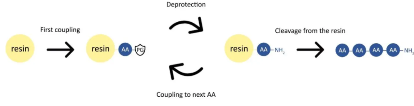
Solid phase peptide synthesis combines the ease of in-solution organic chemistry with simple filtration washing steps. This is achieved by coupling the first amino acid of the desired sequence via its carboxyl group to an insoluble, yet swellable, polymer bead through a cleavable linker. After this initial step, the entire synthesis is based upon a repeated cycle of deprotecting the N-terminal amine group of the growing peptide chain attached to the insoluble resin, followed by performing the coupling reaction between the deprotected N-terminal amine and the carboxylic acid group of the next N-N-terminal protected amino acid in line, resulting in the desired peptide bond. This way, the peptide is built up from the C-terminus of the sequence to the N-C-terminus by repeating the deprotection-coupling cycle until the final amino acid is introduced. At this point, the peptide is ready to be removed from the polymer bead by cleaving off the linker and resin. A schematic overview of the general SPPS procedure can be found in Figure 18.
22 The key benefit of this approach to peptide synthesis is the unidirectional growth of the peptide chain, allowing easy sequential control. In addition, equimolarity between the added amino acids and the growing peptide chain is not required, allowing coupling reactions to be driven to full conversion by utilizing an excess of amino acid followed by extensive washing with dimethylformamide (DMF) and dichloromethane (DCM) of the polymer beads on a filter.
4.2.2 Polymer resin and linker
The choice of an adequate polymer resin to attach the peptides to is essential. Preferably, the polymer bead should exhibit a high degree of swelling in the solvent that will be used throughout the synthesis. When the polymer that contains the attached growing peptide chains is swollen properly, the required reagents for both the deprotection and coupling reactions can easily reach the N-terminal site, leading to higher efficiency of the reactions. In this thesis, polystyrene polymer beads were used containing 1% of the divinylbenzene crosslinker. Higher crosslinker concentration would cause a lower degree of swelling, yet some crosslinker is required to ensure mechanical rigidity of the polymer beads.
The first amino acid of the sequence (counting from the C-terminus) is attached to the resin using a pre-installed chemical moiety on the polymer bead, called the linker. A wide variety of linkers exist, allowing for tunable cleavage conditions and different C-terminal functionalities of the finalized peptide after cleavage. Some more exotic linker molecules can only be cleaved when activated in a prior step (safety catch linker) or can even be cleaved by enzymes or upon UV irradiation.
In this thesis, the 2-chlorotrityl chloride linker was used for the apelin peptides. This is an acid labile linker that results in a carboxylic acid functionality at the C-terminus after cleavage. For the opioid peptides, the Rink amide linker was used in order to obtain an amide functionality at the C-terminus after acidic cleavage (Figure 19). The resins were purchased with the linker molecule pre-attached. Coupling the first amino acid to the 2-chlorotrityl chloride resin is achieved through an SN1 reaction in alkaline conditions, while the Rink amide linker is bought
in a Fmoc protected form, which allows for the immediate start of the regular peptide synthesis (after initial Fmoc deprotection).
23
4.2.3 Fmoc strategy and protecting groups
As amino acids are intrinsically at least bifunctional, preventing uncontrolled polymerization resulting in a random polyamide is key to allow sequential control during the peptide synthesis. The way this is achieved is by utilizing protecting groups, which temporarily shield the amine functionality of the amino acids. The two most frequently used protecting groups for this purpose are the tert-butyloxycarbonyl (Boc) group, which is removed by addition of an acid (e.g. TFA) and the fluorenylmethoxycarbonyl (Fmoc), which is removed by addition of a base (e.g. piperidine).
On top of protecting the amine functionality of the amino acid, many sidechains of amino acids contain reactive moieties that can interfere in the peptide synthesis process. These sidechains also require protecting groups. As these protecting groups are only supposed to be removed at the very end of the peptide synthesis, they should withstand the conditions used for deprotecting the Boc or Fmoc group during the repeated deprotection-coupling cycles. For this reason, orthogonal chemistries are used. In case of the acid-labile Boc, a common sidechain protecting group is benzyl, which requires higher acidity (e.g. HF) to be removed and thus withstands the acidic conditions used to remove the Boc. In the case of the base-labile
Figure 20: The two main protection strategies in SPPS: Boc/Bn (left) and Fmoc/tert-Bu (right). Figure 19: 2-Chlorotrityl chloride linker (left) and Rink amide linker (right).
24 Fmoc, tert-butyl groups are often used, which are removed by acid treatment (e.g. trifluoroacetic acid (TFA)) (Figure 20).
The more pronounced chemical orthogonality and more efficient cleavage reaction of the Fmoc/tert-butyl strategy (base/acid) compared to the Boc/benzyl strategy (acid/stronger acid) is the reason why, during this thesis, the Fmoc/tert-butyl strategy was utilized for the synthesis of all peptides. All amino acids used in the synthesis were purchased in a Fmoc protected, sidechain protected form.
During the synthesis, the base-labile Fmoc protecting groups were removed by addition of piperidine, the reaction mechanism for deprotection is shown in Figure 21. The sidechain protecting groups used during this thesis (Boc, tBu, Trt and Pbf) are removed by the same acidic conditions required to cleave the peptides from the resin. Cleavage and deprotection of the sidechains is performed simultaneously using a cleavage cocktail containing mainly TFA, more info on cleavage cocktails can be found in section 4.2.5.
Coupling reagents (HBTU + DIPEA)
In order to form the desired amide bond between the N-terminal amine of the growing peptide chain and the carboxylic acid of the added Fmoc-protected amino acid, activation of the carboxylic acid is required to ensure an efficient coupling reaction. The coupling reaction can be categorized as a nucleophilic attack of the N-terminal amine on an activated carboxylic acid, which results in the formation of an amide bond. If the carboxylic acid would not be activated, an ordinary acid-base reaction would occur, yielding an undesired ionic bond between the protonated amine and the deprotonated carboxylic acid. For the activation of the carboxylic acid, so-called coupling reagents are commonly used.
25 Many coupling reagents were developed over the past thirteen decades of peptide synthesis research[68]. For this thesis hexafluorophosphate benzotriazole tetramethyl uronium (HBTU)
was used as this coupling reagent benefits from a low degree of racemization of the activated amino acids. It fulfills its activating role after the carboxylic acid is being deprotonated by a non-nucleophilic base, for which di-isopropylethylamine (DIPEA) was used. After this deprotonation, it forms an activated ester which on its turn can efficiently react with the N-terminal amine to form the amide bond. After the coupling, any excess of coupling reagents and all side-products are efficiently washed away. The reaction mechanism of an HBTU assisted coupling reaction can be found in Figure 22.
4.2.4 TNBS test
When synthesizing a peptide manually, it is essential to be able to evaluate the conversion of the coupling steps throughout the entire synthesis. When a next coupling is performed before full conversion of the previous one, two different peptide chains are obtained: one with the intended sequence and one with a deletion of an amino acid. As these peptides only differ one residue, separation is nearly impossible.
For assessing the conversion of the amide coupling reactions, the trinitrobenzene sulphonic acid (TNBS) test was utilized, which is a qualitative color test where TNBS acts as coloring
26 agent. When free primary amines are present in the solution (or on the swollen beads in this case), they will react with the TNBS through nucleophilic aromatic substitution (Figure 23). This reaction product has a distinct bright red color. In practice, a very small amount of polymer beads is used to perform the test. When the coupling has not reached full conversion, the beads turn bright red. If the beads remain pale yellow, the coupling is finished. When red beads are observed, the previous coupling step and the subsequent TNBS-test are typically repeated, until no more red colored beads are observed.
4.2.5 Cleavage cocktail optimization
Cleavage of the peptide from both the 2-chlorotrityl chloride and the Rink amide linkers is generally performed with cleavage cocktails containing 90% or more TFA with addition of certain scavengers. In the Fmoc/tert-butyl strategy used in this thesis, cleavage of the peptide happens simultaneously with the deprotection of the sidechain protecting groups. This deprotection generates stabilized carbocation compounds, which can subsequently react with electron rich sidechains leading to undesired byproducts. To mitigate this problem, scavenger molecules are typically used in the TFA-cleavage cocktails, which irreversibly capture these stabilized carbocations.
A typical cleavage cocktail for the linkers used in this thesis is a mixture of 95% TFA, 2.5% triisopropylsilane (TIS)and 2.5% H2O, the last two being the scavengers. However, the furan
residue contained in the peptide is known to degrade under harsh acidic conditions, most notably when the furan is close to one of the ends of the peptide chain. Introduction of the furan in the middle part of the peptide protects the furan to some extent from the degrading conditions. As for the apelin peptides, the furan is located at the C-terminus of the peptides, previously published work on this specific degradation issue from within the OBCR group was consulted[69].
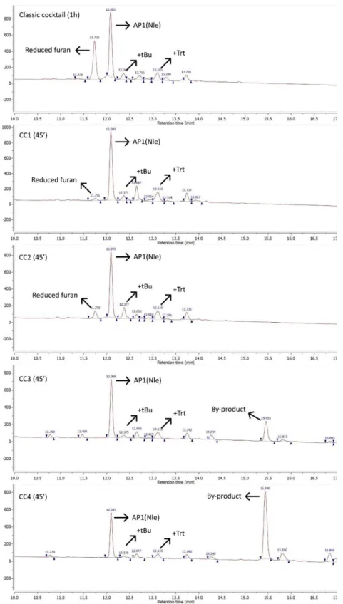
27 In order to optimize the yield of the cleavage reactions, it was decided to perform test cleavages for comparing the classic 95% TFA, 2.5% TIS and 2.5% H2O cocktail to four different
cleavage cocktails (Table 5), which contained two commonly used scavengers that were not examined before in the published paper. In practice, a small number of beads from the AP1(Nle) synthesis was subjected to the different cleavage cocktails for 45 minutes and analyzed via reversed phase high performance liquid chromatography (RP-HPLC) and liquid chromatography followed by mass spectrometry (LC-MS) for determination of the yield and identification of by-products respectively. Not all by-products could be identified, yet the most prevalent by-products could be identified as the peptide with a reduced furan moiety, or peptide-protecting group adducts (+tBu, +Trt). The obtained HPLC and LC-MS spectra of all test cleavages can be found in the experimental data section (section 10.1.3.12-10.1.3.18).
Table 5: Tested cleavage cocktails
Cleavage cocktail Concentrations
Classic 95% TFA, 2.5% TIS, 2.5% H2O
CC1 95% TFA, 2.5% Thioanisole, 2.5% TIS
CC2 92.5% TFA, 2.5% Thioanisole, 2.5% TIS, 2.5% H2O
CC3 95% TFA, 2.5% thioanisole, 2.5% m-cresol
28 To summarize, both CC1 and CC3 initially showed promising results, resulting in higher yields compared to CC2, CC4 and the classic cocktail. CC1 allowed to obtain the highest yield of the two, as can be seen in Figure 24. CC3 led to a slightly lower yield, but the resulting crude mixture did not contain the reduced furan by-product which is located in close proximity
Figure 24: Close-up of most relevant test cleavage HPLC data, identification based on LC-MS data, more details can be found in section 10.1.3.
29 (HPLC-wise) to the main product in the RP-HPLC chromatogram, which is typically difficult to remove during purification. In CC3 and CC4, an unidentified by-product was formed (peak at minute 15.5), which is not present after cleavage with the classic cocktail, CC1 or CC2.
A second test cleavage was performed with a longer cleavage time of one hour and 45 minutes in order to determine more clearly the winner between CC1 and CC3. CC1 came out on top, as upon CC3 treatment, an increased amount of an unidentified by-product was formed after this longer cleavage time, as can be seen in Figure 25. It has to be noted that these spectra have a slight shift in elution time (x-axis) compared to the spectra from Figure 24. This is caused by usage of a larger loop in the HPLC system, resulting in a larger dead volume and therefore later peak elution. In addition, the CC3.2 sample was more diluted than the CC1.2 sample, as can be deduced from the y-axis displaying UV absorption. Nevertheless, relative peak size does not depend on sample concentration and therefore the conclusions drawn are valid.
In conclusion, CC1, consisting of 95% TFA, 2.5% thioanisole and 2.5% TIS, yielded the best results. Therefore, it was used for all cleavages with exclusion of AP1(Met) and AP2(Met), as these were cleaved before this cleavage cocktail optimization was performed.
30
4.2.6 Work-up and purification
The last stage in obtaining high purity peptides consists of the work-up and purification process after cleavage. Work-up of all peptides happened in a very traditional manner, namely using methyl-tert-butylether (MTBE) precipitation. The concept of this process is simple, after cleavage, the peptide is present in the cleavage cocktail solution, together with the TFA, scavengers and cleaved protecting groups. To separate this mixture, the TFA is evaporated under a flux of nitrogen, followed by addition of cold MTBE to the crude mixture. The peptide precipitates, while the residual TFA, scavengers and cleaved protecting groups remain dissolved in the MTBE. After centrifugation at cold temperatures, the supernatant is discarded and new MTBE is added for further washing. This is repeated three times.
Final purification is performed utilizing a preparative reversed phase HPLC. In Figure 26, the purity of the crude after MTBE work-up is compared to the purity after RP-HPLC for SAP1(Nle). For all furan-modified peptides, a purity exceeding 95% was achieved.
Figure 26: RP-HPLC chromatogram of crude SAP1(Nle) mixture after MTBE work-up (top), RP-HPLC chromatogram of SAP1(Nle) after purification (bottom).
31
4.2.7 SPPS applied to furan-modified apelin peptides
As mentioned before, a C-terminal carboxylic acid functionality was desired for the furan-modified apelin peptides. For this reason, the first amino acid was linked to the 2-chlorotrityl chloride resin, an acid-labile resin for solid phase immobilization of carboxylic acids. To do so, a simple SN1 reaction was performed with 1 equivalent of the desired amino acid using DIPEA
as a base, before capping the resin with methanol to block remaining reactive sites on the resin that have not reacted with the first amino acid. After coupling of the first amino acid to the resin through the linker, the remaining part of the apelin peptides was synthesized using an automated peptide synthesizer, performing the deprotection-coupling cycles fully autonomous, using 4 equivalents of amino acid and 4 equivalents of HBTU. After completion of the sequence, the peptides were manually coupled to biotin-PEG4-propionic acid using standard coupling conditions. The only steps remaining at this point are the cleavage (which simultaneously leads to deprotection of the sidechains), followed by work-up and purification. Some apelin peptides were difficult to purify, fractions of the first purification where sufficient
32 separation was not achieved, were repurified after lyophilization. In Figure 27, a schematic overview of the AP2(Met) synthesis procedure is given as an example.
4.2.8 SPPS applied to furan-modified opioid peptides
For the opioid peptides, a C-terminal amide functionality was desired. In order to obtain this, a polystyrene resin with the Rink amide linker was utilized. This linker contains a Fmoc protected amine functionality which is used for attaching the first amino acid after Fmoc deprotection using piperidine. This first coupling is performed just like all other peptide couplings in the synthesis. As mentioned in the part on sequence design, the first amino acid starting from the C-terminus is the unnatural Glu(biotinyl-PEG). For all furan-modified opioid peptides, the entire peptide was synthesized manually, as expensive unnatural amino acids are incorporated in the sequence. Cleavage, work-up and purification was performed in an identical way to the apelin peptide syntheses. In Figure 28, a schematic overview of the synthesis of OP2 is given as an example.
33
4.3 Results & discussion
Synthesis of all envisaged furan-modified peptides was successful. The commercially available Fmoc protected 3-(2-furyl)alanine was included seamlessly in regular SPPS procedures, both in automated peptide synthesis and in manual synthesis. The only challenging hurdle to overcome was the acid-induced furan degradation, which occurred in the cleavage step. Cleavage cocktail optimization allowed partial mitigation of this issue. Yields of all synthesized peptides can be found in Table 6.
Of the nine apelin peptides, seven were obtained with high purity and moderate yields ranging from 2.9% to 28.1% with an average yield of 14.5%. When the furan is inserted at the first position counting from the C-terminus, the yield is substantially lower (indicated in red in Table 6) Two of the envisaged apelin peptide sequences have not been finished in view of the COVID-19 outbreak.
Of the six opioid peptides, four were obtained with high purity and moderate yields ranging from 13.8% to 45.6% with an average yield of 34.6%. These yields are remarkably higher compared to the apelin peptides. This can be attributed to the smaller number of amino acids in the opioid sequences and thus less reactions required in the synthesis. A second explanation for the higher yield is the fact that less side-reactions occurred with the furan during cleavage, as it is protected by the peptide backbone (with the apelin peptides, the furan was closer to the end of the peptide). Two of the envisaged opioid peptide sequences have not been finished in view of the COVID-19 outbreak
Furan-modified apelin peptides Furan-modified opioid peptides
Code
Yield (%)
Yield
(mg) Code Yield (%) Yield (mg) AP1(Met): 2.9 5.9 OP1: unfinished
AP2(Met): 14.4 29.9 OP2: unfinished
AP1(Nle): 7.6 14.5 OP3: 42.8 42.3 AP2(Nle): 28.1 55.1 OP4: 45.6 37.5 AP3: 15.4 30.1 OP5: 36.3 26.2 SAP1(Nle): 11.8 22.7 OP6: 13.8 10.7 SAP2(Nle): unfinished SAP3(Nle): unfinished Native AP(Nle): 21.1 40.6
34
5 Crosslinking experiments
The crosslinking experiments encompass two distinct phases. The first phase covers the treatment of the living cells with the synthesized furan-modified peptides at different concentrations during different incubation periods. During this phase, the endogenously activated crosslinking event takes place. The second phase consists of a Western blot experiment in order to examine if crosslinking of the furan-modified peptide to the GPCRs has occurred. In order to perform the Western blot, the cells need to be lysed, which is achieved by addition of a lysis buffer to the cells. Western blotting covers a series of sample processing steps, starting with gel electrophoresis to separate the different cell lysate components by size. This is followed by a blotting step (transfer of the proteins from the gel to a membrane) and finally the separated compounds are visualized by means of dual immunodetection. Due to the COVID-19 outbreak, only a single crosslinking experiment using three different apelin peptides and subsequent Western blotting was performed by me. Results of a crosslinking experiment performed by my thesis supervisor, Laia Miret Casals, with 4 different opioid peptides are also included. Many planned experiments were cancelled due to the COVID-19 lockdown measures.