Mutagenicity of chemicals in genetically modified animals | RIVM
47
0
0
Hele tekst
(2) TNO rapport V99.1097 / RIVM rapport 650210 002. Page 2 van 47. Abstract The strategy for assessing human health risks of chemicals consists of a large number of tests in different research disciplines. Tests include acute and chronic toxicity, genotoxicity, reproduction toxicity and carcinogenicity. Genotoxic properties of chemicals are assessed in short-term in vitro and in vivo genotoxicity tests. There are two main endpoints for genotoxicity: gene mutations and chromosome aberrations. Under in vitro conditions, there are sufficient assays for both endpoints. Testing under in vivo conditions is essential to confirm in vitro data since it is impossible to mimic, in a petri dish, all the complex factors determining whether a chemical will induce mutations in a specific tissue in animals in vivo. Moreover, in a regulatory context, a relevant negative in vivo result from an adequately performed test overrules positive in vitro results. There are appropriate assays in existence to investigate in vivo chromosome aberration; however, problems occur when a compound induces gene mutations in vitro. In the absence of reliable in vivo gene mutation assays, a justified assessment of the genotoxic potential of chemicals may be hampered. Introduced in this report, based open literature data up to August 2000, are several promising new in vivo gene mutation assays. The report is not restricted to assays with the commercially available transgenic models; all assays whether using transgenes or endogenous genes as reporter genes – are incorporated. In reviewing the current state of the art in evaluating these assays, the advantages and the disadvantages of the assays are discussed. This is to determine the feasibility of the routine use of these new in vivo gene mutation tests for health risk estimation. Gene mutation assays with transgenic animals have already been used on a small scale for legislation of chemicals. However, to allow the routine use of these assays for regulatory purposes, they will have to be validated further and an official OECD guideline prepared..
(3) TNO rapport V99.1097 / RIVM rapport 650210 002. Page 3 van 47. Mailing list 1 2 3 4 5 6-12 13 14-20 21-25 26 27 28 29 30 31 32 33 34 35 36 37 38 39 40 41 42 43 44 45 46 47 48 49 50 51 52 53 54 55 56 57 58 59 60. Prof. Dr. H.J. Schneider Dr. Ir. B.C.J. Zoeteman Dr. J.J. Ende Dr.Ir. M.M.H. Mols Prof. Dr. P. Peters Dr. Ir. P.C. Bragt. Directeur-generaal Volksgezondheid Plv. Directeur-generaal Milieubeheer VWS, Hoofd Consumenten-Veiligheid en Omgevingsrisico's VWS, Directeur Keuringsdienst van Waren VWS, Keuringsdienst van Waren, Hoofdinspecteur Food VWS, Keuringsdienst van Waren, Hoofdinspecteur Nonfood Drs. H. Verburg, VWS, Keuringsdienst van Waren, Veterinair Hoofdinspecteur Dr. H. Roelfzema VWS, Consumenten-Veiligheid en Omgevingsrisico's Dr. C.L. Maas SZW, A&O Onderzoek Drs. A.A. Vijlbrief SZW, ARBO, Arbeidsmilieu Prof. Dr. J.J. Sixma Voorzitter Gezondheidsraad Dr. Ir. E. van Vliet Gezondheidsraad Depot van Nederlandse Publikaties en Nederlandse Bibliografie Drs. H.J. Jeuring VWS, Keuringsdienst van Waren Dr. D.G. Groothuis VWS, Keuringsdienst van Waren Dr. Ir. G. Kleter VWS, Keuringsdienst van Waren Dr. J. Nieuwenhuijs VWS, Keuringsdienst van Waren Dr. Ir. J.M. de Stoppelaar VWS, Directie Gezondheidsbeleid, afd. VVB Dr. D.W.G. Jung VROM-DGM, SAS Dr. J.A. van Zorge VROM-DGM, SAS Dr. F.X.R. van Leeuwen WHO-European Center for Environment and Health, Bilthoven, The Netherlands Dr. R.A. Baan Unit of Carcinogen Identification and Evaluation, IARC, Lyon, France Dr. H. Koeter OECD, France Dr. G. Douglas Health Canada, Ottawa, Canada Dr. D. Casciano FDA/DGRT/HFT, Jefferson, USA Dr. A. Smith HSE Toxicology Unit, UK Dr. S. Madle GBVV, Germany Dr. S. Munn European Chemicals Bureau, Ispra, Italy Dr. K. Schneider FoBiG GmbH, Freiburg, Germany Dr. I. Mangelsdorf Fraunhofer Institute for Toxicology, Hannover, Germany Prof. Dr. E. Dybing National Institute of Public Health, Oslo, Norway Prof. Dr. T. Sanner Institute for Cancer Research, Oslo, Norway Prof. Dr. J. Parry University of Wales, Swansea, UK Dr. J.C. Larsen Danish Veterinary and Food Administration, Institute of Food safety and Toxicology, Soborg, Denmark Dr. S. Flodstrom National Chemicals Directorate, Solna, Sweden Dr. B. Meek Environmental Health Directorate, Health Canada, Ottawa, Canada Dr. P. Muller Standard Development Branch, Ontario Ministry of Environment and Energy, Toronto, Canada Dr. J. Vijg University of Texas, USA Dr. D. Anderson TNO-BIBRA, UK Dr. H.G. Keizer Solvay Duphar BV Ing. E.J. van de Waart Notox, Safety and Environmental Research BV Prof. Dr. Elzinga Directeur Volksgezondheid RIVM Dr. Ir. G. de Mik Directeur sector 3/4 RIVM SBD/Voorlichting en Public Relations.
(4) TNO rapport V99.1097 / RIVM rapport 650210 002. 61 62 63 64 65-69 70 71 72 73 74 75 76 77 78 79 80 81 82 83 84 85 86 87 88 89 90 91 92-101 102-106 107 108 109-135. Dr. N.J. Snoeij Dr. E.D. Kroese. Page 4 van 47. TNO Voeding, Hoofd Divisie Toxicologie en Voeding TNO Voeding, Hoofd afdeling Toxicologische Risicobeoordeling Dr. J.J. van Hemmen Hoofd ACCA TNO Dr. B. van Ommen TNO Nutrition and Food Research Secretariaat, afdeling Toxicologische Risicocobeoordeling Archief TNO Voeding, rapportenbeheer Dr. A. Opperhuizen Hoofd LEO Dr. C.F. van Kreijl LEO Dr. H. van Steeg LEO Dr. H.J. van Kranen LEO Dr. A. de Vries LEO Prof. Dr. J.G. Vos Hoofd LPI Dr. R.B. Beems LPI Dr. P.W. Wester LPI Dr. E. Lebret Hoofd LBM Dr. Ir. H.J.G.M. Derks Hoofd LGO Dr. W.H. Könemann Hoofd CSR Prof. Dr. C.J. van Leeuwen CSR Drs. T.G. Vermeire CSR Drs. A.G.A.C. Knaap CSR Dr. M. van Raay CSR Dr. J.G.M. van Engelen CSR Dr. W.C. Mennes CSR Dr. G.J.A. Speijers CSR Dr. Ir. J.F. van Sonderen Hoofd LGM Dr. J.-W. van der Laan LGM Prof. Dr. G.R. Mohn Dr. M.I. Willems TNO Voeding, afdeling Toxicologische Risicobeoordeling Dr. J. van Benthem LEO Bureau Rapporten registratie Bibliotheek RIVM Reserve exemplaren t.b.v Bureau Rapportenbeheer.
(5) TNO rapport V99.1097 / RIVM rapport 650210 002. Page 5 van 47. Contents Summary ................................................................................................................................................................... 6 Samenvatting ............................................................................................................................................................ 7 1. Introduction ...................................................................................................................................................... 8. 2. Overview of novel gene mutation tests in vivo.............................................................................................. 11. 3. 2.1. Models using endogenous genes ........................................................................................................ 11. 2.1.1. Hypoxanthine-guanine phosphoribosyltransferase (hprt) mouse model ............................................ 11. 2.1.2. Dlb-1 specific locus test..................................................................................................................... 12. 2.1.3. Adenine phosphoribosyl transferase (aprt) mouse model .................................................................. 12. 2.1.4. Thymidine kinase (tk) mouse model .................................................................................................. 12. 2.2. Models using transgenes .................................................................................................................... 13. 2.2.1. Bacteriophage vector mice................................................................................................................. 13. 2.2.2. Plasmid vector mice ........................................................................................................................... 19. Validation issues ............................................................................................................................................. 22 3.1. Reproducibility within and between laboratories, methodological aspects........................................ 23. 3.1.1. Spontaneous mutation frequency, influence of age and tissue specificity.......................................... 23. 3.1.2. Induced mutations, influence of manifestation time and treatment protocol...................................... 24. 3.1.3. Pooling of tissues and organs and clonal expansion of mutants......................................................... 24. 3.1.4. Evaluation of test results and the role of statistical analysis............................................................... 25. 3.2. Does the system measure what it is intended to measure? ................................................................. 25. 3.2.1. Are the spontaneous or induced mutants from prokaryotic or murine origin? ................................... 25. 3.2.2. Comparability of transgenes and endogenous mammalian genes as mutational targets ..................... 26. 3.2.3. Comparability of transgenic and nontransgenic mice in terms of toxicokinetics and dynamics......... 30. 3.3. Predictivity of the novel gene mutation assays for carcinogenicity and for heritable effects ............. 33. 3.3.1. Validation assessment: mutagen/carcinogen correlations .................................................................. 33. 3.3.2. Heritable effects in rodents and results obtained in lacI/lacZ mutagenicity tests............................... 35. 4. Discussion ........................................................................................................................................................ 36. 5. Novel gene mutation assays: implications for regulatory bodies ............................................................... 40. 6. References ....................................................................................................................................................... 42.
(6) TNO rapport V99.1097 / RIVM rapport 650210 002. Page 6 van 47. Summary The assessment of the potential genotoxicity of chemicals in vivo plays an important role both for verification and confirmation of intrinsic mutagenicity and for establishing the mode of action of chemical carcinogens. Genotoxic endpoints measured in vitro include gene mutations and chromosome aberrations, but until recently reliable in vivo tests for gene mutations were lacking. The present paper gives an overview of the recently developed novel gene mutation tests for assessment of potential mutagenicity in animals in vivo: 1) assays using endogenous genes as reporter genes, e.g., hprt, aprt, tk or Dlb-1 and 2) assays using transgenic animals, i.e., animals that possess an exogenous bacterial reporter gene, a so-called transgene, e.g., lacZ or lacI. Only a few endogenous genes and tissues are suitable for measuring mutations in animals in vivo. In contrast, the exogenous reporter genes, which are transmitted via the germ cells, can be measured in every tissue of the transgenic rodents. The transgenic animal assays include bacteriophage- and plasmid-vector based models. The bacteriophage-based models can detect point mutations, small deletions, and insertions, while the plasmid-based models also enable detection of large deletions. The discussion on validation is restricted mainly to the commercially available MutaTMMouse (lacZ) and Big Blue® (lacI) models, since only for these two systems there are sufficient data for validation. Comparison of mutation induction in endogenous and exogenous genes simultaneously in the same animals and in the same tissues showed that, despite differences in mutation properties of the various model mutagens used, the responses of the exogenous loci (lacI, lacZ transgene) and the endogenous loci (Dlb-1, hprt) were generally comparable upon acute dosing, when expressed in terms of absolute increases in mutation frequency. However, when expressed as relative increase over controls, the tests with transgenic animals are generally less sensitive than those with endogenous genes as a consequence of a lower background mutation frequency in endogenous as compared to exogenous reporter genes. Several studies indicate that transgenic mice respond as expected for wild type mice with a similar genetic background, e.g., in terms of toxicokinetics and toxicodynamics. Moreover, it has been shown that the mutations in the lacl or lacZ genes are neutral, i.e., that the mutations do not give selective advantage or disadvantage. In a recent validation study on the predictive value of the novel in vivo gene mutation tests for carcinogenicity, which included a total of 33 model compounds, it was shown that the Big Blue® (n = 21) and MutaTMMouse (n = 12) systems perform well with regard to positive predictivity, specificity, sensitivity, and overall accuracy. The negative predictivity for carcinogenicity was, however, very low (33-50%). It is not clear to what extent the results of this validation study are influenced by the selection of the chemicals and the limited set-up of this study. Moreover, the study did not assess questions such as target organ specificity for mutagenicity in the transgenic animals with respect to target organ specificity for carcinogenicity or on the possible relationship between genotoxicity and carcinogenicity profile in relation to the presumed mode of action of the carcinogen in question. In conclusion, the results obtained until now indicate that the novel in vivo gene mutation assays are suitable for mechanistic and fundamental studies on the occurrence of (point) mutations. Although these novel assays are not yet fully validated and a test guideline is not yet available, the bacteriophage λ-based lacI and lacZ models may already be used for legislation. However, the assays should not be used on a routine base and only carried out with a clear purpose keeping in mind the uncertain negative predictivity for carcinogenicity..
(7) TNO rapport V99.1097 / RIVM rapport 650210 002. Page 7 van 47. Samenvatting Het onderzoek naar mogelijke mutagene eigenschappen van chemische stoffen in vivo speelt een belangrijke rol zowel voor verificatie en/of bevestiging van intrinsieke mutagene eigenschappen als voor het vaststellen van het werkingsmechanisme van kanker verwekkende stoffen. De genotoxische eindpunten die in vitro worden gemeten zijn genmutaties en chromosoomafwijkingen. Tot voor kort waren er geen betrouwbare in vivo testen op genmutaties beschikbaar. Dit rapport geeft een overzicht van recent ontwikkelde in vivo genmutatietesten. Er wordt onderscheid gemaakt tussen: 1) toetsen met normale wild type muizen en een endogeen gen als reporter gen, bijvoorbeeld, hprt, aprt, tk of Dlb-1 en 2) toetsen die gebruik maken van transgene dieren die over een exogeen, bacterieel reporter gen, een transgen, beschikken, zoals bijvoorbeeld lacZ of lacI. Slechts een paar endogene genen en weefsels zijn geschikt voor het meten van genmutaties in vivo. Exogene reporter genen, die via de geslachtscellen worden overgeërfd, kunnen daarentegen worden onderzocht in ieder willekeurig weefsel van transgene dieren. Transgene diermodellen zijn gebaseerd op het gebruik van bacteriofaag dan wel plasmide vectoren. De laatste modellen kunnen puntmutaties, kleine deleties en inserties aantonen terwijl modellen gebaseerd op een plasmide vector daarnaast ook grote deleties kunnen meten. De validatie discussie beperkt zich hoofdzakelijk tot de commercieel verkrijgbare MutaTMMouse (lacZ) en Big Blue® (lacI) modellen. Bepaling van de mutatieinductie in endogene en exogene genen in hetzelfde dier, toonde aan dat ondanks verschillen in mutagene eigenschappen van de gebruikte modelstoffen, bij acute blootstelling de resultaten verkregen met exogene (lacI, lacZ) en endogene loci (Dlb-1 en hprt) in termen van absolute toename in mutatie frequenties vergelijkbaar waren. Echter de toets met transgene muizen blijkt in het algemeen minder gevoelig als de resultaten worden uitgedrukt als een relatieve toename ten opzichte van onbehandelde dieren. De oorzaak is waarschijnlijk de lagere achtergrond mutatie frequentie in endogene dan in exogene reporter genen. In termen van toxicokinetiek en toxicodynamiek hebben verschillende studies aangetoond dat transgene muizen niet verschillen van wild type muizen met een identieke genetische achtergrond. Daarnaast is aangetoond dat de mutaties in de lacI en lacZ genen neutraal zijn; mutaties leiden niet tot een selectief voor- of nadeel. In een recente validatie studie naar de voorspellende waarde van de nieuwe in vivo genmutatietesten voor carcinogeniteit voldeden de Big Blue® (n = 21) en MutaTMMouse (n = 12) modellen goed wat betreft de positieve voorspelbaarheid, specificiteit, gevoeligheid en nauwkeurigheid. De negatieve voorspelbaarheid voor carcinogeniteit was echter erg laag (33-50%). Het is onduidelijk in hoeverre de resultaten zijn beïnvloed door de keuze van de modelstoffen en de beperkte opzet van de studie. Bovendien werd er geen rekening gehouden met de doelorgaan specificiteit voor carcinogeniteit van de verschillende modelstoffen noch met de mogelijke relatie tussen genotoxiciteit en carcinogeniteit wat betreft het veronderstelde werkingsmechanisme van de onderzochte modelstof. Er kan worden geconcludeerd dat de resultaten die tot dusver met de nieuwe in vivo genmutatietesten zijn verkregen aantonen dat deze toetsen geschikt zijn voor mechanistisch en fundamenteel onderzoek naar het voorkomen van mutaties. Hoewel deze nieuwe testsystemen nog niet volledig zijn gevalideerd en er nog geen OECD-richtlijn beschikbaar is, kunnen deze nieuwe toetsen reeds beperkt gebruikt worden voor beleidsdoeleinden. Vanwege de slechte negatieve voorspelbaarheid voor kanker, moeten ze echter nog niet routinematig maar alleen met een specifiek doel gebruikt worden..
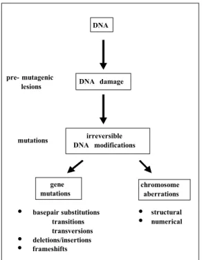
(8) TNO rapport V99.1097 / RIVM rapport 650210 002. 1. Page 8 van 47. Introduction. Governments are daily confronted with regulation of new chemicals, re-evaluation of existing chemicals and, increasingly more, with risk estimation of putative genotoxic or carcinogenic compounds. The strategy for the assessment of human health risks of chemicals consists of a large number of tests on different research disciplines. The potential genotoxicity of chemicals is assessed in short-term in vitro and in vivo genotoxicity tests. For genotoxicity two main endpoints exist, gene mutations and chromosome aberrations (Fig. 1). Most compounds have a preference for one of both, although there are no chemicals that induce exclusively either gene mutations or chromosome aberrations. This implies that one has to test compounds on both endpoints. Moreover, genotoxicity testing should be performed not only in somatic but also in germ cells to assess the potential genetic risks. Unfortunately, a quantitative assay for genetic risk assessment is not available.. DNA. pre- mutagenic lesions. mutations. DNA damage. irreversible DNA modifications. gene mutations. • • •. basepair substitutions transitions transversions deletions/insertions frameshifts. chromosome aberrations. • •. structural numerical. Figure 1: Endpoints in genotoxicity gene mutations and chromosome aberrations. Under in vitro conditions, there are sufficient assays for both endpoints (Table 1). Gene mutation assays in bacteria (Ames test) and mammalian cells as well as assays for chromosome aberrations in mammalian cells are routinely performed. Next to these, there are different kinds of indicator tests such as tests for DNA damage and repair. However, the value of these latter tests is limited compared to those for mutagenicity, i.e., tests for gene mutations and chromosome aberrations. Testing under in vivo conditions is essential to confirm in vitro data since it is impossible to mimic in a petridish all complex factors that determine whether a chemical will induce mutations in a specific tissue in animals in vivo. In vivo tests take into account whole animal processes like absorption, tissue distribution, metabolism and excretion of the chemical and its metabolites, and repair of lesions. Moreover in a regulatory context a relevant negative in vivo result from an adequately performed test.
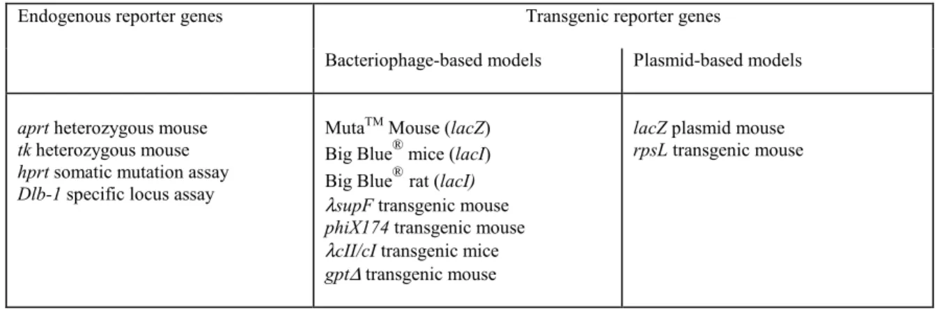
(9) TNO rapport V99.1097 / RIVM rapport 650210 002. Page 9 van 47. overrules positive in vitro results. It is obvious that in vivo confirmation makes only sense when in an animal model the endpoint is evaluated which showed positive results under in vitro conditions. For in vitro clastogenic chemicals, an in vivo chromosome aberration test or a micronucleus test allows evaluation of this endpoint. However, an important limitation of these assays is that testing is predominantly done in cells collected from peripheral blood or bone marrow. Exposure to a compound does not automatically mean that the reporter cells, i.e., bone marrow cells, peripheral blood cells, or their predecessors, have been exposed. Besides, these assays suffer from a tissue restriction and the value of a negative result obtained in a non-target tissue is questionable.. Table 1:. Available genotoxicty assays in mammalian cells or mammals. cytogenetic assay. gene mutation assay. DNA effects/indicator assays. hprt-test mouse lymphoma assay. sister chromatid exchange assay unscheduled DNA synthesis assay. mouse spot test. sister chromatid exchange assay unscheduled DNA synthesis assay. in vitro: chromosome aberration assay in vivo micronucleus test chromosome aberration assay. Problems occur when a compound induces gene mutations in vitro (Table 1). To date, some gene mutation tests in Drosophila melanogaster are available. However, the value of these tests for risk assessment in humans is at least questionable. In mammals the mouse spot test exists, which is a rather insensitive, animal consuming and expensive type of test. Often as a surrogate an in vivo test for unscheduled DNA synthesis (UDS test) is performed. The occurrence of unscheduled DNA synthesis does not directly point to the induction of DNA mutations, but merely to an increase in DNA repair as a consequence of DNA damage. In other words, an increase in unscheduled DNA synthesis points to the occurrence of DNA damage, which does not result in DNA mutations per se.. Table 2:. Promising novel in vivo gene mutation assays in mammals. Endogenous reporter genes. aprt heterozygous mouse tk heterozygous mouse hprt somatic mutation assay Dlb-1 specific locus assay. Transgenic reporter genes Bacteriophage-based models. Plasmid-based models. MutaTM Mouse (lacZ) ® Big Blue mice (lacI) ® Big Blue rat (lacI) λsupF transgenic mouse phiX174 transgenic mouse λcII/cI transgenic mice gpt∆ transgenic mouse. lacZ plasmid mouse rpsL transgenic mouse.
(10) TNO rapport V99.1097 / RIVM rapport 650210 002. Page 10 van 47. This lack of a well-validated in vivo gene mutation test often hampers a justified assessment of the genotoxic potential of chemicals. Recently there have been a number of promising new developments, which may solve the problem of this lack of in vivo gene mutation tests. These novel gene mutation tests for assessment of potential mutagenicity in animals in vivo are listed in Table 2. They include i) assays using endogenous genes as reporter genes, e.g., hprt, aprt, tk or Dlb-1 and ii) assays using transgenic animals, i.e., animals that possess an exogenous bacterial reporter gene, a so-called transgene, e.g., lacZ or lacI. On a small scale, gene mutation assays with these models have been already used. However, to allow the use of these models for regulatory purposes they have to be validated further and an official OECD guideline has to be prepared. In this report, the various models are introduced and the actual state of the art concerning the evaluation of the models is reviewed. Finally, the advantages and the disadvantages of the models will be discussed in order to determine the feasibility of the routine use of these new in vivo gene mutation tests for health risk estimation..
(11) TNO rapport V99.1097 / RIVM rapport 650210 002. 2. Page 11 van 47. Overview of novel gene mutation tests in vivo. The novel in vivo gene mutation tests all have in common the use of selectable reporter genes to determine the mutation frequency. Only few selectable reporter genes are endogenously available in mammalian cells, e.g. hprt, aprt, tk, or Dlb-1. Moreover, the endogenous reporter genes that are suitable for mutagenicity testing, demonstrate a certain tissue restriction; application of tests is, therefore, restricted to only a few specific target tissues. Next the mutation frequencies can be determined via selective growth on certain culture media in vitro. This selection procedure is hampered by the fact that the selective markers are only present twice; many copies of a reporter gene would dramatically improve the detection of gene mutations. Modern molecular biological techniques enabled the development of transgenic animals, which carry many copies of a reporter gene. These reporter genes are transmitted by the germ cells, and thus present manifold in all cells including the germ cells. Consequently, these novel gene mutation tests are divided into models using endogenous genes and those using transgenes.. 2.1. Models using endogenous genes. As already mentioned the models using endogenous reporter genes demonstrate a certain tissue restriction. Determination of the mutation frequency of Dlb-1 is restricted to the small intestine and eventually the colon; determination of the mutation frequency of hprt, aprt and tk to those tissues which express the reporter gene and which can be subcultured in vitro. Against the disadvantage of endogenous reporter genes that selection is hampered by the presence of only one selective marker stands the advantage that these models detect not only point mutations, frameshifts, small insertions and small deletions but also intragenic, large deletions and loss of heterozygosity (LOH; aprt and tk).. 2.1.1 Hypoxanthine-guanine phosphoribosyltransferase (hprt) mouse model The hprt gene is one of the few genes that are suitable as a reporter gene for mutation induction in animals and humans and that can be performed in un-modified species. The hprt gene, which has a coding region of 657 bp (Skopek et al., 1995), is located on the X chromosome and spans 32 kb in rodent and 46 kb in human cells. However, both male and female cells carry only a single active copy of the hprt gene. Hprt is a non-essential enzyme for cells in culture. Although hprt is an endogenous gene present in all tissues, mutant selection is predominantly performed in splenocytes (van Dam et al., 1992; Skopek et al., 1992; Tates et al., 1994) or (human) peripheral T lymphocytes. Mutants are selected by culture in the presence of 6-thioguanine, which is a substrate for the enzyme. It is converted into the corresponding monophosphate, which in turn is toxic to cells. Hprt mutants have lost this enzyme activity, are consequently resistant to the toxic 6-thioguanine and can grow in medium containing 6-thioguanine. Structural and theoretical considerations as well as experimental evidence indicate that the hprt gene is relatively deficient in recovering large genetic alterations (Dobrovolsky et al., 1999a) due its location on the hemizygous X-chromosome..
(12) TNO rapport V99.1097 / RIVM rapport 650210 002. Page 12 van 47. 2.1.2 Dlb-1 specific locus test An assay system which allows scoring of mutations in vivo in the small intestine (and possibly in the colon) of the mouse involves the Dlb-1 locus (Winton et al., 1990). Dlb-1 is a polymorphic genetic locus on mouse chromosome 11 with two alleles: Dlb-1b (most mouse strains) which determines expression of a binding site for the lectin Dolichos biflorus agglutinin (DBA) in mouse intestinal epithelium and Dlb-la (SWR mice) which determines expression in vascular endothelium. DBA binding is co-dominant. In heterozygote Dlb-1a/Dlb-1b mice, mutations affecting the single Dlb-1b allele in an intestinal stem cell are recognised by the histochemical detection of its clonal descendants with a peroxidase conjugate of Dolichos biflorus agglutinin. The mutated clones, in contrast to most of the epithelium, do not stain. Because the Dlb-1 gene has not yet been cloned, the molecular nature of the mutations cannot be determined in DNA sequences.. 2.1.3 Adenine phosphoribosyl transferase (aprt) mouse model The human aprt gene, which is involved in the nucleotide salvage pathway of DNA synthesis, is located on chromosome 16 and is 2.6 kilobases in length; the mouse aprt gene is located near the telomere at chromosome 8. It codes for a protein that converts adenine into AMP. In the C57Bl/6 aprt mouse model the gene was knocked out by homologous recombination in embryonic stem cells; a part of the promotor region as well as the ATG start codon were deleted (Engle et al., 1996; van Sloun et al., 1998). This location sensitises the model to proximal chromosomal events like mitotic recombination and translocations. Because of the recessive nature of aprt mutations, for genotoxicity testing heterozygous aprt mice are used. Mutants occur when the second allele is mutated or lost (LOH). Aprt mutants can be selected due to their resistance to the toxic purine analogues 8-azaadenine or 2,6-diaminopurine; only aprt-/- cells survive culture on medium containing these analogues. The aprt model detects small mutations, intragenic large deletions and LOH.. 2.1.4. Thymidine kinase (tk) mouse model. Dobrovolsky et al. (1999a) reported the development of a novel in vivo gene mutation assay in mice using the autosomal, recessive thymidine kinase (tk) gene, which participates like aprt in the nucleotide salvage pathway of DNA synthesis. In the C57Bl/6 tk mouse the gene was knocked out by homologous recombination in embryonic stem cells (Dobrovolsky et al., 1996). Due to the recessive nature of tk mutations, heterozygous tk mice have to be used for genotoxicity testing. Mutants occur when the second allele is mutated or lost (LOH). Culture in the presence of trifluorothimidine (TFT) or 5-bromo-2’-deoxyuridine (BrdU), which are substrate for the enzyme, results in mutant selection. Exclusively tk-/- cells survive culture on medium containing these analogues. The advantage of the tk model is its sensitivity for large deletions, large chromosomal alterations and LOH. A disadvantage of the tk model is the use of BrdU as selective agent. BrdU itself is a mutagen and it is therefore impossible to absolutely rule out the possibility that some BrdU resistant mutants were produced ex vivo due to exposure to BrdU..
(13) TNO rapport V99.1097 / RIVM rapport 650210 002. 2.2. Page 13 van 47. Models using transgenes. To date, transgenic mice and rats are already used in in vivo gene mutation assay. These animals have in common that they contain multiple copies of a transgene in a shuttle vector as reporter gene, which is transmitted by the germ cells, and thus present manifold in all cells including the germ cells. The key problem for using transgenic animals in gene mutation assays was the rescue of the integrated vector from the genome and the detection of gene mutations in vitro. Based on the shuttle vector used transgenic mice models for mutagenicity testing can be divided in two main approaches. The first using a transgene in a bacteriophage vector whereas in the second one the transgene is in a plasmid vector (Fig. 2).. 2.2.1 Bacteriophage vector mice These transgenic mice are developed by microinjection of a bacteriophage shuttle vector. Principally, the determination of the mutation frequency is identical in the various models. Mice, bearing the shuttle vector, are treated with a chemical and after a certain manifestation period in which the DNA damage is fixed into stable mutations, the mice are killed, the tissues dissected and the genomic DNA isolated (fig. 2). The next step is in vitro packaging into bacteriophages. The use of λ bacteriophages as a shuttle vector was first developed in mouse fibroblasts by Glazer et al. (1986) and was applied to transgenic mice by Gossen et al. (1989). Proteins in the packaging extract cleave the shuttle factor at the cos sites and package it in the phage heads. These phages are then used to infect E. coli deficient for the reporter gene to produce plaques. Mutants are quantified either by color selection or by selective growth on plates containing a medium composition on which non-mutated bacteria can not grow. An aliquot of the infected bacteria is plated on normal minimal plates for the measurement of packaging efficiency. The mutation frequency is calculated by dividing the total number of colored or resistant plaques for the tissue of the individual animal by the total number of plaques with rescued shuttle vectors from the same tissue or from the same DNA sample.. take tissues. Treat mice. isolate DNA. packaging efficiency mutant selection. rescue efficiency. Figure 2:. infection of E. coli. electroporate plasmids into E. coli. Transgenic mice in vivo gene mutation assays. package. ligate plasmids. isolate lacZ plasmids.
(14) TNO rapport V99.1097 / RIVM rapport 650210 002. Page 14 van 47. These transgenic mice models developed with bacteriophage shuttle vectors are suitable for the detection of point mutations, insertions and small deletions. The models do not allow detection of large deletions due to the fact that the cos-sites, at the end of the vector, together with a restrictive length of the vector are essential for excision and packaging into phage heads. The models sometimes suffer from a low packaging efficiency, probably due to the large size of the shuttle vector, which should be isolated intact and the rather high copy number per haploid genome (about 2 million bp per phage DNA at one locus). This requires large amounts of packaging extracts to collect a sufficiently high number of reporter genes. 2.2.1.1 lacI transgenic mouse model To date lacI models are commercially available; the Big Blue® mouse (B6C3F1) and Big Blue® rat (F344) from Stratagene. The lacI mouse model developed by Short and coworkers (Kohler et al., 1991) contains about 30-40 copies of the λ LIZα shuttle vector (Fig. 3) in a head to tail fashion at a single locus on chromosome 4 without detectable rearrangements. In the rat model (Dycaico et al., 1994) 15-20 copies are present per haploid genome (Gollapudi et al., 1998). The λLIZα shuttle vector is 45.6 kb long whereas the reporter gene lacI counts 1080 bp. The vector contains the entire lacIq gene and the αlacZ gene (Kohler et al., 1991). The lacI gene codes for a homotetrameric protein that binds to the lacO operator sequence, which negatively regulates lacZ expression. The αlacZ gene codes for the αamino portion of β galactosidase.. f1 lacZ. lacI. ampr. c1857. f1. OR. Pr Cro. cII. OP Q. Pre Figure 3:. The λLizα shuttle factor with the lacI and cII reporter gene (Stratagene: http://www.stratagene.com/cellbio/toxicology/big_blue_system.htm#liz [December 15, 2000]. After packaging the phage is absorbed to E. coli SCS-8 cells (lacZ∆M15). The colE1 and ampR genes allow replication and selection of the vector in E. coli. The bacteria are then seeded on a selection medium containing 5-bromo-4-chloro-3-indolyl β-D-galactopyranoside (X-gal). The lac I gene allows.
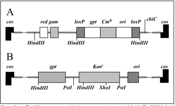
(15) TNO rapport V99.1097 / RIVM rapport 650210 002. Page 15 van 47. the lac repressor to bind to the lac operator which inhibits αlacZ expression and thus β-galactosidase activity resulting in white (colorless) plaques. If a mutation in lacI does occur the lac repressor protein is inactive or unable to bind the lac operator and transcription of the αlacZ gene will occur. The resulting functional β-galactosidase cleaves X-gal thus generating blue plaques. The ratio between blue and white plaques is a measure of the mutagenicity.. 2.2.1.2 lacZ transgenic mouse model The lacZ mouse model (MutaTMMouse) was developed by microinjection of the λgt10-lacZ shuttle vector (fig. 4) in fertilized CD2 oocytes of (BALB/c x DBA/2)CD2 F1 mice (Gossen et al, 1989). In contrast to the lacI model this shuttle vector contains the entire lacZ gene as reporter gene. The vector is about 47 kb long whereas the lacZ gene consists of about 3100 bp. The lacZ mouse model contains about 80 copies of the shuttle vector in a head to tail fashion at chromosome 3. After in vitro packaging the bacteriophage is preabsorbed into E. coli C (lacZ-) cells. The bacteria are seeded on a medium containing X-gal. Plaques containing a normal lacZ are β-galactosidase active and are blue, whereas plaques containing mutated lacZ will be white (colourless). In this case the ratio between colourless and blue plaques is a measure of the mutagenicity.. cos. Figure 4:. lacZ. cos. The λgt10-lacZ shuttle vector (Mirsalis et al., 1995). A selectable system was described by Gossen and Vijg (1993). In this system E. coli lac-, galE- is used for phage infection. To determine the recovery the phages are plated on medium containing Xgal; for mutation selection phenyl β-D-galactoside (P-gal) is used. Since β-galactosidase encoded by the lacZ gene converts lactose into galactose, normal lacZ phages when plated on E. coli lac-, galE- in the presence of lactose or lactose derivatives like P-gal will die. They are unable to metabolize the highly toxic UDP galactose, the product of galactose, into UDP glucose. LacZ mutated phages will survive since they do not form UDP-galactose.. 2.2.1.3 gpt delta transgenic mouse model A distinct feature of the gpt-delta transgenic mouse model by Nohmi et al. (1996) is the incorporation of two different positive selection methods in the transgene: the gpt gene of E. coli for point mutations and/or short deletions and spi- selection for (larger) deletions (1 – 10000 bp (Horiguchi et al., 1999). The gpt-delta C57Bl/6 mice were obtained after microinjection of C57Bl/6-oocytes obtained after superovulation which resulted in about 80 copies of the vector per chromosome 17. The injected λEG10 shuttle vector (Fig. 5A) is about 48 kb long and composed of the λ2001 vector and a linearilised plasmid. The λ2001 vector carries the red and gam genes together with a ΧC mutation involved in spi- selection. The plasmid possesses the gpt gene of E. coli and two direct repeat sequences of loxP which are recognition sequences for Cre recombinase. The coding region of gpt is 456 basepairs, which is convenient for the rapid identification of gene mutations by sequencing..
(16) TNO rapport V99.1097 / RIVM rapport 650210 002. Page 16 van 47. For point mutation or small deletion selection the E. coli strain YG6020 (gpt-) is used which expresses Cre recombinase. Thus when the phage is introduced into strain YG6020 the linearized plasmid is excised, circularized and propagated as a multicopy-number plasmid pYG142 carrying the gpt gene. Detection of gpt mutations was performed by culture on medium containing 6-thioguanine (6-TG), the packaging efficiency was determined by culture on medium without 6-TG. The ratio between the number of mutants and the packaging efficiency is the mutation frequency for point mutations and/or small deletions. Spi- selection (sensitive to P2 interference) requires inactivation of both red and gam function; mutants are positively identified as spi- plaques in E. coli P2 lysogen. For spi- selection rescued phages were infected into E. coli XL-1 Blue MRA (P2), poured on λ trypticase plates according to Shimizu et al. (1995) and spi- plaques were counted. To enumerate the rescued phages a diluted aliquot was infected into E. coli XL-1 Blue MRA. The ratio between these treatments is the mutation frequency for deletions. The possibility to study two different selection methods one for point mutations and/or short deletions and spi- selection for (larger) deletions is a distinct advantage of this model. Another transgenic mouse model that uses gpt as the reporter gene was developed by Yamada et al. (1999). They used the pCGK shuttle vector (Fig. 5B) which contains next to the E. coli gpt gene, the kanamycin-resistant gene, an origin of replication and a cos region derived from the bacteriophage λ. This vector was linearized with EcoR1 before micro injection into fertilised eggs from Crj:CD-1 mice, which in turn were implanted into pseudopregnant CD-1 mice. The mice used for mutagenicity tests contain about 50 copies of the vector per haploid genome.. A cos. red gam. HindIII. loxP. gpt CmR. ori loxP. HindIII. chiC cos. HindIII. B cos. gpt. HindIII. Figure 5:. Kanr. PstI HindIII. XhoI. ori. cos. PstI. The different vectors used in the gpt transgenic mouse models. (A) The λEG10 shuttle vector used to generate the model developed by Nohmi et al. (1996). B: The pCGK shuttle vector (slightly modified drawn) used by Yamada et al. (1999). After in vitro packaging into phages these were used to infect gpt-deficient E. coli ZXR15. For the detection of mutants in the gpt gene the bacteria were plated on plates with 6-TG; for the determination of the packaging efficiency on plates without 6-TG. The mutant frequency is the ratio.
(17) TNO rapport V99.1097 / RIVM rapport 650210 002. Page 17 van 47. between the total number of mutant plaques for the individual animal and the total number of plaques with rescued shuttle vectors for the individual animal. An advantage of both transgenic models is that the coding region of gpt is only 456 basepairs, which makes it convenient for the rapid identification of gene mutations by sequencing. Next it is a positive selection system which is much more convenient than conventional color selection (Masumura et al., 1999).. 2.2.1.4 phiX174 transgenic mouse model The phiX174 model described by Burkhart et al. (1993) uses the bacteriophage phiX174am3cs70, which has a length of about 5 kb, as a recoverable shuttle vector in transgenic mice. The C57Bl6/J mice are homozygous for the phiX174am3cs70 shuttle vector and contain about 50 copies in a tandem array per haploid genome. The insert has no apparent effect on the health or breeding capacity of the mice. The reporter gene is am3 (amber3) located in Gene E, which codes in phiX174 for a lysis inducing protein. The am3 mutation, a nonsense mutation, renders the mutant phage unable to grow in bacteria that do not carry an amber suppressor such as E. coli C (non-permissible host, su-). It is a reverse mutation model since the model selects on the reversion of the nonsense mutation in am3. The mutation in am3 reverts with a single transition (AT→GC: wild type) or two transversions (AT→TA or AT→CG: both pseudowildtype). It differs from the earlier mentioned model since the bacteriophages are not preabsorbed by the bacterial host but electroporated into electroporesis competent E. coli’s. The survival was determined by plating on E. coli CQ-2 (sup+) which contains a suppressor for am3, whereas the number of revertants was determined by plating on E. coli (sup-) without the am3 supressor. The ratio between the number of revertants and the survival is a measure for mutagenicity. The advantage of this transgenic mouse model is that phiX174 has been extensively used and therefore provides a useful background of data and experimental design for transition to in vivo mutagenicity test. Moreover, since reversion can be accomplished by only three possible base pair substitutions, there is little requirement for sequencing in order to determine the nature of the mutation. The use of vectors that detect only a narrow array of genetic alterations may reveal specific mutagenicity which may go undetected in systems responding to a wide range of genetic alterations (Malling et al., 1998). However, comparing the transgenic am3 target with the endogenous hprt indicated that the potential sensitivity of the am3 assay is not achieved using the published protocols (Chen et al., 1999).. 2.2.1.5 supF transgenic mouse model The λsupF transgenic mice model (Leach et al., 1996b) carries 80-100 copies of a λ phage vector supF (λsupF; Fig. 6) with the supF amber suppressor tRNA gene of E. coli along with the c1857 allele of the λ repressor gene as reporter genes. The vector, which is about 48.5 kb long, was microinjected into fertilized oocytes, which in turn were implanted into foster mothers. FISH with λsupF DNA probes indicated that the vectors were integrated at a single spot on chromosome 7 (Leach et al., 1996b). The reporter gene supF is rather small having a coding region of only 85 bp. After in vitro packaging the phages were infected into E. coli PG901 [Cla lacZ125(am)] which are without β galactosidase activity due to an amber mutation in the lacZ gene which codes for β galactosidase. E. coli are cultured on medium containing X-gal and IPTG. Phages with a normal supF overcome the amber mutation leading to transcription and translation of β galactosidase which.
(18) TNO rapport V99.1097 / RIVM rapport 650210 002. Page 18 van 47. metabolizes X-gal resulting in blue plaques. Phages with supF mutations produce white (colorless) plaques since they cannot suppress the amber mutation in the lacZ gene of E. coli and consequently do not have β galactosidase activity. The ratio between white and blue plaques is a measure of the mutagenicity. Both the supF gene and transgenic supF cell lines have been extensively used and therefore provides a useful background of data and experimental design for transition to in vivo mutagenicity test with transgenic supF mice. An second advantages of the model is that the coding region of supF is only 85 bp, smaller than any other of the reporter genes used, which makes it very convenient for the rapid identification of gene mutations by sequencing.. pBR322 amp/ori. supF. satI. cos. satI BamHI. att. EcoR1 Figure 6:. mmt-neo. c1857. cos. satI satI. The λsupF shuttle vector (Leach et al., 1996a, 1996b). 2.2.1.6 cII/cI transgenic mouse model Jakubczak et al. (1996) described an assay for mutants in the cII gene present in bacteriophage λ shuttle vectors (Fig. 3). However, also mutations at the cI locus can give rise to plaques under the selective conditions of the assay; consequently the assay is referred to as cII/cI. This gene, with a coding region of 294 bp, plays a critical role in the decision between lysis or lysogeny of λ phages following infection of E. coli. The gene is present in all transgenic models using bacteriophage λ shuttle vectors. Accumulation of the product of the cII gene shortly after infection induces transcription of the cI repressor gene leading to lysogeny, and host Hfl integrases, which degrade the cII protein. Apparently a balance between lysis and lysogeny is maintained. In Hfl- bacteria the cII protein is not degraded but accumulated resulting in a continuous lysogeny and will not give rise to plaques. Bacteria with a mutated cII or cI gene can proceed through a lytic cycle and consequently can form plaques. To determine the packaging efficiency, the phages were infected into the non-selective E. coli G1217 (hfl+). Mutants were identified by preabsorbing the phages to E. coli G1225 or G1250 (hfl-), on which only mutant bacteria form plaques. Since the cI gene product appeared to be temperature sensitive, it is also possible to determine the packaging efficiency on E. coli G1225 or 1250 (hfl-) (Zimmer et al., 1998). Wild type phages will grow lytically in hfl- strains when incubated at 37ºC. This has the advantage that the selection of mutants and the determination of the packaging efficiency are done in the same strain..
(19) TNO rapport V99.1097 / RIVM rapport 650210 002. Page 19 van 47. CII has a coding region of only 294 bp, which makes also this model convenient for gene mutation identification by sequencing. The positive selection system of the model is much more convenient than conventional color selection and makes it less subjective in scoring mutants. Another advantage of cII is that it is a λ phage gene; the cII gene can be used as reporter with any λ based mouse or rat mutagenicity assay. In other words any λ based mouse or rat mutagenicity assay has two different reporter genes. By assessment of the mutation frequency of both these genes, false positives/negatives, clonal expansion or Jackpot mutations (Swiger et al., 1999) may be recognized. A final advantage is that ex vivo mutations are never a problem because of the immediate commitment to lysogeny/lysis following infection. The decision for lysogeny/lysis is already made before any DNA replication takes place (Watson et al., 1998).. 2.2.2 Plasmid vector mice At first instance the assays using a plasmid vectors are identical to that using bacteriophage vectors. However, after the isolation, the genomic DNA is enzymatically cut to release the monomeric plasmid sequences. The plasmid is then purified from the genomic DNA, circulized by ligation and electroporated in E. coli deficient for the transgene (fig. 2). Mutants are again quantified by selective growth. An advantage over the bacteriophage vector models is its high rescue efficiency mainly due to the transformation efficiency of the E. coli host. In contrast to the bacteriophage-based models, these plasmid vector based models detect next to point mutations, insertions and small deletions also large deletions. Detection of the latter ones is possible since the system does not depend on packaging. Intra-plasmid deletions or even deletions containing part of the murine genome are detectable as long as the ampR gene and the origin of replication both present on every copy of the plasmid are available.. (lacZ). A. HindII PstI. Figure 7:. lacZ. A. HindII PstI. lacZ. (A). HindII PstI. mouse genome. The pUR288 shuttle plasmid (Gossen et al., 1993a). 2.2.2.1 plasmid lacZ transgenic mouse model The lacZ plasmid mouse also known as the pUR 288, IM1 or even Xenomouse was made by Gossen et al., (1993b) carries lacZ gene of E. coli as reporter gene in the pUR288 shuttle plasmid vector (Fig. 7) in C57Bl/6 mice. Approximately 20 copies of the pUR288 plasmid have been integrated head to tail. Integration of the plasmid was observed at different chromosomes, 3, 4, 11 and 12; “line 60” harbors plasmids at chromosomes 3 and 4 (Vijg et al., 1997). The plasmid is about 5 kb long and the lacZ reporter gene 3100 bp. Isolated genomic DNA was digested with HindIII or PstI, circulated with T4 DNA ligase and electroporated into E. coli C (lacZ-,galE-). Using the same galE- E. coli C strain the mutant selection is identical as those described for the bacteriophage λ lacZ model (§ 2.2.1.2). To determine the plasmid.
(20) TNO rapport V99.1097 / RIVM rapport 650210 002. Page 20 van 47. recovery X-gal and for mutation selection P-gal is used. P-gal is metabolized into phenol and galactose by β-galactosidase, the product of lacZ. Since galactose in turn is metabolised into the highly toxic UDP-galactose only mutants lacking β-galactosidase will form plaques on this medium.. 2.2.2.2 rpsL transgenic mouse model Gondo et al. (1996) developed a transgenic mouse model in a background of C57Bl/6J mice with about 350 copies of the rpsL gene in a shuttle plasmid. The complete plasmid is only 3000 bp long. An E. coli shuttle plasmid pML4 (Fig. 8) carrying next to the rpsL (strA) gene the kanamycineresistant gene, was used. The rpsL gene carries an amber mutation and expresses the wild-type phenotype of streptomycin sensitive in E. coli harboring supE or supF mutation. Plasmids with normal rpsL expression transform streptomycin-resistant E. coli to streptomycin-sensitive ones. Isolated genomic DNA is digested with BanII, circulated with T4 DNA ligase and electroporated into E. coli RR1. The RR1 strain is resistant to streptomycin (rpsL-) but sensitive to kanamycin. The rescue efficiency was determined by counting the number of plaques after culture on medium containing kanamycin. Mutants were selected by culture on medium containing kanamycin and streptomycin. Since electroporation of rpsL+ plasmids make RR1 E. coli streptomycin-sensitive, only E. coli cells harboring a mutated rpsL gene can grow on this medium. The mutation frequency equals the number of mutated rpsL bacteria per the total rescued number of bacteria.. KanR. ApaLI. Figure 8:. rpsL ori. BanII EcoRI ApaLI HhoI XbaI. KanR. rpsL ori. BanII EcoRI HhoI XbaI. mouse genome. The shuttle plasmid pML4 (Gondo et al., 1996). Because the reporter gene is only 375 bp long, the rpsL transgenic mouse model is very suitable for monitoring mutation spectra. It is possible to sequence the entire reporter gene coding region by setting one pair of primers. Finally it is a positive selection system which is much more convenient than conventional color selection..
(21) TNO rapport V99.1097 / RIVM rapport 650210 002. Table 3:. Page 21 van 47. Overview of mutagenicity test systems in mammal in vivo target size. detection system of mutants. DNA damage detected. remarks. models using endogenous genes hprt model hprt. 657 bp. bps, ins, del. tk model. tk. ?. aprt model. aprt. ?. 6-thioguanine resistance trifluorothymidine or 5-bromo-2’deoxyuridine resistance 8-azaadenine or 2,6diamino-purine resistance. Dlb-1 specific locus assay. Dlb-1. ?. histochemical detection. bps, ins, del, LOH. restricted to tissues which can be subcultured restricted to tissues which can be subcultured; heterozygous mice are used; one tk allele is knocked out. restricted to tissues which can be subcultured; heterozygous mice are used; one aprt allele is knocked out. restricted to epithelial cells of the small intestine (and colon). X-gal resistance phenyl β-Dgalactoside resistance X-gal resistance. bps, fs, ins, dels. no tissue restriction. bps, fs, ins, dels bps ins, del. no tissue restriction. model. reporter gene. models using transgenes in bacteriophage vectors LacZ model lacZ 3100 bp. LacI model. LacI. 1080 bp. gpt-delta model. gpt red, gam. 456 bp. gpt model. 456 bp. supF model phiX174 model. gpt kanr supF am3. 85 bp 1 bp. cII/cI model. cII. 294 bp. models using transgenes in plasmid vectors lacZ plasmid model lacZ 3100 bp rpsL plasmid model. rpsL (strA). 375 bp. 6-thioguanine resistance spi- (sensitive to P2 interference) selection 6-thioguanine resistance X-gal resistance selection by am3 suppression selection by hfl expression. phenyl β-D-galactoside resistance streptomycine selection. bs, ins, del, LOH bps, ins, del, LOH. no tissue restriction; two selection systems: one for bps and one for del. bps, ins. no tissue restriction. bps, ins, dels bp. no tissue restriction no tissue restriction; reverse mutation assay no tissue restriction; present in all models using λ bacteriophage as vector. bps, ins, dels. bps, ins, del bps, ins, del. no tissue restriction; detects large deletions no tissue restriction; detects large deletions. Abbreviations bp, base pair(s); bps, base pair substitution; fs, frame shift; del, deletions; dels, small deletions up to 100 bp; ins, insertions; tran, translocation; LOH, loss of heterozygosity; X-gal: 5-bromo-4-chloro-3-indolyl β-D-galactopyranoside.
(22) TNO rapport V99.1097 / RIVM rapport 650210 002. 3.. Page 22 van 47. Validation issues. Important aspects of the validation procedure for the novel-gene mutation assays with transgenic mice include: (1) assessment of the reproducibility of the assay through interlaboratory trials, (2) comparison of effects induced in the exogenous transgene with those in endogenous genes, and (3) comparison with in vitro and in vivo assays currently used to assess the genotoxic or carcinogenic potential of a chemical, with respect to sensitivity, specificity, predictivity, and accuracy. A number of working groups on various aspects of mutagenicity testing in transgenic animal models have been organised and regularly meetings take place where new developments of gene-mutations assays in transgenic models are discussed. Reports and proceedings of these meetings and working groups have been published. See for example Environ Mol Mutagen 25(3), 1995; Environ Mol Mutagen, 34(2-3), 1999 (Special Issue: Transgenics, Eds Heddle JA, Glickman BW, Nohmi T, van Steeg H); and IARC Scientific Publications no 146, 1999. In addition information on mutation assays with transgenic animal models including a description of the models, data on identification of mutations by DNA sequencing, compounds tested with results and authors are stored in a computer data base (accessible via the InterNet URL: http://darwin.ceh.uvic.ca/bigblue/bigblue.htm). Most of the novel in vivo mutation assays listed in Table 3 suffer from a limited number of data. Only the commercially available MutaTMMouse (lacZ) and Big Blue® (lacI) models have been explored by a number of investigators resulting in sufficient knowledge and data for validation. Therefore, the discussion of validation issues in this report is restricted mainly to the two rodent gene mutation assays based on λ shuttle vector systems that are commercially available and have been explored by a number of investigators, i.e., the MutaTMMouse (lacZ) and the Big Blue® (lacI) system. Chapter 3.1 gives a survey of various test characteristics of the MutaTMMouse (lacZ) and the Big Blue® (lacI) system known to have influence on the outcome of the tests and playing a role with respect to the differences that are found within and between laboratories. Chapter 3.2 focuses on the question whether or not the system measures what it is intended to measure. This is elaborated by means of 3 questions, namely: (1) with regard to the origin of the spontaneous and induced mutants, are they from murine or host origin? (2), do the exogenous genes accurately reflect mutations at endogenous loci? And (3), do transgenic and nontransgenic mice respond in a comparable way in terms of metabolism, spontaneous and agent-induced genotoxicity and carcinogenesis. Finally, chapter 3.3 pays attention to the question whether the endpoint studied is predictive of the endpoints of interest, namely, rodent carcinogenicity and heritable effects. Next to the above mentioned literature sources a number of well-known review articles were used as starting point to discuss the validation issues. The main review articles consulted are those of Gorelick 1995; Heddle and Swiger, 1996; Josephy et al., 1997; Schmezer and Eckert, 1999. In addition, as indicated in the text, a number of more recent, original papers have been included as well..
(23) TNO rapport V99.1097 / RIVM rapport 650210 002. 3.1. Page 23 van 47. Reproducibility within and between laboratories, methodological aspects. 3.1.1 Spontaneous mutation frequency, influence of age and tissue specificity Mutation frequency versus mutation rate1 Heddle and Swiger (1996) pay attention to the meaning of the expressions ‘mutation frequency’ and ‘mutation rate’. What we measure is the frequency of mutants, i.e., the number of mutated cells /number of cells examined or the number of mutant colonies or plaques per number of colonies or plaque forming units. From that we infer the mutation rate. There are two mutation rates, the spontaneous rate (mutations per cell division or mutations/unit of time) and the induced rate (mutations/unit dose). Since there are substantial numbers of pre-existing (spontaneous) mutants in mice, it is important to recognise that a doubling of the spontaneous mutation rate/cell division will not lead to a doubling of the mutant frequency. The minimum increase in mutation rate that is needed to double the mutant frequency in one cell division depends on the number of spontaneous mutants per cell division and the number of pre-existing mutants. The rate of mutations per cell division has not been determined in vivo. Although there are estimates of the increase in mutant frequency as a function of age, the number of cell divisions over this interval is unknown (Heddle and Swiger, 1996). Spontaneous mutation frequency Gorelick (1995) gives an overview of studies aimed at comparing the reproducibility of spontaneous mutation frequencies in untreated transgenic animals within and between laboratories, and the interanimal variability. The data were analysed for sources of variability and it was found that the largest contribution was from the variability between animals. Age is one of the factors underlying interanimal variability. In untreated transgenic mice, the mutant frequency increases very rapidly with age from conception to birth, more slowly from birth to adulthood, and very slowly thereafter (Heddle and Swiger, 1996). The interanimal variability was fairly good reproducible between labs (Gorelick, 1995, Heddle and Swiger, 1996). The influence of age on the spontaneous mutation frequency is an important research issue in various groups, but is not further explored in this paper. The spontaneous background mutant frequencies in transgenic animals are relatively high compared to the spontaneous background mutant frequencies found in models using endogenous genes. The spontaneous frequency influences the sensitivity of these assays. Induced mutations arise as an absolute increase in mutant frequency, so the lower the spontaneous frequency, the more sensitive the assay is to the same increase. This is apparent in studies in which exogenous and endogenous genes are compared. Thus the high spontaneous background frequency reduces the sensitivity of the transgenic assays. Therefore it is important to optimise the experimental set-up as far as possible to maximise the sensitivity of the assays. See also in section 3.1.2 the paragraph entitled “Treatment protocol and experimental set-up”. Tissue specific background mutation frequencies Transgenic mouse tissues that have been sampled for determination of the mutation frequency, include liver, lung, nasal mucosa, kidney, urinary bladder, bone marrow, spleen, skin, fore- and glandular stomach, large intestine, colon, brain and heart (Schmezer and Eckhart, 1999). In addition mutation frequencies have been determined in splenic and peripheral blood T-lymphocytes, and in male germ cells (Gorelick, 1995). The spontaneous background mutant frequency in transgenic animals is rather similar in somatic tissue but one order of magnitude higher in intestine and one order lower in germ cells. The significance of the tissue-specific background mutation frequencies is not 1. MF, mutation frequency, i.e., the number of mutated cells /number of cells examined or the number of mutant colonies or plaques per number of colonies or per number of PFU (= plaque forming units); MR, mutation rate expressed as mutations per cell division, mutations per unit of time or mutations per unit dose.
(24) TNO rapport V99.1097 / RIVM rapport 650210 002. Page 24 van 47. clear. For tissue-specific spontaneous background mutation frequencies see Gorelick (1995), Morrison and Ashby (1994), and de Boer et al. (1998). A relation between mitogenic activity and tissue specific mutation frequency is suggested.. 3.1.2 Induced mutations, influence of manifestation time and treatment protocol Manifestation time Manifestation time is defined as the time required after exposure of the animal before mutants can be detected. The manifestation time is one of the important experimental variables. The minimum time required for expression varies from one tissue to another and may, moreover, be influenced by chemical treatment of the animal. To be valid, comparisons between tissues and treatments must be made after complete expression of the mutations. Factors involved in the length of the manifestation time comprise toxicokinetics, such as uptake and metabolic conversion, DNA repair and replication, and cellular factors such as the turnover time. Cell turnover is considered the most critical factor with regard to the length of the manifestation time. The manifestation time has not been characterised for most tissues. To the extent that measurements are made prior to complete manifestation of the mutations, the mutation frequency may be underestimated substantially. The significance of a negative result is uncertain unless the manifestation time is known (Heddle and Swiger, 1996, Heddle, 1999a, Sun and Heddle, 1999). Treatment protocol and experimental set-up Treatment protocols vary considerably between experiments. Variables that affect the outcome of the assay include duration of exposure, way of administering the test substance, and the dose levels selected in relation to the maximum tolerated dose of the chemical in question. Experimental data strongly suggest that chronic treatment protocols are most effective at maximising the sensitivity of the assays (Heddle and Swiger, 1996, Cosentino and Heddle, 1999). This is in concordance with the assumption that mutations in the transgenes seem to be neutral, i.e., that they provide the cell containing them with neither an advantage nor a disadvantage, multiple treatments should thus lead to an additive effect provided the manifestation time is taken into account (Heddle and Swiger, 1996). Another important point regards the selection of tissues for the assessment of mutagenicity after treatment with a specific chemical. Many chemical carcinogens and mutagens show a tissue specificity, but the relation between target organ specificity for mutagenicity and carcinogenicity has not been explored up to now. Logically, both these target tissues have to be isolated and evaluated next to tissues that are taken routinely such as the liver. Moreover, for validation purposes of mutation tests in transgenic animal models, data from target tissues for carcinogenicity should count more heavily than data from non target tissues. In the validation reviews reported this feature was not always taken into account. It is clear that conflicts may arise when a negative result is found in the target tissue for carcinogenicity but a positive one in the non-targets.. 3.1.3 Pooling of tissues and organs and clonal expansion of mutants In several studies it is reported that the mutation frequency is determined in pooled organ samples because from a single animal not enough genomic DNA could be collected for a reliable determination of the mutation frequency. Krebs and Favor (1997) obtained meaningful results in liver but not in germ cells due to the low amount of genomic DNA extracted which was not packageable in the bacteriophage lacZ assay. One prerequisite for pooling of DNA samples is that the number of independent measure-points for non-pooled and pooled tissues in control and exposure groups have to be in the same order, which implies inclusion of extra animals in the experimental set-up. Another.
(25) TNO rapport V99.1097 / RIVM rapport 650210 002. Page 25 van 47. objection against pooling of tissues of different animals within one treatment group is the occurrence of the so-called ‘jackpot effect’. Jackpot mutations are mutations which arise from a single mutational event which most likely occurs during development and early growth. Such an event may result in an exceedingly high mutant frequency in all tissues showing the same mutation. Obviously a large jackpot is easily identified among individual animals in control groups. However, identification becomes problematic among animals in treatment groups or when only small jackpots occur because they have arisen later during development. The latter may lead to a false positive conclusion (Heddle and Swiger, 1996). Recently it has been published that jackpot mutations can be easily identified without sequencing in the cII MutaTMMouse or the cII Big Blue® mouse, because cII and lacZ or lacI are independent of each other on the same bacteriophage vector (Heddle, 1999b; Swiger et al., 1999). In case a high mutation frequency is found at lacI, the same DNA’s can be repackaged and cII analysis can be applied. If the high mutation frequency is due to treatment the mutation frequency will be increased in both genes. In case of a jackpot mutation, the mutation frequency in the lacI gene is high, whereas that in the cII gene is not increased. If the sample is an outlier with respect to mutation frequency at both loci in multiple tissues then the mutator-phenotype may explain such results (Heddle, 1999b; Swiger et al., 1999).. 3.1.4 Evaluation of test results and the role of statistical analysis For assessment of the biological and/or toxicological relevance of an increase in mutation frequencies it is important to consider dose-response relationships and reproducibility, while statistical analysis may be important especially when the levels of induced mutation are low. For discussions on methods to be used for statistical analysis of data obtained in mutation assays see Piegorsch et al., 1997, Delongchamp et al., 1999, Schmezer and Eckert, 1999. In most cases, an increase in mutation frequency of twofold or more over the control value is taken as an indication for a mutagenic effect. However, a twofold increase is not the only criterion for a positive effect. So a 1.3-1.9-fold increase in mutation frequency is reported as a positive result in some experiments after statistical analysis, while in other experiments, increases of twofold and more are not considered to be significant, mainly because of high inter-animal variation.. 3.2. Does the system measure what it is intended to measure?. An important validation issue, which raises a number of different questions, is the point whether or not the system measures what it is intended to measure. Three of these questions are discussed below. The first discussion point regards the origin of the spontaneous and induced mutants, the second question discussed below is whether or not the exogenous genes accurately reflect mutations at endogenous loci and the third discussion point regards the question whether transgenic mouse lines respond as nontransgenic mice in terms of metabolism, DNA adduct formation, DNA repair, other genotoxicity endpoints, and spontaneous and agent-induced carcinogenesis.. 3.2.1 Are the spontaneous or induced mutants from prokaryotic or murine origin? A major concern in evaluating mutations with a model that requires bacteria for identification of these mutations is that DNA damage (e.g. DNA adducts) formed in vivo in the mouse may be introduced into E. coli and subsequently converted into a mutation in vitro. Theoretically mutations may occur (1) as mouse derived premutagenic DNA adducts, lesions, mismatches that are fixed in vitro during.
(26) TNO rapport V99.1097 / RIVM rapport 650210 002. Page 26 van 47. replication in E. coli; (2) as artifactual lesions or adducts produced during DNA extraction or processing, and (3) during replication of λ phage within E. coli. Although the first effect does occur it is largely dependent on the manifestation time after treatment. If the manifestation time is long enough to allow repair of DNA damage in the mouse then the incidence of E. coli derived mutations is reduced significantly (Mirsalis et al., 1994). Postmitotic cells like those of brain of transgenic mice treated with ENU were investigated at several times after treatment. An increased mutation frequency was only observed several days after treatment. If the mutations had originated in E. coli than an increase whould have been expected immediately after treatment when the DNA damage levels are highest (Gossen and Vijg, 1993; Dollé et al., 1999). Moreover, lacZ mutation spectra from these cells isolated from the brain showed a pattern different from spectra obtained from ENU-treated E. coli cells (Gossen and Vijg, 1993). Dollé et al. (1999) confirmed the possibility that most mutants rescued from the mouse had really originated in the bacterial host by comparing the lacZ mutant frequency in plasmids obtained from the mouse with those in plasmids derived from E. coli. These authors found that although E. coli derived mutations may contribute up to 20% to the mouse spectrum, most of the mutations seem to be derived from the same precursor, suggesting a much lower contribution. Indeed they reported mutations in E. coli which could not be identified among those found in the mouse suggesting that the mutations detected arose in E. coli during the growth period necessary for preparing the plasmids and probably represent a jackpot effect. Moreover, only mutations occurring in the E. coli host during the first round of replication have a chance of detection. A replication-derived mutation would likely be present on only one strand of the transgene and could give rise to mosaic mutant plaques or colonies. A mosaic plaque or colony consists of a mixture of mutants and wild type cells resulting in a sectored appearance. Hill et al. examined to what degree the Big Blue® assay generates mutations that occur outside of the mouse by investigation of plaque morphology. Generally four different types of mutant plaque morphologies are observed in the standard Big Blue® assay, i.e., circular (i.e. the plaque circumference is at least 50% blue), pinpoint (a dot of blue colour peripherally located in a wild type plaque), sectored and noncircular plaques. The circular mutant plaques are analysed in the Big Blue® as murine-derived events. The most direct evidence for the murine derived origin of circular mutants was the similarity in the types of mutations found in jackpot and nonjackpot mutations of circular mutant plaques. In addition, about half of the spontaneous mutations in the lacI transgene were transitions and transversions at CpG dinucleotides, a mammalian-specific feature. The mutation pattern observed at lacI is consistent with AT mutation pressure operating in a GC rich DNA and approaches that reported for observed germline human factor IX mutations. Furthermore, the spontaneous mutation pattern of circular Big Blue® mutants differed significantly from that of an endogenous lacI gene in E. coli. Pinpoint mutants, which a priori were not expected to be mouse-derived, have a mutation pattern consistent with the mutation pattern of an endogenous E. coli lacI gene. Moreover, analysis of induced mutagenesis studies revealed mutation frequencies and patterns for the Big Blue® circular mutants which were comparable to endogenous mouse genes. In reconstruction experiments, blue plaques derived from a superinfection with wild type and mutant phage produced approximately 50% blue and 50% clear plaques on replating. This phenomenon was not seen when plaques derived from mouse were replated in the Big Blue® assay. Hill et al. concluded that several lines of evidence indicated that the circular mutants were derived primarily from the mouse (Hill et al., 1999).. 3.2.2 Comparability of transgenes and endogenous mammalian genes as mutational targets An assumption in the development and the use of transgenic assays is that mutations at these loci accurately reflect mutations at endogenous loci. However, the transgenic targets differ in several ways from the endogenous loci. First, the sequences and location in the genome differ between transgenes.
Afbeelding


+7


GERELATEERDE DOCUMENTEN
Second, SPDC takes a formalistic approach towards its obligations to contribute to achieving SD: (i) it does no operate in the spirit of informal commitments of the
Moreover, several associations between miRNAs and other, well-known and novel heart failure-related biomarkers were identified in patients with worsening heart failure, and
In Nederland zijn deze standplaatsen soortenarmer, al vond Reijnders (1958) niet minder dan 22 mossoorten op de stronk van een Kaukasische spar (Picea orienta- lis)
Van allemaal losse programma’s binnen het Beleidsondersteunende Onderzoek Verduurzaming Plantaardige Productie (VPP) één geheel maken.. Dat is de taak die José Vogelezang en
publication, it soon surpassed the Tamerlane tradition in the number of plays produced on the nineteenth-century stage, 114 and secondly, Byron’s influence in creating a
dataset van Murdock, waarin tal van sociaal-culturele karakteristieken zijn opgenomen voor ongeveer 400 verschillende Afrikaanse etnische groepen in de prekoloniale tijd om
Als kapstok voor het indelen van de gemeentelijke strategieën die de gemeente gevoerd heeft in de te onderzoeken casussen zal een overzicht worden gegeven van
The purpose of this study was to systematically investigate the effect of variations in dose and duration of labeling on label incorporation, distribution, retention and
