Biology Department
Research Group Mycology
_____________________________________________________________________________
A BIG WHITE MESS: DELIMITING
SPECIES WITHIN RUSSULA
SUBGENUS BREVIPEDUM
Florence Beel
Studentnumber: 01401156
Supervisor(s): Prof. Dr. Annemieke Verbeken
Scientific tutor:
Ruben De Lange
Master’s dissertation submitted to obtain the degree of Master of Science in Biology Academic year: 2019 - 2020
2 © Faculty of Sciences – research group Mycology
All rights reserved. This thesis contains confidential information and confidential research results that are property to the UGent. The contents of this master thesis may under no circumstances be made public, nor complete or partial, without the explicit and preceding permission of the UGent representative, i.e. the supervisor. The thesis may under no circumstances be copied or duplicated in any form, unless permission granted in written form. Any violation of the confidential nature of this thesis may impose irreparable damage to the UGent. In case of a dispute that may arise within the context of this declaration, the Judicial Court of Gent only is competent to be notified.
3
Contents
1. Corona preambule ... 4 2. Introduction ... 5 2.1. Taxonomy ... 5 2.2. Russula ... 52.3. Russula subgenus Brevipedum and species delineation ... 7
2.4. Species descriptions ... 9
3. Objectives ... 9
4. Materials and Methods ... 10
4.1. Specimens ... 10 4.2. Morphology ... 10 4.3. Molecular work ... 11 5. Results ... 13 5.1. Species description ... 13 5.2. Molecular work ... 23 6. Discussion ... 25 6.1. Comparison ... 25 6.2. Species description ... 49 6.3. Molecular analysis ... 49 6.4. Zebroid incrustations ... 50 6.5. American species ... 50 6.6. Improvements ... 50 7. Conclusion ... 51
8. Comparisons in table form... 53
8.1. R. macrostigma ... 53 8.2. R. zebrihyphis ... 56 8.3. Russula boeykensii ... 60 8.4. Russula hampei ... 64 9. Glossary ... 66 10. Summary ... 66 11. Samenvatting ... 69 12. Acknowledgement ... 72 13. Reference list ... 73
4
1. Corona preambule
Under normal circumstances spore drawings are done using a Zeiss Axioscop 2 microscope and camera lucida with an enhancement of 6000x. And there would have been the possibility of having SEM (Scanning electronic microscopy) photos taken of the spores.
Another 3 extra markers would have been used for the fruitbody samples, namely LSU, rpb2 and Tef1-α.
5
2. Introduction
2.1. Taxonomy
Taxonomy is the science dealing with delimitation, naming and describing of species, and categorize them. In these days of modern taxonomy and advancing insight it seems that the nomenclatural changes are never ending. The introduction of modern molecular techniques made it possible to untangle and clarify a lot of relationships between taxa. Fungi were traditionally grouped based upon macroscopic (shape of the fruiting body, hymenophore type, spore print colour etc.) and microscopic features (spore, basidia and cystidia size, chemical reactions etc.). The Morphological Species Concept was used, species were diagnosed and grouped by morphological characters. Nowadays the Biological and Phylogenetic Species Concept are mainly used and DNA sequences are used to make phylogenies (Taylor et al., 2000). Since 2010 the use of molecular techniques has significantly increased the number of new species described (Hawksworth & Lücking, 2017). New fungal species are mostly found through inventory in poorly studied areas or habitats or through environmental sequencing. Another source of unknown fungal species is discovered when known taxa are revised using molecular techniques (Hawksworth & Lücking, 2017). By using multiple molecular markers, we are now able to make more correct phylogenies and find hidden correlations or untangle species complexes. Nowadays even whole genomes can be sequenced, the future of taxonomy probably lies in using these full genome sequences to make phylogenies (Wu et al., 2019).
Environmental sequencing is a technique which allows to detect multiple organisms in environmental samples, like soil and water samples. This technique can detect trace amounts of DNA of the whole community present in the sample. For (ecto)mycorrhizal fungi this technique can give us insights in the diversity present in the soil in contrast with the above ground fungal diversity which is visible through the presence of fruitbodies. As well as provide us with information about the mycorrhizal fungi-host connection by analysing DNA found in root tips covered in mycorrhiza. The family of the Russulaceae (Taylor & Bruns 1999) was early differentiated from the other Agaricomycetes, mainly because sphaerocytes in their trama make their fruitbodies structure brittle unlike other fungi. Russula is the largest ectomycorrhizal genus within the Russulaceae (over 4500 species estimated), with over 3000 estimated species (He et al., 2019). Russula differentiates from the other ectomycorrhizal Russulaceae by its species that never produce latex. Some Multifurca Buyck & V. Hofst. species produce latex, this genus contains species that were previously placed within either Lactarius or Russula. Lactifluus (Pers.) Roussel and Lactarius Pers. species always produce latex.
2.2. Russula
Russula is a genus of ectomycorrhizal (ECM) fungi with a cosmopolitan distribution given that their host plants occur (Buyck et al., 2018). In many ecosystems worldwide it is one of the dominant ECM genera. There is a high diversity in macroscopic, microscopic and even chemical features that can be found within the Russula fruitbodies (e.g. Sarnari 1998; Singer 1986; Romagnesi 1967; Bon 1988). Despite the extensive research that has been done within this genus there is still an unknown diversity. There are several reasons contributing to this unknown diversity. One being unequal sampling, most mycologist focussing on Russula are based within Europe and this has
6
caused an undersampling and restricted knowledge of tropical species and species in North America (Buyck & Adamcik, 2013; Buyck et al., 2018). Another reason is the existence of species complexes, cryptic and pseudo-cryptic species. Species complexes are often composed by species with high resemblance despite showing genomic differences. The high resemblance makes it impossible to differentiate these species in the field, and even microscopically. Cryptic species only showcase molecular differences. Pseudo-cryptic species have high resemblance however there are microscopic differences that can be found. Furthermore some Russula species have host specificity on top of having a habitat preference and vegetation successional stage (Bigg, 2000; Geml et al., 2010). The knowledge about host connections is very limited, these connections are made underground and the occurring fruitbodies can be situated quite a distance from their host plant.
In this work, we will mainly focus on unravelling species complexes of pseudo-cryptic species and identification of the host tree connection. All selected species belong to Russula subg. Brevipedum (Buyck & V. Hofst., 2015). R. subg. Brevipedum was described in 2015 as R. subg. Brevipes after the American type species R. brevipes, but this was an invalid name and is changed into Brevipedum in 2020 (Buyck et al., 2020). The species within R. subg. Brevipedum were formerly classified within R. subg. Compactae (Fr.) Bon. A short overview of how the taxonomy of the groups of interest for this workhave evolved over the years is given here.
As mentioned before, there have been several changes within the Russulaceae, likewise within Russula and its subgenus Compactae (Fr.) Bon. This subgenus is a basal group within Russula and is mainly characterized by the presence of lamellulae, firm, compact and large fruitbodies which lack the colour diversity that is so typical for the genus, instead they mainly have black and white pigments.
Fries grouped species in R. subg. Compactae as primitive species closely related to the “Lactaria”, the milkcaps. R. subg. Compactae is a basal group within Russula, the species within this group portray ancestral characteristics like pale spores and pale or brownish cap colour. Subsequent the split between Russula and the Lactaria (Lactarius, Lactifluus and Multifurca), R. subg. Compactae is assumed to be the second group to diversify within Russula after R. sect. Heterophyllae (Looney et al., 2016). R. subg. Compactae is characterised by the abundant presence of lamellulae, the lamellae are white, cream or yellow. The fruitbody is fleshy, firm at least in the juvenile phase, whitish in the beginning, later stained with ochre, brown, blackish colours. The cap has an acute margin, is rigid and never furrowed, little differentiated and has a smooth surface. The spores barely have an amyloid spot. The basidia are remarkably narrow. The epicutis often has poorly characterized dermatocystidia, which are little to not septate. The epicutis has late-setting brown vacuolar pigment, particularly striking in black-discolouring forms. There is never a veil.
In the classification of Romagnesi (1967, amendment 1985, 1987) R. subg. Compactae was then divided in the sections Nigricantinae (Bataille), Plorantinae (Bataille) and Archaeinae (Heim ex Bataille). R. sect. Nigricantinae is characterized by reddening and/or blackening of the flesh of the fruitbody when damaged or by old age. R. sect. Plorantinae is characterized by white flesh, that slowly (multiple hours) discolours brown but not red or black, and some have a green or blue coloration of the lamellae or at the top of the stipe.
7
Later, in the classification of Bon (1988), R. subg. Compactae was seen as a subgenus with sections Compactae, the Plorantes Bataille & Singer and the section Archaeinae Heim. Russula sect. Plorantes was divided in the subsections Delicinae Bataille and Pallidosporinae Bon. In fact, no new groups were created, but they were renamed. Russula sect. Compactae is the former section Nigricantinae, and R. sect. Plorantes is the former section Plorantinae.
Mauro Sarnari’s classification used in ‘Monografia illustrate del Genere Russula in Europa’ (1998) divides the subgenus Compactae in 3 sections; Compactae Fries, Archaeinae Heim ex Buyck & Sarnari and Lactarioides Bataille, Konrad & Josserand. Another name change has occurred here, this time R. sect. Plorantes is renamed as R. sect. Lactarioides.
Recent DNA analysis with multiple markers has changed the phylogeny of Russula overall and R. subg. Compactae (Fr.) Bon in specific (Miller & Buyck, 2002; Looney & Matheny, 2016; Buyck et al., 2018). The species of the former R. subg. Compactae (Fr.) Bon are now divided over 5 subgenera; R. subg. Glutinosae Buyck & X.H. Wang (Buyck et al., 2020), R. subg. Archaeae Buyck & V. Hofst. (Hongsanan et al., 2015),R. subg. Compactae1 (Fr.) Bon, emend. Buyck & V. Hofst.
(Hongsanan et al., 2015),R. subg. Malodorae Buyck & V. Hofst. (Hongsanan et al., 2015) and R. subg. Brevipedum Buyck & V. Hofst. (Hongsanan et al., 2015; Buyck et al., 2020). R. subg. Brevipedum contains the species that were previously placed within R. sect. Lactarioideae and this is the group of interest off this research.
2.3. Russula subgenus Brevipedum and species delineation
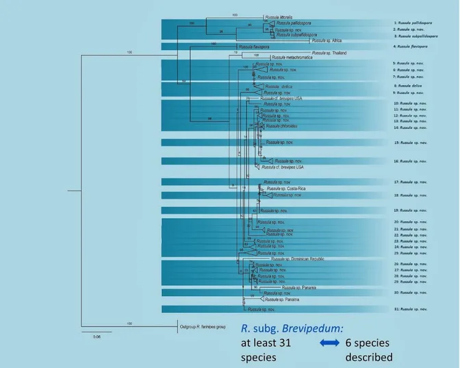
The species within Russula subgenus Brevipedum have a white cap, that can have yellow to red-brown stains. This is unlike most Russula, this genus is famous for the broad variety of cap colours. The flesh of these fungi is white but turns slowly yellow to rusty brown when exposed to the air. The spore print is white to yellow, the lamellae or the top of the stipe can have a blue or green hue. At the moment only 7 species within the subgenus Brevipedum are described in Europe: R. chloroides Krombholz, R. delica Fries, R. flavispora (Blum in Romagn.) Romagn., R. pallidospora (Blum in Romagn.) Romagn., R. littoralis, R. pseudodelica Lange (nec J. Schaef.) sec. Blum and R. laevis Kälviäinen, Ruotsalainen & Taipale (Adamčík et al., 2019). Within these 7 species, varieties are known in the species R. chloroides and the species R. delica and these varieties can differ between the different authors. However, when phylogenetic trees based on ITS (internal transcribed spacer) sequences are made, there seem to be at least 31 species within R. subg. Brevipedum (figure 1). This finding implicates that a lot of research is still needed within this subgenus to sort out the species complexes and describe the new species. To be able to differentiate these species without using DNA, macroscopic or microscopic differences need to be found and described.
8
Figure 1. Phylogenetic tree of European species within Russula subgenus Brevipedum by Ruben De Lange (unpublished)
In North and South America 11 species are known within R. subg. Brevipedum.(Singer, 1952, 1963; Shaffer, 1964; Buyck & Ovrebo, 2002; Kong, Montoya, & Estrada-Torres, 2002; Buyck & Adamčík, 2013). The four species described into detail by Buyck and Adamcik (2013) are R. brevipes Peck, Ann. Rep. N.Y. St. Mus. Nat. Hist. 43: 20. 1890, R. inopina Shaffer (Shaffer, 1964), R. romagnesiana Shaffer (Shaffer, 1964) and R. vesicatoria Burl. (Burlingham, 1944). The other species are R. littoralis, R. fuegiana Singer, NA Sing., Rev. Mycol. Paris 15:125. 1950 , R. cascadensis Shaffer (Shaffer, 1964), R. delicula Romag., Bull. Soc. Mycol. Fr. 61: 30 1946, R. idroboi Singer (Singer, 1963), R. austrodelica, R. herrerae Kong, Montoya et Estrada (Kong et al., 2002) and R. aucarum Singer (Singer, 1975). R. herrerae is characterised by the presence of a marginal veil and this character differentiates this species from all other Brevipedum species. R. aucarum is a species of the section Delicoarchaeae found in Panama. The distinction between R. sect. Delicoarchaeae and the former R. sect. Lactariodeae are unclear, some suggest R. sect. Delicoarchaeae is a synomym of R. sect. Lactariodeae (Buyck & Ovrebo, 2002).
In his monograph ‘Les Russules d’Europe et d’Afrique du Nord’, Romagnesi (1967) already mentioned that R. section Plorantinae (now R. subg. Brevipedum) is a tricky group, since the
9
characteristics which are used to distinguish and differentiate species in other Russula groups have little to no use in this group. The descriptions of R. delica even differs between Fries, Singer and Kühner & Romagnesi (Shaffer, 1964).
2.4. Species descriptions
Most of the descriptions of Russulales species are not complete, most have information about spores, basidia and cystidia sizes and ornamentation, but not of density of these structures and differences between the pileipellis margin and centre or between lamellae sides and edges (Adamčík et al., 2019). Information about the mycorrhizal structures, their accompanied host plant is missing in most descriptions. Besides being incomplete, the descriptions are not consistent between different continents, and are often author specific. The combination of these factors makes comparing descriptions difficult to even impossible. With their paper, Adamčík et al. (2019) are now encouraging others to make a consistent description of Russula species. They created a standard template, with a manual and examples, which is universally applicable.
The below-ground features of fungi, the mycorrhizae, are often not, or in restricted amounts, examined. There is still a lot to discover about ectomycorrhizae and about the correlation between above and below-ground parts of the fungi (Buyck et al., 2018). Russula species have contact exploration type ectomycorrhizae, this type of ectomycorrhizae have a smooth mantle with a small number of emanating hyphae (Agerer, 2001). Russula subg. Brevipedum has ectomycorrhizae whose cystidia have exclusively russuloid forms (Agerer, 2006).
The key to unravel species complexes of pseudo-cryptic species could lay in more detailed description of macroscopic and microscopic characteristics, including those of the mycorrhizae. Differences between lamellae sides and edges and between pileus margin and centre, are often not described while this could potentially be a discriminating factor. Cystidal density is another characteristic that is absent in descriptions found in Romagnesi (1967) and Sarnari (1998), while these are still the principal works for the European species.
3. Objectives
A first objective is to make a complete description of possible new taxa within the Russula subgenus Brevipedum. Those taxa are suggested by the molecular phylogeny based on ITS markers and were chosen based upon availability of specimens. These taxa were initially recognised to be other species within this subgenus, but molecular analysis shows they are different species (De Lange et al., unpublished).
The second objective is to compare these descriptions carefully to find microscopic differences to delimitate species within species complexes of pseudo-cryptic species. This master thesis frames in the research project done by Ruben De Lange on the former R. subg. Compactae (Fr.) Bon which is now known not to be a monophyletic group. The aim of his project is to delimit species within the subgenera Archaeae, Compactae, Malodorae, Glutinosae and Brevipedum based on morphological, molecular and ecological characters.
10
4. Materials and Methods
4.1. Specimens
The specimens were collected fresh by Ruben De Lange (R. macrostigma, 2 and 3), Jesko Kleine (R. macrostigma), Ronny Boeykens (Russula boeykensii), Felix Hampe and Cathrin Manz (Russula hampei). Three species were collected within Europe and the other species was collected in Panama, Central America, by Felix Hampe and Cathrin Manz. These collections were dried and stored in the Herbarium Universitatis Gandavensis (GENT). A small fragment of each specimen was deposited in a strong detergent, 2*CTAB buffer (2% cetyltrimethylammonium bromide). For all four species, all available specimens at the GENT Herbarium were microscopically examined. At the finding location of each specimen (only R. macrostigma & 2) roots were collected by carefully removing the upper soil layers and uncovering plant roots. These plant roots are then collected and preserved in aluminium foil together with some surrounding soil, to prevent desiccation. Later these were soaked in water and studied under a binocular microscope with small magnification to determine whether ectomycorrhiza was present on these root tips. The root tips without ectomycorrhiza were discarded. For each collection of root tips, a sample is preserved in 70% ethanol and another sample is preserved in 2*CTAB buffer.
4.2. Morphology
4.2.1. Macroscopy
While collecting, short macromorphological descriptions were made and photographs were taken of the specimens by the collectors. The colour codes used are from the Methuen book of colours (Kornerup & Wanscher, 1978). Spore deposits were available for the specimens collected by Ronny Boeykens (Russula boeykensii).
4.2.2. Microscopy
Microscopy was done on dried specimens. Spores were observed, measured and photographed in Melzer’s reagent, elements of the hymenium and pileipellis were observed and measured in Congo-Red. The hymenial elements were observed and measured both at the lamellae edge and the lamellae sides. Hyphal terminations and pileocystidia examined and measured near the pileus margin and the pileus centre. Spore measurements were done using a Zeiss Axioscop 2 microscope and pictures were taken with a Nikon Eclipse Ni-U microscope at 1000x magnification and a Bresser MikroCamII Full HD HSP camera. Bresser MikroCamLabII software was used to make stacking images, these images were used to create spore drawings. Basidia measurements are without the sterigmata. Drawings of the pileipellis and hymenial elements were made with an Olympus CX21 microscope with a drawing tube at 1000x magnification. Chemical reactions in Cresyl Blue (Buyk, 1989), carbolfuchsin (Romagnesi, 1967) and sulfovanillin (Caboň et al., 2017) were examined to respectively observe the presence of metachromatic incrustations in the pileipellis, incrustations on primordial hyphae and colouring of cystidia contents.
Per described species there were at least 2 specimens (maximum 4). Statistics for all microscopic characteristics, except for spores, were based on average on 10 measurements per specimens. Per specimen 20 spores were measured in side view excluding ornamentation. Measurements are given as (minimum –) average minus standard deviation (SD) – average – average plus SD (– maximum). Q indicates the length/width ratio of the spores. The spore ornamentation density is
11
computed following (Adamcik & Marhold, 2000).The density of hymenial cystidia is computed following (Buyck, 1991).
4.3. Molecular work
This part was performed by Ruben De Lange.
Little fragments of fresh material of the fruitbodies and root tips were preserved in small tubes with CTAB (Cetyl trimethyl-ammonium bromide). Afterwards DNA was extracted from these samples using the CTAB extraction method described in Nuytinck and Verbeken (2003). For collections of which no fresh fragments were preserved in CTAB, a modified CTAB protocol (Tel-Zur et al. 1999; mod. by Agentschap Plantentuin Meise) was used.
4.3.1. Fruitbodies
The marker that was amplified for the fruitbody samples is the internal transcribed spacer region of ribosomal DNA (ITS), specifically the ITS1 and ITS2 spacer regions and the ribosomal gene 5.8S, using primers ITS-1F and ITS4 (White et al., 1990; Gardes & Bruns, 1993) and protocols for PCR amplification follow Le et al. (2007). An automated ABI 3730 XL capillary sequencer at Macrogen was used to sequence the PCR products. Assembly of the forward and reverse sequences into contigs and where needed edited with BioloMICS (BioAware SA NV).
4.3.2. Root tips
The internal transcribed spacer region of ribosomal DNA (ITS) was amplified, more specifically ITS1, both for plant and fungal DNA, and ITS2 spacer, solely for fungal DNA, regions. The forward primers ITS1-F and fITS7 and reverse primers ITS2 and ITS4 were used respectively for the fungal ITS1 and ITS2 markers (White et al., 1990; Gardes & Bruns, 1993; Tedersoo et al., 2013). The forward primer ITS-p5 and reverse primer ITS-u2 were used for the plant ITS1 marker (Cheng et al., 2016). Amplification was done using a two-step PCR process. In the first step of PCR, the above mentioned primers prolonged with NexteraTM tails (Illumina) were used with the setting
following the description of (Le et al., 2007). Subsequent a DNA quantity and quality check, the PCR product was polished with the NucleoMag NGS Clean-up and Size Select kit (Machery-Nagel). In the second PCR step, a Nextera™ XT label (Illumina) was added to the amplicon under the following quantities: 3 μL of template DNA, 1 μL of each primer (10 pmol/μL), and 15 μL of Master Mix for a final volume of 20 μL. Amplification conditions were: 95 °C for 10 min, 8 cycles of 30 s at 95 °C, 60 s at 55 °C and 30 s at 72 °C, followed by 7 min at 72 °C. Subsequent quantification and clean-up, the sample was sent to BaseClear (Leiden, the Netherlands) for paired-end sequencing using the Illumina MiSeq technology (2 × 300 bp) amongst a batch of other amplicons with different Nextera™ labels.
4.3.3. Dataprocessing
Performed by me and Ruben De Lange
The Naturalis Galaxy v.19.01 instance was used to process the Illumina sequence reads. The reads for each specific specimen were isolated by demultiplexing the reads based upon their unique tags. Merging of the R1 and R2 reads from the paired-end sequencing was done using FLASH (Magoc
12
& SL, 2011) with the minimum overlap size set at 100 bp. We discarded the reads shorter than 250 bp or with more than 8 consecutive N’s or a Phred score lower than 28 and trimmed the primers utilizing Cutadapt (Martin, 2011). The sequences were dereplicated subsequent quality control with PRINSEQ (Schmieder & Edwards, 2011). The dereplicated sequences were arranged by size and clustered in zero-radius OTU’s with the UNOISE algorithm (Edgar & Flyvbjerg, 2015; Edgar, 2016) to denoise the amplicon reads. The VSEARCH UCHIME algorithm (Edgar et al., 2011)was used to discard the chimera sequences. A BLASTN search (Altschul et al., 1997) against the UNITE and GenBank databases was used to create an OTU abundance and taxonomic assignment table. The online MAFFT v7 program (Katoh & Toh, 2008) was used to align the sequences, using the E-INS-I strategy. Trimming of the trailing ends and manual edits of the alignment where necessary were done using Mega 6 (Tamura et al., 2013). The ITS alignment was divided into partial 18S, ITS1, 5.8S , ITS2 and partial 28S. RAxML v8.0.24 (Stamatakis & A, 2014) was used to perform maximum likelihood (ML) analyses. These were then combined with the Rapid Bootstrapping algorithm with 1000 replicates under the GTRCAT option (Stamatakis, Hoover, & Rougemont, 2008).
All analyses were conducted on the CIPRES Science Gateway (Miller, Pfeiffer, & Schwartz, 2010). The fungal and plant DNA from the root tip samples is combined to find the most probable mycorrhizal fungi-host connection based on the OTU-abundance table. Only the samples which contain compactoid Russula are used.
13
5. Results
5.1. Species description
5.1.1. Russula macrostigma
Russula macrostigma Beel & De Lange nom. prov.
Holotype: EUROPE, Italy, Tuscany, 8 Nov 2016, R. De Lange (RDL 16-032/1) Etymology: ‘macrostigma’ refers to the large and very amyloid suprahilar spot.
Pileus medium to large sized,77–
127 mm diam., when mature infundibuliform with a deep depression in the centre; margin smooth, not striate, cuticle smooth, matt, usually retaining some debris; white/yellow-white (3A2, 4A2) to light yellow (4A3, 4A4, 4A5), brownish orange (5C6) with spots of light brown (6D8), orange brown (5C6) and dark yellow (4A8).
Lamellae up to 3 mm deep, dense
(6–9L + 3–5l/cm), with decurrent tooth, white to yellow-white (3A2, 4A2); lamellulae general, of
different lengths, furcation’s absent or rare. Stipe 25–26 х 25–29 mm, cylindrical or narrowly clavate, white to yellow-white (3A2, 4A2) at the top and darker at the base, brownish orange (5C6); medulla solid. Context firm in all parts of the basidiomata, white-pale cream, on damaged places turning brownish orange; taste mild, afterwards slightly acrid; odour fruity. Spore print white to pale cream (Ib–IIa). FeSO4 brown-orange. Guaiac stipe, lamellae and context all strong and fast reaction. KOH stipe and lamellae yellowish, context yellow to slightly reddening.
Spores (7.1–)8.3–9.1–9.8(–10.8) × (5.3–)6.7–7.2–7.8(–8.4) µm, broadly ellipsoid to ellipsoid, Q =
(1.15–)1.19–1.25–1.31(–1.43), n= 60; ornamentation small sized, moderately distant to dense [3– 8 in a 3 µm diam. circle] amyloid warts, 0.2–0.5 µm high, locally subreticulate, sometimes fused in chains (3–5 fusions in a 3 µm diam. circle), connected with occasional line connections [0–3(–7) line connections in a 3 µm diam. circle] suprahilar spot large, amyloid. Basidia (38–) 45.2–54.3– 63.4 (–80) × (5–) 8.9–11.4–13.9 (–19) µm, narrowly clavate to clavate, 4–spored; basidiola first cylindrical, then clavate, ca. 4–12 µm wide. Hymenial cystidia numerous to abundant, ca. 38000/mm², (43–) 56.5–72.4–88.3 (–110) × (4–) 6.1–7.7–9.3 (–11) µm, narrowly cylindrical to narrowly clavate or narrowly fusiform, often slightly moniliform and flexuous, apically acute or obtuse, with or without appendage 2–6.63(–10) µm long; contents strongly heteromorphous (refringent), reacting weakly (greyish) in sulfovanillin; near the lamellae edges numerous, usually smaller, (38–)11.1–45.12–(–80) × (5–)6.1–8.0–9.9(–15) µm, fusiform, clavate or subcylindrical, apically often obtuse, mostly with an appendage 3–4.5(–5); contents similar. Lamellae edges fertile;
marginal cells not distinctive. Pileipellis orthochromatic in Cresyl Blue, not sharply delimited from
the underlying context, 140–250 µm deep, vertically almost homogeneous, composed of irregularly oriented, non-gelatinized hyphae that become denser and more horizontally oriented towards the
14
context; longer hyphal terminations forming conical fascicules near the surface. Acid-resistant
incrustations absent. Hyphal terminations near the pileus margin very flexuous, thin-walled,
terminal cells (12–) 20–28–36(–42) × 3–5.2–7.4(–11) µm, cylindrical or clavate, apically obtuse; subterminal cells very irregular, sometimes branched, often covered by strong glutinous coating. Hyphal terminations near the pileus centre different in length, terminal cells (10–)16.1–27.2–38.3(– 60) × (2–)2.9–3.9–4.9(–7) µm, usually cylindrical, apically obtuse; subterminal cells very irregular, flexuous and often covered with glutinous hyaline coating. Pileocystidia near the pileus margin 1– to–5 celled, (14–)34.8–58.5– 82.3(–131) × (2–)3.5–5.2–6.9(–10) µm, narrowly clavate to subcylindrical, often flexuous, apically obtuse or acute, with or without appendage, contents refringent, yellowish, no reaction in sulfovanillin. Pileocystidia near the pileus centre often longer, (20–)39.7–65.9–92.1(135) × 3–5.2–7.3(–11) µm, usually clavate and with similar contents.
Habitat: Specimens found in Italy by evergreen oaks, specifically Quercus ilex and Quercus suber. Additional material studied: EUROPE, Italy, Tuscany, 7 Nov 2016, R. De Lange (RDL 16-026/2);
15
R. macrostigma; a) basidia and cystidia of the lamellae sides; b) basidia and cystidia of the lamellae edges; c) & f) spores; d) pileocystidia and hairs of the pileipellis centre; e) pileocystidia and hairs of the pileipellis centre. The scale is 10µm.
e d c b a f
16
5.1.2. Russula zebrihyphis
Russula zebrihyphis Beel & De Lange nom. prov.
Holotype: EUROPE, Sweden, Medelpad (Province), Borgsjö parish, Sodra Sillre, 62°34'22.4"N 15°47'46.7"E, 31 Aug 2018, R. De Lange (RDL_18_043).
Etymology: ‘zebrihyphis’ refers to the zebroid incrustations of the pileipellis hyphae.
Pileus medium to large sized, 85–160 mm diam.
when mature, infundibuliform with a deep depression in the centre; margin smooth, not striate; yellow–white (4A2, 4A3, 4A4), with spots of brownish orange (5D8) and yellow (4A6).
Lamellae up to 5 mm deep, dense (5–8L + 1–
2l/cm), with decurrent tooth, white to yellow-white (3A2, 4A2); lamellulae general, of different lengths; furcation’s absent or rare. Stipe 20–40 х 25–55 mm, cylindrical or narrowly clavate, white to yellow-white (3A2, 4A2), turning brownish orange (5D8) and yellow (4A6) on damaged places; medulla solid. Context firm in all parts of the basidiomata; taste mild, sometimes very light sharp tinge; odour fruity,
flowery, like R. pectinatoides (fishy when old). Spore print white to pale cream (Ib–IIa). FeSO4 no
reaction. Guaiac stipe and lamellae strong and fast reaction. KOH stipe, lamellae and context no reaction.
Spores (7.7–)8.5–9.4–10.2(–11.6) × (6.6–)7.2–7.9–8.6(–10.2) µm, subglobose to broadly ellipsoid,
Q = (1.1–)1.12–1.19–1.25(–1.33) n= 80; ornamentation normal to high, moderately distant to very dense [4–12 in a 3 µm diam. circle] amyloid warts, 0.9–1.6 µm high, locally subreticulate, sometimes fused in chains (0–9 fusions in a 3 µm diam. circle), connected with occasional to frequent line connections [0–2(–6) line connections in a 3 µm diam. circle] suprahilar spot large, amyloid. Basidia (37–) 43.9–52.4–60.8 (–69) × (10–) 11.1–13.4–15.9 (–19) µm, narrowly clavate to clavate, 4–spored; basidiola first cylindrical, then clavate, ca. 4–12 µm wide. Hymenial cystidia abundant, 5000–20000/mm², (45–) 50.9–65.9–80.8 (–108) × (7–) 7.9–9.1–10.1 (–11) µm, narrowly cylindrical to narrowly clavate or narrowly fusiform, often slightly flexuose, apically acute or obtuse, mostly with appendage 1–4.1(–9) µm long; contents strongly heteromorphous, refringent, reacting strong (blackening) in sulfovanillin; near the lamellae edges numerous, usually smaller, (36–)47.8– 56.4–65.0(–71) × (5–)7.0–8.8–10.5(–12) µm, fusiform, clavate or subcylindrical, apically often obtuse, without or with an appendage 2–3(–5); contents similar. Lamellae edges fertile; marginal
cells not distinctive. Pileipellis orthochromatic in Cresyl Blue, not sharply delimited from the
underlying context, 150–540 µm deep, vertically almost homogeneous, composed of irregularly oriented, non-gelatinized hyphae that become denser and more horizontally oriented towards the context. Acid-resistant incrustations absent. Hyphal terminations near the pileus margin very flexuous, thin-walled, terminal cells (10–) 18.4–37.3–56.3(–118) × (2–)3.4–6.5–9.6(–13) µm, cylindrical or clavate, apically obtuse; subterminal cells very irregular, sometimes branched. Hyphal terminations near the pileus centre, terminal cells 18–41.8–65.6(–122) × 2.9–5.1–7.3(–12) µm, usually cylindrical, apically obtuse; subterminal cells very irregular, flexuous and often covered with
17
glutinous hyaline coating, sometimes with zebroid incrustation (see figure e). Pileocystidia near the pileus margin 1–to–4 celled, (28–)45.5–91.4–137(–237) × (3–)3.8–7.8–11.7(–23) µm, narrowly clavate to subcylindrical, often flexuous, apically obtuse or acute, rarely with appendage, contents refringent, yellowish, no reaction in sulfovanillin. Pileocystidia near the pileus centre often shorter and less broad, (27–)30.7–47–63.3(–84) × (3–) 3.5–5.6–7.7(–11) µm, usually clavate and with similar contents.
Habitat: Specimens found in Sweden in proximity to Picea, Betula, Populus, (Alnus and Pinus). Additional material studied: EUROPE, Sweden, Medelpad (Province), Borgsjö parish, Sodra
Sillre, 62°34'22.4"N 15°47'46.7"E, 31 Aug 2018, R. De Lange (RDL 18-041, RDL 18-042); ibid., 62°31'20.6"N 15°57'04.7"E, 31 Aug 2018, R. De Lange (RDL 18-038).
18
R. zebrihyphis; a) basidia and cystidia of the lamellae sides; b) basidia and cystidia of the lamellae edges; c) & f) spores; d) pileocystidia and hairs of the pileipellis centre; e) pileocystidia and hairs of the pileipellis centre. The scale is 10µm.
b d
c
a
e
19
5.1.3. Russula boeykensii
Russula boeykensii Beel & De Lange nom. prov.
Holotype: EUROPE, Belgium, Limburg, Vliermaalroot (Kortessem), Jongenbos, 50°52'45.5"N 5°26'21.1"E, R. Boeykens (RB 15 08 17 08).
Etymology: ‘boeykensii’ refers to the name one of the collectors of this species: Ronny Boeykens.
Pileus when mature, infundibuliform with a deep depression in the centre; white/yellow with brown
spots (4A8: dark yellow; 5D8: yellowish brown; 5DF: umber). Lamellae yellowish white; lamellulae general, of different lengths; furcation’s absent or rare. Stipe white to yellowish-white (34A, 4A2).
Context not observed; taste acrid; odour fruity. Spore print whitish to pale cream (Ia–IIb). FeSO4
orange or pink and afterwards grey; Guaiac stipe and lamellae strong and fast reaction; KOH not observed.
Spores (7.2–)7.7–8.2–8.6(–9.0) × (6.0–)6.4–6.8–7.2(–7.5) µm, subglobose to broadly ellipsoid, Q
= (1.14–)1.16–1.20–1.24(–1.29), n= 40; ornamentation normal to high, distant to very dense [2–12 in a 3 µm diam. circle] amyloid warts, 0.6–2.1 µm high, locally subreticulate, sometimes fused in chains (0–6 fusions in a 3 µm diam. circle), connected with occasional to frequent line connections [0–2(–6) line connections in a 3 µm diam. circle] suprahilar spot irregular, faintly amyloid to amyloid.
Basidia (40–) 52.5–58.9–65.2 (–72) × (9–) 10.7–12.1–13.5 (–14) µm, narrowly clavate to clavate,
4–spored; basidiola first cylindrical, then clavate, ca. 5–12 µm wide; near the lamellae edges usually smaller. Hymenial cystidiaabundant, 120000–220000/mm², (57–) 62.2–71.9–81.5 (–94) × (7–) 7.2–8.2–9.2 (–10) µm, narrowly cylindrical to narrowly clavate or narrowly fusiform, often slightly flexuose, apically acute or obtuse, often with appendage 1–4(–5) µm long; contents strongly heteromorphous, refringent, reacting faintly (greying) in sulfovanillin; near the lamellae edges abundant, usually smaller and slender, (32–)40.7–53.7–66.7(–96) × (6–)6.2–7.6–9.0(–13) µm, fusiform, clavate or subcylindrical, apically often obtuse, without or with an appendage 3–5.3(–6); contents similar. Lamellae edges fertile; marginal cells not distinctive. Pileipellis orthochromatic in Cresyl Blue, not sharply delimited from the underlying context,75–200 µm deep, vertically almost homogeneous, composed of irregularly oriented, non-gelatinized hyphae that become denser and more horizontally oriented towards the context. Acid-resistant incrustations absent. Hyphal terminations near the pileus margin very flexuous, thin-walled, terminal cells (14–) 16.9–28.7– 40.4(–56) × 3.6–4.9–6.2(–9) µm, cylindrical or clavate, apically obtuse; subterminal cells very irregular, sometimes branched. Hyphal terminations near the pileus centre, terminal cells (12–) 16.8–24.4–31.6(–39) × (3–)3.3–4.5–5.6(–7) µm, usually cylindrical, apically obtuse; subterminal cells very irregular, flexuous and often covered with glutinous hyaline coating. Pileocystidia near the pileus margin 1–to–3 celled, (26–)38.2–71.6–105(–158) × 4–5.1–6.2(–8) µm, narrowly clavate to subcylindrical, often flexuous, apically obtuse or acute, often with appendage, contents refringent, yellowish, no reaction in sulfovanillin. Pileocystidia near the pileus centre often shorter and less broad, (32–)33.9–59.3–84.8(–106) × (3–)3.2–4.3–5.4(–7) µm, usually clavate and with similar contents.
Habitat: Species collected on sandy loam soil.
Additional material studied: EUROPE, Belgium, Limburg, Vliermaalroot (Kortessem),
20
Diepenbeek/Kortessem, Netelbroekstraat, Nietelbroek (Netelbroek), 50°53'02.4"N 5°22'40.5"E, 23 Aug 2014, R. De Lange (RDL-19-23-08-2014).
Russula boeykensii; a) basidia and cystidia of the lamellae sides; b) basidia and cystidia of the lamellae edges; c) & f) spores; d) pileocystidia and hairs of the pileipellis centre; e) pileocystidia and hairs of the pileipellis centre. The scale is 10µm.
a b c d e f a
21
5.1.4. Russula hampei
Russula hampei Beel & De Lange nom. prov.
Holotype: Central America, Panama, Paso Ancho, Parque Nacional Volcan Baru, Chiriqui, 8°48'56.0"N 82°34'45.9"W, 16 June 2018, Felix Hampe & C. Manz, (FH 18-070).
Etymology: ‘hampei’ refers to the collector of this species: Felix Hampe.
Pileus when mature, infundibuliform with a deep depression in the centre; white to pale yellowish
white (2A2), yellow white (3A2, 4A2), light yellow (4A5) and dark yellow (4A8) with light yellow spots. Lamellae pale yellowish white (2A2) to yellow-white (3A2, 4A2); lamellulae general, of different lengths; furcation’s absent or rare. Stipe white to pale yellowish white (2A2), yellow white (3A2, 4A2), light yellow (4A5) and dark yellow (4A8) with light yellow spots. Context not observed.
Spore print not observed.
Spores (6.5–)7.1–7.6–8.2(–8.9) × (5.7–)6.0–6.5–7.1(–7.7) µm, subglobose to broadly ellipsoid, Q
= (1.05–)1.13–1.17–1.22(–1.26), n= 40; ornamentation normal to high, moderately distant to very dense [4–13 in a 3 µm diam. circle] amyloid warts, 1.0–2.0 µm high, locally subreticulate, sometimes fused in chains (0–6 fusions in a 3 µm diam. circle), connected with occasional to abundant line connections [0–6(–9) line connections in a 3 µm diam. circle] suprahilar spot irregular, faintly amyloid to amyloid. Basidia (38–) 45.1–49.5–53.8 (–58) × (9–) 11.0–12.1–13.2 (–14) µm, narrowly clavate to clavate, 2–4–spored; basidiola first cylindrical, then clavate, ca. 4–11 µm wide; near the lamellae edges usually smaller (34–) 35.4–40.8–46.1 (–47) × (11–) 11.2–12.7–14.3 µm.
Hymenial cystidia numerous to abundant, 2100–4200/mm², (50–) 56.8–64.5–73.2 (–75) × (6–)
6.9–8.5–10.0 (–12) µm, narrowly cylindrical to narrowly clavate or narrowly fusiform, often slightly flexuose, apically acute or obtuse, mostly without appendage 1–3.5(–7) µm long; contents strongly heteromorphous, refringent, reacting strongly (blackening) in sulfovanillin, weakly metachromatic walls in Cresyl Blue; near the lamellae edges more abundant, usually smaller and slender, (35– )41.4–51.0–60.6(–69) × (6–)6.3–7.4–8.5(–10) µm, fusiform, clavate or subcylindrical, apically often obtuse, (mostly) without appendages; contents similar. Lamellae edges fertile; marginal cells not distinctive. Pileipellis slightly metachromatic in Cresyl Blue, not sharply delimited from the underlying context, 210–300 µm deep, vertically almost homogeneous, composed of irregularly oriented, non-gelatinized hyphae that become denser and more horizontally oriented towards the context. Acid-resistant incrustations absent. Hyphal terminations near the pileus margin very flexuous, thin-walled, terminal cells (12–) 13.4–26.0–38.5(–71) × (3–)4.8–6.6–8.4(–10) µm, cylindrical or clavate, apically obtuse; subterminal cells very irregular, sometimes branched. Hyphal terminations near the pileus centre usually less broad, terminal cells (15–) 16.0–26.2–36.3(–50) × (3–)3.7–4.8–5.9(–7) µm, usually cylindrical, apically obtuse; subterminal cells very irregular, flexuous and often covered with glutinous hyaline coating, sometimes branched. Pileocystidia near the pileus margin 1–to–4 celled, (23–)33.3–63.3–93.2(–124) × (3–)3.4–5.9–8.4(–12) µm, narrowly clavate to subcylindrical, often flexuous, apically obtuse or acute, rarely with appendage, contents refringent, yellowish, no reaction in sulfovanillin. Pileocystidia near the pileus 1–to –2 celled centre often shorter and less broad, (18–)28.1–48.9–69.6(–90) × (3–)3.6–4.4–5.1(–6) µm, usually clavate and with similar contents.
Additional material studied: Central America, Panama, Paso Ancho, Parque Nacional Volcan
22
Russula hampei; a) basidia and cystidia of the lamellae sides; b) basidia and cystidia of the lamellae edges; c) & f) spores; d) pileocystidia and hairs of the pileipellis centre; e) pileocystidia and hairs of the pileipellis centre. The scale is 10µm.
a b c d e f
23
5.2. Molecular work
The PCR success from the root tip samples are relatively low. From all the root tip samples, the ITS1 PCR-amplification was only successful for 45.6%. For ITS2 and the plant ITS this number is even lower, 24.9% and 11.4% respectively. The percentage of samples for which both ITS1 and ITS2 amplification was successful was just little lower than the success rate of ITS2, namely 21.4%. However, the success rate for all three amplification processes is very low, for merely 4.3% of all root tip samples all three amplification processes were successful.
Only 24.4 % and 11.3% of respectively the ITS1 and ITS2 Illumina reads from the root tips matched with OTU’s assigned to the subgenus Compactae sensu lato (all compactoïd Russula).
In only 1 of the 4 newly described species all three primer PCR-amplification was successful. This was for R. zebrihyphis, the plant OUT’s assigned to the root tip samples of R. zebrihyphis are Salix caprea, Quercus dentata and Quercus petrea.
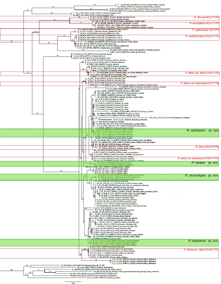
The phylogenetic tree in figure 2 was compiled using ITS sequences of R. subg. Brevipedum species available in the Herbarium Universitatis Gandavensis and supplemented with other ITS sequences from R. subg. Brevipedum from GenBank.
Bootstrap values over 75 are seen as well-supported, values between 50 and 75 are seen as moderately supported and values lower than 50 are seen as little supported.
The first split within tis phylogenetic tree has moderate support, the next splits are well-supported except for the large group in which all the new described species of this work are placed. Support ranges from well-supported to barely supported.
24
25
6. Discussion
6.1. Comparison
A comparison of the newly described R. subg. Brevipedum species with the previously described species will be made in this section. The newly described species which are collected within Europe will be compared in depth with the previously described species of Europe and more briefly compared to those previously described in North America. Only the differences between species are described.
The conscious choice was made to compare the newly described species with all species descriptions from this subgenus from Romagnesi, Sarnari and Shaffer. Because of this a lot of the comparisons with European species are sectioned in Romagnesi, Sarnari and Shaffer, these sections refer to the comparison with the description found respectively in Romagnesi (1967), Sarnari (1998) and Shaffer (1964).
6.1.1. R. macrostigma
European species
R. macrostigma VS R. delica
1. RomagnesiMacroscopic: R. delica var. puta has a longer and less broad stipe (35–65 × 13–17 mm) compared to R. macrostigma (25–26 х 25–29 mm). R. delica and R. delica var. trachyspora has a complex odour of fruit and fish (one can dominate, var. puta only has very faint odour) while R. macrostigma has a fruity odour. R. delica and R. delica var. trachyspora has a slow, faint pink-orange reaction to FeSO4, R. delica var. puta shows a pink-red after fifteen
minutes, while R. macrostigma has a brown-orange reaction. Guaiac has a positive reaction, but not always immediate with R. delica, while R. macrostigma shows an immediate and strong positive reaction.
Microscopic: R. delica spores are slightly larger (8–10–11.5 × 6.5–8.7 µm) compared to R. macrostigma spores ((7.1–)8.3–9.1–9.8(–10.8) × (5.3–)6.7–7.2–7.8 (–8.4) µm). Spore ornamentation of R. delica is slightly larger (0.5–0.7–1.0 µm, var. trachyspora 1.0–1.5 µm) than those of R. macrostigma (0.2–0.5 µm). The hymenial cystidia of R. delica are longer (65–150 µm, var. trachyspora 78–135 µm, var. puta 100–120 µm) compared to these of R. macrostigma ((43–)56.5–72.4–88.3 (–110) µm) and they have a strong reaction to sulfovanillin while these of R. macrostigma only show a faint reaction.
2. Sarnari
Macroscopic: R. delica var. puta has a slender stipe (15–20 mm) compared to R. macrostigma (25–26 х 25–29 mm). R. delica var. delica has a longer stipe (25–48 mm) compared to R. macrostigma (25–26 х 25–29 mm). R. delica var. delica has a strong and unpleasant odour, like peach or salt with fruity components when young and a peaty flavour in the lamellae, while R. macrostigma has a fruity odour and has a mild and later slight acrid taste. R. delica var. delica reacts pale pink with FeSO4, R. macrostigma has a brown-orange
26
Microscopic: R. delica var. delica has larger spores (8.5–11.2 × 7–9 µm) which are subglobose with higher ornamentation (0.8–1.0 µm, var. puta 1–1.2 µm) compared to the broadly ellipsoid to ellipsoid spores from R. macrostigma ((7.1–)8.3–9.1–9.8(–10.8) × (5.3– ) 6.7–7.2–7.8 (–8.4) µm and ornamentation height 0.2–0.5 µm high). The hymenial cystidia of R. delica var. delica are longer and thicker (78–150 × 9–13 µm) compared to those from R. macrostigma ((43–) 56.5–72.4–88.3 (–110) × (4–)6.1–7.7–9.3 (–11) µm).
3. Shaffer
Microscopic: Spore ornamentation of R. delica is higher (0.4–1.0 µm) compared to R. macrostigma (0.2–0.5 µm). Suprahilar spot weakly amyloid for R. delica, while suprahilar spot is large and strongly amyloid for R. macrostigma.
R. macrostigma VS. R. chloroides
1. RomagnesiMacroscopic: R. chloroides can have a smaller pileus (var. chloroides 45–130 mm, var. parvispora 45–100 mm) and longer stipe (var. chloroides (15–)30–50(–90) mm, var. parvispora 25–40 mm) compared to R. macrostigma (49–81 mm pileus diameter and 10– 25 mm stipe length). R. chloroides var. chloroides FeSO4 dirtyred reaction, R. chloroides
var. parvispora pink-orange reaction, R. macrostigma brown-orange.
Microscopic: R. chloroides var. parvispora has smaller spores (6.5–8.0 × 6.0–6.7 µm) compared to the spores of R. macrostigma ((7.1–)8.3–9.1–9.8(–10.8) × (5.3–)6.7–7.2–7.8(– 8.4) µm). R. chloroides var. chloroides has higher spore ornamentation (1–1.5 µm) compared to R. macrostigma (0.2–0.5 µm high). R. chloroides var. parvispora hymenial cystidia colouring black in SV.
2. Sarnari
Macroscopic: R. chloroides can have a smaller pileus (var. chloroides 65–150 mm) compared to R. macrostigma (49–81 mm). FeSO4 R. chloroides var. trachyspora pinkish
reaction, R. chloroides var. chloroides slow (sometimes faint) pink-orange reaction. Guaiac R. chloroides var. chloroides slow, green reaction.
Microscopic: R. chloroides spores are slightly bigger (var. chloroides 8–11.2 × 7.2–8.8 µm, var. trachyspora 9.5–11.4 × 8–10.5 µm), higher spore ornamentation (1.3–1.6 µm) compared to R. macrostigma ((7.1–)8.3–9.1–9.8(–10.8) × (5.3–)6.7–7.2–7.8(–8.4) µm, 0.2– 0.5 µm high). R. chloroides var. parvispora has smaller spores (6.4–8.0 × 6.0–6.7 µm) compared to the spores of R. macrostigma ((7.1–)8.3–9.1–9.8(–10.8) × (5.3–)6.7–7.2–7.8(– 8.4) µm).
R. macrostigma VS. R. pseudodelica
1. RomagnesiMacroscopic: R. pseudodelica has an acrid taste, while R. macrostigma’s taste starts of mild taste and afterwards just slightly acrid. The spore print of R. pseudodelica is darker (pale, ochraceous cream) compared to R. macrostigma (Ib–IIa).
27
Microscopic: The spore ornamentation of R. pseudodelica reaches up to 1.25 µm which is higher compared to these of R. macrostigma (0.2–0.5 µm). The suprahilar spot is faintly amyloid in R. pseudodelica while being strongly amyloid in R. macrostigma.
2. Shaffer
Macroscopic: R. pseudodelica has a pungent taste, while R. macrostigma’s taste starts of mild taste and afterwards just slightly acrid. The spore print of R. pseudodelica is darker (custard-ochraceous) compared to R. macrostigma (Ib–IIa).
Microscopic: The spores of R. pseudodelica are less broad (6.9–9.3 × 6.3–7.0 µm) compared to the spores of R. macrostigma ((7.1–)8.3–9.1–9.8(–10.8) × (5.3–)6.7–7.2–7.8(– 8.4) µm). The spore ornamentation of R. pseudodelica is higher (0.5–1.3 µm) compared to these of R. macrostigma (0.2–0.5 µm). The suprahilar spot of R. pseudodelica is ornamented like the remainder of the spore but the ornamentation is lower and often weakly amyloid, while the suprahilar spot of R. macrostigma is amyloid but not ornamented. The hyphae in the pileipellis are less broad in R. pseudodelica (hyaline hyphae 1.0–2.6 µm broad and oleiferous hyphae 2.0–5.3 µm broad).
R. macrostigma VS. R. pallidospora
1. RomagnesiMacroscopic: R. pallidospora has a longer stipe (40–45 mm) compared to R. macrostigma (pileus diameter 49–80 mm, stipe length 10–25 mm). R. pallidospora has a complex smell, with a fruity component (R. macrostigma fruity smell) and it has a mild (slightly refreshing) taste, later bitter (R. macrostigma mild taste, later slightly acrid). R. pallidospora reacts pinkish with FeSO4, while R. macrostigma reacts brown-orange. R. pallidospora has a
darker spore print (IId) compared to R. macrostigma (Ib–IIa).
Microscopic: The spores of R. pallidospora are smaller (especially more slender; 7.2–9.0 × 5.9–6.8 µm) compared to R. macrostigma ((7.1–)8.3–9.1–9.8(–10.8) × (5.3–)6.7–7.2–7.8(– 8.4) µm). The basidia of R. pallidospora are slender (8–11 µm) compared to those of R. macrostigma ((5–) 8.9–11.4–13.9 (–19) µm).
2. Sarnari
Macroscopic: R. pallidospora can have a bigger pileus (60–150 mm) and longer stipe (25– 50 mm) compared to R. macrostigma (pileus diameter 77–127 mm, stipe length 25–29 mm). R. pallidospora reacts pale pink-orange with FeSO4, while R. macrostigma reacts
brown-orange. R. pallidospora reacts slow and dirty blue with Guaiac, while R. macrostigma has an immediate strong reaction. R. pallidospora has a darker spore print (IIc) compared to R. macrostigma (Ib–IIa).
Microscopic: The spores of R. pallidospora are smaller (especially more slender; 7.5–8.75 × 6.0–7.0 µm) compared to R. macrostigma ((7.1–)8.3–9.1–9.8(–10.8) × (5.3–)6.7–7.2– 7.8(–8.4) µm). The basidia of R. pallidospora are slender (9–11 µm) compared to those of R. macrostigma ((5–) 8.9–11.4–13.9 (–19) µm). The hymenial cystidia of R. pallidospora are longer (65–165 µm) compared to those of R. macrostigma ((43–) 56.5–72.4–88.3 (– 110) µm).
28
R. macrostigma VS. R. flavispora
1. Romagnesi
Macroscopic: R. flavispora has a remarkable odour with fruity notes, the taste is bitter in the stipe and acrid in the lamellae while R. macrostigma has a fruity odour and mild, later acrid taste. FeSO4 reacts an intense dirty pink for R. flavispora and brown-orange for R.
macrostigma. Spore print (light golden yellow IVb) and lamellae (ochre)darker than spore print R. macrostigma (Ib–IIa).
Microscopic: R. flavispora has smaller spores (7.5–8 × 6.2–6.7 µm) compared to these of R. macrostigma ((7.1–)8.3–9.1–9.8(–10.8) × (5.3–)6.7–7.2–7.8(–8.4) µm). The spore ornamentation of R. flavispora is larger (up to 0.75 µm, R. macrostigma 0.2–0.5 µm) the warts are more isolated and the suprahilar spot is vague and very faintly amyloid (R. macrostigma has a distinct suprahilar spot which is strongly amyloid). The hymenial cystidia of R. flavispora are longer (72–120 µm) than these of R. macrostigma ((43–) 56.5–72.4– 88.3 (–110) µm). R. flavispora pileipellis has elements which have a greying reaction to sulfovanillin while the pileipellis elements of R. macrostigma show no reaction.
2. Sarnari
Macroscopic: R. flavispora has a complex odour (stronger when cut) with notes of fish, boiled herbs and fruit, it has an acrid and bitter while R. macrostigma has a fruity odour and mild, later acrid taste. FeSO4 reacts pink-orange for R. flavispora and brown-orange for R.
macrostigma. Spore print (bright yellow IVb) and lamellae (ochre) darker than spore print R. macrostigma (Ib–IIa).
Microscopic: R. flavispora has smaller spores (6.4–8.6 × 5.5–6.8 µm) compared to these of R. macrostigma ((7.1–)8.3–9.1–9.8(–10.8) × (5.3–)6.7–7.2–7.8(–8.4) µm). The warts of R. flavispora are more isolated and the suprahilar spot is vague and very faintly amyloid (R. macrostigma has a distinct suprahilar spot which is strongly amyloid). The hymenial cystidia of R. flavispora are longer (70–140 µm) than these of R. macrostigma ((43–) 56.5–72.4– 88.3 (–110) µm).
R. macrostigma VS. R. littoralis
1. RomagnesiMacroscopic: R. littoralis has an odour similar to those of some Lactarius species and a mild taste in the stipe, very bitter in the lamellae (R. macrostigma mild, later acrid). R. littoralis reacts pink with FeSO4, R. macrostigma has a brown-orange reaction. The spore
print of R. littoralis is darker (creme IIc) compared to R. macrostigma.
Microscopic: The spores of R. littoralis are less broad (5.5–6.5(–6.7) µm) compared to the spores of R. macrostigma ((5.3–)6.7–7.2–7.8(–8.4) µm). R. littoralis does not have a clearly differentiated suprahilar spot in contrast to R. macrostigma. The basidia of R. littoralis are small and slender (46–56 × 8.5–10 µm) compared to those of R. macrostigma((38–) 45.2– 54.3–63.4 (–80) × (5–) 8.9–11.4–13.9 (–19) µm). The hymenial cystidia of R. littoralis show a strong blackening with sulfovanillin, while those of R. macrostigma merely have a faint greying reaction.
29
R. macrostigma VS. R. laevis
Macroscopic: R. laevis has a smaller, shiny pileus (40–75 mm) compared to the matt pileus of R. macrostigma (77–127 mm). The spore print of R. laevis is slightly darker (IIb–d) compared to R. macrostigma (Ib–IIa).
Microscopic: R. laevis has slightly larger spores ((9.2–)9.5–10–10.5(–11.3) × (7.6–)8–8.5– 8.9(–9.6) µm) and lower Q ((1.14–)1.16–1.18–1.21(–1.26)) compared to R. macrostigma ((7.1–)8.3–9.1–9.8(–10.8) × (5.3–)6.7–7.2–7.8(–8.4) µm, Q = (1.15–)1.19–1.25–1.31(– 1.43)). The ornamentation of R. laevis (0.8–1.1(–1.3) µm) is higher compared to R. macrostigma (0.2–0.5 µm). The suprahilar spot of R. laevis is small, partly amyloid to amyloid, while these of R. macrostigma is large and distinctly amyloid. The hymenial cystidia of R. laevis (moderately numerous, 850–900/mm²) are considerably less dense and have a stronger reaction in SV (dark grey-brown) compared to R. macrostigma (abundant 30000–50000/mm², light grey in SV). The hyphal termination of R. laevis are longer, both at the margin ((20–)36.5–53.6–71.5(–116) × (4–)5–6–7(–9) µm) and the centre of the pileus ((20–)23.5–31.9–40.5(–56) × (3.5–)4–5–6(–7) µm, compared to R. macrostigma (pileus margin (12–) 20–28–35.9(–42) × 3–5.2–7.4(–11) µm, pileus centre (10–)16.1–27.2–38.3(– 60) × (2–)2.92–3.91–4.91(–7) µm). R. laevis has pileocystidia which are slightly metachromatic and are always 1–celled while these of R. macrostigma are orthochromatic and can consist of up to 5 cells.
North American species
R. macrostigma can be ruled out not to be one of the American species based upon a few clear differences.
R. brevipes has spores with a wider width, resulting in an average lower Q value (Q= (1.09–)1.11– 1.16–1.21(–1.29)) compared to R. macrostigma (Q= (1.15–)1.19–1.25–1.31(–1.43)). The spore ornamentation of R. brevipes is higher (0.7–1.7 µm) than these of R. macrostigma (0.2–0.5 µm). The hymenial cystidia of R. brevipes have weakly metachromatic walls while R. macrostigma is orthochromatic. Of the 2 variations described by Shaffer (1964) R. brevipes var. acrior has higher spore ornamentation (0.7–1.7 µm), and the suprahilar spot is weakly amyloid. R. brevipes var. megaspore has larger spores (9.3–14.1 × 8.0–12.0 µm).
R. inopina has smaller spores ((6.5–)6.9–7.2–7.5(–7.8) × (5–)5.2–5.5–5.7(–6) μm) and higher spore ornamentation (0.4–0.7 μm) compared to R. macrostigma ((7.07–)8.31–9.05–9.78(–10.84) × (5.32–)6.71–7.24–7.77(–8.42) µm, 0.2–0.5 µm high). The suprahilar spot is small and inamyloid to partly amyloid for R. inopina, compared to fully amyloid for R. macrostigma. The basidia of R. inopina are slender (8–9,5 µm) compared to these of R. macrostigma ((5–)8.89–11.4–13.91(–19) µm). The hymenial cystidia of R. inopina do not have appendices while these of R. macrostigma often do.
R. romagnesiana has smaller spores ((5.8–)6–6.3–6.7(–7.3) × (5.1–)5.2–5.4–5.7(–6.1) μm), a lower Q–value ((1.09–)1.12–1.16–1.2(–1.25)) and a smaller suprahilar spot compared to R. macrostigma (((7.07–)8.31–9.05–9.78(–10.84) × (5.32–)6.71–7.24–7.77(–8.42) µm, Q=(1.15–
30
)1.19–1.25–1.31(–1.43)). The hymenial cystidia of R. romagnesiana have weakly metachromatic walls while R. macrostigma is orthochromatic. R. romagnesiana has shorter and less broad basidia (40–44–48 × 8.5–9.7–11 µm) compared to R. macrostigma ((38–) 45.2–54.3–63.4 (–80) × (5–) 8.9–11.4–13.9 (–19) µm).
R. vesicatoria has smaller spores ((7–)7.3–7.6–7.9(–8.2) × (5.8–)6–6.3–6.7(–7) μm), a lower Q value ((1.12–)1.15–1.19–1.23(–1.31)) and a smaller suprahilar spot which is not amyloid compared to R. macrostigma ((7.07–)8.31–9.05–9.78(–10.84) × (5.32–)6.71–7.24–7.77(–8.42) µm, Q=(1.15– )1.19–1.25–1.31(–1.43)) with a large, strongly amyloid suprahilar spot. R. vesicatoria is distinctly metachromatic in the subhymenium, while R. macrostigma is orthochromatic. R. vesicatoria has moderately numerous hymenial cystidia, which are insensitive to sulfovanillin, the hymenial cystidia of R. macrostigma are numerous to abundant and have a weak reaction to sulfovanillin. R. vesicatoria has shorter and less broad basidia (41–46–50 × 8–9.5–10.5 µm) compared to R. macrostigma ((38–) 45.2–54.3–63.4 (–80) × (5–) 8.9–11.4–13.9 (–19) µm).
R. fuegiana spores (6.8–8.5 × 5.3–7.3 µm) are smaller compared to R. macrostigma ((7.07–)8.31– 9.05–9.78(–10.84) × (5.32–)6.71–7.24–7.77(–8.42) µm). Ornamentation height similar, but rarely forming a partial to broken reticulum while R. macrostigma is often subreticulate. Suprahilar spot of R. fuegiana is finely and faintly ornamented while these of R. macrostigma are large and amyloid. The hymenial cystidia of R. fuegiana do not have appendices while these of R. macrostigma often do.
R. cascadensis is easily discernibly different by its intense acrid taste. R. cascadensis spores (6.7– 8.2 × 4.8–6.7 µm) are smaller compared to R. macrostigma ((7.07–)8.31–9.05–9.78(–10.84) × (5.32–)6.71–7.24–7.77(–8.42) µm). Suprahilar spot of R. cascadensis has weakly amyloid to almost no ornamentation while these of R. macrostigma are large and amyloid. R. cascadensis has shorter and less broad basidia (40–52 × 8–10.6 µm) compared to R. macrostigma ((38–) 45.2– 54.3–63.4 (–80) × (5–) 8.9–11.4–13.9 (–19) µm). The hymenial cystidia of R. cascadensis do not have appendices while these of R. macrostigma often do.
R. delicula both spores (8.1–10.6 × 7–9.4 µm) and spore ornamentation (0.5–1.6 µm) are big compared to R. macrostigma ((7.07–)8.31–9.05–9.78(–10.84) × (5.32–)6.71–7.24–7.77(–8.42) µm and ornamentation height 0.2–0.5 µm). Suprahilar spot of R. delicula is weakly amyloid, while these of R. macrostigma is strongly amyloid.
Intermediate conclusion: R. macrostigma
R. macrostigma is most similar to R. delica as described in Shaffer (1964), R. delica var. trachyspora and R. delica var puta. R. macrostigma differs from R. delica by lower spore ornamentation and a distinct amyloid suprahilar spot. R. macrostigma differs from R. delica var. trachyspora by lower spore ornamentation and shorter hymenial cystidia, a less strong reaction to sulfovanillin and an odour that lacks a fish component. R. macrostigma differs from R. delica var. puta by a broader stipe, lower spore ornamentation and shorter hymenial cystidia which react less to sulfovanillin.
31
R. macrostigma is can be distinguished mostly by a light spore print, a large and distinctly amyloid suprahilar spot, quite large spores and broad basidia.
32
6.1.2. R. zebrihyphis
European species
R. zebrihyphis VS R. delica
1. RomagnesiMacroscopic: R. delica and R. delica var. trachyspora has a slow, faint pink-orange reaction to FeSO4, R. delica var. puta shows a pink-red after fifteen minutes,while R. zebrihyphis
has no reaction. Guaiac has a positive reaction, but not always immediate with R. delica, while R. zebrihyphis shows an immediate and strong positive reaction.
Microscopic: Spore ornamentation of R. delica is smaller (0.5–0.7–1.0 µm, var. puta up to 0.85 µm) than those of R. zebrihyphis (0.9–1.6 µm). The hymenial cystidia of R. delica are longer and slender (65–150 × (6.5–)7.2–11.5(–13.5) µm, var. trachyspora 78–135 × 6–11.5 µm, var. puta 100–120 × 6.5–10 µm) compared to these of R. zebrihyphis ((45–) 50.9– 65.9–80.8 (–108) × (7–) 7.9–9.1–10.1 (–11) µm).
2. Sarnari
Macroscopic: R. delica var. delica has a strong and unpleasant odour, like peach or salt with fruity components when young and a peaty flavour in the lamellae, while R. zebrihyphis has a fruity, flowery, pectinatoides odour (fishy when old) and has a mild taste, sometimes a very light sharp tinge. R. delica var. delica reacts pale pink with FeSO4, R. zebrihyphis
has no reaction.
Microscopic: R. delica has smaller spore ornamentation (var. delica 0.8–1.0 µm, var. puta 1.0–1.2 µm) compared to R. zebrihyphis (0.9–1.6 µm). The hymenial cystidia of R. delica var. delica are longer and thicker (78–150 × 9–13 µm) compared to those from R. zebrihyphis ((45–) 50.9–65.9–80.8 (–108) × (7–) 7.9–9.1–10.1 (–11) µm).
3. Shaffer
Microscopic: R. delica has more slender spores (8.2–10.8 × 6.9–8.1 µm) and lower spore ornamentation (0.4–1.0 µm) compared to R. zebrihyphis 2 ((7.7–)8.5–9.4–10.2(–11.6) × (6.6–) 7.2–7.9–8.6(–10.2) µm, ornamentation 0.9–1.6 µm). Suprahilar spot weakly amyloid for R. delica, while suprahilar spot is large and strongly amyloid for R. zebrihyphis.
R. zebrihyphis VS. R. chloroides
1. RomagnesiMacroscopic: R. chloroides has a mild taste in the stipe, but acrid and unpleasant in the lamellae, while R. zebrihyphis has a mild taste (sometimes very faint sharp tinge). R. chloroides can have a longer stipe (var. chloroides (15–)30–50(–90) mm) compared to R. zebrihyphis (25–55 mm). R. chloroides var. chloroides FeSO4 dirty red reaction, R.
chloroides var. parvispora pink-orange reaction, R. zebrihyphis no reaction.
Microscopic: R. chloroides var. parvispora has smaller spores (6.5–8.0 × 6.0–6.7 µm) compared to the spores of R. zebrihyphis ((7.7–)8.5–9.4–10.2(–11.6) × (6.6–)7.2–7.9–8.6(– 10.2) µm).