Fracture risk during extraction of well-fixed extended cementless stems: porous versus hydroxyapatite coated.
Frederik Matthys, MD1
Christophe Pattyn, MD, PhD1
Affiliations:
1 Department of Orthopaedic Surgery Ghent University Hospital
Corneel Heymanslaan 10, 9000 Gent
Corresponding author:
Frederik Matthys; frederik.matthys@ugent.be Telephone Belgium: 0032 9 332 58 55
Ghent University Hospital
Corneel Heymanslaan 10, 9000 Gent
2 Abstract
Background and study aims
The concern of extensive fracturing and bone damage during implant removal has been reported for ingrowing stems, in particular in extended porous coated stems, potentially impeding successful re-implantation of a femoral revision implant and consequently handicapping patients for life.
The aim of the present study is to describe this particular complication and comparing the occurrence in porous coated and hydroxyapatite (HA) coated femoral implants.
Materials and methods
Sixty-two consecutive revision hip replacements were performed between January 2010 and December 2016 at a single academic institution by one surgeon. Clinical follow-up involved examination with Harris Hip Score (HHS) at 2 years post-surgical intervention. Fracture occurrence and severity were compared between groups by means of the Vancouver classification for intraoperative fractures.
Results
Overall, higher rates of fracturing were observed in the porous coated group (81.8%,
P<0.05) compared to the HA coated group (43.5%, P<0.05). Of these fractures, the
majority (72.7%) were B3 fractures. There was a significant difference between the mean HHS of both groups (P<0.05). in the porous-coated group versus the group with HA coating (mean Harris Hip Scores of 68.45 vs 86.17).
3 Conclusion
An increase in the number of revision hip arthroplasty procedures is expected in the future. Due to bad results associated with porous coated stem removals, surgeons have to be careful with these type of uncemented stems in primary hip arthroplasty, and this especially in young patients who have a likelihood of revision surgery.
Keywords
Hydroxyapatite coating, porous coated, fracture risk, revision surgery, hip replacement, Harris Hip Score.
4 Introduction
Primary hip arthroplasty is likely the most successful and cost-efficient procedure in orthopaedics. Patients and surgeons can expect a hip replacement to last 25 years in around 58% of patients according to National joint registries.[1] With changing patient demographics, increasing life expectancy and active lifestyle at older ages, the future demand for joint replacement surgery is expected to increase by 175% by 2030 in the USA, parallel in the UK and Wales and in the Netherlands by 53 percent during the same period [2-4]. A similar growth in revision surgery procedures can be expected, of which the outcome is often less favourable [4-6].
Revision surgery is generally more expensive, provides a decreased prognostic outcome and can further have an impact on the quality of life and activity [4]. Furthermore, revision hip replacements fail much earlier than do primary hip replacement, necessitating further revisions. [1] Although the outcome of primary surgery is well documented, revision surgery is far less popular in terms of survival and outcome studies [6-8]. A particular concern that has been raised over the last two decades is the complicated removal of cementless stems, potentially impeding successful re-implantation of a femoral revision implant and consequently handicapping patients for life [9-12]. Despite the important impact on patients quality of life and related social costs, there is almost no literature available describing the risk factors.
The concern of extensive fracturing and bone damage during implant removal has been reported for ingrowing stems, in particular in extended porous coated stems [12,13]. The aim of the present study is to describe this particular complication and to
5 compare the occurrence in hydroxyapatite (HA) coated and porous coated femoral implants.
Material and Methods
Study population
Sixty-two consecutive revision hip replacements were performed between January 2010 and December 2016 at a single academic institution. Only revisions of a primary total hip replacement were included: these were all done by the same experienced surgeon- and performed through a posterolateral approach. Exclusion criteria were revisions of already revised total hip replacement, periprosthetic fractures and loosening of the femoral stem.
The patient’s medical record was reviewed to identify the date and type of primary hip procedure. The preoperative radiographs of all hips were assessed to determine the femoral bone type with use of the isthmus ratio described by Dorr. [14]
All fractures caused by the intervention were identified and scored by a retrospective review of the operative record and verified by examination of the postoperative radiographs (standard anteroposterior pelvis, anteroposterior and lateral hip x-rays) by consensus of 2 clinical experts (FM,CP).
6 We used an adapted Vancouver classification system for intraoperative fractures. Although it was originally invented for the description of fractures that originate in the placement of primary prostheses, the classification is valuable in describing fractures that arise during extraction of the femoral component. In contrast to the Vancouver classification system for postoperative, periprosthetic fractures, its reliability and validity has not been tested [15]. According to this classification system, the femur was divided into three anatomical areas: A (the pertrochanteric region), B (the diaphysis), and C (the distal metaphyseal, or supracondylar, region). In each anatomic location, the fracture was categorized as 1 (a cortical perforation), 2 (an undisplaced linear crack), or 3 (a displaced or unstable fracture).
Clinical follow-up involved examination with Harris hip score at 2 years post surgical intervention.
Ethical approval was obtained from the university and hospital internal review boards prior to the commencement of the study (UZ Gent, registration number 2019/1847). All patients gave informed consent to participate in the study.
Statistical analysis
Statistical analysis was conducted by using SPSS Statistics 23 software. Significance level has been set at P ≤ 0.05. Fracture occurrence and severity were compared between groups by means of the Pearson Chi-square Test.
7 Results
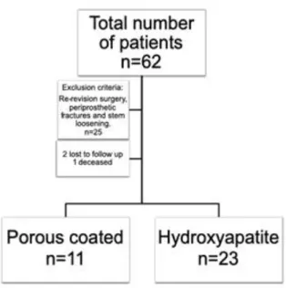
Fig 1 describes the flowchart of patients:
Thirty-four patients were included in the cohort, 11 of them in the non-cemented porous coated group and 23 in the hydroxyapatite group. In the porous coated group there were 10 women and one man. In the hydroxyapatite group there were 12 women and 11 men. The mean age of the cohort at the time of the procedure was 62.5 years. The mean age in the hydroxyapatite group was 64 year and the mean age in the porous coated group was 59.2 year. There was no significant difference between the mean age of the groups (p=0.33). (Table 1)
Two patients were lost to follow up because they did not want to participate in this study and one patient had deceased due to pulmonary complications arising after surgery.
Our dataset consisted of patients who had undergone revision surgery for the following reasons: aseptic loosening, metallosis, infection, wear and osteolysis, luxation and chronic pain.
Radiographic Results
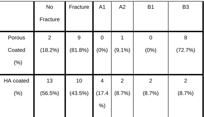
Overall, higher rates of fracturing were observed in the porous coated group (81.8%, p < 0.05) then in the HA coated group (43.5%). Of these fractures, the majority were B3 fractures. 72.7% of the patients in the porous coated group suffered from a B3 fracture. And only 18.2% had no fractures after removal. A detailed overview of the findings is presented in Table 2.
8 Clinical Results
The mean HHS at 2 years was 80.44 (34-100). A significant decrease in HHS was observed (p < 0.05) in the porous coated group (68.45) when compared to the patients in the HA coated group (86.17). This result was significant (p = .004). (Table 3)
Discussion
The etiology of intraoperative fractures associated with cementless fixation in primary hip arthroplasty has been well documented [16]. Several intrinsic and extrinsic factors like poor bone stock, the use of a straight stem and underreaming lead to an increase in intraoperative fractures which in turn is associated with increased morbidity and mortality, However, little literature is available about the prevalence and risk factors of intraoperative femoral fractures in revision hip arthroplasty. Our study is the first comparing the risk of intraoperative femoral fractures in revision surgery between HA coated and porous coated femoral implants during femoral implant removal.
In the late 1980s, hydroxyapatite was applied on the implant surface in uncemented total hip arthroplasty because of its biocompatibility and osteoconductive potential [17]. Some studies have shown that the use of HA coating on porous-coated stems improved clinical and radiographic outcomes compared to porous coated stems. It has been mentioned that HA coating improves the postoperative Harris Hip Score (HHS), reduce the incidence of thigh pain and reduce the incidence of femoral
9 osteolysis [18]. Other articles have demonstrated no clinical or radiographic advantages with use of HA coating [19].
We conclude that the fracture risk of stem removal is highly influenced by the type of coating. Overall, the intraoperative fracture risk was double as high in porous-coated stems compared to HA coated (81.8% vs 43.5%). Of these fractures, the majority (72.7%) were B3 fractures which are unstable fractures of the pertrochanteric region extending to the diaphysis (Figure 1). This type of fractures can compromise future surgical options. In reimplantation surgery these fractures can oblige the use of long revision stems and sometimes allow no other option than a definitive girdlestone. In our study this results in significantly worse clinical outcomes after 2 years in the porous-coated group versus the group with HA coating (mean Harris Hip Scores of 68.45 vs 86.17).
For the orthopedic surgeon, the removal of osseointegrated cementless porous-coated stems is more challenging due to the very irregular surface that stimulates bony ingrowth in gaps. It is difficult to find a proper resection plane between the implant and the femur. A thin osteotome cannot reach the osseointegrated portion distal to the metaphysis of the femur, making extraction of the stem sometimes extremely difficult.
Whereas hydroxyapatite implants allow a faster closure of the gaps between stem and bone. These stems can stimulate bone ingrowth early, and after this ingrowth the coating is resorbed [20]. This results in a fixation on a relatively smoother stem surface, resulting in easier stem removal in case of revision surgery.
10 Cementless fixation in primary hip arthroplasty is associated with progressive stress shielding [21]. An insufficient load transfer between bone and implant can be influenced by the difference in coating on a hip implant. There is evidence that suggest that HA-coated stems have significantly less stress shielding and superior osseous remodeling (22). Can this be an explanation why we find that HA coated stems are less prone to fracture?
A weakness of this study is the small size of the study population and the uneven distribution in the different groups with more women in de porous coated group. (10/11): this can probably bias our results.
Further studies are needed to confirm this hypothesis. If confirmed, we should rethink the usage of these implants in primary fixation, especially in young people who may undergo revision surgery at an older age.
Conclusion
An increase in the number of revision hip arthroplasty procedures is expected in the future. Due to bad results associated with porous coated stem removals, surgeons have to be careful with these type of uncemented stems in primary hip arthroplasty, and this especially in young patients who have a likelihood of revision surgery.
11 1. JT Evans, JP Evans, RW Walker, AW Blom et all How long
does a hip replacement last? A systematic review and meta-analysis of case series and national registry reports with more than 15 years of follow. The Lancet, 2019
2. Otten R, van Roermund PM, Picavet H.S.J. Trends in aantallen knie- en heupartroplastieken. Nederlands tijdschrift voor Geneeskunde 2010, 154:A1534
3. Kurtz S, Ong K, Lau E, Mowat F, Halpern M. Projections of primary and revision hip and knee arthroplasty in the United States from 2005 to 2030. J Bone Joint Surg Am. 2007;89(4):780-5.
4. Patel A, Pavlou G, Mujica-Mota RE, Toms AD. The epidemiology of revision total knee and hip arthroplasty in England and Wales: a comparative analysis with projections for the United States. A study using the National Joint
Registry dataset. Bone Joint J. 2015;97-b(8):1076-81.
5. Clohisy JC, Calvert G, Tull F, McDonald D, Maloney WJ. Reasons for revision hip surgery: a retrospective review. Clin Orthop Relat Res. 2004(429):188-92.
6. Mardones R, Gonzalez C, Cabanela ME, Trousdale RT, Berry DJ. Extended femoral osteotomy for revision of hip arthroplasty: results and complications. J Arthroplasty. 2005;20(1):79-83.
12
7. Badarudeen S, Shu AC, Ong KL, Baykal D, Lau E, Malkani AL. Complications After Revision Total Hip Arthroplasty in the Medicare Population. J
Arthroplasty.2017;32(6):1954-8.
8. Postler AE, Beyer F, Wegner T, Lutzner J, Hartmann A, Ojodu I, et al. Patient-reported outcomes after revision surgery compared to primary total hip
arthroplasty.Hip Int. 2017;27(2):180-6.
9. Adelani MA, Crook K, Barrack RL, Maloney WJ, Clohisy JC. What is the
prognosis of revision total hip arthroplasty in patients 55 years and younger? Clin Orthop Relat Res. 2014;472(5):1518-25.
10. Laffosse JM. Removal of well-fixed fixed femoral stems. Orthop Traumatol Surg Res. 2016;102(1 Suppl):S177-87.
11. Nagoya S, Sasaki M, Kaya M, Okazaki S, Tateda K, Yamashita T. Extraction of well-fixed extended porous-coated cementless stems using a femoral longitudinal split procedure. Eur Orthop Traumatol. 2015;6(4):417-21.
12. Meek RM, Garbuz DS, Masri BA, Greidanus NV, Duncan CP. Intraoperative fracture of the femur in revision total hip arthroplasty with a diaphyseal fitting stem. J Bone Joint Surg Am. 2004;86-a(3):480-5.
13
13. Adolphson PY, Salemyr MO, Skoldenberg OG, Boden HS. Large femoral bone loss after hip revision using the uncemented proximally porous-coated Bi-Metric prosthesis: 22 hips followed for a mean of 6 years. Acta Orthop. 2009;80(1):14-9.
14. Dorr LD. Total hip replacement using APR system. Tech Orthop , 1986;1: 22-34.
15. Brady OH, Garbuz DS, Masri BA, Duncan CP. The reliability and validity of the Vancouver classification of femoral fractures after hip replacement. J Arthroplasty. 2000;15:59-62
16. Long-Term Outcome and Risk Factors of Proximal Femoral Fracture in
Uncemented and Cemented Total Hip Arthroplasty in 2551 Hips. The Journal of Arthroplasty. 21(6 Suppl 2):53-9 ·
17. Antonio Herrera,Jesús Mateo,Jorge Gil-Albarova. Cementless
Hydroxyapatite Coated Hip Prostheses. Biomed Res Int. 2015; 2015: 386461.
18. Yun-Lin Chen, Tiao Lin, An Liu, Ming-Min Shi, Bin Hu, Zhong-li Shi and Shi-Gui Yan. Does hydroxyapatite coating have no advantage over porous coating in primary total hip arthroplasty? A meta- analysis. J Orthop Surg Res. 2015 Jan 28;10:21.
14
19. Gandhi R, Davey JR, Mahomed NN. Hydroxyapatite coated femoral stems in primary total hip arthroplasty: a meta-analysis. J Arthroplasty. 2009
Jan;24(1):38-42.
20. Alphons J. Tonino, Bart C. H. van der Wal, Ide C. Heyligers, et al. Bone Remodeling and Hydroxyapatite Resorption in Coated Primary Hip Prostheses. Clin Orthop Relat Res. 2009 Feb; 467(2): 478–484.
21. Bobyn JD, Mortimer ES, Glassman AH, et all. Producing and avoiding stress shielding. Laboratory and clinical observations of noncemented total hip arthroplasty. Clin Orthop Relat Res. 1992 Jan; (274):79-96.
22. Chambers B, St Clair SF, Froimson MI. Hydroxyapatite-coated tapered cementless femoral components en total hip arthroplasty. J Arthroplasty. 2007;22:71–74.
15 Fig. 1. Flowchart
Fig. 2. Extensive damage to the proximal femur after removal of a porous coated femoral implant
16 Table 1: Demographic Characteristics
Porous coated hydroxyapatite
Mean age at 2 years follow up (SD) (P = 0.331) 59.27 (13.108) 64.09 (13.389) Sex M/F 1/10 (9.1%/90.9%) 11/12 (47.8%/52.2%) Dorr A/B/C 5/6/0 (45.5%/54.5%/0%) 11/12/0 (47.8%/52.2%/0%) Mean HHS (SD) (P = 0.004) 68.45 (14.067) 86,17 (15.882)
17 Table 2: Fracture occurrence and severity by means of between the porous-coated and HA coated group the Vancouver classification for intraoperative fractures.
No Fracture Fracture A1 A2 B1 B3 Porous Coated (%) 2 (18.2%) 9 (81.8%) 0 (0%) 1 (9.1%) 0 (0%) 8 (72.7%) HA coated (%) 13 (56.5%) 10 (43.5%) 4 (17.4 %) 2 (8.7%) 2 (8.7%) 2 (8.7%) (p<0.05)
Table 3: hip score (HHS) at 2 years post-surgical intervention between the porous-coated and HA porous-coated group.
Mean HHS (SD)
Porous coated
68.45 (14.067)
18 Total 80.44 (17.289)