Effect of administration route on
biodistribution and shedding of
replication-deficient viral vectors
used in gene therapy
A literature study
Report 320001001/2008RIVM Report 320001001/2008
Effect of administration route on biodistribution and
shedding of replication-deficient viral vectors used in
gene therapy
A literature study
E.F.A. Brandon B. Tiesjema J.C.H. van Eijkeren H.P.H. Hermsen Contact: E.F.A. BrandonCentre for Substances and Integrated Risk Assessment esther.brandon@rivm.nl
This investigation has been performed by order and for the account of Office for Genetically Modified Organisms of the National Institute for Public Health and the Environment
© RIVM 2008
Parts of this publication may be reproduced, provided acknowledgement is given to the 'National Institute for Public Health and the Environment', along with the title and year of publication.
Abstract
Effect of administration route on biodistribution and shedding of replication-deficient viral vectors used in gene therapy
A literature study
In gene therapy, genes (heredity material) are introduced in patients to treat diseases caused by deletions or alterations in genes. With adapted viruses it is possible to direct the gene of interest to a desired place in the body. This modified virus is called a viral vector.
The gene of interest can also spread, via the viral vector, to a site outside the patient. The gene can then possibly be transferred to other people, animals and other organisms where it may cause undesired effects. To evaluate the risks of viral spreading, knowledge of how the viral vector behaves inside the body and subsequently leaves it, for example via faeces, urine or saliva, is crucial.
In this report, the distribution in and the excretion from the body of two viral vectors (HAdV-5 and AAV2) per administration route (i.e. via blood or muscles) have been critically evaluated. It can be concluded that as well as the type of viral vector involved, the administration route also influences the distribution and excretion of the viral vector. The viral vector remains local through some routes and is then only excreted via only one or a few routes. Other routes result in distribution throughout the entire body and excretion via several routes. These processes are described in qualitative models. The information from these models is relevant for researchers, risk assessors and regulators in the development of viral vectors for gene therapy.
Key words:
Rapport in het kort
Effect van de toedieningsroute op de biodistributie en excretie van replicatie-deficiënte virale vectoren welke gebruikt worden bij gentherapie
Een literatuur studie
Bij gentherapie worden genen (erfelijk materiaal) bij patiënten ingebracht om ziektes te behandelen waarvan de oorzaak een ontbrekend of veranderd gen is. Met aangepaste virussen is het mogelijk om het te introduceren gen naar de gewenste plek in het lichaam te brengen. Dit veranderde virus wordt een virale vector genoemd.
Het te introduceren gen kan zich via de virale vectoren ook buiten de patiënt verspreiden. Dat kan zowel in mensen, als in dieren en andere organismen, waar het eventueel ongewenste effecten kan veroorzaken. Om de risico’s van verspreiding te beoordelen is kennis nodig over de manier waarop het virus zich binnen het lichaam gedraagt en over hoe het vervolgens het lichaam verlaat, via bijvoorbeeld ontlasting, urine of speeksel.
In dit rapport wordt van twee virale vectoren (HAdV-5 en AAV2) per toedieningsroute, via bijvoorbeeld het bloed of de spieren, kritisch geëvalueerd hoe zij zich in het lichaam verspreiden en hoe zij zich uitscheiden. Het blijkt dat behalve het type virale vector, de toedieningsroute invloed heeft op de verdeling en uitscheiding van de vector. Via sommige toedieningsroutes blijft de virale vector lokaal en wordt hij via één of enkele routes uitgescheiden. Andere toedieningsroutes resulteren in verspreiding over het hele lichaam en uitscheiding via meerdere routes. Deze processen zijn beschreven in kwalitatieve modellen. De informatie uit deze modellen is relevant voor onderzoekers, risicobeoordelaars en het beleid bij de ontwikkeling van virale vectoren voor gentherapie.
Trefwoorden:
Contents
ABBREVIATIONS ...11 SUMMARY ...13 1 INTRODUCTION ...15 1.1 GENE THERAPY...15 1.2 GOAL...15 1.3 ADENOVIRUS (AD)...161.3.1 Wild type virus ...16
1.3.2 Attachment and entry of adenoviral vectors ...16
1.4 ADENO-ASSOCIATED VIRUS (AAV) ...17
1.4.1 Wild type virus ...17
1.4.2 Attachment and entry of AAV vectors ...17
1.5 QUANTITATIVE MODELLING...18 2 METHOD...21 3 RESULTS...23 3.1 ANALYSIS...23 3.1.1 Viral titres ...23 3.1.2 Assays ...23 3.1.3 Administration route ...24 3.2 RESULTS HADV-5 ...24
3.2.1 Biodistribution and shedding per administration route ...26
3.2.1.1 Intravenous administration ... 26
3.2.1.2 Intraperitoneal and intra-pleural administration... 26
3.2.1.3 Intra-tumoral administration ... 26
3.2.1.4 Dermal administration ... 27
3.2.1.5 Intra-prostatic administration... 27
3.2.1.6 Brain administration ... 27
3.2.1.7 Intra-muscular administration... 28
3.2.1.8 Inhalatory and intranasal-bronchial administration... 28
3.2.1.9 Administration via other routes ... 28
3.2.2 Shedding per excretion route ...29
3.2.2.1 Urine... 29
3.2.2.2 Faeces ... 30
3.2.2.3 Mouth and nose secreta... 30
3.2.2.4 Semen ... 31
3.2.2.5 Various smaller excretion routes ... 31
3.3 RESULTS AAV2...32
3.3.1 Biodistribution and shedding per administration route ...33
3.3.1.1 Intravenous administration ... 33
3.3.1.2 Intraperitoneal administration... 34
3.3.1.3 Intra-muscular administration... 34
3.3.1.4 Inhalatory and intranasal-bronchial administration... 35
3.3.2 Shedding per excretion route ...36
3.3.2.1 Urine... 36
3.3.2.2 Faeces ... 36
3.3.2.3 Mouth and nose secreta... 37
3.3.2.4 Semen ... 37
3.3.2.5 Various smaller excretion routes ... 37
3.4 QUANTITATIVE MODELLING...38
4 DISCUSSION...39
4.1 ISSUES IN DATA INTERPRETATION...39
4.2 QUANTITATIVE MODEL...40
4.3 QUALITATIVE MODEL...41
4.3.1 Adenoviral 5 vector...42
4.3.1.1 Shedding of HAdV-5 via urine ... 42
4.3.1.2 Shedding of HAdV-5 via faeces ... 43
4.3.1.3 Shedding of HAdV-5 via mouth and nose secreta ... 45
4.3.1.4 Shedding of HAdV-5 via semen ... 47
4.3.1.5 Shedding of HAdV-5 via smaller excretion route... 48
4.3.1.6 Shedding of HAdV-5 via menstrual blood... 48
4.3.2 Adeno-associated 2 vector ...48
4.3.2.1 Shedding of AAV2 via urine ... 49
4.3.2.2 Shedding of AAV2 via faeces... 50
4.3.2.3 Shedding of AAV2 via mouth and nose secreta ... 51
4.3.2.4 Shedding of AAV2 via semen ... 53
4.3.2.5 Shedding of AAV2 via smaller excretion routes ... 54
4.3.2.6 Shedding of AAV2 via menstrual blood... 54
5 CONCLUSIONS AND RECOMMENDATIONS...55
5.1 CONCLUSIONS...55
5.2 RECOMMENDATIONS...58
ACKNOWLEDGMENT ...59
REFERENCES...61
APPENDIX A. BIODISTRIBUTION AND SHEDDING OF HADV-5 AND AAV2 PER ADMINISTRATION ROUTE ...73
Table A.1. Intravenous and intra-arterial administration of HAdV-5 ...74
Table A.2. Intraperitoneal and intra-pleural administration of HAdV-5...81
Table A.3. Intra-tumoural administration of HAdV-5...88
Table A.4. Dermal administration of HAdV-5...98
Table A.5. Intra-prostatic administration of HAdV-5 ...100
Table A.6. Brain administration of HAdV-5...104
Table A.7. Intra-muscular administration of HAdV-5 ...109
Table A.8. Inhalatory and nasal-bronchial administration of HAdV-5...114
Table A.9. Ocular administration of HAdV-5...121
Table A.10. Salivary gland administration of HAdV-5...124
Table A.11. Para-aortic lymph node administration of HAdV-5...131
Table A.12. Spinal administration of HAdV-5 ...132
Table A.14. Bladder administration of HAdV-5 ...134
Table A.15. foetal administration of HAdV-5 ...135
Table A.16. Intravenous administration of AAV2 ...137
Table A.17. Intraperitoneal administration of AAV2...152
Table A.18. Intra-muscular administration of AAV2...153
Table A.19. Inhalatory and nasal-bronchial administration of AAV2...162
Table A.20. Ocular administration of AAV2...164
Table A.21. Salivary gland administration of AAV2...170
Table A.22. Intra-cochlear and intra-articular administration of AAV2 ...172
Table A.23. Intra-cerebral lateral ventricle administration of AAV2 ...173
Table A.24. Isolated limb perfusion administration of AAV2...174
APPENDIX B. DETAILED BIODISTRIBUTION AND SHEDDING TABLES OF AD AND AAV (CD-ROM)...175
Table B.1. Intravenous and intra-arterial administration of ...179
Table B.2. Intra-peritoneal and intra-pleural administration of Ad...186
Table B.3. Intra-tumoral administration of Ad...187
Table B.4. Dermal administration of Ad...193
Table B.5. Intra-prostatic administration of Ad...197
Table B.6. Administration into the brain of Ad...199
Table B.7. Intra-muscular administration of Ad...200
Table B.8. Inhalatory and intranasal-bronchial administration of Ad...202
Table B.9. Ocular administration of Ad...207
Table B.10. Salivary gland administration of Ad...208
Table B.11. Other routes of administration of Ad...212
Table B.12. Intravenous and intra-arterial administration of AAV...214
Table B.13. Intraperitoneal administration of AAV...222
Table B.14. Intra-muscular administration of AAV...226
Table B.15. Inhalatory and intranasal-bronchial administration of AAV...229
Table B.16. Ocular administration of AAV...230
Table B.17. Other administration routes of AAV...232
Abbreviations
Ad adenovirus
HAdV-5 human adenovirus serotype 5
AAV adeno-associated virus
AAV2 adeno-associated virus serotype 2
ADME absorption, distribution, metabolism and excretion
BAL bronchoalveolar lavage
CAR coxsackie/adenovirus receptor
c-Met hepatocyte growth factor receptor
CPE cytopathic effect assay
DNA deoxyribonucleic acid
drp deoxyribonuclease-resistant particles
GI gastrointestinal tract
iu infectious unit
kg kilogram
LGN lateral geniculate nucleus
PBK physiology based kinetic
PCR polymerase chain reaction
pfu plaque forming unit
Q-PCR quantitative polymerase chain reaction
RGD integrin binding arginine–glycine–aspartate
RT-PCR reverse transcriptase polymerase chain reaction
ru replication units
Summary
Gene therapy is a rapidly developing field in which genes are introduced to treat diseases caused by deletions or alterations in DNA sections (e.g. haemophilia). Viral vectors are a frequently used form of gene therapy to introduce therapeutic genes. A wide range of viral vectors is used in the field of gene therapy, for example HAdV-5 (an adenovirus) and AAV2 (an adeno associated virus). Each viral vector has its specific safety profile with respect to the risks of infection with the viral vector for the patient and his environment, including people in close contact with the patient. To evaluate this risk, knowledge on the biodistribution and potential shedding (= excretion) of the viral vector is crucial. The current public literature was analysed for biodistribution and shedding data for adenovirus and adeno-associated virus from preclinical and clinical studies with a focus on the influence of the administration route on spreading. Studies describing ex vivo gene therapy are not included and their biodistribution and risk of shedding are beyond the scope of this report.This report will only focus on HAdV-5 and AAV2, the serotypes mostly used in viral gene therapy. Based on biodistribution and shedding data related to the used administration route, descriptive qualitative models were formulated for the biodistribution and shedding of viral vectors.
This report shows that biodistribution and shedding via excreta depend on the route of administration. Some routes lead to local biodistribution (e.g. intraperitoneal) and thus to no shedding or local shedding only. Other routes lead to systemic biodistribution (e.g. intra-muscular) and to shedding via several excretion routes (e.g. urine and faeces). The most striking differences between both vectors were observed for shedding via semen en the transport over the blood-brain barrier. Shedding via semen can be expected for AAV2, but not for HAdV-5. In addition, transport across the blood-brain barrier is expected for AAV2 but not for HAdV-5, given that the blood-brain barrier is intact.
Based on the obtained biodistribution and shedding data, it is not possible to formulate a quantitative model at this moment, because it is not possible to generate kinetic input parameters of HAdV-5 or AAV2 for such a model. For a quantitative modelling approach, there is need for more experimental data on biodistribution and shedding, e.g. from sampling blood, tissues (if possible) and excreta at multiple time points from shortly after administration to a period of several weeks or months. However, it was possible to construct qualitative models for HAdV-5 and AAV2 and describe the biodistribution and shedding after administration via various routes. These models can help researchers and risk assessors in predicting the different shedding routes after a certain administration route. In addition, the qualitative models can help researchers to setup relevant pre-clinical in vivo experiments and clinical trials. In addition, they can help risk assessors to determine the risk of shedding via the different excretion routes. Finally, it can help regulators in setting up guidance for non-clinical studies and clinical trials.
Biodistribution of gene therapy viral vectors is mostly investigated in animal models. Differences between species in receptors needed for viral infection, erythrocyte binding or the presence of neutralising antibodies can influence the biodistribution profile and the shedding. It is important to be aware of these differences when the spreading of an adenoviral vector from an animal model is extrapolated to humans. However, good (pre-)clinical kinetic studies are needed, in which biodistribution and shedding is investigated in the relevant tissues and excreta routes and for a relevant time frame.
1
Introduction
1.1 Gene therapy
Gene therapy is a rapidly developing field in which genes are introduced to individuals to treat diseases caused by deletions or alterations in DNA (e.g. haemophilia, cystic fibrosis, cancer). Viral vectors are a frequently used form of gene therapy to introduce therapeutic genes in the tissue or at cellular level. Although gene therapy testing in humans has advanced rapidly, many questions surround its use. A wide range of viral vectors is used in the field of gene therapy. Among those are vectors derived from adenovirus, retrovirus, adeno-associated virus (AAV) and poxvirus like vaccinia virus and canary pox virus (http://www.wiley.co.uk/genmed/clinical). Each viral vector shows a specific safety profile with respect to the risks that might occur when a vector is administered to an animal or a patient. Essential vector properties such as the molecular design, the expression cassette and expression of the cloned transgene will affect this safety profile. In order to generate a safety profile for a particular use of a certain vector, it is essential to evaluate the risks based on all essential elements, including the administration route. In addition, not only the risk for the patient needs to be assessed, but also the risk for the workers involved and other people in close contact needs to be evaluated. To evaluate this risk, knowledge on the potential spreading of the viral vector is crucial.
A recently published definition of biodistribution and shedding is applied in this report (1). Biodistribution is defined as the spreading of a viral vector in tissues and organs of the treated individual (animal or human) after administration of the viral vector. Shedding is defined as the dissemination of a vector outside the patient and thus into the environment. Shedding routes include urine, faeces, sweat, saliva, nasopharyngeal fluids, breath, semen, menstrual blood and wound exudates. Plasma and blood are not excreta, because under normal conditions it is not shed spontaneously. Except for shedding via wound exudates and menstrual blood, it is expected that shedding into the environment can only occur after the vector is delivered to organs that play a role in excretion. Therefore, biodistribution data are not only useful to evaluate patient safety, but may also predict shedding of a viral vector into the environment.
To evaluate this potential spreading, information on biodistribution and shedding should be assessed for each viral vector. Spreading of viral vectors is influenced by several factors like: 1) the quantity of the vector delivered to the subject and 2) the susceptibility of different tissues to viral infection. To evaluate the safety profile of viral derived vectors, frequently used in gene therapy, knowledge on wild type virus should be included.
1.2 Goal
Regarding safety and risk assessment of viral vectors used in gene therapy, biodistribution and shedding play a pivotal role. In this report, biodistribution and shedding data from pre-clinical and clinical studies obtained with two non-replicating viral vectors (HAdV-5 and AAV2, was analysed. These viral vectors were selected, because they are the most frequently used non-replicating viral vector serotypes. Current public literature was analysed with a focus on the influence of the administration route on spreading. Studies describing ex vivo gene therapy are not included and their biodistribution and risk of shedding are beyond the scope of this report.
This report presents a critical overview on biodistribution and shedding data related to the used administration route. Based on these data, it will be attempted to formulate a model for the biodistribution and shedding of viral vectors. These models can help researchers and risk assessors in predicting the different shedding routes after a certain administration route. However, a modelling approach for the biokinetics of viral vectors, which are by no means ‘simple’ chemical compounds, is more difficult because of the more complex behaviour of the viral vectors relative to the well understood physicochemical processes of chemical compounds. Inaddition, there is a great number of different viruses and serotypes used in gene therapy. Therefore, ‘a’ model for viral vectors, considered together as a ‘compound’ (be it biological of nature rather than chemical) is not realistic, but a model per viral vector serotype should be constructed. If a quantitative model per viral vector serotype is not feasible then a descriptive qualitative model will be formulated.
1.3 Adenovirus (Ad)
1.3.1 Wild type virus
There are 51 serotypes of human adenoviruses, classified into 6 species (A-F) (2;3). Adenovirus group C infections occur worldwide in humans and HAdV-5 infections are usually acquired during childhood. Subgroup B serotype infections in the human population are rare, resulting in a much lower seroprevalence (4). Although epidemiologic characteristics of the adenoviruses vary by type, all are transmitted by direct contact, faecal-oral transmission, and occasionally by waterborne transmission. Adenoviruses mostly enter their hosts by mouth, the nasopharynx or the ocular conjunctiva. Most adenovirus infections are locally in the eyes, the pharynx or lungs (5). Adenoviruses can be present for a longer period in intestine and faeces are considered a common source of shedding. Latency of adenoviruses was observed in tonsils and lymphocytes, because re-isolation of latent adenovirus could be demonstrated 24 months after initial infection. Persistence of adenoviruses in lymphocytes of a human host can last for years (6). Two viral gene products (E1A and E3) facilitate persistence by antagonising antiviral responses of the host (7). Replication of adenoviruses preferentially occurs in cells of the respiratory epithelium, although limited replication in lymphocytes, the gastrointestinal tract, urinary bladder and liver has been reported. Additionally, culturing of adenoviruses from the blood suggests a viremic spread (8).
Adenoviruses are species specific; however, some crosses of the species barrier have been reported. For HAdV-5, infection of experimental animals (cotton rats) can occur through lungs (9) and eyes (10). Upon infection in these animals, also shedding of adenovirus was observed. In most other species, like mice, adenoviruses fail to produce new virus particles, although the animals could be infected and showed expression of viral proteins (5).
1.3.2 Attachment and entry of adenoviral vectors
Attachment and entry of wild type adenovirus and adenoviral vectors requires binding of viral proteins to cellular receptors. Attachment of most human adenoviruses (including HAdV-5) involves high affinity binding of the viral capsid protein to the coxsackie/adenovirus receptor (CAR) (11-14). Cell entry through internalisation in coated pits is then mediated by the binding of adenovirus to cell surface RGD-binding integrins (14-16). The role of CAR and RGD binding integrins is supported by the observation that various cell lines which lack CAR or RGD binding integrins are not effectively infected with adenoviral vectors (11;16;17). Also in vivo spreading routes are influenced by the presence or absence of CAR (13). To extrapolate the spreading of an adenoviral vector from an animal model to humans, it is important to be aware of the differences in CAR distribution. CAR expression is widespread in both human and mice, being present in many organs including heart, brain, pancreas, liver, lung, kidney, small intestine, colon and prostate (12;13;17-19). However, it has been reported that while CAR is present on human erythrocytes, it is not on mice erythrocytes (20). This would explain a
high adenoviral vector concentration in mouse plasma while, as a result of vector binding to human erythrocytes, human plasma contains a significantly lower adenoviral vector concentration. Therefore, it should be noted that in order to determine the actual amount of adenoviral vectors in human blood, analysis of erythrocyte samples is required in addition to only plasma material.
Unlike HAdV species A, C, D E and F, species B and some species D do not bind to CAR, but use the membrane cofactor CD46 as attachment receptor (4;21). CD46 is ubiquitously expressed in almost all human cells and although there is a high conservation between human CD46 and CD46 of monkeys (both old and new world), conservation between human and pigs, guinea pigs, rats and mice is very low (4).
The differences in humans and animals with regard to receptor expression and conservation emphasize that one should be careful when results obtained with HAdVs in animal studies are extrapolated to the human situation.
1.4 Adeno-associated virus (AAV)
1.4.1 Wild type virus
AAV belongs to the Parvoviridae family which belongs to the smallest animal DNA viruses. Because of the frequent association with adenoviruses these viruses were named adeno-associated virus (22). Different AAV serotypes have been isolated from several avian and mammalian hosts. Human infections with AAV are very common and AAV serotype 2 is prevalent in humans. It has been reported that 85-90% of the human population is sero-positive for antibodies against AAV (23;24). Tissue specificity is determined by the capsid serotype. AAV2 presents natural tropism towards skeletal muscles, neurons, vascular smooth muscle cells and hepatocytes (25)
AAV2 is detected in blood, genital tract, cervical epithelium and semen (26-28). Additionally Burguete
et al. presented evidence for infection of the human embryo with AAV during pregnancy (29). The
evidence was based on the presence of AAV virions in amnion fluid. In mice, transplacental transmission of AAV1 has been reported (30;31).
Replication of AAV is dependent on the cellular function and requires the cell to go through S phase. Furthermore, the replication of AAV is dependent on co-infection with a helper virus such as adenovirus or herpes virus. In the absence of a helper virus co-infection the AAV genome can integrate into the cellular DNA and as such establish a latent infection. Latent AAV infection can be reactivated upon infection with a helper virus.
1.4.2 Attachment and entry of AAV vectors
Heparan sulphate proteoglycans serve as primary attachment receptor for AAV2 (32). The presence of heparan sulphate proteoglycans on the cell surface directly correlates with the efficiency by which AAV can infect cells. The use of heparan sulphate proteoglycans as receptor explains the broad tropism of AAV, which include human, non-human primate, canine, murine and avian cell types. It should be noted that removal of heparan sulphate moieties from the cell surface did not completely abolish AAV infectivity. AAV still exhibits some specific binding to cell lines that do not produce heparan sulphate proteoglycans, indicating that in the absence of this receptor AAV attachment and infection can occur although inefficiently (32).
Upon receptor binding of AAV2, a co-receptor is required to enable internalisation. It has been reported that αvβ5 integrin heterodimers, hepatocyte growth factor receptor c-Met and fibroblast growth factor receptor 1 serve as suitable co-receptors (33;34). The αvβ5 integrins also act as a co-receptor for HAdV-5. c-Met is predominantly expressed in epithelial cells and in several non-epithelial cells, such as liver, neural, and skeletal muscle cells (35). Those cells are successfully transduced by AAV2 (36). Fibroblast growth factor receptor 1 is expressed in many organs and tissues, however, the relative abundance in skeletal muscle, neuroblasts and glioblasts in the brain correlates with the documented high efficiency of AAV-mediated transduction in these tissues (37;38).
1.5 Quantitative modelling
Prediction of the human in vivo kinetics based on in silico, in vitro and animal data can be very helpful in the field of risk assessment (species to species extrapolation, high dose to low dose extrapolation, route to route extrapolation, in vitro-in vivo extrapolation). In the field of gene therapy, it can help researchers in estimating the concentration of the viral vector at the site of action and possible adverse effects in the patient. In addition, it can help risk assessors in predicting the risk for the patients environment. Human in vivo kinetics can be predicted with quantitative models. There are several different modelling methods, for example physiologically based kinetic (PBK) modelling.
PBK modelling can predict in vivo kinetics by combining data from various sources (in vivo, in vitro and in silico). PBK models are compartment models where transfer of a compound between the body compartments and its elimination from the body system is described in terms of the physiological properties of the species of interest (e.g. human or laboratory animal) and physicochemical and biochemical properties of the compound. An example of such a PBK model is shown in figure 1. PBK modelling accounts at least for the gross physiological aspects of the biological system such as organ volumes, regional blood flow and alveolar flow. But these models are flexible enough to incorporate more refined structural compartimentation and biochemical processes. In addition, detailed kinetic input, such as information on the distribution rate and complexation with the immune system, is needed to make a model which can make a reliable prediction for the human in vivo kinetics. The different ADME (absorption, distribution, metabolism and excretion) processes are incorporated into these models. Distribution of a substance is generally assumed to be determined by tissue composition and physicochemical properties of the substance such as solubility in water and octanol (as a representation of body fat) and binding to plasma proteins. However, the distribution of substances that are not only governed by the physicochemical properties of the substance and tissue components, but also by active processes through membrane transporters, can be modelled. Further elimination of the parent compound is by renal, bile and respiratory clearance. Also formation of immune complexes and thus the elimination via an immune reaction can be incorporated in a quantitative model. The level of detail of a PBK model highly depends on the availability of experimental data and/or detailed knowledge of the physical-chemical properties of a compound.
To generate a PBK model for the viral vectors HAdV-5 and AAV2, kinetic input parameters for absorption, distribution and excretion are needed per administration route. For input on the absorption and distribution, quantitative information on the biodistribution versus time has to be extracted from the current public literature. It is not expected that viral vectors are metabolised such as chemical compounds. Shedding data from various relevant time points and data on immune-complex formation are needed to be incorporated in the excretion part in the PBK model.
blood tissue blood adipose slowly perfused richly perfused liver
Figure 1. Example of a PBK model for oral exposure. The squares represent different tissues (compartments) relevant for the kinetics of a certain compound. The arrows represent the transport of the compound between the different tissues.
2
Method
PubMed was searched for articles using the following keywords: gene therapy, viral, viral vector, virus, vaccine, adenovirus/adenoviral/Ad and adeno-associated virus/adeno-associated viral/AAV in combination with kinetic/kinetics, shedding, excretion, biodistribution/distribution, model and modeling/modelling published until 31 December 2007. Based on the title and the abstract, a selection was made retrieving all articles that described pre-clinical or clinical trials with adenoviral or adeno-associated viral vectors and modelling of viral kinetics.
For adenoviral and adeno-associated viral vectors 151 and 89 articles were selected, respectively. The content of these articles was analysed for information on biodistribution and shedding and also for relevant references. A second selection was on replication-deficient vectors and on serotype HAdV-5 and AAV2. Thus, replication competent adenoviral vectors were excluded. Articles on other serotypes and on viral vectors designed to change the vector tropism were excluded, due to the possibility of altered biodistribution and shedding properties. The information on biodistribution and shedding was categorised based on the applied administration route.
Sixteen articles were found describing a model for viruses or vaccines, not selectively on adenovirus and adeno-associated virus. These articles were screened for relevance, quantitative biodistribution and shedding data at several time points per viral vector and administration route, for the development of a PBK model for adenoviruses and adeno-associated viruses used in gene therapy.
3
Results
3.1 Analysis
3.1.1 Viral titres
Viral titres were expressed in various ways in the reviewed studies used. Basically three types of titres can be distinguished: vector particles, DNase resistant particles and infecting units. Vector particles (vp) or genome particles are measured with PCR and only provide information concerning the amount of genome copies. This can include unpackaged DNA and DNA from defective viral particles. DNase resistant particles (drp) on the other hand only provide information regarding correctly folded viral capsids. This can again include defective viral particles as well as empty capsids. Unpackaged DNA, empty capsids and defective viral particles are not able to deliver vector DNA to (mammalian) cells and therefore overestimate the ‘real’ viral titre. Infectious units (iu) give the most accurate information about the amount of viral particles that are able to introduce their therapeutic genes at the tissue or cellular level. Various infectious assays can be used to determine a viral titre; the assay mostly used is the plaque forming assay, which provides a titre in plaque forming units (pfu).
3.1.2 Assays
Furthermore, biodistribution and shedding were measured using a variety of assays, ranging from PCR to in vivo imaging. The various assays used in the references can be found in Table 1. When data from infectious assays were available, these were used since they have the most relevance for risk assessment purposes. If infectious assay data were not available, the following data were used in order of importance: Q-PCR, non-Q-PCR, PCR not specified, RT-PCR, Southern blot, transgene expression and in vivo imaging. Antibody assay data were not used, because they do not provide information on the presence of viral particles in specific biodistribution or shedding samples. In addition, one has to keep in mind that in most cases transgene expression and RT-PCR only give information on the expression of the gene product and not on the localisation of the viral vector itself.
Table 1. Assays used in references to determine biodistribution and shedding HAdV-5
(n = 53 references)
AAV2 (n = 31 references)
type of assay biodistribution shedding biodistribution shedding
PCR (total) 38 (72%) 16 (30%) 22 (71%) 9 (29%) PCR not specified 25 (47%) 14 (26%) 12 (39%) 6 (19%) Q-PCR 7 (13%) 7 (23%) 2 (6%) non-Q-PCR 1 (3%) RT-PCR 6 (11%) 2 (4%) 3 (10%) Southern blot 2 (4%) 1 (2%) 6 (19%) antibody assay 2 (4%) 2 (4%) 2 (6%) transgene expression 13 (25%) 4 (8%) 1 (3%) infectious assay 14 (26%) 14 (26%) 3 (10%) 3 (10%) in vivo imaging 2 (4%)
3.1.3 Administration route
Currently, HAdV-5 derived vectors have been administered using different administration routes, such as intravenous, intra-muscular, intraperitoneal, intra-prostatical, dermal, intra-tumoral, brain, inhalatory and intranasal-bronchial. In addition, data on some minor routes of administration were found. The following administration routes were found in literature for AAV2 derived vectors: intravenous, intraperitoneal, intra-muscular, inhalatory and intranasal-bronchial. Also data on other routes of administration were found, but for these routes only limited literature data was available.
3.2 Results HAdV-5
Biodistribution and shedding of HAdV-5 viral vectors are dependent on the route of administration. In table 2 the biodistribution and shedding routes of HAdV-5 viral vectors following various administration routes are described. Detailed information on the biodistribution and shedding per experiment can be found in Appendices A (Table A.1 to A.15) and B.
Table 2. Biodistribution and shedding of HAdV-5
Administration route Biodistribution Shedding
intravenous positive: blood, blood cells, plasma,
serum, heart, kidney, liver, lung, spleen, prostate, GI-tract, muscle, bladder, ureter, bone marrow, eye, gonads, lymph nodes and pancreas
negative: brain
positive: bile, rectal swab, urine and
sperm (probably due to blood in sample)
negative: oral rinse
intraperitoneal and intra-pleural
positive: ascites, pleural fluid,
kidney, liver, prostate and spleen
negative: lung and blood negative: sputum, stool, urine,
nasal swab, urethral swab and rectal swab
intra-tumoral positive: plasma and proximal
lymph node
negative: distal lymph nodes, liver,
kidney and testis
positive: urine, saliva, sputum,
throat swab, faeces and bronchial fluid
dermal positive: axillary lymph node
negative: blood, brain, gonads, heart,
kidney, liver, lung, spleen and mesenteric lymph nodes
positive: wound bed
intra-prostatic positive: blood, prostate, heart,
kidney, liver, lung, spleen, GI-tract, bladder, testis, epididymis, rectum, muscle, thyroid and salivary glands
positive: urine
negative: stool, saliva, ear swab and
Table 2. Biodistribution and shedding of HAdV-5 (continued)
Administration route Biodistribution Shedding
brain positive: brain
negative: blood, serum and plasma negative: urine, faeces, sputum,
nasal swab and rectal swab intra-muscular positive: blood, liver, lung, spleen
and heart
negative: urine, faeces, stool, semen
and throat swab inhalatory and
intranasal-bronchial
positive: blood, lung, tonsil and
nostril
positive: saliva, sputum, faeces,
nasal brush, rectal swab, pharynx swab and bronchial fluid
negative: urine
ocular positive: blood, heart, kidney, liver,
spleen, bone marrow, conjunctiva, LGN, optic nerve and retina
negative: lung, gonads and brain negative: sputum and urine
salivary gland positive: blood, gonads, heart, liver,
lung, spleen, mandibular lymph nodes and sublingual, parotid and
submandibular glands
negative: brain, kidney, intestine,
tongue and oral mucosa
no data available
para-aortic lymph node positive: serum
negative: urine
spine positive: serum
negative: urine
ileum positive: serum
negative: urine
bladder positive: bladder, liver, kidney,
heart, gonads, lung, adrenal gland and ureter
no data available
foetal positive: heart, liver, spleen,
adrenals, brainstem, digestive tract, kidney, lung, muscle, placenta,
peritoneum and skin
negative: cerebral cortex
no data available
GI-tract = gastrointestinal tract LGN = lateral geniculate nucleus
3.2.1 Biodistribution and shedding per administration route
3.2.1.1 Intravenous administration
HAdV-5 viral vectors that are administered intravenously or intra-arterially are rapidly distributed via the bloodstream, irrespective of the dose injected. Already 10 minutes following administration in mice, viral vector DNA can be detected in the liver, lung and spleen and at several time points between 72 hours and 2 weeks after injection all major organs of the body contain vector DNA (39-42). Also in rabbits, viral DNA and/or RNA could be detected in eye, heart, kidney, liver, lung, ovary and spleen, one day after injection (43). Two weeks after intra-arterial administration, viral RNA was detected in the bone marrow, white blood cells, liver, lung, lymph nodes, muscles and testes of rabbits, but not in the brain and epididymis. Furthermore, depending on the infused artery, viral DNA was also found in the heart, kidney and spleen (44). In addition, in plasma samples of rabbits injected intravenously with recombinant HAdV-5 vectors, infectious particles were found from 10 minutes up to 48 hours after injection (45). In humans, vector DNA could be found in plasma up to 28 days, following injection of 1*1012 vp or more (46). When doses of 1*1011 vp or lower were injected, plasma samples became negative 5 days following administration (46). Organ samples on the other hand can remain positive in experimental animals for at least 49 weeks (last time point analysed) (47). Despite extensive distribution in rabbits, no HAdV-5 could be found in the brain, indicating that HAdV-5 is not transported over the blood-brain barrier (44). Only injection of a very high dose of viral vector, irrelevant for human therapy (2*1012 vp/kg) into the femoral vein of baboons induced spreading of viral DNA to the brain (48).
Since Ad 5 is distributed to all organs that play a role in excretion (lungs, bladder, GI-tract and gonads) following intravenous administration (39-44;47;49-52), it can be expected that shedding will occur via several routes. Indeed, traces of viral vectors have been found in bile (10 minutes up to 24 hours) following administration of an HAdV-5 viral vector in mice (40). Sperm samples of rabbits have been tested positive, however; this was presumably caused by contamination with blood (44). In a human clinical study, occasional positive findings were reported in rectal swabs and urine somewhere between 1 and 28 days following repeated injections of 3*1010 – 3*1012 vector particles in the bloodstream (46). The presence of viral vector genome particles in blood, plasma or serum was found between 1 minute until a maximum of 28 days (41;43;45;46;53;54). However, shedding data for periods over 1 month are not present for the intravenous administration route.
3.2.1.2 Intraperitoneal and intra-pleural administration
Only limited data are available concerning the biodistribution of HAdV-5 viral vectors following intraperitoneal administration. In mice, viral DNA was found in the liver, kidney, spleen, prostate and peritoneal and pleural fluid 2 weeks after viral delivery, but not in the lung (39). In human subjects, no evidence was found for shedding via sputum, urine or faeces within 28 days after administration (55;56). Also following intra-pleural delivery of HAdV-5 vector particles, blood samples and nasal, urethral and rectal swabs remained negative for 28 days (57).
3.2.1.3 Intra-tumoral administration
It can be expected that biodistribution and shedding patterns depend on the origin of the injected tumour. In tumour models in mice, viral particles were already detected in plasma 25 seconds after injection in the tumour. Plasma values peaked during infusion and 10 minutes after the infusion few viral vectors remained in the blood. Viral DNA was also detected in the liver 10 minutes after infusion (58). Clinical trials in lung cancer patients reported the presence of viral DNA in blood or plasma samples from 30 minutes up to 7 days following injection (59-62). Later time points were not analysed. In two patients that died 25 and 151 days after the start of the gene therapy, viral DNA could not be detected in kidneys, liver, testis and distal lymph nodes. However, in 1 patient viral DNA was detected in the proximal lymph nodes (61). Shedding was consistently detected in sputum up to 60 days (59;60;62) and gargle up to 15 days (61). In some of the studies, shedding in urine was reported even
up to 14 days (59;61), however, this was not confirmed by a third study (60). After two days, shedding was detected in faeces and throat swabs of patients injected with a high dose, but not with a low dose of HAdV-5 (60). In addition, viral presence was detected in saliva in the first 24 hours, but not 9 days after viral delivery (59).
Following administration of HAdV-5 viral particles into other tumours in the interior of the body (breast, colon, liver, adrenals) virus was detected in blood or plasma between 8 and 24 hours after injection (63-65). Shedding analysis in stool, throat or nasal swabs and urine were negative (63-65). Cunningham et al. (2005) showed that the injection site remains positive for 30 days after injection of 2*1010 to 2*1012 vp in humans with carcinomas (66). Blood, sputum and urine samples taken at 2 weeks were negative when HAdV-5 viral vector particles were delivered into soft tissue sarcomas of the extremities (67). Repeated administration of doses larger than 3*1010 pfu in patients with squamous cell carcinomas of head and neck resulted in the appearance of infectious particles in blood 30 minutes up to 24 hours after injection of the virus into the tumour. Viral DNA was detected in a dose dependent manner. In addition, infectious HAdV-5 was detected in urine from patients who received doses of 3*109 pfu or greater and HAdV-5 was detected in sputum and saliva of patients who received doses of 1*1011 pfu (68). After direct injection into a brain tumour, no viral genome particles could be detected in blood or serum or in the shedding routes urine and nasal swab (69;70). This confirms that HAdV-5 is not transported over the blood-brain barrier.
3.2.1.4 Dermal administration
In rabbits, where 4 weekly dosages were administered to the surface of a wound, data indicate that the viral particles did not reach the bloodstream and remained near the surface of the wound. Organs remained negative and only the wound bed and nearby lymph nodes tested positive 22 days after the first injection (71). Data with regard to shedding following dermal administration are not available, but no viral DNA in blood and organs was detected.
3.2.1.5 Intra-prostatic administration
In humans, after vector doses up to 1*1011 iu, no viral DNA could be detected in blood, plasma or serum (54;72;73). No human biodistribution data are available however. Data in mice showed biodistribution of intra-prostatic administered HAdV-5 to the liver and prostate, but not to lung, kidney or spleen 14 days after injection, whereas in studies with dogs, spreading to a variety of organs including prostate, kidney, lung and spleen was reported between 1 and 7 days (39;74). Despite the observation that viral DNA was present in the salivary glands in dogs following intra-prostatic administration of HAdV-5 (74), in humans shedding via saliva could not be detected (72). In addition, shedding did not occur via faeces or ear and nasal swabs (72;75). Excretion of viral particles via urine on the other hand was observed in humans up to 32 days following injection depending on the dose (54;75).
3.2.1.6 Brain administration
In general, no viral DNA was detected in whole blood or plasma samples following administration of HAdV-5 to the brain in humans (69;70;76-78). Only in one study, where an HAdV-5 viral vector was injected into the wound bed after resection of a brain tumour, adenovirus was detected in plasma samples of 2 patients 3 days after administration (79). However, in several other studies, samples of earlier (day 1-2) as well as later time points (day 5-1 month) were negative, as were samples of day 3 itself (69;70;76-78). As mentioned earlier, this indicates that HAdV-5 is not transported over the blood-brain barrier and most likely the biodistribution to the systemic circulation in the study by Di Pasquale and Chiorini (79)is due to the surgery and damage to the blood-brain-barrier. Data regarding spreading to peripheral organs are not available, but can be expected to be negative, considering the absence of viral DNA in the bloodstream. Conform the lack of viral particles in the bloodstream, no indications of shedding were found. Samples of urine, faeces, sputum and nasal swabs were tested several times from
24 hours until 1 month following administration of HAdV-5 viral vectors to the brain of human patients and no presence of viral vector particles was detected (69;70;76-78).
3.2.1.7 Intra-muscular administration
Biodistribution of viral vector particles following intra-muscular administration of HAdV-5 depends on the muscle group that is injected. Whereas viral genome copies could be detected in venous blood 1 hour following intra-coronary injection of doses larger than 2*109 vp in humans, probably via direct diffusion to the pulmonary arteries, no evidence of viral spreading to blood was observed 2 days up to 12 weeks following injection of up to 2.9*1010 vp in muscles of the leg (80;81). One day after intra-coronary injection of an HAdV-5 viral vector in pigs, viral DNA was found in liver, lung and spleen (51). Nevertheless, in another study, no shedding via faeces or urine could be detected in pigs on day 2 or 7 (82). In addition, adenovirus was not detected in urine, faeces and semen samples or throat swabs of human subjects injected either intra-coronary or in leg muscles up to 12 weeks after viral delivery (80;81).
3.2.1.8 Inhalatory and intranasal-bronchial administration
Following administration of 1*107 – 9.4*108 pfu to the nose and lungs of cystic fibrosis patients, blood samples were regularly analysed for the presence of viral DNA. Up to 28 days after administration, no viral DNA was detected (83). Results on shedding appear to depend on the exact area of administration. Incidental findings of shedding via faeces and urine are reported following nasal administration of HAdV-5 viral particles.
Wild type adenovirus shedding in urine was measured, but no recombinant adenovirus shedding was detected (84). Following delivery of viral vector to the lower airways of cystic fibrosis patients, no evidence of shedding via urine, faeces or sputum could be found within 2 days (85). When virus was delivered to both the nasal and bronchial epithelium, the presence of viral DNA was found in saliva (only within 1 week), nasal brush (within 2 weeks) and bronchial fluid samples (up to 3 weeks) (83). Extensive data on biodistribution following inhalatory administration of HAdV-5 are lacking. Occasional presence of viral vector in the tonsils was reported in humans after 1 or 4 days (83). In addition, viral DNA was found in the lungs of mice 10 minutes up to 24 hours following viral delivery of 1*109 pfu into the trachea (86).
3.2.1.9 Administration via other routes
Ocular administration of HAdV-5 in monkeys resulted in detection of viral DNA in blood from 15 minutes up to 7 days after injection. At day 6, viral DNA was present in bone marrow, eye, heart, kidney, liver and spleen, but not in gonads and lungs. At day 29, viral DNA was still found in the liver and spleen, but not anymore in blood and bone marrow (87). In humans, no shedding via sputum or urine was detected 3 weeks after ocular delivery (88).
When a recombinant HAdV-5 vector was delivered into the submandibular gland of rats, spreading to blood, lung, liver and gonads was observed at doses of 6*109 vp or higher, but no viral DNA was found in these organs for longer than 29 days. Mandibular lymph nodes and injected glands tested positive for up to 92 days. Brain, kidney, intestines, buccal oral mucosa, palatal oral mucosa and tongue remained negative for viral DNA, and only occasional (non dose-related) positive results were found in heart and mucosa of the floor-of-mouth (89;90). Shedding data were not present.
After viral delivery of HAdV-5 vectors at a dose of 2.5*1010 pfu to the para-aortic lymph node, spine or ileum in human subjects, infectious virus was found in blood samples at day 2 and sometimes day 3. At a dose of 2.5*108 pfu, however, no viable virus was detected in all 3 administration routes. Shedding in urine was not detected 2-10 days after injection (54).
One day after administration of HAdV-5 viral particles into the bladder of mice, viral DNA could be detected in the bladder, liver, heart, gonads, lung, ureter, kidneys and adrenal gland (50).
In one study, the distribution following in utero administration of an HAdV-5-LacZ viral vector was analysed. Seventy-two hours after injection of the viral vector into the amniotic fluid, transgene expression was observed in the digestive epithelium, lungs and surface of the skin, whereas administration to the intra-peritoneal space of the foetus resulted in positive X-gal staining in the peritoneum and the upper part of the digestive tract. However, when the virus was delivered to the placental parenchyma, β-galactosidase expression could not be detected in the foetal organs (91). In the same study, the HAdV-5-LacZ viral vector was distributed via the umbilical vein to foetal guinea pigs. After 24 hours, foetuses were taken out by caesarean section and analysed for virus distribution. Presence of viral genomic DNA was found in liver, heart and spleen, and to a lesser extent also in the adrenals, brainstem, kidney, small intestine, placenta, lung and muscle, but not in the cerebral cortex (91).
3.2.2 Shedding per excretion route
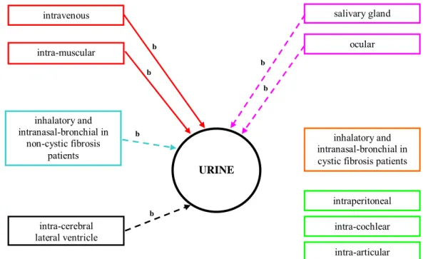
3.2.2.1 Urine
Shedding in urine was detected when HAdV-5 was administered intravenously (46), intra-tumorally (59;61) and intra-prostatical (54;75). Shedding in urine or urethral swabs could not be detected after intraperitoneal (55;56), intra-pleural (57), intra-muscular (80-82), inhalatory/intranasal-bronchial (85) or ocular (88) administration of HAdV-5, or when HAdV-5 was administered directly into the brain (70;76-78), para-aortic lymph node (54), spine (54) or ileum (54). Urine has not been tested for the presence of HAdV-5 after dermal administration or administration in the salivary gland, bladder or maternal urine after administration to the foetus.
The presence of HAdV-5 in urine and plasma (46) suggests the possible presence of HAdV-5 in kidney. This is confirmed in several pre-clinical studies that reported an HAdV-5 signal in kidney after intravenous delivery in mice (39;42). In non-clinical studies also the long term presence of HAdV-5 in plasma is confirmed. One study indicated that the expression of the cloned transgene in serum is gradually decreasing and finally undetectable at 63 days after administration (42). This is in agreement with the observation that at day 70 no HAdV-5 could be detected in kidney (42). It is therefore expected that also shedding in urine after day 70 could not have been detected, although this has not been investigated.
After intra-tumoral administration of HAdV-5 (1*106 to 1*1011 pfu) in patients with non-small-cell lung cancer, shedding in urine is reported within 24 hours of injection in all patients (n=24). After 9 days, HAdV-5 could not be detected in urine (59). Shedding in urine was also reported by Fujiwara et al. (2006), after intra-tumoral administration of the same tumour type (1*109 to 1*1010 pfu) (61). However, these authors detected a positive signal on days 3 to 14 in 3 out of 12 patients. In this study, no shedding was detected on days 1 and 2 and all patients tested negative on day 15. After day 15, no tests for shedding were performed (61). Kidney samples obtained from the 2 deceased patients on day 25 and 151 were negative (61). One of these patients did show shedding in urine until day 14. The negative kidney signals are consistent with the fact that after day 14 no shedding in urine was observed anymore. Together, the data suggest that the vector is cleared from the kidney within 15 days following intra-tumoral administration of HAdV-5. In addition, no HAdV-5 was detected in urine 2 days following intra-tumoral administration to the same tumour type with a vector dose of (1*107 to 1*109 pfu) (60). Administration into other tumour types demonstrated the absence of shedding in urine, e.g. administration into liver tumours (63), mammary tumours (64), melanoma (64;65) and soft tissue sarcoma (67).
Intra-prostatic administration of HAdV-5 results in shedding of HAdV-5 via urine (54;75). Shedding via urine was observed when HAdV-5 was injected in the prostatic fossa (54) or into the prostate gland (75). When patients received HAdV-5 by injection in the prostatic fossa, no HAdV-5 material was detected in blood serum (54). In addition, the presence of replicating adenovirus in blood was not detected following injection into the prostate gland (75). Also kidney remained negative for HAdV-5
material in a study investigating the biodistribution of intra-prostatic injected HAdV-5 in mice, but showed biodistribution to the liver (39).
3.2.2.2 Faeces
Shedding of HAdV-5 via faeces has been observed following several administration routes. Following intravenous injection of an HAdV-5 viral vector, shedding was detected in the faeces of 1 out of 6 patients, somewhere between 1 and 28 days after injection (46). In addition, following intravenous injection in mice, HAdV-5 was detected in bile from 10 minutes up to 24 hours after delivery of the virus (84), indicating that the virus is shed via faeces. Intra-tumoral injection of low doses of HAdV-5 (≤ 107 pfu) did not result in faecal shedding 1-2 days after injection in patients (60;63), whereas in the same period, at doses ≥ 108 pfu, viral DNA was detected in stool samples (60). Similarly, faecal shedding following the inhalatory administration route seems to be dose-dependent, with shedding only observed at doses of 1010 pfu (84;85). In addition, data indicate that faecal shedding is time-dependent, and only occurs (at doses of 1010 pfu) in the first 2 days after nasal administration, but not afterwards (84).
Shedding of HAdV-5 via faeces could not be detected when administered via the intraperitoneal (55;56), intra-pleural (57), intra-prostatic (72), or intra-muscular route (81;82) or after administration to the brain (76;77) and was not analysed following the other administration routes (dermal, ocular, salivary gland, para-aortic lymph node, spine, ileum, bladder and in the mother after foetal administration).
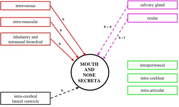
3.2.2.3 Mouth and nose secreta
Shedding of HAdV-5 via mouth and/or nose has not been investigated systematically. Indications for shedding of HAdV-5 via mouth and nose were reported following intra-tumoral (59-62;68), inhalatory and intranasal administration (83). Shedding via nose and mouth after intravenous, intra-prostatic, brain, intra-muscular and ocular administration has not been observed. No data is published indicating the presence or absence of shedding via mouth and nose regarding dermal administration and administration directly to the salivary gland, para-aortic lymph node, spine, ileum and bladder.
Following intra-tumoral administration to different tumour types like lung cancer tumours (59-62) and head and neck squamous cell carcinoma (68), HAdV-5 material was detected in several mouth and nose secreta samples which might indicate HAdV-5 shedding via mouth and/or nose. Positively tested samples were sputum (59;60;68), bronchial fluid (60), saliva (59;68), throat swabs (60) and gargle (61). In a clinical study where HAdV-5 was administered intra-tumorally in lung cancer tumours, sputum and saliva tested positive for HAdV-5 within the first 24 hours of injection in all tested patients (n=18) that received dose levels between 1*106 to 1*1011 pfu. Nine days after injection no positive results were obtained. Testing was based on a CPE assay, meaning no infectious HAdV-5 particles were present (59). In another study, 21 lung cancer patients were injected intra-tumoral with 107 to 109 pfu HAdV-5. Positive sputum samples (days 1 to 60) were detected by PCR in 15 patients. With a CPE assay, the number of positively tested samples was strongly reduced compared to the previous study; only two HAdV-5 positive samples on day 1 and 2 were found (60). In this study, throat swabs were also positive between days 1 and 11 in six out of twenty-one patients. A CPE test performed on these samples did not reveal the presence of HAdV-5 infectious particles. Bronchial fluid samples collected (up to 90 days) were HAdV-5 positive (PCR) on days 1 (20 patients) to 90 (4 patients). With a CPE assay, HAdV-5 positive bronchial fluid was only detected on day 0 just after administration (60). In a recent study, HAdV-5 was detected by PCR in 29 of 39 gargle samples obtained 1 day after vector injection, regardless of dose level (109 to 1011 pfu) and declined to undetectable levels within 15 days for most patients. In total, 90 gargle samples (14.4%) were positive for vector DNA (61). Infectious HAdV-5 was also detected in sputum and/or saliva samples of patients who received HAdV-5 intra-tumorally injected in head and neck squamous cell carcinomas (1011 pfu) (68). At lower doses all samples remained negative. The highest titer found was 106 pfu per 0.5 ml sputum or saliva.
Shedding via nose and mouth secreta has also been reported following inhalatory and intranasal administration used for treatment of cystic fibrosis patients with dose levels ranging from 105 to 5.4*108 pfu (83). CPE testing did not reveal the presence of HAdV-5 in cultures derived from nasal brush, bronchial brush or saliva. Using PCR, HAdV-5 was detected in nasal brush (up to 21 days post administration), bronchial brush (up to 14 days post administration) and in saliva (up to 14 days post administration) (83).
HAdV-5 shedding via nose and mouth has not been reported following intra-tumoral administration of HAdV-5 to other tumour types like metastatic breast cancer tumours (64), melanoma (64), liver cancer tumours (63), soft tissue sarcomas of the extremity (67) and recurrent malignant gliomas (70). In these studies no HAdV-5 was detected in throat swabs (63;64), sputum (67) and nasal swabs (70).
3.2.2.4 Semen
The presence of HAdV-5 material was detected in sperm of rabbits 2 weeks following intravenous administration, although according to the authors, this was probably due to the presence of (positive) blood in the sample (44). In addition, following intravenous administration distribution of HAdV-5 could also be found in the testes of rabbits and baboons (44;48), as well as in the prostate and ureter of mice (39;50). However, adenovirus was not detected in sperm 8 weeks following intra-muscular administration in human, but biodistribution to organs involved in the production or excretion of semen were not analysed (80). For the other administration routes described in this review, shedding via semen was not analysed.
3.2.2.5 Various smaller excretion routes
After dermal application, only local distribution was found and shedding was observed via the wound bed (71). No other excreta were measured. The other HAdV-5 administration routes did not investigate biodistribution to the skin or shedding via a wound bed or skin.
Shedding via ear swabs was only studied by Herman et al (1999) and did not occur within 4 days after intra-prostatic administration of 1*108 to 1*1011 iu (75). However, the detection method was based on culturing for wild type adenovirus.
After intravenous administration of 9.5*1011 vp HAdV-5 to rabbit, the eyes were positive on day 1 using quantitative PCR. No other biodistribution studies investigated the distribution to the eye or the shedding via lachrymal fluid.
3.3 Results AAV2
Biodistribution and shedding of AAV2 viral vectors are dependent on the route of administration. In table 3 the biodistribution and shedding routes of AAV2 viral vectors following various administration routes are described. Detailed information on the biodistribution and shedding per experiment can be found in Appendices A (Table A.16 to A.24) and B.
Table 3. Biodistribution and shedding of AAV2
Administration route Biodistribution Shedding
intravenous positive: plasma, brain, liver, lung,
spleen, kidney, gonads, heart, bladder, muscle, GI-tract, salivary gland, lymph node, retina,
tonsil, bone marrow, gallbladder, thyroid, pancreas, skin, thymus,
trachea, adrenal gland and peripheral blood mononuclear cells
negative: spinal cord
positive: saliva, urine and semen
intraperitoneal positive: abdominal tissue,
diaphragm and intercostal muscle
negative: heart, liver, lung, kidney,
spleen, other muscles and testis
no data available
intra-muscular positive: serum, heart, kidney, liver,
lung, spleen, gonads, injected muscle, lymph node and
epididymal fluid
negative: intestine, other muscle,
adrenal gland, brain, blood cells, thymus, thyroid and spinal cord
positive: saliva, urine, semen,
faeces, lachrymal swab and nasal swab
negative: stool
inhalatory and intranasal-bronchial
positive: blood positive: sputum negative: urine, stool, BAL,
nasal swab and nose secretion
ocular positive: diaphragm, heart,
lymph node, kidney, eye, visual cortex and LGN
negative: gonads, jejunum, liver,
lung, muscle, pancreas, spleen, cerebellum and thalamus
no data available
salivary gland positive: salivary gland, liver, spleen
and testis
negative: heart, lung, kidney and
lymph node
Table 3. Biodistribution and shedding of AAV2 (continued)
Administration route Biodistribution Shedding
intra-cochlear and intra-articular
positive: cerebellum, cochlea negative: serum, cortex, heart,
kidney, liver and lung
no data available
intra-cerebral lateral ventricle
positive: brain, meninges and
pial surface
no data available isolated limb perfusion positive: serum and perfused muscle
negative: kidney, liver, lung, control
muscle, gonads and spleen
no data available
LGN = lateral geniculate nucleus BAL = broncho-alveolar lavage GI-tract = gastrointestinal tract
3.3.1 Biodistribution and shedding per administration route
3.3.1.1 Intravenous administration
In a clinical trial for the treatment of haemophilia, AAV2 was delivered to the liver via the hepatic artery which resulted in unexpected transient vector dissemination to the semen which was detected by PCR. Results from serum and urine suggested that clearance was dose dependent. Urine was the first body fluid to clear. There was no association between dose and time for the clearance of semen. The last positive semen samples in the lowest dose cohort were at 12 weeks, whereas the last positive samples in the five subjects in the two higher- dose cohorts occurred at a mean of ~5 weeks. Age of the subject seemed to be the best predictor for the time needed for the semen to clear, with young men on average clearing more quickly than older men. Semen fractionation, carried out for a single subject, showed no evidence of vector sequences in motile sperm. Peripheral blood mononuclear cells showed the longest duration of positive results of any body fluid or tissue analysed (92). In pre-clinical studies following administration of AAV2 to the hepatic artery, biodistribution to the gonads was analysed in rats after 50 and 92 days. A dose-dependent response was observed; 50 days after injection the percentage of animals with a positive signal in the gonads increased with increasing doses of AAV2 and after 92 days positive results were only observed in rats that had received the highest dose (1*1013 vp/kg) (93). The liver was positive for at least 7 weeks in rats treated with 5*1012 vp/kg (94). However, in this study no other organs or shedding were investigated. Also in rabbit, administration of AAV2 to the hepatic artery resulted in detectable vector sequences after more than 18-20 months in the gonads of animals that were administered a dose of 1*1012 or 1*1013 vp/kg, but not in animals that received 1*1011 vp/kg. The liver was positive for more than 1 year after injection in all dosage groups. At 4 days after administration of the virus, infectious particles were detected in semen, but not anymore after 7 and 14 days. The presence of viral DNA however, was found up to 6, 8 or 13 weeks after doses
of 1*1011, 1*1012 and 1*1013vp/kg, respectively (95). In mice administered AAV2
(2.1*1010-8.4*1010 vp) via the portal vein or via tail vein infusion, the liver remained positive for 49 days, while the muscle and spleen were negative within 7 days (96). In mice treated with 2*1011 vp, the brain, heart, kidney, liver, lung and spleen (investigated organs) were still positive at 6 weeks after administration (97). Biodistribution to the gonads or shedding in the semen was not detected between 7 and 90 days after injection (3.7*1012 or 7*1012 vp/kg) in dogs into the hepatic artery (93).
Four biodistribution studies have been performed in monkeys (98-101). Following administration of AAV2 into the hepatic artery or portal vein of Macaques, viral DNA was found in plasma after 3 and 6 days, but not after 8 days. In addition, after 11 months, viral DNA was found in the liver and spleen, but not in the kidneys or gonads (100). A previous study by Nathwani et al. (2001) showed that the heart, liver and spleen were also positive for viral genome particles after 22 weeks of administration (98). However, kidney and lung were negative at 22 weeks. Mori et al. (2006) performed an extensive study concerning the biodistribution of AAV2 following injection of several doses in the femoral vein of Cynomolgus monkeys (101). Doses varied from 1*1010 to 2.5*1011 vp/kg and follow up time after viral injection varied from 2 days to 7 months. Colon, spleen and several lymph nodes (axillary, hilar, iliac and mesenteric) tested positive for viral DNA for all doses and time points. In addition, liver, tonsil, bladder and inguinal lymph nodes were positive for all doses and time points, except 3 months after injection of the lowest dose. At doses of 2.5*1010 or higher, viral DNA was detected in bone marrow, gall bladder, lung and pancreas for 2 days up to 5 months after injection. At such doses viral DNA was also present in heart and kidney after 3 months (negative on day 2) and in the trachea and cerebrum 2 days and 3 months after injection. Adrenals and muscle were only observed to be positive after doses of 1*1011 vp/kg or higher (3 and 5 months) and retina was found positive (7 months) after the injection of 1*1010 vp/kg. Occasionally, not dose- and/or time-related positive findings were obtained in the cerebellum, gonads, ileum, oesophagus, stomach, salivary glands, skin, thymus and thyroid. In the jejunum and spinal cord, evidence for the presence of viral particles was never detected (101). Evidence for shedding in monkeys was only observed in saliva and urine, up to 3 and 6 days respectively, following administration to the hepatic artery or portal vein (100). Other shedding routes were not analysed.
3.3.1.2 Intraperitoneal administration
Only one study reported the biodistribution after intraperitoneal administration of AAV2 (102). Two months after viral delivery (2*1011 vp) in neonatal mice, viral DNA was found in the intercostal muscles, abdominal tissue and diaphragm, but not in the heart, kidney, liver, lung, spleen, testis and other muscle tissue (triceps and masseter) (102). Shedding was not studied, but data indicate that AAV2 remains locally situated.
3.3.1.3 Intra-muscular administration
Biodistribution of viral vector particles following intra-muscular administration of AAV depends on the muscle group that is injected. After intra-myocardial administration of AAV2 (4*1011 vp) to rat, biodistribution of AAV2 was observed to the testes, heart, kidney and liver, but not to the spleen (103). Blood or plasma was not studied, but because AAV2 vectors were measured outside the heart, it is expected that the viral vector is transported throughout the body via the systemic circulation.
Two limited biodistribution studies were performed in mice with AAV2 injections into muscle of the leg. AAV2 was present in the liver after 56 days after a dosage of 1*1011 vp and in the gonads up to 91 days after dosages of 1.7*1011 and 1.7*1012 vp/kg (93;104). In another biodistribution study administering 5*1010 vp in mice, it was shown that after 22 weeks the measured organs were still positive for viral genomic copies (98).
One very limited biodistribution study after intra-muscular administration was performed in rat by Arruda et al., 2001 (2.8*1011 and 2.8*1013 vp/kg) (93). Epididymal effluent was positive at 15 days after injection into the leg muscle. No other tissues were studied.
Arruda et al., 2001 also studied the biodistribution of AAV2 in rabbits (93). Serum was positive up to 48 hours and negative at 7 days and gonads were positive up to 90 days. However, semen was negative throughout the study, indicating that in contrast to in rats, semen is not a shedding route of AAV2 in rabbit.