Focus cardiac ultrasound: the European
Association of Cardiovascular Imaging viewpoint
Aleksandar N. Neskovic
1*
, Thor Edvardsen
2, Maurizio Galderisi
3, Madalina Garbi
4,
Giuseppe Gullace
5, Ruxandra Jurcut
6, Havard Dalen
7,8, Andreas Hagendorff
9, and
Patrizio Lancellotti
10, for the European Association of Cardiovascular Imaging
Document Reviewers: Bogdan A. Popescu, Rosa Sicari and Alexander Stefanidis
1
Clinical Hospital Center Zemun, Faculty of Medicine, University of Belgrade, Belgrade, Serbia;2
Department of Cardiology, Oslo University Hospital and University of Oslo, Oslo, Norway;
3
Department of Translational Medical Sciences, Federico II University Hospital, Naples, Italy;4
King’s Health Partners, King’s College Hospital NHS Foundation Trust, London, UK;
5
Cardiovascular Department of Azienda Ospedaliera della Provincia di Lecco, Lecco, Italy;6
Department of Cardiology, University of Medicine and Pharmacy ‘Carol Davila’, Emergency Institute of Cardiovascular Diseases, Bucharest, Romania;7
MI Lab and Department of Circulation and Medical Imaging, Norwegian University of Science and Technology, Trondheim, Norway;8
Department of Internal Medicine, Levanger Hospital, Nord-Trøndelag Health Trust, Levanger, Norway;9
Department of Cardiology-Angiology, University of Leipzig, Leipzig, Germany; and10
Department of Cardiology, University of Lie`ge Hospital, GIGA Cardiovascular Sciences, Heart Valve Clinic, CHU Sart Tilman, Lie`ge, Belgium Received 6 April 2014; accepted after revision 10 April 2014; online publish-ahead-of-print 27 May 2014
The concept of point-of-care, problem-oriented focus cardiac ultrasound examination (FoCUS) is increasingly applied in the settings of medical emergencies, including cardiac diseases. The European Association of Cardiovascular Imaging (EACVI) recognizes that cardiologists are not the only medical professionals dealing with cardiovascular emergencies. In reality, emergency cardiac diagnostics and treatment are also carried out by a wide range of specialists. For the benefit of the patients, the EACVI encourages any medical professional, sufficiently trained to obtain valuable information from FoCUS, to use it in emergency settings. These medical professionals need to have the necessary knowledge to understand the obtained information entirely, and to use it correctly, thoughtfully and with care. In this document, the EACVI underlines major differences between echocardiography and FoCUS, and underscores the need for specific education and training in order to fully utilize advantages and min-imize drawbacks of this type of cardiac ultrasound examination in the critically ill patients.
Keywords EACVI † Viewpoint † Focus cardiac † Ultrasound
“Truth is one, paths are many” - Mahatma Gandhi “The truth is rarely pure, and never simple” - Oscar Wild
Introduction
There is a trend of rapidly growing use of echocardiography in emer-gency settings by non-cardiologists or cardiologists without specific expertise. Therefore, the European Association of Cardiovascular Imaging (EACVI) has been urged to address this important issue and set the standards for adequate education and training, for the safe and efficient use of echocardiography in emergency care.1It has been recognized that performing echocardiographic examinations in emergency situations is challenging and that both interpretation errors and missed findings are likely to occur more frequently. Con-sequently, the position of the EACVI is that the level of competence in echocardiography required for emergency cases is at least the
same as for elective cases. Accordingly, the EACVI recommended competence requirements for emergency echocardiography are the same for cardiologists and non-cardiologists, i.e. anaesthesiologists, emergency physicians, intensive care specialists, cardiac surgeons, cardiac physiologists (individuals owing a cardiac physiology degree and subsequent appropriate echocardiography training and certifica-tion, working in team with cardiologists and cardiac surgeons). It was proposed that non-cardiologists should reach the same level of ex-pertise through a training programme similar with the training pro-gramme for general cardiologists, with additional theoretical learning on certain cardiovascular diseases/conditions.1
We are also witnessing an evolving trend of using cardiac ultra-sound examination as a bedside, point-of-care diagnostic test in emergency settings, examination named ‘Focus Cardiac Ultrasound’ (FoCUS).
Current EACVI recommendations refer to emergency echocardi-ography and not to FoCUS.1Therefore, the EACVI, as the reference
*Corresponding author. Tel:+381 641216359; Fax: +381 112107057, Email: neskovic@hotmail.com
Published on behalf of the European Society of Cardiology. All rights reserved.&The Author 2014. For permissions please email: Journals.permissions@oup.com.
by guest on October 1, 2015
European echocardiographic community representative, should briefly address this issue.
Of note, this document is not a systematic review of current FoCUS practice.
Purpose
The purpose of this document is to outline the position of the EACVI on the use of FoCUS in the emergency settings, to underline major differences between echocardiography and FoCUS, and to reinforce the need for specific education and training in order to fully utilize advantages and minimize drawbacks of this type of cardiac ultrasound examination in the critically ill patients.
Terminology
Echocardiography is a comprehensive investigation, requiring maximum technical skills along with expertise in cardiovascular pathophysiology and cardiovascular diseases. Thus, the term echocar-diography refers to comprehensive standard echocarechocar-diography in emergency settings, i.e. emergency echocardiography, which always represents a full echocardiographic investigation of cardiac morphology and function, using fully equipped echocardiographic machines, performed by a sufficiently trained operator who is able to independently perform and interpret the study.1
The term FoCUS defines the point-of-care cardiac ultrasound exam-ination, performed according to standardized but restricted scanning protocol to add information to the physical examination, by an oper-ator not necessarily fully trained in echocardiography but appropriate-ly trained in FoCUS, who is at the same time usualappropriate-ly responsible for immediate decision-making and/or treatment.2,3
Both cardiologists and non-cardiologists can perform either echo-cardiography examinations or FoCUS, depending on clinical circum-stances, existing equipment, and expertise. FoCUS is typically used by non-cardiologists who underwent minimal training, but can also be performed by fully trained cardiologists or cardiac physiologists in emergency settings.
Imaging devices
The whole spectrum of echocardiographic machines, from high-end, fully equipped platforms to pocket-size imaging devices, can be used for FoCUS.
However, due to logistics as well as to the narrow scope of the FoCUS examinations, smaller, especially hand-held and pocket-size imaging devices are used more frequently. Current pocket-size imaging devices offer diagnostic-quality two-dimensional and, in part, colour Doppler cardiac imaging in real-time providing the ability to measure only linear dimensions.4The small size makes them attractive for all operators performing FoCUS examinations. Purely, qualitative assessment performed with pocket-size imaging devices has demon-strated good level of diagnostic accuracy for both experienced and non-experienced operators.5–8However, it should be acknowledged that image quality is generally inferior when compared with high-end systems, and that this might be particularly important in difficult patients, resulting in technically suboptimal studies, leading to less re-liable findings.4,6,9–11 The operator must be fully aware of these
technical limitations and examinations performed with pocket-size imaging devices must be only reported as a complement to physical examination, and not as a complete echocardiogram.4
Expectations and limitations of the
FoCUS
The crucial difference between echocardiography and FoCUS in emergency settings, with the assumption that both are performed and interpreted in a competent manner, is the amount of information obtained. FoCUS examination provides sufficient information for mostly qualitative gross assessment of cardiac morphology and func-tion, reported as ‘absent/present’, or ‘yes/no’ (i.e. qualitative assess-ment). Being an inherently limited approach, FoCUS examination carries additional risks of overlooking important abnormalities and of false readings of an incomplete dataset. Although these types of errors could certainly occur also during echocardiography, their expected rates might be considerably higher for the FoCUS, and can be especially high in situations where the operators are not fully trained in echocardiography and/or cardiology.1It should be emphasized that failure to appreciate the limitations of the FoCUS (Table1) may lead to serious misinterpretation of the findings with potentially devastating clinical consequences. Recognition of these limitations, therefore, must be incorporated in any training protocol for the FoCUS.
Finally, a FoCUS operator should not overestimate his own expertise and need to refer the patient to echocardiographic exam-ination whenever possible, particularly in situations in which FoCUS, although diagnostic, is not sufficient for accurate decision-making.
Education and training frame
The concept of point-of-care, problem-oriented FoCUS examin-ation has been actively promoted by different professional organiza-tions,12–14with a number of initiatives aiming to set up, standardize and support education and training, and to facilitate research related to the application of this concept in in- and out-of-the-hospital emergency situations, including cardiac emergencies.
Increasing interest among a wide range of physicians of different specialities involved in managing patients in emergency settings has
Table 1 Clinically relevant limitations of the FoCUS when compared with comprehensive echocardiography
1. Technical inferiority of the imaging devices typically used for FoCUS examination compared with fully equipped high-end
echocardiographic machines
2. Limited skills and experience of the operators
3. Typically unfavourable settings (emergencies, critically ill, time constrains)
4. Narrow list of detectable evidence-based targets
5. ‘Absent/Present’ or ‘Yes/No’ reporting style (related to 1 – 4) 6. Subtle/complex cardiac abnormalities (i.e. regional asynergy)
difficult to assess (related to 1 – 4)
by guest on October 1, 2015
been driven further by the availability of high-quality small or pocket-size imaging devices. On the other hand, there is an obvious shortage of cardiologists fully trained in echocardiography at all locations where medical emergencies occur. Therefore, we believe that the EACVI should encourage any medical professional, sufficiently trained to obtain valuable information from FoCUS, to use it in emer-gency settings. Thus, all attempts at individual or professional organ-ization levels, aiming to narrow this existing logistic gap between obvious needs and available human resources are praised by the EACVI.
The EACVI recognizes that cardiologists are not the only medical professionals dealing with cardiovascular emergencies.1In reality, emergency cardiac diagnostics and treatment are also carried out by wide range of specialists, including emergency physicians, intensive care specialists, anaesthesiologists, and cardiac surgeons. In addi-tion, in life-threatening scenarios, fellows in training in respective specialities and even sonographers/cardiac physiologists can find themselves in need of performing cardiac ultrasound scanning in an attempt to obtain essential diagnostic information.
There are undisputable differences between existing educational and training programmes for emergency echocardiography1 and FoCUS.12,15–21 The differences are substantial, not only with regard to content and duration of echocardiographic training,1,22–25 but also to specific content on emergency cardiovascular disease for non-cardiologists. A variety of non-cardiology specialists perform FoCUS in a variety of specific emergency situations within specific patient populations. Being predominantly responsible for developing education and training programmes for echocardiography,1,22–25the EACVI is not primarily in charge with the development and running of FoCUS educational and training programmes. The key responsibility for this lies in the hands of clinician’s/operator’s respective speciality organizations and/or scientific bodies, which are fully aware of re-spective specialty-specific FoCUS use circumstances.
Currently, a variety of recommendations, statements, and proto-cols for education and training in FoCUS provided by respective societies/organizations exists.2,3,12–21,26,27 Teaching courses for FoCUS in emergency or critical care situations have been developed in order to qualify individuals, after short, intensive and narrow scope-oriented training28,29meant to allow them to identify basic but critical cardiac conditions and pathologies (Table2). Studies have shown that these findings may beneficially modify patient man-agement,30–41 and predict outcome.42,43,44,45,46 We believe that further expansion of the list of teaching and training targets for FoCUS (i.e. valvular abnormalities, intracardiac masses, regional wall motion analysis) would be associated with an increased risk of improper use and errors.8Assessment of these conditions in the vast majority of cases requires significantly higher level of expertise and should indicate referral for echocardiographic evaluation.
Recently, the EACVI has provided a list of emergency cardiovas-cular diseases/conditions to be included in additional learning programmes for non-cardiologists in training on emergency echocar-diography.1We believe that this list should be incorporated in the theoretical/didactic part of the FoCUS training programmes for non-cardiologists. To which extent the listed conditions1 will be addressed in particular training programmes for FoCUS should be decided by each respective speciality responsible for running the programme. Such specific theoretical knowledge will aid physicians
who perform the FoCUS to integrate cardiac ultrasound findings in clinical context, for the best possible patient management.
Knowing the complexity of the topic and diversity of medical pro-fessionals who undergo training in FoCUS, it seems unlikely that strictly predefined minimal number of hours of hands-on image ac-quisition training or the number of personally performed and/or interpreted cases, would ever fit for all. The available literature clearly reflects this diversity.2,3,12–21However, since the competency in FoCUS should be the minimal net result of the training process, the number of required hours/studies might be adjusted for each trainee according to the results of the competency evaluation incorporated into the ongoing training process. General tools for the assessment of competency in FoCUS currently do not exist and each respective specialty should be encouraged to work on defining and implement-ing them in the trainimplement-ing process.
It appears logical to recommend that a significant number of FoCUS examinations should be performed with the same imaging device and under similar real-life clinical scenarios as for the ones encountered when using FoCUS,3 instead of practising on stable patients using fully equipped echocardiographic machines. This is very important, since higher technical skills may be needed for optimal image acquisition in unfavourable emergency settings com-paring to echo scanning in stable, elective conditions.1
It is also important that the case mix of performed FoCUS examinations during the training process covers the full range of critical/life-saving scenarios representative for the trainee’s scope of practice.
Finally, we recommend that FoCUS training programmes ensure that the operators are aware of their own capacity and limitations, given the specific equipment and different situations. Only in this
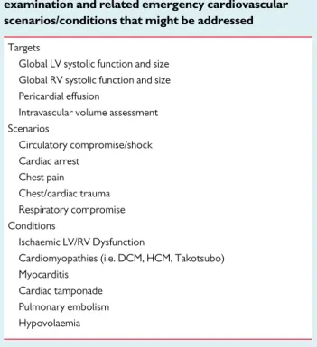
Table 2 Evidence-based targets of FoCUS examination and related emergency cardiovascular scenarios/conditions that might be addressed
Targets
Global LV systolic function and size Global RV systolic function and size Pericardial effusion
Intravascular volume assessment Scenarios Circulatory compromise/shock Cardiac arrest Chest pain Chest/cardiac trauma Respiratory compromise Conditions Ischaemic LV/RV Dysfunction
Cardiomyopathies (i.e. DCM, HCM, Takotsubo) Myocarditis
Cardiac tamponade Pulmonary embolism Hypovolaemia
DCM, dilated cardiomyopathy; HCM, hypertrophic cardiomyopathy; LV, left ventricular; RV, right ventricular.
by guest on October 1, 2015
way, it is ensured that FoCUS can improve diagnostics. The use of FoCUS or simplified ultrasound devices should never deprive the patient of the opportunity of a better diagnostic test.
Supervision, quality control,
echocardiographic service
Considering all features of FoCUS, referral for echocardiographic examination must follow FoCUS as soon as possible, in all cases with abnormal findings or non-diagnostic studies, and continuous supervision and quality control are essential. Established emergency echocardiography service in the hospital1may provide professional, educational, and training support for non-cardiologists performing FoCUS, through 24 h availability of second opinion/consultative or on-call service, team work (professional help, consultations, regular reviewing of cases), and continuous supervision. Accredited echocardiography laboratories should preferably be the hub for quality control and supervision, where FoCUS cases should be reviewed, re-evaluated, and discussed together with fully trained and/or expert cardiologists.
Reporting and storage
As recommended for emergency echocardiography,1FoCUS exam-ination should be well and retrievably stored, including both the report and the acquired images/cineloops. These data should be stored permanently to be used for case reviews, consultations, quality control purpose, and as evidence of findings in acute setting for medico-legal purposes.
Since the time available to elaborate a FoCUS examination report may be extremely brief, a concise report (written and/or verbal), concentrating on critical findings and integrated into the decision-making process may be issued. However, this must be followed by a final written report, interpreted, approved, and signed by the opera-tors with adequate formal education.
Co-operation
Both reference echocardiography community representatives and respective speciality societies/organizations currently involved in FoCUS educational and training activities have the common aim to expand and improve cardiac ultrasound examination practice for the benefit of patients. Although approaches and proposed standards to reach this aim may not always be the same, differences should be debated and positions harmonized whenever possible.
The EACVI position is that all current and future activities related to FoCUS should be done in close co-operation between respective speciality societies/organizations already engaged in FoCUS educa-tional and training activities, and the reference echocardiography community representatives. This could be done by means of prepar-ing recommendations and consensus documents,2,3endorsing docu-ments, adjusting educational and training programmes, running research projects, and organizing joint professional and scientific meetings.
This approach would certainly provide the mileu for true partner-ship and the basis for future developments in the field.
Conclusions
The viewpoint of the EACVI on FoCUS is summarized in Table3. The EACVI fully acknowledges the efforts and achievements of individuals and professional organizations that dedicate their activities to improve emergency cardiac care using point-of-care FoCUS. At the same time, the EACVI believes that only full implementation of edu-cation and training requirements for all professionals performing FoCUS, will secure the accuracy and quality of the information obtained with cardiac ultrasound in emergency settings. These requirements should be defined by the respective specialty profes-sional organizations/regulatory bodies in collaboration with the ref-erence echocardiography community representatives.
The EACVI promotes and strongly supports systematic training in echocardiography and emergency echocardiography, as a general pre-requisite for full competence in collecting high-quality informa-tion with cardiac ultrasound and, consequently, optimal patient management.
For critically ill patients, it is not important whether the life-saving information is acquired by non-cardiologist performing FoCUS, or by the expert cardiologist performing echocardiography. When such information is available, it has to be used. However, for the benefit of the patients, the involved medical professionals should have the
Table 3 Summary of the EACVI viewpoint on FoCUS
FoCUS should only be used as a point-of-care cardiac ultrasound examination, aimed to detect limited number of critical cardiac conditions
FoCUS may provide key clinical information regarding the presence of pericardial effusion/cardiac tamponade, left and right ventricular size and function, intravascular volume status, and may aid
decision-making during cardiopulmonary resuscitation
FoCUS should never be considered or reported as echocardiographic examination
Educational curriculum and training programme for FoCUS should be designed and conducted by the specialty professional organizations/ societies involved in treating medical emergencies, including cardiac, with continual collaboration with reference echocardiographic communities
FoCUS should only be used by the operators who have completed appropriate education and training programme, and who fully understand and respect its scope and limitations
All patients with cardiovascular abnormalities detected by FoCUS should be referred to echocardiographic examination as soon as appropriate
If the underlying cardiovascular disease could not be definitively ruled out in critically ill by FoCUS, echocardiography should be considered FoCUS examinations should be recorded and permanently stored and
reports issued in a timely manner
Continual supervision and quality control of the FoCUS examinations are essential, provided preferably by accredited echocardiographic laboratories and emergency echocardiography services
Reference echocardiographic community representatives should actively follow developments in the field and, whenever appropriate, work on improving educational and training curricula in concert with respective specialities professional societies/organizations, to deliver the best possible care for the patients
by guest on October 1, 2015
necessary knowledge to understand the obtained information entire-ly, and to use it correctentire-ly, thoughtfully and with care.
Conflict of interest: none declared.
References
1. Neskovic AN, Hagendorff A, Lancellotti P, Guarracino F, Varga A, Cosyns B et al. Emergency echocardiography: the European Association of Cardiovascular Imaging recommendations. Eur Heart J Cardiovasc Imaging 2013;14:1 – 11. 2. Labovitz AJ, Noble VE, Bierig M, Goldstein SA, Jones R, Kort S et al. Focused cardiac
ultrasound in the emergent setting: a consensus statement of the American Society of Echocardiography and American College of Emergency Physicians. J Am Soc Echo-cardiogr 2010;23:1225 – 30.
3. Spencer KT, Kimura BJ, Korcarz CE, Pellikka PA, Rahko PS, Siegel RJ. Focused cardiac ultrasound: recommendations from the american society of echocardiography. J Am Soc Echocardiogr 2013;26:567 – 81.
4. Sicari R, Galderisi M, Voigt J-U, Habib G, Zamorano JL, Lancellotti P et al. The useof pocket-size imaging devices: a position statement of the European Association of Echocardiography. Eur J Echocardiogr 2011;12:85 – 7.
5. Galderisi M, Santoro A, Versiero M, Lomoriello VS, Esposito R, Raia R et al. Improved cardiovascular diagnostic accuracy by pocket size imaging device in non-cardiologic outpatients: the NaUSiCa (Naples Ultrasound Stethoscope in Cardiology) study. Cardiovasc Ultrasound 2010;26:51.
6. Prinz C, Voigt JU. Diagnostic accuracy of a hand-held ultrasound scanner in routine patients referred for echocardiography. J Am Soc Echocardiogr 2011;24:111 – 6. 7. Andersen GN, Haugen BO, Graven T, Salvesen O, Mjølstad OC, Dalen H. Feasibility
and reliability of point-of-care pocket-sized echocardiography. Eur J Echocardiogr 2011;12:665 – 70.
8. Mjølstad OC, Andersen GN, Dalen H, Graven T, Skjetne K, Kleinau JO et al. Feasi-bility and reliaFeasi-bility of point-of-care pocket-size echocardiography performed by medical residents. Eur Heart J Cardiovasc Imaging 2013;14:1195 – 202.
9. Scholten C, Rosenhek R, Binder T, Zehetgruber M, Maurer G, Baumgartner H. Hand-held miniaturized cardiac ultrasound instruments for rapid and effective bedside diagnosis and patient screening. Journal of Evaluation in Clinical Practice 2005;11:67 – 72.
10. Liebo MJ, Israel RL, Lillie EO, Smith MR, Rubenson DS, Topol EJ. Is pocket mobile echocardiography the next-generation stethoscope? A cross-sectional comparison of rapidly acquired images with standard transthoracic echocardiography. Ann Intern Med 2011;155:33 – 8.
11. Liu SC, Chang WT, Huang CH, Weng TI, Ma Matthew HM, Chen WJ. The value of portable ultrasound for evaluation of cardiomegaly patients presenting at the emer-gency department. Resuscitation 2005;64:327 – 31.
12. Price S, Via G, Sloth E, Guarracino F, Breitkreutz R, Catena E et al. World Interactive Network Focused On Critical UltraSound ECHO-ICU Group. Echocardiography practice, training and accreditation in the intensive care: document for the World Interactive Network Focused on Critical Ultrasound (WINFOCUS). Cardiovasc Ultrasound 2008;6:49.
13. Mayo PH, Beaulieu Y, Doelken P, Feller-Kopman D, Harrod C, Kaplan A et al. Ameri-can College of Chest Physicians/La Societe de Reanimation de Langue Francaise statement on competence in critical care ultrasonography. Chest 2009;135: 1050 – 60.
14. American College of Emergency Physicians. Emergency ultrasound guidelines 2008. Available at: http://www.acep.org (20 February 2014, date last accessed). 15. Hellmann DB, Whiting-O’Keefe Q, Shapiro EP, Martin LD, Martire C,
Ziegelstein RC. The rate at which residents learn to use hand-held echocardiography at the bedside. Am J Med 2005;118:1010 – 8.
16. Vignon P, Mucke F, Bellec F, Marin B, Croce J, Brouqui T et al. Basic critical care echo-cardiography: validation of a curriculum dedicated to noncardiologist residents. Crit-ical Care Medicine 2011;39:636 – 42.
17. Jones AE, Tayal VS, Kline JA. Focused training of emergency medicine residents in goal-directed echocardiography: a prospective study. Acad Emerg Med 2003;10: 1054 – 8.
18. Martin LD, Howell EE, Ziegelstein RC, Martire C, Shapiro EP, Hellmann DB. Hospi-talist performance of cardiac hand-carried ultrasound after focused training. Am J Med 2007;120:1000 – 4.
19. Royse CF, Seah JL, Donelan L, Royse AG. Point of care ultrasound for basic haemo-dynamic assessment: novice compared with an expert operator. Anaesthesia 2006; 61:849 – 55.
20. Mayo PH. Training in critical care echocardiography. Ann Intens Care 2011;1:36. 21. Expert Round Table on Ultrasound in ICU. International expert statement on
training standards for critical care ultrasonography. Intens Care Med 2011;37: 1077 – 83.
22. Update of the Echocardiography Core Syllabus of the European Association of Car-diovascular Imaging (EACVI). Available at: http://www.escardio.org/communities/ EACVI/education/Pages/core-syllabus.aspx (20 February 2014, date last accessed). 23. Cosyns B, Garbi M, Separovic J, Pasquet A, Lancellotti P. Education Committee of the European Association of Cardiovascular Imaging Association (EACVI). Update of the echocardiography core syllabus of the European Association of Cardiovascular Imaging (EACVI). Eur Heart J Cardiovasc Imaging 2013;14:837 – 9.
24. Gillebert TC, Brooks N, Fontes-Carvalho R, Fras Z, Gueret P, Lopez-Sendon J et al. ESC core curriculum for the general cardiologist (2013). Eur Heart J 2013;34: 2381 – 411.
25. Popescu BA, Andrade MJ, Badano LP, Fox KF, Flachskampf FA, Lancellotti P et al. on behalf of the European Association of Echocardiography. European Association of Echocardiography recommendations for training, competence, and quality improve-ment in echocardiography. Eur J Echocardiogr 2009;10:893 – 905.
26. Gullace G, Demicheli G, Monte I, Colonna P, Carerj S, Caso P et al. Reclassification of echocardiography according to the appropriateness of use, function- and competence-based profiles and application. J Cardiovasc Echogr 2012;22:91 – 8. 27. Gullace G, Demicheli G, Monte I, Colonna P, Carerj S, Caso P et al. Educational
pathway, indication, quality process of the novel Echo classification according to ap-propriateness of use and application. J Cardiovasc Med 2014 (in press).
28. Price S, Ilper H, Uddin S, Steiger HV, Seeger FH, Schellhaas S et al. Periresuscitation echocardiography: training the novice practitioner. Resuscitation 2010;81:1534 – 9. 29. Jensen MB, Sloth E, Larsen KM, Schmidt MB. Transthoracic echocardiography for
cardiopulmonary monitoring in intensive care. Eur J Anaesthesiol 2004;21:700 – 7. 30. Breitkreutz R, Price S, Steiger HV, Seeger FH, Ilper H, Ackermann H et al. Emergency
Ultrasound Working Group of the Johann Wolfgang Goethe-University Hospital, Frankfurt am Main. Focused echocardiographic evaluation in life support and peri-resuscitation of emergency patients: a prospective trial. Resuscitation 2010;81: 1527 – 33.
31. Rozycki GS, Feliciano DV, Ochsner MG, Knudson MM, Hoyt DB, Davis F et al. The role of ultrasound in patients with possible penetrating cardiac wounds: a prospect-ive multicenter study. J Trauma 1999;46:543 – 51.
32. Mayron R, Gaudio FE, Plummer D, Asinger R, Elsperger J. Echocardiography per-formed by emergency physicians: impact on diagnosis and therapy. Ann Emerg Med 1988;17:150 – 4.
33. Tayal VS, Kline JA. Emergency echocardiography to detect pericardial effusion in patients in PEA and near-PEA states. Resuscitation 2003;59:315 – 8.
34. Chardoli M, Heidari F, Rabiee H, Sharif-Alhoseini M, Shokoohi H, Rahimi-Movaghar V. Echocardiography integrated ACLS protocol versus conventional cardiopulmonary resuscitation in patients with pulseless electrical activity cardiac arrest. Chin J Traumatol 2012;15:284 – 7.
35. Jones AE, Craddock PA, Tayal VS, Kline JA. Diagnostic accuracy of left ventricular function for identifying sepsis among emergency department patients with nontrau-matic symptonontrau-matic undifferentiated hypotension. Shock 2005;24:513 – 7. 36. Disney PJ, Da Costa R, Hutchison SJ. Transthoracic echocardiography to identify or
exclude cardiac cause of shock. Chest 2004;126:1592 – 7.
37. Leung JM, Levine EH. Left ventricular end-systolic cavity obliteration as an estimate of intraoperative hypovolemia. Anesthesiology 1994;81:1102 – 9.
38. Zengin S, Al B, Genc S, Yildirim C, Ercan S, Dogan M et al. Role of inferior vena cava and right ventricular diameter in assessment of volume status: a comparative study: ultrasound and hypovolemia. Am J Emerg Med 2013;31:763 – 7.
39. Feissel M, Michard F, Faller JP, Teboul JL. The respiratory variation in inferior vena cava diameter as a guide to fluid therapy. Intensive Care Medicine 2004;30:1834 – 7. 40. Barbier C, Loubie`res Y, Schmit C, Hayon J, Ricoˆme JL, Jardin F et al. Respiratory
changes in inferior vena cava diameter are helpful in predicting fluid responsiveness in ventilated septic patients. Intens Care Med 2004;30:1740 – 6.
41. Moore CL, Rose GA, Tayal VS, Sullivan DM, Arrowood JA, Kline JA. Determination of left ventricular function by emergency physician echocardiography of hypotensive patients. Academic Emergency Medicine 2002;9:186 – 93.
42. Blaivas M, Fox JC. Outcome in cardiac arrest patients found to have cardiac standstill on the bedside emergency department echocardiogram. Acad Emerg Med 2001;8: 616 – 21.
43. Salen P, O’Connor R, Sierzenski P, Passarello B, Pancu D, Melanson S et al. Can cardiac sonography and capnography be used independently and in combination to predict resuscitation outcomes?. Acad Emerg Med 2001;8:610 – 5.
44. Prosen G, Krizˇmaric´ M, Zavrsˇnik J, Grmec S. Impact of modified treatment in echo-cardiographically confirmed pseudo-pulseless electrical activity in out-of-hospital cardiac arrest patients with constant end-tidal carbon dioxide pressure during com-pression pauses. J Int Med Res 2010;38:1458 – 67.
45. Plummer D, Brunette D, Asinger R, Ruiz E. Emergency department echocardiog-raphy improves outcome in penetrating cardiac injury. Ann Emerg Med 1992;21: 709 – 12.
46. Tomruk O, Erdur B, Cetin G, Ergin A, Avcil M, Kapci M. Assessment of cardiac ultra-sonography in predicting outcome in adult cardiac arrest. J Int Med Res 2012;40: 804 – 9.
by guest on October 1, 2015