39-week carcinogenicity study with cyclosporin A in XPA-/- mice, wild type mice and
XPA-/-.P53+/- double transgenic mice
Part of the ILSI/HESI Program on Alternative Methods for Carcinogenicity Testing
RB Beems, CF van Kreijl and H van Steeg
December 2001
This investigation has been performed by order and for the account of the Direcorate General of RIVM, within the framework of project 650080, Carcinogenic risk evaluation of drugs in transgenic mice.
Abstract
The objective of this study was to evaluate the carcinogenic response of cyclosporin A in XPA -/-mice having a C57BL/6 background. XPA-/- mice are deficient in nucleotide excision repair and have shown increased susceptibility to genotoxic carcinogens and uv-light. The study was part of a world-wide evaluation program of alternative carcinogenicity assays, including assays with transgenic mice. Cyclosporin A is an immunosuppressive drug and a non-genotoxic human carcinogen, inducing lymphomas and Kaposi sarcomas in transplantation patients. The study protocol also enables a reduction of the number of laboratory animals as compared with the conventional carcinogenicity study.
Groups of 15 male and 15 female XPA-/- mice or wild type (WT) mice were fed diets containing 0, 3, 10, 30 or 80 mg/kg bw cyclosporin A for 9 months (ad lib). Groups of 15 male and 15 female XPA-/-,P53+/- double transgenic (DT) mice were fed 0, 30 or 80 mg/kg bw for 9 months. Parameters included body weight, food intake, cyclosporin concentration in the blood (day 10), haematology, organ weights, gross and microscopic pathology. Histopathology was performed on all protocol organs (ca 45) of the controls and 80 mg/kg bw animals, all possible target organs (lymphoid organs, small intestine, heart, kidneys, liver, stomach, lungs, testes) of 30 mg/kg bw animals and the
lymphoid organs only of the 10 and 3 mg/kg bw animals. In a concurrently running study positive control groups (XPA-/-, WT and DT) were fed 0 or 300 ppm 2-acetamido-fluorene (2-AAF) for 9 months. Tumour incidences were statistically analysed with the Fishers’s exact test and with the method of Peto.
In the 80 mg/kg bw groups statistically significant treatment-related changes were observed in all genotypes, including reduced body weights (not in WT females), strongly increased relative spleen weights, increased relative heart and liver weights and reduced relative thymus weights. Furthermore there was evidence of anaemia and leukocytosis. Gross pathology revealed enlarged spleen and lymph nodes and distended and overfilled intestinal tract. Treatment-related microscopic observations included mild degrees of hepatocellular degeneration, myositis, myocardial
inflammation and renal changes and mild to severe acidophilic macrophage pneumonia. Proliferative changes included atypical hyperplasia of duodenal cryptal epithelium and hyperkeratosis of the forestomach in a few animals. The lymphoid system was the main target for cyclosporin A showing lymphoid hyperplasia of the lymph nodes, especially involving plasma cells, cortification of the thymic medulla, proliferative changes in the white pulp of the spleen and an increased incidence of lymphomas, probably of B-cell type, that was statistically significant in XPA-/- and DT mice of both
sexes. In WT females the incidence of lymphomas did not reach the level of statistical significance (Fisher’s exact test), however, the Peto trend-test was positive. In WT males lymphomas were absent.
In the 30 mg/kg bw group there were decreased body weights in several groups, distinct hyperplastic lymphoid alterations of the splenic white pulp, accompanied by increased splenic weight and a minimal tendency towards haematological changes. In this group a statistically significant increase in lymphomas was observed in female DT mice only (Fisher’s exact test). In the 3 and the 10 mg/kg bw group no treatment-related changes were found. Based on decreased body weights this dose is considered to be the maximum tolerated dose (MTD).
Lymphomas belong to the spontaneous background tumours of C57BL/6 mice, so a statistically significant increase of this tumour type is required for a positive response. Noting that 30 mg/kg bw appears to be the MTD and considering that carcinogenicity should be evaluated at doses not exceeding the MTD, it is concluded that cyclosporin A is negative in the short-term alternative carcinogenicity assay in male and female XPA-/- and WT mice and in male DT mice. It is positive in female DT mice at the MTD (30 mg/kg bw).
At a dose of 80 mg/kg bw that distinctly exceeds the MTD, cyclosporin A is positive in XPA-/- mice and in DT mice of both sexes. The positive response is stronger in DT mice than in
XPA-/- mice. At 80 mg/kg bw cyclosporin A is also considered to be positive in WT females (based on a positive Peto trend test) and it is negative in WT male mice.
The positive control compound 2-AAF was positive in XPA-/- and DT mice showing liver tumours and transitional cell tumours of the urinary bladder.
Samenvatting
Het doel van dit onderzoek was de evaluatie van de carcinogene potentie van cyclosporine A in XPA-/- muizen. XPA-/- muizen zijn deficiënt in DNA-repair, met name in nucleotide excision repair (NER), en hebben een grotere gevoeligheid voor genotoxische carcinogenen en
ultraviolet licht dan wild-type muizen. Het onderzoek was een onderdeel van een wereldwijd evaluatie programma van alternatieve carcinogeniteitsstudies, waaronder studies met transgene muizen, gecoördineerd door het ILSI Health and Environmental Sciences Institute (USA). Cyclosporine A is een immunosuppressief geneesmiddel en een niet-genotoxisch humaan carcinogeen dat lymfomen en Kaposi sarcomen induceert in orgaantransplantatiepatiёnten. Cyclosporine A was één van de modelstoffen binnen het evaluatie programma van alternatieve carcinogeniteitstesten. Het gebruikte proefprotocol maakt tevens een reductie van het aantal proefdieren mogelijk, vergeleken met een conventionele carcinogeniteitsstudie.
Groepen van 15 mannelijke en 15 vrouwelijke XPA-/- muizen en wild type muizen kregen een dieet met 0, 3, 10, 30 of 80 mg/kg lichaamsgewicht cyclosporine A voor een periode van 9 maanden. Groepen van 15 mannelijke en 15 vrouwelijke XPA-/- , P53+/- dubbeltransgene muizen kregen een dieet met 0, 30 of 80 mg/kg lichaamsgewicht cyclosporine A. Onderzochte parameters waren lichaamsgewicht, voerverbruik, concentratie cyclosporine A in het bloed (dag 10), haematologie, orgaangewichten en macroscopische en microscopische pathologie. Histopathologie werd uitgevoerd van alle protocolorganen (ca. 45 per dier) van de onbehandelde controles en de 80 mg/kg
lichaamsgewicht dieetgroep, van mogelijke targetorganen (lymfoїde organen, dunne darm, hart, nieren, lever, maag, testes, longen) in de 30 mg/kg lichaamsgewicht dieetgroep en alleen de lymfoïde organen in de 3 en 10 mg/kg lichaamsgewicht dieetgroep. Separaat werd een positieve controlestudie uitgevoerd met 0 en 300 ppm 2-acetamido-fluoreen (2-AAF) in XPA-/- muizen, wild type muizen en dubbel transgene muizen. Incidenties van tumoren werden statistisch geanalyseerd met de Fisher’s exact test en met de methode van Peto.
In de 80 mg/kg lichaamsgewicht dieetgroep werden in alle genotypen behandelingsgerelateerde veranderingen gevonden, waaronder afgenomen lichaamsgewicht (niet in wild type vrouwtjes), sterk verhoogd relatief miltgewicht, verhoogd relatief hart- en levergewicht en afgenomen relatief
thymusgewicht. Ook waren er aanwijzingen voor anaemie en leukocytose. Macroscopische observaties waren een vergrote milt en lymfeklieren en een verwijde en overvulde dunne darm. Behandeling-gerelateerde histopathologische veranderingen omvatten lichte hepatocellulaire degeneratie, myositis, onsteking van het myocardium, nierveranderingen en acidofiele macrofaag pneumonie. Proliferatieve veranderingen waren atypische hyperplasie van het cryptepitheel in het
duodenum en hyperkeratose van de voormaag in enkele dieren. Het lymfoide systeem was het belangrijkste doelwit van cyclosporine A intoxicatie. De veranderingen omvatten lymfoide hyperplasie, vooral plasmacytair, van de lymfeklieren, cortificatie van de medulla van de thymus, proliferatieve veranderingen in de witte pulpa van de milt en een verhoogde incidentie van
lymfomen, waarschijnlijk van B-cel type, die statistisch significant was in XPA-/- en dubbeltransgene muizen van beide geslachten. In wild type muizen was de stijging in lymfoma-incidentie niet
significant, hoewel de Peto trend test positief was.
In de 30 mg/kg lichaamsgewicht dieetgroep werden verminderde lichaamsgewichten waargenomen in verschillende groepen, alsmede hyperplastische veranderingen in de witte pulpa van de milt en verhoogde miltgewichten en een minimale tendens tot haematologische veranderingen. In deze groep werd in dubbel transgene vrouwelijke muizen een statistisch verhoogde incidentie van lymfomen waargenomen. In de 3 en 10 mg/kg lichaamgewicht groepen werden geen behandeling-gerelateerde veranderingen gezien. In dit experiment wordt 30 mg/kg lichaamsgewicht beschouwd als de
‘maximum tolerated dose’ (MTD).
Lymfomen horen tot de spontane achtergrondstumoren van C57BL/6 muizen. Daarom is, volgens afspraak binnen het ILSI project, een statistisch verhoogde incidentie vereist voor een positieve respons. Er van uitgaande dat 30 mg/kg lichaamsgewicht de MTD is en overwegende dat
carcinogeniteit eigenlijk niet moet worden geëvalueerd bij doseringen die boven de MTD uitgaan, zou men kunnen concluderen dat cyclosporine A negatief is in het kortdurende alternatieve carcinogeniteitsassay met mannelijke en vrouwelijke XPA-/- muizen en wild type muizen en in mannelijke dubbeltransgene muizen. Het is positief in vrouwelijke dubbeltransgene muizen bij de MTD van 30 mg/kg lichaamsgewicht.
Bij een dosering van 80 mg/kg lichaamsgewicht, die duidelijk hoger is dan de MTD, is cyclosporine A overtuigend positief in XPA-/- muizen en in dubbeltransgene muizen van beide geslachten. De carcinogene respons is groter bij dubbeltransgene dan bij XPA-/- muizen. Bij deze dosering is cyclosporine A ook positief in wild type vrouwtjes, doch negatief in mannelijke wild type muizen. Het positieve controle carcinogeen 2-AAF was positief in XPA-/- en dubbeltransgene muizen en induceerde tumoren van de lever en de urineblaas.
Contents
1. INTRODUCTION 7
1.1 Objective of the study 7
1.2 Testing facility 7
1.3 Responsible personnel 8
1.4 Contributors 8
1.5 Time schedule 8
2. MATERIALS AND METHODS 9
2.1 Characterisation of the test substance 9
2.2 Animals 9
2.3 Dose levels 10
2.4 Experimental design 11
2.5 Observations, analyses and measurements 13
2.6 Statistical analysis of the results 16
2.7 Retention of records, samples and specimen 16
3. RESULTS 17 3.1 Toxicokinetics 17 3.2 Mortality 17 3.3 Body weights 18 3.4 Food intake 18 3.5 Haematology 19 3.6 Organ weights 19 3.7 Gross pathology 20 3.8 Histopathology 20 4. DISCUSSION 27
5. DEVIATIONS FROM THE PROTOCOL 30
Tables 1 – 17b 32
1. INTRODUCTION
1.1
Objective of the study
The purpose of this study was to evaluate the carcinogenic response of cyclosporin A in C57BL/6 XPA-/- transgenic mice. XPA-/- mice (XPA) are deficient in DNA nucleotide excision repair (NER) and are expected to have a higher susceptibility to genotoxic carcinogens than wild type mice. In addition cyclosporin A was tested in C57BL/6 wild type (WT) mice and in XPA-/-, P53+/- double transgenic (DT) mice. The study is part of an international initiative concerning evaluation of alternative carcinogenicity assays, including transgenic mouse models, that is co-ordinated by ILSI/HESI, Washington, USA. Within the ILSI/HESI program several classes of carcinogens are tested in various alternative models. The rationale for selecting cyclosporin A as a test compound was that it is a representant of a known human carcinogen with a non-genotoxic working
mechanism. Its carcinogenic potential is supposed to be based on immunosuppression and reduced immunosurveillance. The dietary route was chosen in order to reliably mimic human exposure. Since there was no obvious need for gavage as exposure route, the test compound was mixed into the diet. A study with a positive control substance (2-AAF) in XPA mice, which run closely simultaneously, will be reported separately. An additional advantage of the ILSI study protocol is the reduction of the number of laboratory mice from 50/sex/group as used in conventional carcinogenicity studies, to 15/sex/group.
1.2
Testing facility
The study was conducted by:Laboratory of Pathology and Immunobiology (LPI), in close co-operation with
Laboratory of Health Effects Research (LEO) and Central Animal Laboratory (CDL) of the National Institute of Public Health and the Environment (RIVM), PO box 1, 3720 BA Bilthoven, Netherlands.
Analyses for the concentration of cyclosporin A in plasma were performed by:
Diagnostic Laboratory for Infectious Diseases and Perinatal Screening (LIS), RIVM, Netherlands. Analysis of the concentration of cyclosporin A in the diet was conducted by Laboratory for Analytical Residue Research (ARO), RIVM, Netherlands.
1.3
Responsible personnel
Study director : Dr R.B. Beems (LPI)
Deputy study director : Dr H. van Steeg (LEO)
Project leader : Dr H. van Steeg (LEO)
Deputy project leader : Dr C.F. van Kreyl (LEO) Responsible for animal care: : C. Schot
Responsible for laboratory determinations : Dr H. van Loveren (Haematology) (LPI)
: L.H. Elvers (Determination cyclosporin in blood) (LIS)
: Drs A.A.M. Stolker (Determination cyclosporin in food) (ARO)
Responsible for pathology : Dr R.B. Beems (LPI) Responsible for histotechniques : Dr P.J.M. Roholl (LPI)
Management LPI : Prof Dr J.G. Vos (LPI)
Quality Assurance Manager LPI : A.N. de Klerk (LPI) The study is part of the RIVM MAP project 650080 (1998-2000)
1.4
Contributors
Animal care A. de Liefde, P. Reulen, C. Schot, M.J. Vlug-Poelen
Autopsy A. de Liefde, P. Reulen, C. Schot, M.J. Vlug-Poelen, J. Beenen, S. Luypen, H.K. Urk, R.F. Vlug, H.W. Verharen
Haematology L de la Fonteyne-Blankestijn, Y.C. Wallbrink-de Dreu Cyclosporin in blood A. Esmeijer
Cyclosporin in food K.L. Wubs
Histotechniques F.K. Gielis-Proper, G. van Leuveren, J. Loendersloot,
J.E. Robinson, J.P. Vermeulen F.M. de Vlugt-van den Koedijk, S.G.P. de Waal- Jacobs, B. Nagarajah, J van den Berg-Wijnands, histotechnicians TNO Zeist
Pathology Dr R.B. Beems
QA A.N. de Klerk
Archives K.F. van Mourik, S.G.P de Waal-Jacobs, F.M. de Vlugt-van de Koedijk, A.N. de Klerk
1.5
Time schedule
a. arrival of the animals: June 15, 1998 (acclimatization),
July 7, 1998 for the 80 mg/kg bw groups b. experimental start date (day 1): June 22, 1998
July 16, 1998 for the 80 mg/kg bw groups c. end of Cyclosporin A treatment: March 22, 1999
April 15, 1999 for the 80 mg/kg bw groups c. experimental termination date (autopsy): April 5, 1999
April 22, 1999 for the 80 mg/kg bw groups d. toxicokinetics: Start at Juli 6, termination at Juli 16, 1998 Date of the report December 2001
2.
MATERIALS AND METHODS
2.1
Characterisation of the test substance
At 12 January 1998 an amount of 25 g cyclosporin A and a pertaining certificate of analysis was received from Novartis, Summit, NY, USA. The cyclosporin A was supplied under supervision of the cyclosporin A compound co-ordinator of the ILSI/HESI project. From these 25 g, about 3 g was used for the preceding 4-week dose range finding study. The remaining 22 g was used for the first part of the 9-months carcinogenicity study.
Test substance: Cyclosporin A Cas: 059865-13-3
Batch number: 96049
Purity: > 98 %
Appearance: White to off-white fine crystalline powder
Storage conditions stock: Refrigerator at 2-8oC (maximum permissible temperature 400C, minimum permissible temperature -50C (stock))
At June 26, 1998 a second batch of 9.06 g cyclosporin A of the same batch number was received from Novartis. A third batch of 40 g cyclosporin A was received in August 1998 (no specific
identification number). At October 14, 1998 the last batch of 50 g cyclosporin A, identified as lot nr 97800004 was received from Novartis.
2.2
Animals
The study was conducted with male and female transgenic mice (XPA-/-), wild type mice and double transgenic mice (XPA-/-, P53+/-), all with a C57BL/6 background. In this report these genotypes are referred to as XPA, WT and DT respectively. The mice, including the WT C57BL/6 animals, were obtained from the Central Animal Laboratory of the RIVM, Bilthoven, the Netherlands. At the commencement of the treatment period, the age of the mice was 6 – 9 weeks. Due to availability of the mice and the later start of the additional high dose group it was unavoidable that there were some differences in the mean age of the groups at the start of the study.
Mean age of the mice at the start of the study
XPA WT DT
mg/kg bw/day 0 3 10 30 80 0 3 10 30 80 0 30 80
weeks 7 7 7 7 9 7 7 7 7 8 6 6 8
2.3
Dose levels
In order to establish the dose levels for the present study the toxicity of cyclosporin A was
investigated in a 28-day (range-finding) study (study 9800174, date final report 26 January 2000). In this range finding the test substance was administered in the diet at (intended) levels of 0, 3, 10, 30 and 80 mg/kg bw/day. Each dose group consisted of 6 male and 6 female XPA-/- mice. In addition groups of 6 male and 6 female XPA-/-, P53+/- double transgenic mice were fed diets containing 0 or 80 mg/kg bw/day. The concentration of cyclosporin A in the blood was determined at day 3 and day 24 in a subset of mice. Cyclosporin A in the diet induced reduced body weights, red blood cell changes, reduced white blood cell counts and histopathological changes in thymus and spleen at 80 mg/kg bw. At 30 mg/kg bw histopathological changes of the thymus and changes in some red blood cell parameters were still present. It was concluded that 30 mg/kg bw cyclosporin in the diet is a LOAEL and 10 mg/kg bw is a NOAEL. At 80 mg/kg bw there were distinct, though modest, toxic effects. Based on these data, and considering that other participants of the ILSI/HESI program used 30 mg/kg bw cyclosporin A as the high dose level in a study with TG.AC transgenic mice, it was initially decided to use 0, 3, 10 and 30 mg/kg bw as dose levels in the present carcinogenicity study. When the carcinogenicity study was about to start the data on blood concentration of cyclosporin A from the 4-week dose range finding became available and were considered to be unexpectedly low. Therefore it was decided to add an additional high dose group of 80 mg/kg bw to the study. Thus, the present study was conducted with 0, 3, 10, 30 and 80 mg/kg bw as dose levels. The additional 80 mg/kg bw group was treated exactly the same way as the main study and was housed in the same animal room. However all actions, including autopsy, took place 3 weeks beyond those of the main study. Unfortunately, due to availability of the mice, to slight differences in their mean age and to the additional time needed to prepare the high dose diets, the body weights of the XPA males and
females, WT females and DT males and females of the 80 mg/kg bw groups were slightly higher than those of the main study.
2.4
Experimental design
Upon arrival the mice were housed in their experimental room and checked for overt signs of illness and anomalies, and acclimatised to the laboratory conditions for about one week. On the
experimental start date (day 1), the animals were allocated (males and females separately) to the various groups by randomisation.
During the study, each treatment group was coded by a number and a colour. The mice were housed individually. Each cage was provided with a card showing the unique animal identification number, the cage number, the group identification, the dose identification and the study number.
The mice were housed individually under conventional conditions in one room in type II macrolon cages with sterilised dust-free wood shavings as bedding material. The room was ventilated with about 15 air changes per hour and was maintained at a temperature of 22 ± 3°C and a relative humidity of at least 40% and not exceeding 70% other than during room cleaning. Lighting was artificial with a sequence of 12 hours light and 12 hours dark.
All housing conditions were according to applicable provisions of the pertaining legislation.
Food and water were provided ad libitum from the arrival of the mice until the end of the study. The food was provided as pellets in food hoppers. The mice were fed a commercial rodent diet obtained from Altromin, Lage, Germany. The diet was analysed by the supplier for nutrients and
contaminants.
The test substance was mixed (as a powder) through the diet by Altromin at levels of 0, 18.7, 62.5, 190 and 500 mg/kg diet. The diet was pelleted. The food was stored at -20°C until use and samples of each dose-level were taken at least once of each batch received from the supplier, and retained in order to be analysed for the concentration of the test compound. Determination of cyclosporin A in the diet was performed by the Laboratory for Analytical Residue Research (ARO), RIVM,
Netherlands, using a method based on Khoschsorur et al. (1997) J of Chromatography B, 690, 367-372.
The drinking water was given in glass bottles, which were cleaned weekly and filled up when necessary. Municipal tap water (in accordance with quality guidelines based on the Dutch
legislation and the EEC Council Directive 80/778/EEC) was supplied by N.V. Waterleidingbedrijf Midden-Nederland (WMN).
The test substance was administered in the diet for 39 consecutive weeks (7 days/week) followed by a 1-2 -week treatment-free period.
For XPA and WT mice there were five groups, four test groups receiving different levels of cyclosporin A and one control group. DT mice were given diets containing 0, 30 or 80 mg/kg bw. Satellite groups intended for the analysis of the concentration of cyclosporin A in plasma of treated mice were given 0, 30 or 80 mg/kg bw for 10 days. These satellite groups consisted of 42 mice, 3 male and 3 female XPA mice fed control diet, 9 male and 9 female XPA mice fed 30 mg/kg bw cyclosporin in the diet and 9 male and 9 female XPA mice fed 80 mg/kg bw cyclosporin A in the diet. The diets were the same as those of the carcinogenicity study.
For computertechnical reasons and reasons of study administration within the RIVM the additional high dose group of 80 mg/kg bw was set up as a separate study with study number 9800603. In the present report both studies, the main study 9800518 and the additional study 9800603 are combined as if they were one study. This implied that the animals of study 9800603 had to be renumbered in order to provide a clear listing of individual data and meaningful summary tables, and to enable statistics on both studies as a whole. All archived raw data and the histological materials have the original numbers. A cross-reference list is kept in the study file. Also the animals used for
toxicokinetics had to be renumbered.
The various groups are presented in the scheme below.
Groups and animal numbers in the groups
XPA-/- Wild type XPA-/- P53
+/-mg/kg bw/day * 0 3 10 30 80 0 3 10 30 80 0 30 80 male / female 15 / 15 15 / 15 15 / 15 15 / 15 15 / 15 15 / 15 15 / 15 15 / 15 15 / 15 15 / 15 15 / 15 15 / 15 16/ 14 males numbers 1-15 16-30 31-45 46-60 301-315 121-135 136-150 151-165 166-180 331-345 241-255 256-270 361-375 females numbers 61-75 76-90 91-105 106-120 316-330 181-195 196-210 211-225 226-240 346-360 271-285 286-300 376-390**
* Intended dose levels, corresponding to 0, 18.7, 62.5, 190 and 500 mg/kg diet, assuming a food intake of 4g/day and a bw of 25 g.
Animals for toxicokinetics (only XPA-/- mice) Count and animal numbers
mg/kg bw/day males females
0 3 (401-403) 3 (422-424)
30 9 (404-412) 9 (425-433)
80 9 (413-421) 9 (434-442)
2.5
Observations, analyses and measurements
Clinical signsThe general condition and behaviour of all animals was checked daily. All abnormalities, signs of ill health or reactions to treatment were recorded.
Body weight
The body weight of each animal was recorded from the start of the exposure once every week up to week 13 and once every 2 weeks up to week 39. Body weights were also recorded at the day of scheduled autopsy in order to determine the correct organ to body weight ratios.
Food consumption
The quantity of food consumed per animal was assessed in week 1 and 2 and once every 4 weeks thereafter up to week 38. Food consumption was measured over one-week periods.
Test substance intake
The mean intake of the test substance per kg body weight per day was estimated from the food-intake figures and the measured cyclosporin A concentration in the various batches of food that were
prepared during the study. For each of the (one-week) periods that the food intake was measured, the cyclosporin intake per kg bw was calculated. The actual dose was expressed as the arithmetic mean of these data per group and over the whole experimental period.
Haematology
At autopsy, blood was collected from all surviving mice by unilateral eye-extirpation whilst under ether anaesthesia. Blood was collected in tubes containing K2-EDTA as anticoagulant. In each
-blood cell morphology -haemoglobin (HGB)
-packed cell volume (haematocrit) (HCT) -red blood cell count (RBC)
-total white blood cell count (WBC) -differential white blood cell count -thrombocyte (platelet) count (PLT) -mean platelet volume (MPV) Erythrocyte indices:
-red blood cell arithmetical distribution width (RDW) -mean corpuscular volume (MCV)
-mean corpuscular haemoglobin (MCH)
-mean corpuscular haemoglobin concentration (MCHC). -haemoglobin distribution width (HDW)
The analyses of white and red blood cells were performed with an H1-E analyser Bayer diagnostics, Miles Inc, Tarrytown, NY, USA equipped with multi-species software.
Toxicokinetics
The blood of the subgroup intended for toxicokinetics was analysed for the concentration of cyclosporin A. Whole blood was collected from all mice of this subgroup by eye extirpation whilst under ether anaesthesia. Blood of 3 mice/sex/ timepoint was pooled in glass EDTA tubes and assayed for drug concentration. The samples were taken at three time points (09:30, 12:00 and 16:00hr) on day 10 of mice of the 30 and 80 mg/kg bw /day group and of one time point of untreated control mice. Determination of cyclosporin A in the serum was done at the Diagnostic Laboratory for Infectious Diseases and Perinatal Screening (LIS), RIVM, Netherlands, using Cyclo-Trac SP-Whole Blood Radioimmunoassay for Cyclosporin (Incster, Minnesota, USA), product nr 23000. Tracer and antiserum were diluted with 10% PBS before use. No organs were weighed or preserved from the animals intended for toxicokinetics.
Pathology
Early in week 42 and the following 2 weeks, the surviving animals of the carcinogenicity study were killed in such a sequence that the average time of killing during the day was approximately the same for each group. Blood was collected by eye-extirpation whilst under ether anaesthesia and death was ensured by cervical dislocation. Then the animals were examined macroscopically for pathological changes.
The following organs of all surviving animals were weighed:
-adrenals -liver
-brain -spleen
-heart -thymus
-kidneys -testes.
Samples of the following tissues and organs of all animals were preserved in a neutral aqueous phosphate-buffered 4 per cent solution of formaldehyde (10% neutral buffered formalin):
-adrenals -ovaries
-aorta -pancreas
-bone marrow (femur) -pituitary
-bone marrow (sternum) -prostate
-brain (cerebrum, cerebellum, pons) -salivary glands
-epididymides -sciatic nerve
-oesophagus -seminal vesicles
-eyes -skeletal muscle
-gall bladder -skin
-gross lesions -spinal cord
-Harderian/lachrymal glands -spleen
-heart -stomach (glandular/forestomach)
- intestines small (3x) -testes
-intestines large (colon, caecum, rectum) -thymus
- kidneys -thyroid with parathyroids
- liver -trachea
- lungs -urinary bladder
- lymph nodes (mesenteric, mandibular) -uterus
- mammary glands -vagina/cervix
- nasal cavity
The tissues required for microscopic examination were embedded in paraffin wax, sectioned at 5 µm and stained with haematoxylin and eosin. Detailed microscopic examination was performed on all tissues listed above of all animals (3 genotypes) of the control group and the 80 mg/kg bw group. Histopathology was extended to the thymus, spleen, mesenteric lymph node and mandibular lymph node of the intermediate and low dose groups. In addition the kidneys, liver, heart, stomach, lungs, testes and duodenum of all mice of the 30 mg/kg bw groups were processed in order to screen for
toxic effects in this group for estimation of the maximum tolerated dose (part of the animals from the 30 mg/kg bw group were processed completely). Gross and histopathological data were processed using the PATHOS pathology data acquisition system (version 5.01). Neoplastic and non-neoplastic proliferative lesions were scored. In addition some other types of lesions were scored in order to correlate macroscopic findings with microscopy or, in case of cyclosporin A related changes, to establish the maximum tolerated dose. For each tumour it was indicated whether the tumour was considered ‘fatal’ or ‘incidental’, in order to enable the Peto trend-test.
Positive control study
A concurrent positive control study with groups of male and female XPA, WT and DT mice fed diets containing 0 or 300 ppm 2-acetamido-fluorene (2-AAF) for 9 months is reported separately.
2.6
Statistical analysis of the results
The results of the various determinations and measurements were analysed statistically, using conventional statistical methods including analysis of (co)variance followed by an appropriate comparison test, or by a non-parametric test (level of significance P=0.05). For body weights both analysis of variance (ANOVA) and analysis of covariance were used (body weight on day 1 as co-variable) followed by the Bonferroni multiple comparison test. For food intake (males) and the absolute and relative organ weights Kruskal-Wallis one-way analysis of variance on ranks was performed followed by the Bonferroni multiple comparison test. For haematological parameters analysis of variance was used followed by a Student’s T-test and outliers (Dixon’s test) were
excluded from the means and from the analyses. Mortality was analysed with the Peto-Wilcoxon test and the log-rank test for survival analysis (NCSS 6.0.22). The Fisher’s exact test (one-sided) was used for pathological changes, including tumours. The Peto trend-test (included in the PATHOS pathology data acquisition system) was performed on lymphomas.
2.7
Retention of records, samples and specimen
All raw data and other information relevant to the quality and integrity of the study will be retained. They will be filed in the archives of the Laboratory of Pathology and Immunobiology (RIVM) after termination of the study (reporting) for a period of 15 years. Wet specimens and paraffin blocks will be stored for a period of 8 years and slides for a period of 15 years following the date of the final report. At the end of the storage period the materials will be discarded.
3.
RESULTS
3.1
Toxicokinetics
The body weights of the subgroup used for the determination of cyclosporin A in the blood were reduced after one week of treatment, especially in 80 mg/kg bw animals (table 1). From table 2 it appears that the blood-cyclosporin concentrations sampled at day 10 tended to increase during the morning and to decrease in the afternoon (except for females of the 80 mg/kg bw group that showed the lowest value at 12:00 hr). The values are higher than those observed in the preceding dose range-finding. The half-life of cyclosporin A in humans is reported to be 6 – 12 hr (data for mice are not available). In order to obtain steady-state blood drug levels, day 10 was selected as sampling date. Cyclosporin was measured in whole blood in order to permit direct comparison with available human data that are generally determined in whole blood. Since this study did not include a single (gavage) exposure with subsequent blood sampling over the day, it was not considered useful to determine the area under the curve. The figures presented here give an impression of the blood-concentrations of cyclosporin upon feeding the substance in the diet, but cannot be used to establish a toxicokinetic profile of cyclosporin in the XPA mouse. Since the concentrations reported in humans treated with cyclosporin are reported to be between 100 and 500 µg/l (occasionally up to 650 µg/l), the internal dose reached in this study is considered to be sufficiently high.
3.2
Mortality
Mortality occurred in the 80 mg/kg bw groups of all genotypes, especially in the last few months of the study (table 3a-b). Highest mortality was seen in the double transgenics and in WT males. There were no distinct differences between XPA and WT mice. In the lower dose groups only a few animals died, without any dose response relationship. When the survival distributions were analysed with the Peto-Wilcoxon test or the log-rank test, the mortality at 80 mg/kg bw is highly significant in all genotypes and both sexes, except for the female wild type mice which showed no significant difference with untreated controls.
3.3
Body weights
Mean body weights (table 4a-f) were reduced at 80 mg/kg bw/day and to a lesser extent at
30 mg/kg bw at most time points, except in high-dose WT females which showed increased body weights. This may partly be due to the relatively high weights of these females at the start of the study. In XPA and WT males, body weights were also reduced at the 3 mg/kg bw level, although the difference was only occasionally statistically significant in the second part of the study.
Since the body weights of the 80 mg/kg bw group were higher (especially XPA males and WT females) than those of the other groups at the start of the experiment (being actually a separate group, added to the study) these data were analysed statistically using analysis of covariance with the initial body weights as covariate as well as with a standard ANOVA method. These methods revealed similar trends towards a decrease in body weights in the two highest dose groups. No explanation was found for the statistically significant decrease in body weights of XPA and WT males at the 3 mg/kg bw level that was occasionally observed. However, this observation seems not relevant to the outcome of the study.
3.4
Food intake
Food intake (table 5a-c) showed variations between the groups that several times reached the level of statistical significance. The figures of treated animals appeared to be increased as well as decreased as compared to controls. There was generally no distinct dose response relationship and there were no obvious differences between the 3 genotypes. Only the food intake figures of the female DT mice were low in the last months of the study. The decreased food intake at week 38 in treated groups was due to an exceptional high intake of all control animals. Based on the levels of cyclosporin measured in the various batches of diet, the mean food intake figures and the mean body weights at the time of food intake determinations, the mean actual intake of cyclosporin A was estimated (see below). The figures represent the mean of the data measured over the whole experimental period.
Actual dose of cyclosporin A in the various groups
intended dose (mg/kg bw) 0 3 10 30 80
actual dose XPA male 0 2.7 7.8 27.6 74.0 female 0 3.2 9.8 34.1 87.2 actual dose WT male 0 2.6 7.7 29.2 79.1 female 0 3.6 10.8 36.8 92.8 actual dose DT male 0 - - 29.2 76.7 female 0 - - 37.8 89.3
3.5
Haematology
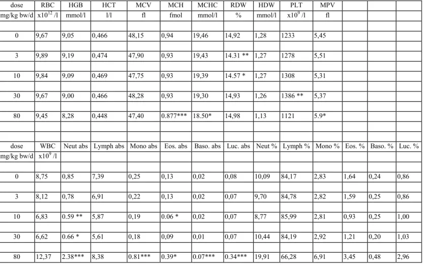
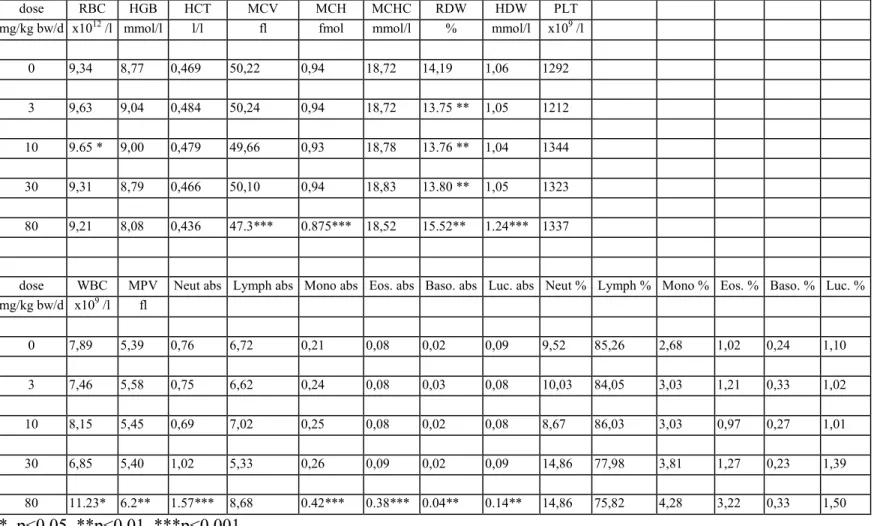
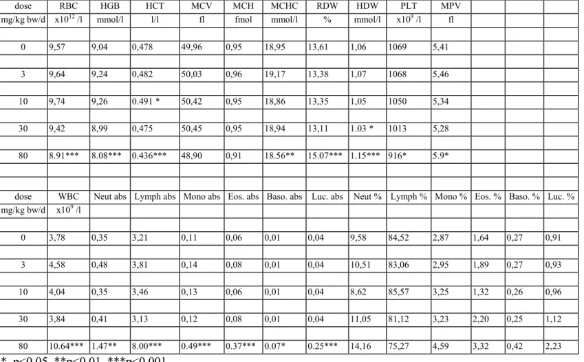
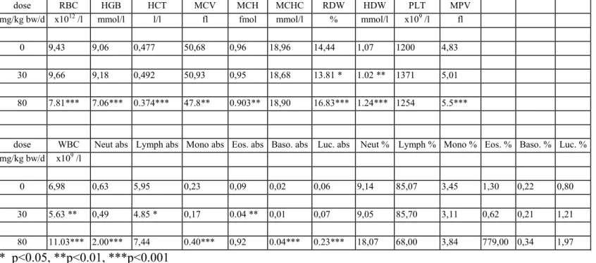
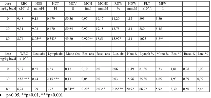
The results of the haematological determinations are summarised in tables 6a-f. In the 80 mg/kg bw group there was anaemia, seen as decreased rbc, hgb, htc, mvc, mch and mchc in varying degrees of severity, which were most pronounced in DT mice. At 30 mg/kg bw isolated parameters showed already a tendency to anaemia, though not to a statistically significant degree. There were no convincing haematological effects in the lower dose groups.
In the 80 mg/kg bw group white blood cells (lymphocytes, neutrophils, monocytes, eosinophiles and basophiles) were distinctly increased in all genotypes. The difference with the controls was
statistically significant in most cases. In contrast, in the 30 mg/kg bw group there was an overall decrease of white blood cells which was due to a decrease in lymphocytes. Other white blood cells were generally increased in this dose group, though not to a statistically significant level in most cases. In the 10 and 3 mg/kg bw group several parameters showed some variation without obvious dose-related trend. There were no distinct trends in platelets.
3.6
Organ weights
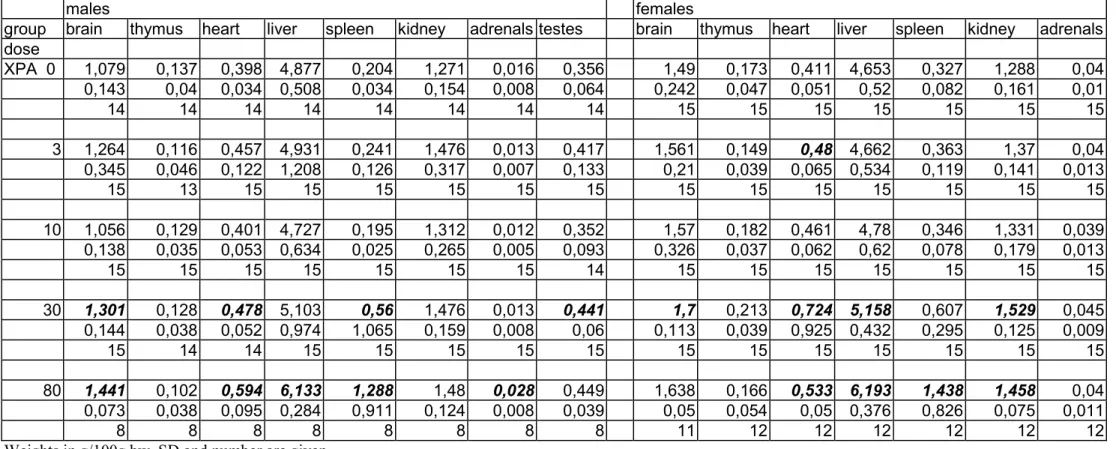
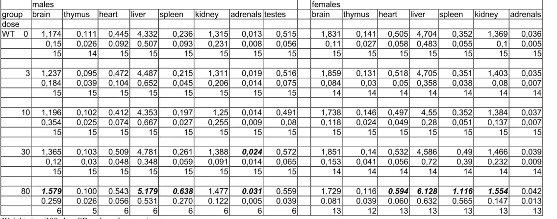
The weight of the spleen, both absolute and relative, was considerably increased at the 80 mg/kg bw level in all genotypes and in both sexes (tables 7a-c and 8a-c). In XPA mice there was also a distinct tendency of increased spleen weights at 30 mg/kg bw. Other trends in organ weight changes included increased liver weights, increased heart weights and decreased thymus weights at the 80 mg/kg bw group and incidentally at 30 mg/kg bw. However, these changes in absolute and/or relative organ weights occurred not always consistently in both sexes, and the severity varied between the genotypes.
The other organ weights showed some variations between the groups that occasionally reached the level of statistical significance. However, since there was no evidence of a
dose-response-relationship, these isolated observations were considered to be fortuitous findings (only differences with concurrent controls are indicated in the tables).
3.7
Gross pathology
Macroscopic observation (table 9a-b) revealed distended and overfilled intestinal tract containing a normal to relatively soft content, splenomegaly and enlarged lymph nodes (generally isolated, occasionally generalised) at 80 mg/kg bw. At 30 mg/kg bw there was a tendency towards enlarged spleen and lymph nodes. Four animals from the 80 mg/kg bw group only, spread over all genotypes showed malocclusion of the incisors. Since gingivitis (not noticed in this study) has been reported as an effect of cyclosporin A in several species, it cannot be excluded that the malocclusion of the incisors observed in the high dose group is treatment-related. No specific study to this potential aetiology was conducted. In the lower dose groups there were no obvious treatment-related gross changes.
3.8
Histopathology
Table 10a-b summarises the non-neoplastic lesions, table 11a-b summarises the incidence of the various tumour types in the groups, table 12 summarises the total number of tumours and the number of tumour bearing animals in the groups, table 13 summarises the treatment-related proliferative lesions (including non-neoplastic) and table 14 the incidence of lymphomas.
Non-proliferative lesions
Although non-proliferative lesions were not specifically investigated in this study, some of these changes were scored in order to correlate gross findings with microscopic observations or to estimate a maximum tolerated dose in case of changes that are known to be induced by cyclosporin A. Non-proliferative lesions that were considered treatment-related included myocardial inflammatory changes and fibrosis, inflammatory changes in the skeletal muscles and the stomach wall, acidophilic macrophage pneumonia in the lungs, frequently of moderate to marked degree, slight mineralisation in several organs, slight single cell necrosis and some brown pigment accumulation in the liver and slight nephropathy, seen as basophilic tubules with interstitial fibrosis and slight mononuclear inflammatory cell infiltrates in the renal cortex (table 10a-b). The incidence of the above changes
was increased in the 80 mg/kg bw group. In the 30 mg/kg bw group they were not seen or were not increased in incidence as compared with the controls. There were no obvious differences between the 3 genotypes. Inflammatory changes that occurred in other organs, e.g. salivary glands and secondary reproductive organs were not specifically scored.
In the 80 mg/kg bw group thymic involution was increased in incidence and/or severity in all genotypes and both sexes.
Non-neoplastic proliferative lesions
Atypical hyperplasia of the duodenal crypt epithelium was observed in several XPA males and one or 2 WT or DT females in the 80 mg/kg bw group and in a single WT female of the 30 mg/kg bw group. Although the difference with the controls was only statistically significant in XPA males, a treatment-relationship could not be excluded.
Hyperkeratosis of the forestomach was observed in several mice of both sexes in the 80 mg/kg bw group, in a single XPA male of the 30 mg/kg bw group and 2 DT controls. The distribution of this change over the groups may also suggest a treatment-relationship.
Histopathology revealed that the lymphoid system was the main target for cyclosporin A toxicity. Affected lymphoid organs included thymus, spleen, and lymph nodes.
Thymic changes consisted of cortification of the thymic medulla, seen as a generally large thymus (for the age of the animal) with an apparently increased cortex/medulla ratio, due to population of the medulla with small, cortex-type lymphocytes, obscuring the normal medullary architecture. This change occurred in both male and female mice at 30 and 80 mg/kg bw/day in XPA and WT
genotypes and in DT males. In DT females there was only cortification of the medulla in 2 controls and 2 30 mg/kg bw animals. However, in the 80 mg/kg bw group this change might be obscured by the thymic involution that was more pronounced in this group.
In the 30 mg/kg bw group the white pulp of the spleen showed an active aspect, seen as the formation of (frequently pronounced) germinal centres and a diffuse lymphoid hyperplasia of the white pulp. The hyperplastic change was observed in many animals, both males and females of all genotypes in the 30 mg/kg bw group and appeared to progress, resulting in disarranged periarteriolic lymphocyte sheaths (PALS) that were occupied by lymphoid cells showing increasing grades of atypia. These lymphoid cells resembled follicular centre cells but also lymphoid cells showing features of plasma cells were regularly observed. The affected areas increased in size, gradually occupied the major part of the white pulp and appeared to invade the red pulp, occasionally merging
with affected areas from the adjacent PALS regions. In these cases distinction from (early)
lymphoma was difficult. The term lymphoma was restricted to lymphoid proliferation with distinct invasion of the red pulp by irregular sheets, clusters or rows of lymphoid cells. The diagnosis lymphoma was not used when there was a more or less sharp demarcation between the proliferating lymphoid cells and the red pulp, showing compression rather than invasion. A disarranged atypical PALS was observed in the 80 mg/kg bw group and to a lesser extent in the 30 mg/kg bw group. It appeared to form a continuum with the diffuse hyperplasia and pronounced germinal centres as mainly observed at 30 mg/kg bw. At 10 and 3 mg/kg bw these splenic changes were absent.
In the lymph nodes proliferative changes included lymphoid hyperplasia, and plasmacytic
hyperplasia. Lymphoid hyperplasia was characterised by proliferation of lymphocytes mainly in the (para)cortical area and in more severe cases in the whole lymph node. Plasmacytic hyperplasia was seen as hyperplasia of lymphoid cells with plasmacytoid features, mainly in the medullary cords. When the process progressed the medullary cords increased in size, occupying increasing parts of the lymph node and plasma cells proliferated also to other compartments of the node. Proliferation was diffuse or in sheets and in the more advanced lesions the plasmacytoid lymphoid cells became increasingly pleomorphic. Finally the organoid structure of the lymph node was gradually lost. Plasmacytic hyperplasia may be a local process involving only one lymph node or even a part of one node. Occasionally both lymphoid hyperplasia and plasmacytic hyperplasia were present in one lymph node. In that case the most advanced change was diagnosed.
The incidence and severity of both types of lymphoid hyperplasia varied considerably in the various genotypes and in both sexes. The incidence of plasmacytic hyperplasia was highly significant in XPA and WT females of the 80 mg/kg bw group, moderate in XPA males of the 80 mg/kg bw group, but absent in WT males and only marginal in DT males and females of the high dose group. In general the most severe cases of the proliferative lymphoid lesions occurred obviously in the 80 mg/kg bw group. The cumulative incidence of hyperplastic lymphoid lesions in the lymph nodes (all nodes taken together) shows about similar incidences in XPA and WT mice of both sexes, but a lower incidence in DT mice (table 13). In male XPA mice distinct effects are also seen in the 30 mg/kg bw group. The relatively low incidence in DT mice is probably due to the high incidence of lymphomas in this group, obscuring the preceding hyperplastic changes. Moreover several autolysed animals in this group precluded examination of (part of) the lymphoid tissues.
Neoplastic lesions
Tumours of the lymphoid system
Since no immunohistochemistry was performed the lymphomas were classified according to a simple system including lymphoma when a single lymphoid organ was affected, early lymphoma when only a part of a lymphnode or the spleen showed neoplastic lymphoid cells, multicentric lymphoma when more than one lymphoid organ was involved and generalised lymphoma when also non-lymphoid organs were invaded. The distinction between atypical lymphoid hyperplasia and (early) lymphoma was occasionally difficult. Criteria for lymphoma were sheets or areas of atypical lymphoid cells (not necessarily occupying the whole node), loss of normal architecture of the lymph node and in the spleen distinct invasion of the red pulp by sheets of atypical PALS-lymphocytes.
Most of the lymphomas appeared (in HE slides) to be of B-cell type, representing one side of a range that starts with plasmacytic hyperplasia in the medullary cords. When the plasmacytic proliferation progressed, the proliferating cells became increasingly pleomorphic and the normal organoid structure of the lymph node was gradually lost. Finally nodular structures of lymphoid cells were formed that compress surrounding lymphoid tissues, or the whole node was occupied by hyperplastic and pleomorphic lymphoid cells. Generally these lymph nodes were considerably enlarged.
Although the thymus was not involved in most cases, typically thymic (T-cell) lymphomas were also observed with involvement of the thymus as major tumour-site and generally a more uniform and malignant cell type.
From table 14 it appears that the incidence of lymphomas is significantly increased in XPA and DT mice of the 80 mg/kg bw group and in DT females of the 30 mg/kg bw group (Fisher’s exact test, one sided). In treated WT females the difference with the controls was not statistically significant (Fisher’s exact test) and in WT males no lymphomas were observed. When the Peto trend-test was performed on the lymphomas there was a statistically highly significant positive trend for all groups and both sexes, except for the WT males (table 15).
Since the 80 mg/kg bw group exceeded the MTD, the Peto trend-test was also performed on the lymphomas of the 0, 3, 10 and 30 mg/kg groups, with exclusion of data from the 80 mg/kg bw group. A statistically significant positive trend was found for WT females and DT females. XPA females approached statistical significance (P=0.0526).
Non-lymphoid neoplasms
Non-lymphoid tumours (table 11a-b) included a hepatocellular adenoma (WT), a haemangiosarcoma (WT) and a haemangioma (DT) of the liver, a haemangiosarcoma (DT) of the spleen, 2 bronchiolo-alveolar adenomas (XPA, WT), a pituitary adenoma (WT), a histiocytic sarcoma of the spleen (XPA), a follicular adenoma of the thyroid (WT), a fibrosarcoma of the seminal vesicles (DT), a granulosa cell tumour of the ovary (DT), a fibrosarcoma of the skin (DT) and an osteosarcoma (DT). The distribution of the tumours over the dose groups and the low incidence indicate these tumours are not treatment-related.
The organs and tissues other than those described above did not show any distinct proliferative changes that could be attributed to the administration of cyclosporin A. The XPA-/- and double transgenic mice showed only modest background pathology, similar to wild type mice and no lesions were identified that could specifically be related to the XPA or DT genotype.
Cause of death
Lymphomas were considered cause of death in DT mice only. In this genotype the incidence of fatal lymphomas was 1, 1 and 4 for males and 0, 2 and 2 for females in the 0, 30 and 80 mg/kg bw groups respectively. In the XPA and WT groups all lymphomas were incidental since they were observed in survivors or they did not cause the death of the mice that died intercurrently. Other fatal tumours, unrelated to treatment, were an osteosarcoma in a DT control female and a histiocytic sarcoma in a XPA control male. It was not possible to establish an explicit cyclosporin-related cause of death. The severity of treatment-related histopathological lesions in intercurrent deaths was not higher than that in survivors. Nephrotoxicity of hepatotoxicity can be excluded as cause of death since they were only of a mild degree. Perhaps deterioration of the immune system and haematological disorders (not determined in decedents) may play an important role.
Positive control study
The results of the concurrent positive control study are reported separately. The study is summarised as follows. p-Cresidine (2500 ppm) and 2-AAF (300 ppm) were administered in the diets of 15 male and 15 female XPA-/- and XPA-/-.p53+/- mice. Furthermore, groups of 10 male and 10 female wild type mice were administered the same diets, whereas 10 males and 10 females of each genotype were
included as untreated controls. Histopathology was performed on the liver (with gall bladder) and urinary bladder of all animals.
p-Cresidine appeared to be rather toxic to XPA-/- and XPA-/-.P53+/- mice, in particular for the liver as shown by the toxic lesions described in table 17 a and b (hepatocellular degeneration and
hypertrophy/pleomorphism). Based on body weight gain, the concentration of 2500 ppm clearly exceeded the MTD. Exposure to 300 ppm 2-AAF appeared to be less toxic (hepatotoxic) and was found to be just near or at the MTD for the two transgenic mouse strains. Besides in the 2-AAF treated XPA-/-.p53+/- mice, the observed intercurrent mortality was low. Both compounds had little or no effect on male and female wild type mice (body weights and liver toxicity). The
histopathological observations are summarised in table 17 a and b.
(i) p-Cresidine was not carcinogenic to WT mice, but gave a positive, although not very robust, carcinogenic response in XPA-/- mice. It was clearly positive in XPA-/-.P53+/- mice.
(ii) 2-AAF appeared marginally positive in the bladder of male wild type mice. For XPA-/- mice a positive carcinogenic effect was observed for both sexes, with a robust response in the livers of the females. A strong positive response was observed with XPA-/-.P53+/- mice, particularly in the urinary bladder of both males and females.
4.
DISCUSSION
The dietary administration of cyclosporin A induced a statistically significant increase in
lymphomas in male and female XPA and DT mice at a level of 80 mg/kg bw and in DT females at 30 mg/kg bw. At 80 mg/kg bw cyclosporin A was also considered as positive in WT females, based on a positive Peto test. It was negative in WT males. The tumorigenic effect was most pronounced in DT mice. At 80 mg/kg bw also hyperplastic changes in the lymph nodes and spleen were induced. However the severity of these hyperplastic changes was not clearly associated with the tumour incidence. In DT mice this is probably due to lymphomas that may have obscured the hyperplastic changes. In addition a number of mice in this group was (partially) lost due to autolysis.
From the body weight figures, the survival data, the haematology, the organ weights, as well as the pathological changes found in the heart, muscles, lungs, kidneys, stomach and small intestines it appears that the MTD was exceeded at the high dose level. Considering the body weights of the 30 mg/kg bw groups in the last months of the study it appears that in XPA, WT and DT males and in XPA females the body weight reduction was at least 10% at several time points, indicating that the MTD may be at, or slightly higher than 30 mg/kg bw. The blood-cyclosporin concentration in the 80 mg/kg bw group at day 10 points to an internal exposure of several times the exposure associated with the human therapeutic dose in transplantation patients.
In addition to hyperplastic changes in the lymph nodes and spleen there were epithelial
non-neoplastic proliferative lesions in the small intestines and the forestomach. However, these changes did not progress to neoplasms within the experimental period of 9 months.
Tumours that are not considered treatment-related since they were seen in untreated controls or occurred in low incidence without dose-response relationship included hepatocellular adenoma, bronchiolo-alveolar adenoma, fibrosarcoma, histiocytic sarcoma, thyroid follicular cell adenoma, liver haemangioma and spleen haemangioma and haemangiosarcoma, ovary malignant granulosa cell tumour, pituitary adenoma and osteosarcoma. Osteosarcomas occur within the background tumour profile of P53+/- mice. There was no obvious difference in spontaneous tumour types between XPA and WT mice. A summary of tumours that were observed in untreated controls (XPA, WT and DT) from other 9-months ILSI/HESI studies is given in table 16.
Since cyclosporin A is a non-genotoxic carcinogen the positive response in NER-deficient XPA mice, as opposed to the negative response in WT mice, was unexpected. The working mechanism was not investigated in this study. Due to background carcinogens and endogenous DNA-reactive metabolites WT mice as well as XPA mice harbour certain levels of initiated cells. One might speculate that the number of these initiated cells may be greater in XPA mice, lacking NER, than in
WT mice with normal functioning DNA-repair. Perhaps in strongly immunosuppressed animals these initiated and subsequently transformed cells may escape the normal immunosurveillance activity of the animal, leading to a higher incidence of tumours in XPA than in WT animals. This would be in line with the tumour type involved since, when this theory is valid, one should indeed expect tumours that occur also spontaneously in the strain of mice. Recent investigations point to a reduced number and activity of NK cells in XPA mice (personal communication University of Rotterdam) which might also lead to reduced immunosurveillance in XPA mice. It is not clear
whether the direct action of cyclosporin A on the induction of transforming growth factor β (TGF-β), as was recently reported in literature (Hojo M et al., Nature (1999) 397: pg 530) plays a role in the higher susceptibility of XPA mice. Similar to depressed immunosurveillance, induction of TGF-β may result in malignant transformation and progression of already initiated cells that may be present in higher numbers in XPA mice than in WT mice, resulting in larger numbers of tumours that occur also ‘spontaneously’ in XPA mice. The observation that untreated XPA mice do not show a higher spontaneous tumour incidence than WT mice may be due to the relatively short experimental period of 9 months. Perhaps such a difference in spontaneous tumour incidence will only be disclosed when the mice age, and data on tumour incidences in ageing XPA mice (above 18 months) are lacking at present.
Lymphomas belong to the spontaneous background tumours of C57BL/6 mice, so a statistically significant increase of this tumour type is required for a positive response. Noting that 30 mg/kg bw appears to be the MTD and considering that carcinogenicity should be evaluated at doses not exceeding the MTD, it is concluded that cyclosporin A is negative in the short-term alternative carcinogenicity assay in male and female XPA and WT mice and in male DT mice. It is positive in female DT mice at the MTD (30 mg/kg bw).
At a dose of 80 mg/kg bw that distinctly exceeds the MTD, cyclosporin A is positive in XPA mice and in DT mice of both sexes. The positive response is stronger in DT mice than in
XPA mice. At 80 mg/kg bw cyclosporin A is also considered to be positive in WT females (based on a positive Peto trend test) and it is negative in WT male mice.
The positive control compound 2-AAF was positive in XPA and DT mice showing liver tumours and transitional cell tumours of the urinary bladder (reported elsewhere).
Review of the report
The report was reviewed by the XPA-Assay Working Group of the ILSI/HESI program and the comments were discussed on May 25, 2000 at RIVM, Bilthoven, Netherlands. Members of the XPA-Assay Working Group were: Dr CF van Kreyl (RIVM), Dr PA McAnulty (Scantox), Dr RB Beems (RIVM), Dr A Vynckier (Janssen), Dr H van Steeg (RIVM), Dr R Franson-Steen (Astra),
Dr C Alden (Monsanto), Dr R Forster (CIT), Dr JW van der Laan (EU/CPMP representative, RIVM) and Dr J Vandenberghe (Janssen).
5.
DEVIATIONS FROM THE PROTOCOL
During the study it was decided to extend the experimental period from 6 months to 9 months. Part of the tissues of animal nr 47 was mistakenly numbered as nr 48 (blocks). Since no proliferative lesions were observed in the tissues and both animals belonged to the same group and sex, this was not considered to have influenced the results of the study.
By mistake the animals were also weighed in week 18. These additional weights were kept in the raw data but were excluded from in the individual data, and the summary tables. Animals were weighed in week 38 instead of in week 37.
By mistake food intake was also determined in week 3, of the groups fed 0, 3, 10 and 30 mg/kg bw (not 80 mg/kg bw). These additional weights were kept in the raw data but were excluded from the individual data, and the summary tables.
The organ weights of a few animals that were killed moribund a few days before the terminal autopsy period were weighed. These weights were kept in the raw data, but were not included in the summary tables of the survivors.
One mouse from the DT 80 mg/kg bw group (nr 388) that was designated as a female, turned out to be a male. Therefore the total number of male mice in the 80 mg/kg bw group is 16 and the number of females in this dose group is 14.
Determination of the blood concentration of cyclosporin A was started at 9:30 hr instead of 8:30 hr. During histotechnical processing of the study it was initially decided to process also all tissues of the 30 mg/kg group, in addition to the controls and the 80 mg/kg bw group. However, due to personnel-technical problems this extension was not continued and processing of the tissues of the remaining 30 mg/kg bw animals was restricted to those tissues mentioned under 2.4 ‘pathology’. So part of this dose group has been processed and examined completely (all tissues preserved) and in another part about 11 tissues per animal were processed and examined .
It was decided not to perform reticulocyte counts.
Part of the histotechniques was performed by TNO-Nutrition, Zeist, Netherlands.
At microscopy several organs and tissues, especially aorta, gall bladder, pituitary and thyroid, appeared to be missing. Upon searching for these organs in the containers with fixed materials twice (the second time by the study pathologist) still a number of organs was missing. In most cases this is believed to be due to cutting the paraffin blocks too far, especially when more than one organ was embedded in the same block. These cases are indicated in the individual animal data as ‘no sample’. In case of (partly) autolysed animals no attempt was made to search for missing tissues.
The results of 2 separate studies, the main study with dose groups 0, 3, 10 and 30 mg/kg bw and an additional study with the high dose group of 80 mg/kg bw were reported in this report.
Table 1. Body weights and food intake of animals for toxicokinetics
Body weights in week 1 Dose (mg/kg bw) sex 0 30 80 f 3 9 9 19,4 18,2 18,5 0,57 1,11 0,71 m 3 9 9 25,3 24,8 24,0 0,38 2,09 1,75 Body weights in week 2
Dose (mg/kg bw) sex 0 30 80 f 3 9 9 20,4 18,7 17,6 0,58 1,08 0,96 m 3 9 9 26,2 24,8 21,8 1,00 2,50 1,56 Food intake in week 1
Dose (mg/kg bw) sex 0 30 80 f 3 9 9 28,6 26,9 29,1 3,39 3,54 6,99 m 3 9 9 33,9 29,3 26,3 0,81 2,84 3,18 Food intake in week 2*
Dose (mg/kg bw) sex 0 30 80 f 3 9 9 10,2 12,6 15,8 2,52 1,28 4,23 m 3 9 9 10,5 11,6 16,9 2,23 4,27 3,74 * Week 2 had only 3 days
Count, mean and standard deviation are given
Body weight at week 1 is the initial body weight, body weight at week 2 is the body weight after one week on the experimental diets. Food intake in week one is the food intake consumed in the first week on the
experimental diets. Food intake in week 2 is the food consumed from day 8 – 10. The animals were killed at day 10.
Table 2. Concentration cyclosporin a in the blood
males females dose mg/kg bw 9:30 12:00 16:00 9:30 12:00 16:00 0 <25 * - - <25 - -30 828 757 644 646 707 540 80 1635 1905 1200 1692 1467 1663Samples taken at day 10
* mean below detection limit of 25 µg cyclosporin/l blood - = not performed
Table 3a. Survival in males
Week 1 2 3 4 5 6 7 8 9 10 11 12 13 15 17 19 21 23 25 27 29 31 33 35 37 39 XPA 0 15 15 15 15 15 15 15 15 15 15 15 15 15 15 15 15 15 15 15 15 15 14 14 14 14 14 3 15 15 15 15 15 15 15 15 15 15 15 15 15 15 15 15 15 15 15 15 15 15 15 15 15 15 10 15 15 15 15 15 15 15 15 15 15 15 15 15 15 15 15 15 15 15 15 15 15 15 15 15 15 30 15 15 15 15 15 15 15 15 15 15 15 15 15 15 15 15 15 15 15 15 15 15 15 15 15 15 80 15 15 15 15 15 15 15 15 15 15 15 15 15 15 15 15 15 15 14 14 14 14 13 13 12 12 WT 0 15 15 15 15 15 15 15 15 15 15 15 15 15 15 15 15 15 15 15 15 15 15 15 15 15 15 3 15 15 15 15 15 15 15 15 15 15 15 15 15 15 15 15 15 15 15 15 15 15 15 15 15 15 10 15 15 15 15 15 15 15 15 15 15 15 15 15 15 15 15 15 15 15 15 15 15 15 15 15 15 30 15 15 15 15 15 15 15 15 15 15 15 15 15 15 15 15 15 15 15 15 15 15 15 15 15 15 80 15 15 15 15 15 15 15 15 15 15 15 15 15 15 15 15 15 15 15 14 13 12 12 12 10 10 DT 0 15 15 15 15 15 15 15 15 15 15 15 15 15 15 15 15 15 15 15 15 15 15 15 14 14 14 30 15 15 15 15 15 15 15 15 15 15 15 15 15 15 15 15 15 15 15 15 15 15 15 15 15 15 80 16 16 16 16 15 15 14 14 14 14 14 14 14 14 13 13 13 13 13 13 12 12 12 12 9 9 Number of animals alive at the end of the week indicated. The first 2 columns represent the genotype and the dose in mg/kg bwTable 3b. Survival in females
Week 1 2 3 4 5 6 7 8 9 10 11 12 13 15 17 19 21 23 25 27 29 31 33 35 37 39 XPA 0 15 15 15 15 15 15 15 15 15 15 15 15 15 15 15 15 15 15 15 15 15 15 15 15 15 15 3 15 15 15 15 15 15 15 15 15 15 15 15 15 15 15 15 15 15 15 15 15 15 15 15 15 15 10 15 15 15 15 15 15 15 15 15 15 15 15 15 15 15 15 15 15 15 15 15 15 15 15 15 15 30 15 15 15 15 15 15 15 15 15 15 15 15 15 15 15 15 15 15 15 15 15 15 15 15 15 15 80 15 15 15 15 15 15 15 15 15 15 15 15 15 15 15 15 15 15 15 15 15 14 12 12 12 12 WT 0 15 15 15 15 15 15 15 15 15 15 15 15 15 15 15 15 15 15 15 15 15 15 15 15 15 15 3 15 15 15 15 15 15 15 15 15 15 15 15 15 15 15 15 15 15 15 15 15 15 15 15 15 14 10 15 15 15 15 15 15 15 15 15 15 15 15 15 15 15 15 15 15 15 15 15 15 15 15 15 15 30 15 15 15 15 15 15 15 15 15 15 15 15 15 15 15 15 15 15 15 15 15 15 15 14 14 14 80 15 15 15 15 15 15 15 15 15 15 15 15 15 14 14 14 14 14 14 14 14 14 14 14 13 13 DT 0 15 15 15 15 15 15 15 15 15 15 15 15 15 15 15 15 15 15 15 15 15 15 15 15 14 14 30 15 15 15 15 15 15 15 15 15 15 15 15 15 15 15 15 15 15 15 15 15 15 15 15 15 15 80 14 14 14 14 14 14 14 14 14 14 14 14 14 14 14 14 14 14 14 14 14 13 11 10 9 8 Number of animals alive at the end of the week indicated. The first 2 columns represent the genotype and the dose in mg/kg bwTable 4a. Mean body weights XPA
-/-mice, males
Wee k 1 2 3 4 5 6 7 8 9 10 11 12 13 15 17 19 21 23 25 27 29 31 33 35 38 39 Dose 0 15 15 15 15 15 15 15 15 15 15 15 15 15 15 15 15 15 15 15 15 15 14 14 14 14 14 22,7 24,5 25,2 25,4 26,7 27,7 27,8 28,7 28,9 29,3 29,5 30,6 30,3 31,6 33,3 34,4 35,6 36,4 38,3 37,5 39,4 39,6 41,6 41,2 41,9 40,5 1,6 1,4 1,9 2,1 2,2 2,3 2,1 2,5 2,5 2,7 2,8 3 2,7 2,7 3,4 3,5 3,9 4,2 4,4 4,6 4,9 4,8 5,5 5,2 5,5 5,6 3 15 15 15 15 15 15 15 15 15 15 15 15 15 15 15 15 15 15 15 15 15 15 15 15 15 15 22,4 23,9 24,1 24,4 25,4 26 25,8 26,6 27,2 27,7 27,5 27,6 28 29,5 30,8 30,8 32 32,4 35 34,2 36,4 35,3 37,4 36,5 37,2 36,7 1,4 1,2 1,2 1,1 1,4 1,3 1,2 1,1 1,2 1,1 1,3 1,5 1,5 1,7 2,3 2 2,4 2,4 3,1 3 3,2 3,3 4 3,6 4,2 3,8 10 15 15 15 15 15 15 15 15 15 15 15 15 15 15 15 15 15 15 15 15 15 15 15 15 15 15 22,5 23,7 24,6 25,1 26,4 26,8 27,4 27,9 28,5 28,8 28,7 30,2 30 30,5 32,5 33,1 34,1 34,3 36,2 35,9 37,2 37 39,4 38,9 40,4 39,5 1,5 1,7 2,1 2,1 2,4 2,4 2,3 2,6 2,6 2,6 2,8 3 3,1 3 3,2 3,2 3,5 3,5 3,6 3,9 4,3 4,4 4,6 4,6 4,8 4,4 30 15 15 15 15 15 15 15 15 15 15 15 15 15 15 15 15 15 15 15 15 15 15 15 15 15 15 23,4 23,9 25 25,9 26,9 27,4 27,8 28,5 28,9 29,1 29,5 29,9 30,2 30,8 31,8 31,9 32,9 32,9 34,6 33,7 34,5 33,8 34,9 34,5 35,2 34,9 1,3 1,4 1,3 1,2 1,7 1,5 1,5 1,6 1,4 1,5 1,6 1,7 1,7 1,8 2,1 1,7 2,4 2,3 2,5 2,5 2,7 2,7 2,8 2,8 3 2,7 80 15 15 15 15 15 15 15 15 15 15 15 15 15 15 15 15 15 15 14 14 14 14 13 13 12 12 25,2 22,4 23,1 23,8 25,4 25,8 25,5 26,3 27,2 27,8 28,1 28,3 28 28,2 28,6 29,2 29,2 28,8 29 28,9 28,9 28,6 29,4 28,6 28,8 28,1 1,5 1,8 1,7 1,8 1,8 1,3 1,4 1,5 1,5 1,9 2 2 1,9 1,9 1,9 2,1 1,9 2 2,4 2,3 2,5 3,1 2,3 2,3 2,2 2,2 Body weights in g. Number, mean and SD are givenDose in mg/kg bw
Table 4b. Mean body weights Wild Type mice, males
Wee k 1 2 3 4 5 6 7 8 9 10 11 12 13 15 17 19 21 23 25 27 29 31 33 35 38 39 Dose 0 15 15 15 15 15 15 15 15 15 15 15 15 15 15 15 15 15 15 15 15 15 15 15 15 15 15 23,3 24,5 24,7 25 26 26,7 26,8 27,7 28,1 29 29 29,6 29,5 30,8 32,2 32,3 33,9 34,5 36,6 36,3 38,9 37,8 39,7 39,3 40,5 38,9 0,7 0,8 1,1 1,1 1,4 1,4 1,3 1,7 1,6 1,7 1,5 1,8 1,8 2 2,4 2,4 2,9 2,8 2,7 2,9 3,2 2,9 3,4 3,5 3,3 3,4 3 15 15 15 15 15 15 15 15 15 15 15 15 15 15 15 15 15 15 15 15 15 15 15 15 15 15 23,6 24,7 24,4 24,9 26,1 26,8 27,1 27,8 28,7 28,8 28,7 29,4 29,6 31 32,1 31,5 32,9 32,8 35,3 33,8 36,3 35,3 37,5 36,6 37,9 37,5 0,7 1,1 1 1 1,4 1,4 1,2 1,7 1,6 1,6 1,7 2,1 2,3 2,1 2,6 2,3 2,9 2,7 3,3 2,9 3,4 3,4 3,2 3,8 4 3,8 10 15 15 15 15 15 15 15 15 15 15 15 15 15 15 15 15 15 15 15 15 15 15 15 15 15 15 23,5 24,3 25 25,7 26,8 27,2 27,6 28,4 28,8 29,3 29,8 30,7 30,9 31,3 33 33,6 34,9 35,3 37,3 36,2 38,4 37,6 40 39,4 40,6 39,9 0,9 0,9 1 1 1,2 1,3 1,2 1,5 1,6 1,6 1,3 1,6 1,8 1,6 2,1 2,2 2,4 2,3 2,5 2,4 2,9 2,6 3,2 3,4 3,1 3,6 30 15 15 15 15 15 15 15 15 15 15 15 15 15 15 15 15 15 15 15 15 15 15 15 15 15 15 23 23,4 23,9 24,4 25,7 26,4 26,6 27,1 27,6 27,5 28,4 28,4 29 29,3 29,8 29,8 31,1 31,5 32,7 32,2 32,9 32,8 34,4 33,6 34,2 33,9 1,1 1,5 1,2 1,2 1,1 1,1 1,1 1,2 1,3 1,4 1,3 1,4 1,5 1,6 1,6 1,8 1,8 2,2 2,4 2,6 2,7 2,7 3,3 2,7 3,2 3 80 15 15 15 15 15 15 15 15 15 15 15 15 15 15 15 15 15 15 15 14 13 12 12 12 10 10 23,9 21,9 22,4 23,4 25,4 25,7 25,2 25,4 26,1 27,6 27,4 27,6 27,7 28,2 28,5 28,9 29,2 28,9 28,9 29 29,4 29,9 30 29,5 28,2 28,3 2,4 3,1 2,7 2,3 2,8 2,7 2,3 2,4 2,3 2,5 2,4 2,4 2,3 2,1 2,2 2,3 2,2 2,7 2,8 2,9 2,1 2,2 2,4 3,2 2,6 3,1 Body weights in g. Number, mean and SD are givenDose in mg/kg bw
Table 4c. Mean body weights XPA
-/-,P53
+/-mice, males
Wee k 1 2 3 4 5 6 7 8 9 10 11 12 13 15 17 19 21 23 25 27 29 31 33 35 37 39 dose 0 15 15 15 15 15 15 15 15 15 15 15 15 15 15 15 15 15 15 15 15 15 15 15 14 14 14 22,4 23,8 24,5 24,8 25,9 26,6 26,8 27,7 28,1 28,4 28,8 29,5 29,4 30,6 32,2 32,1 32,9 33,8 35,8 35,3 38 37 38,7 37,9 38,7 36,5 1,6 1,3 1,4 1,5 1,9 1,8 1,6 2,1 2 2,2 2,2 2,4 2,3 2,3 3 3,3 3,9 3,5 4,3 4,3 4,9 5 5,3 5,7 6,4 6,3 30 15 15 15 15 15 15 15 15 15 15 15 15 15 15 15 15 15 15 15 15 15 15 15 15 15 15 22,2 22,9 23,6 24,3 25,5 26,1 26,2 26,8 27,3 27,6 28,1 28,3 28,2 29,1 29,8 29,8 30,9 31,5 33,1 32,1 32,9 32,8 33,5 32,9 33,5 33,5 1,3 1,1 1 1,1 1,3 1,3 1,3 1,3 1,6 1,6 1,5 1,5 1,6 1,8 2,1 2,2 2,6 2,6 2,8 2,7 2,9 3 3,2 3,3 3,3 3,1 80 16 16 16 16 15 15 14 14 14 14 14 14 14 14 13 13 13 13 13 13 12 12 12 12 9 9 23.6 21.5 22.0 23.6 25.6 25.9 26 26,3 27,3 28,5 28,5 28,4 28,5 28,8 29,2 29,6 29,9 29,8 29 29,3 30,1 29,7 30,5 29,8 30,5 29,9 2.8 2.9 3.2 2.7 3.0 3.2 1,1 0,9 1,2 1,3 1,3 1,9 1 1,1 1,3 1,4 1,3 1,1 1,6 2,5 1,3 1,4 1,6 2,2 1 1,1 Body weights in g. Number, mean and SD are givenDose in mg/kg bw
Table 4d. Mean body weights XPA
-/-mice, females
Wee k 1 2 3 4 5 6 7 8 9 10 11 12 13 15 17 19 21 23 25 27 29 31 33 35 37 39 Dose 0 15 15 15 15 15 15 15 15 15 15 15 15 15 15 15 15 15 15 15 15 15 15 15 15 15 15 18,4 19,3 19,6 19,8 20,5 21,7 22,1 22,3 22,7 23,3 23,3 23,6 23,3 24,5 25 25,2 26,4 26,5 27,7 27,2 28,6 29 29,9 30,5 31,7 30,8 1,4 1,6 1,5 1,2 1,2 1,4 1,2 1,4 1,6 1,5 1,5 2,1 1,7 1,8 2,2 2,5 2,8 2,6 4 3,3 4,3 3,7 5 4,9 5,8 4,5 3 15 15 15 15 15 15 15 15 15 15 15 15 15 15 15 15 15 15 15 15 15 15 15 15 15 15 18,7 20,2 19,9 20,5 21,5 22,1 22,3 22,3 22,9 22,9 23,4 23,2 23,6 24,3 25 24,8 25,8 25,5 26,1 25,8 27,1 26,9 28 28 28,3 28,6 0,8 1 0,7 0,7 0,8 0,7 0,6 0,7 1 1,3 1,9 0,8 0,7 0,8 1,2 1,1 1,2 1,2 1,3 1,5 1,4 1,4 1,9 1,5 1,8 1,6 10 15 15 15 15 15 15 15 15 15 15 15 15 15 15 15 15 15 15 15 15 15 15 15 15 15 15 18,4 19,1 19,6 20,2 21,3 21,6 22,1 22,1 22,5 22,6 22,8 23,5 23,5 24,6 24,9 25,4 25,9 26,2 26,2 26,6 27,8 27,7 29,4 28,8 30,4 29,9 1,8 2,3 2 1,8 2 1,8 1,8 1,8 1,8 1,7 1,9 2,4 2,5 2,5 3,4 3,3 3,9 3,8 3,7 4,3 5 5,1 5,9 5,7 6,9 6,4 30 15 15 15 15 15 15 15 15 15 15 15 15 15 15 15 15 15 15 15 15 15 15 15 15 15 15 18,7 19 19,4 20,3 21,5 22,1 22,4 22,5 22,9 22,5 23,1 23,2 23,4 23,6 23,9 24,2 24,7 25,1 25,8 25,1 25,8 25,7 26,5 26,7 27 27 1 1,2 1,2 1,3 1,3 1,3 1,6 1,4 1,3 1,4 1,3 1,6 1,2 1,2 1,5 1,4 1,5 1,7 1,7 1,7 1,7 1,6 1,8 2,1 2,5 1,9 80 15 15 15 15 15 15 15 15 15 15 15 15 15 15 15 15 15 15 15 15 15 14 12 12 12 12 19,3 18,3 19,3 20,2 21,2 21,2 20,9 21,5 22 23,2 22,7 22,8 23 23,8 24 24,1 24,9 24,5 25 24,3 24,8 24,3 25,8 26 25,9 25,3 1,2 1,2 1,5 1,2 1,3 1,4 1,1 1,1 1,1 1 1,4 1,5 1,3 1,1 1,1 1,3 1,2 1,8 1 2,1 1,8 2,4 1,1 1,1 0,9 1,2 Body weights in g. Number, mean and SD are givenDose in mg/kg bw
