Dutch DisMod. Constructing a set of consistent data for chronic disease modelling | RIVM
117
0
0
Hele tekst
(2) Page 2 of 117. RIVM report 260751 001.
(3) RIVM report 260751 001. Page 3 of 117. Abstract Using the simulation model context we analysed data the consistency of data on incidence, prevalence and mortality for specific chronic diseases so as to construct an appropriate data set for chronic disease models. These simulation models integrate data from different sources and are used to estimate the public health effects of trends in and interventions on disease risk factors. Disease incidence and prevalence data are derived from disease registration in general practice, epidemiological surveys and health-care registration. Disease-specific mortality data are collected from national statistics, while remission and case fatality rates are taken from the scientific literature. The successive steps followed in the consistency analyses were: (visual) comparison of data from different sources, calculation of remission and mortality rates from given disease incidence and prevalence rates and comparison of results with remission and mortality data from other sources. Data was evaluated on lung cancer, asthma and COPD, coronary heart diseases and congestive heartfailure, diabetes mellitus, and dementia and stroke. From the results it can be concluded that differences between data from several sources can often be explained by differences in registration coding rules. For most diseases, calculated excess mortality rates are much greater than the mortality where the disease is the primary cause of death, registered in national statistics. In general, the estimated mortality parameters are in agreement with data from the literature, with the estimation of age-specific rates being complicated by time-trends..
(4) Page 4 of 117. RIVM report 260751 001.
(5) RIVM report 260751 001. Page 5 of 117. Preface Simulation models based on the multistate lifetable method are increasingly used to estimate the effects over time and age of public health intervention programs, for example Weinstein et al., 1987, Gunning-Schepers, 1998, and Barendregt, Bonneux, 1998. These models describe several population risk factors and disease categories simultaneously taking into account some main integrative aspects. These models share the same mathematical methodology, but differ in the selection of risk factors, diseases, model specification and data used. The main model parameters are the initial population numbers, that are specified by gender, age, risk factor classes and disease states, and the transition rates between these states. When restricted to the part of the model describing the disease processes the main parameters are the initial disease prevalence rates, the disease incidence and remission rates, and the disease-specific mortality rates. As data are derived from various sources it is necessary to evaluate their consistency within the context of our model structure. Murray&Lopez (1996) have called this type of evaluation of data consistency ‘Disease Modelling’(DisMod). Disease Modelling is similar to the general process of developing simulation models in public health. The knowledge and expertise are integrated of the fields of epidemiology, public health, and mathematical biology. Many persons have directly and indirectly contributed to the foregoing report. Especially we would like to thank dr HC Boshuizen, drs ME Kruijshaar and drs TL Feenstra, who made several critical but inspiring comments on the analyses..
(6) Page 6 of 117. RIVM report 260751 001.
(7) RIVM report 260751 001. Page 7 of 117. Contents Samenvatting 9 Summary 10 1.. 2.. 3.. 4.. Introduction 11 1.1. Consistent model input 11. 1.2. Research goal and methods 11. 1.3. Modelling mortality in the context of chronic diseases 13. 1.4. Basic model equations 14. 1.5. Data sources 16. 1.6. Data selection criteria 18. 1.7. Reader 19. Lung cancer 21 2.1. Introduction 21. 2.2. Data sources 21. 2.3. Data on incidence and prevalence 21. 2.4. Estimated mortality rates 22. 2.5. Estimated prevalence rates 24. 2.6. Concluding remarks 26. Chronic non-specific lung diseases: asthma and chronic obstructive pulmonary disease 27 3.1. Introduction 27. 3.2. Data sources 27. 3.3. Data on incidence and prevalence 28. 3.4. Estimated remission and mortality rates 28. 3.5. Other data from literature on remission and mortality 31. 3.6. Concluding remarks 33. Coronary heart diseases and congestive heart failure 35 4.1. Introduction 35. 4.2. Data sources 35. 4.3. Data on incidence and prevalence for CHD 37. 4.4. Data on incidence and prevalence for CHF 38. 4.5. Estimated mortality rates for CHD 38. 4.6. Estimated mortality rates for CHF 43. 4.7. Combining CHD and CHF 45. 4.8. Data from literature 46. 4.9. Concluding remarks 47.
(8) Page 8 of 117. 5.. 6.. 7.. 8.. RIVM report 260751 001. Diabetes mellitus 49 5.1. Introduction 49. 5.2. Data sources 49. 5.3. Data on incidence and prevalence 49. 5.4. Estimated mortality rates 50. 5.5. Other data from literature 54. 5.6. Concluding remarks 55. Dementia 56 6.1. Introduction 56. 6.2. Data sources 56. 6.3. Data on incidence and prevalence 57. 6.4. Estimated mortality rates 57. 6.5. Data from literature 60. 6.6. Concluding remarks 60. Stroke 62 7.1. Introduction 62. 7.2. Data sources 62. 7.3. Data on incidence and prevalence 63. 7.4. Estimated mortality rates 63. 7.5. Data from literature 65. 7.6. Concluding remarks 68. Concluding remarks 69. Appendix 1. Mailing list 71. Appendix 2. Tables and figures of disease incidence and prevalence data 73. Appendix 3. The chronic disease model equations 95. Appendix 4. Smoothing the data 103. Appendix 5. Comparing incidence and prevalence ratios for dementia 105. Appendix 6. Symbols and abbreviations 107. References 109.
(9) RIVM report 260751 001. Page 9 of 117. Samenvatting Om te komen tot consistente verzamelingen van invoergegevens voor dynamische modellen van chronische ziekten hebben we de consistentie binnen de modelcontext geanalyseerd van gegevens over incidentie, prevalentie en sterfte voor specifieke ziekten. Dergelijke modellen integreren gegevens uit verschillende bronnen en worden gebruikt om de volksgezondheidseffecten van trends in en interventies op risicofactoren door te rekenen. Bijvoorbeeld, de effecten van anti-rook campagnes in termen van toekomstige oorzaak-specifieke ziekte en sterfte, gespecificeerd naar geslacht en leeftijd. Incidentie- en prevalentiegegevens komen uit huisartsenregistraties, epidemiologische onderzoeken en registraties in de gezondheidszorg, gegevens over overleving uit de wetenschappelijke literatuur, terwijl ziekte-specifieke sterftecijfers uit de CBS-statistieken komen. De opeenvolgende analysestappen zijn: een vergelijking van de gegevens uit verschillende bronnen, de berekening van remissie- en sterfterates uit gegeven incidentie- en prevalentierates, en vervolgens een vergelijking met data uit de literatuur. We hebben data geëvalueerd met betrekking tot longkanker, astma en COPD, coronaire hartziekten en hartfalen, diabetes mellitus, dementie, en beroerte. Omdat ziekte-specifieke cijfers omtrent de ‘excess’ sterfte zelden beschikbaar zijn, zijn de door ons berekende ‘excess’ sterfterates omgezet in ratio’s, die vergeleken zijn met relatieve risico’s en hazard ratio’s uit de literatuur. De gevonden verschillen tussen incidentie- en prevalentiecijfers uit verschillende bronnen zijn meestal terug te vertalen naar verschillen in registratiekarakteristieken. Voor veel aandoeningen is de berekende ‘excess’ sterfte veel groter dan de sterfte met de ziekte als de primaire doodsoorzaak in de CBS-statistieken. Verschillende ‘mechanismen’ kunnen dit verschil verklaren. Personen met de ene ziekte kunnen een vergrote kans hebben op andere, meer fatale ziekten. Deze vergrote kansen kunnen het gevolg zijn van een onderliggend gemeenschappeljk pathophysiologisch proces. Zo vormt bijvoorbeeld diabetes een risicofactor voor coronaire hartziekten. Daarnaast kunnen gemeenschappelijke risicofactoren een rol spelen. Bijvoorbeeld, roken is een belangrijke risicofactor voor COPD en longkanker. Tenslotte kunnen ziekten de algemene gezondheidstoestand zodanig negatief beïnvloeden, dat patiënten vatbaarder worden voor andere ziekten. De geschatte sterfteparameters komen in de meeste gevallen goed overeen met die uit de literatuur. De resultaten voor longkanker, astma, COPD, hartfalen, diabetes en beroerte lijken valide ‘at face value’ en in vergelijking met gegevens uit de literatuur. Voor CHZ en dementie zijn nog verbeteringen mogelijk. De aanwezigheid van trends in de tijd maakt het schatten van leeftijd-specifieke cijfers lastig, omdat de achter de rekenmethode liggende veronderstelling van tijd-constante parameters dan niet meer opgaat..
(10) Page 10 of 117. RIVM report 260751 001. Summary Using the simulation model context we analysed the consistency of data on incidence, prevalence and mortality for specific chronic diseases so as to construct an appropriate dataset for the chronic disease models. These simulation models integrate data from different sources and are used to estimate the public health effects of trends in and interventions on disease risk factors. For example, to calculate the effects of smoking interventions on future cause-specific morbidity and mortality, specified by gender and age. The parameters of the model that describe the disease processes are initial disease prevalence rates, incidence and remission rates, and mortality rates. Mortality rates are specified by cause. Disease incidence and prevalence data are derived from disease registration in general practice, epidemiological surveys and health-care registration. Disease-specific mortality data are collected from national statistics, while remission and case fatality rates are taken from the scientific literature. The successive steps followed in the consistency analyses were: (visual) comparison of data from different sources, calculation of remission and mortality rates from given disease incidence and prevalence rates and comparison of results with remission and mortality data from other sources. Data was evaluated on lung cancer, asthma and COPD, coronary heart diseases and congestive heartfailure, diabetes mellitus and dementia and stroke. Since disease-specific excess mortality rates are rarely reported in literature, we have transformed the calculated excess mortality rates to hazard ratios and compared these ratios to reported cause-specific relative mortality risks and hazard ratios. From the results it can be concluded that differences between disease incidence and prevalence figures from several sources can often be explained by differences in coding rules of registrations. So the types of patients we describe in our model are ‘mean’ patients of all sources that have been selected. For most diseases the calculated excess mortality rates are much larger than the mortality with the disease as the primary cause of death as registered in national statistics. Several ‘mechanisms’ can explain these differences. Patients having one disease may have increased risks for other, more fatal diseases. These increased risks may be the result of shared pathophysiological processes, for example diabetes mellitus being a risk factor for coronary heart diseases. The increased risks also may be the result of joint risk factors, for example smoking being an important risk factor for COPD and lung cancer. Moreover, non-fatal diseases may decrease the general health state of patients such that they are more susceptible to more fatal diseases. In general the estimated mortality parameters are in agreement with data from the literature. The results for lung cancer, asthma, COPD, heartfailure, diabetes and stroke seem to be valid at face value and compared to data from literature. For CHD and dementia the results can still be improved. The estimation of agespecific rates is complicated by time-trends, because the assumption of time-constant model parameters underlying the model equations is not valid..
(11) RIVM report 260751 001. 1.. Introduction. 1.1. Consistent model input. Page 11 of 117. At the RIVM we have developed a modelling approach to calculate chronic diseases morbidity and mortality as a function of risk factor prevalence in the Dutch population. A dynamic multistate model has been constructed to describe demographic and epidemiological processes, such as ageing, changes in risk factor prevalence, and their effect on morbidity and mortality. The model structure will be applied to assess the public health gain of preventive measures or changes in disease treatment. Such applications are performed in the framework of the National Public Health Status and Forecasts Report of the RIVM (Ruwaard&Kramers, 1998). Important model parameters are initial disease prevalence rates and annual disease incidence, remission and mortality rates. The model parameters have to be estimated from data that are available from registrations, epidemiological surveys, and the medical literature. As data are derived from various sources it is necessary to evaluate their consistency within the context of our model structure. Lack of consistency may result from differences in sampling period or population, definition of disease incidence and/or prevalence (systematic differences) as well as from random error. Thus, apart from analysing data on incidence, prevalence, remission, and mortality separately, simultaneous analyses within the framework of the chronic disease model are required. Using the model equations and given disease incidence and prevalence data, disease remission and mortality rates can be calculated. Subsequently consistency can be checked by comparing the results to mortality and remission data from registrations and literature. The approach we have applied here is similar to the approach Murray and Lopez used to ascertain consistency within their disease projections in the Global Burden of Disease project, called Dis(ease) Mod(elling) (Murray&Lopez, 1993).. 1.2. Research goal and methods. Our goal was to obtain consistent input figures for our chronic disease modelling activities. Data on disease incidence and prevalence were available from registrations in general practice, epidemilogical surveys and registrations in health care. The data sources have been selected applying a set of explicit criteria (see §1.6). Disease remission and mortality rates have been estimated from these data employing the chronic disease model equations. Subsequently, consistency was evaluated by comparing the model results to data from other sources, e.g. cause-specific mortality data from Statistics Netherlands (CBS), and mortality hazard ratios, relative risks and remission rates from the scientific literature. We have evaluated a series of relevant disease categories in the Netherlands with respect to mortality,.
(12) Page 12 of 117. RIVM report 260751 001. health care costs, and healthy life years lost, including lung cancer, coronary heart disease, asthma and chronic obstructive pulmonary disease (COPD), dementia, stroke, and diabetes mellitus. All data refer to the starting year of the chronic disease model, i.e. 1994. All disease incidence, prevalence and mortality data have already been assessed in the framework of the RIVM Public Health Status and Forecasting Document (VTV-1997; Ruwaard&Kramers, 1998). The evaluation of the disease data consisted of the following steps: Step 1 Comparison of data from different sources, for instance through visualisation of gender- and age-specific data. To adjust for random fluctuation data on disease incidence and prevalence were smoothed, using the method described in appendix 4. Step 2 Calculation of excess mortality rates, mortality hazard ratios and disease duration times given disease incidence, prevalence and remission data. Step 3 Comparison of results to data on mortality. We estimated all parameters by solving the chronic disease model equations with diseasespecific data (see §1.4). So in step 2 the excess mortality rate is the parameter to be estimated given data on disease incidence, remission and prevalence. Since in the case of lung cancer reliable excess mortality data are also available, we estimated the prevalence rates as well given data on disease incidence and mortality. Subsequently we compared the estimated prevalence rates with available prevalence data from disease registration. The estimation of excess mortality and prevalence rates respectively may apply to data from the same source as well as to aggregated data. Results are compared to data from literature, whenever possible and appropriate, to check the consistency. We have scanned the literature databases for relevant publications with respect to each disease category using the key-words ‘excess mortality’,’prognosis’, ‘hazard ratio’, ‘relative risk’ and ‘remission’. This search was restricted to articles published over the last 10 years dealing with populations in Western societies. If large (at visual comparison) differences between model results and data from the literature were found, we have tried to explain these inconsistencies. The explanations that have generally been found are the following. (1) Differences between the patient population inclusion criteria, e.g. total patient population versus hospitalised patients. (2) Differences between definition of disease-specific mortality, i.e. excess mortality versus registered causespecific mortality. (3) Past time trends in disease prevalence rates that result in nonequilibrium situations. That means, the model assumption of time-constant prevalence rate values is unvalid. Adjustment for time trends is possible, but leads to some difficulties. Timetrends may be different for different ageclasses, and the calculated excess mortality rates are.
(13) RIVM report 260751 001. Page 13 of 117. sensitive to prevalence time trends (see Appendix 3.5.). In case of differences we have generally concluded that the disease incidence and prevalence data are valid (sometimes after omitting data from one or two sources). Sometimes we concluded that calculated genderspecific excess mortality rates were unvalid. Most often we decided to choose mean values or values for males, when no significant differences between both genders were reported in the literature. The chronic disease modelling approach has been documented in a recent RIVM-publication (Hoogenveen et al., 1998). We have not developed new, more detailed disease models for our analyses. However, in some cases suggestions for further model development are given that may be utilised to answer more detailed research questions.. 1.3. Modelling mortality in the context of chronic diseases. In mortality statistics one can distinguish primary and secondary causes of death. The primary cause is the underlying condition leading to death and the second cause is the condition directly leading to death. In practice this distinction is not always easy to define. For instance many patients with diabetes will eventually die from complications such as coronary heart disease. Sometimes it is far from obvious which of both diseases will be registered as the primary cause of death. It depends on the physician who fills in the death certificate, the age of the patient and the coding rules of the central coding administration. In chronic disease modelling one is confronted with similar problems. Patients with a specific disease have a higher mortality risk than persons without the disease, all other variables equal. The difference in mortality is expressed here as excess mortality. However in general only part of this excess mortality can be attributed to the specific disease. Thus the difference between the excess mortality and ‘primary cause’ mortality can be interpreted as being due to competing death risks. The higher the ‘fatality’ of a given disease, the smaller the mortality due to competing death risks. Competing death risks can be modelled using covariables (joint risk factors, see Appendix 3). The mortality due to competing death risks is especially important on high ages for chronic diseases, such as COPD for which smoking is an important risk factor. So part of the excess mortality must be attributed to coronary heart disease, lung cancer etc. (Zahl, 1997). We will elaborate on this aspect of dependent competing death risks in more detail in another report (Hoogenveen et al., 1999). In our chronic diseases model disease morbidity is described in terms of disease incidence and prevalence. As a consequence mortality can be related to both disease variables. Mortality that is proportional to disease incidence is called ‘acute mortality’ here; mortality that is proportional to disease prevalence is called ‘chronic mortality’. The example of myocardial infarction may clarify this. In the literature several types of mortality after myocardial infarction are distinguished, like the mortality within one month after the.
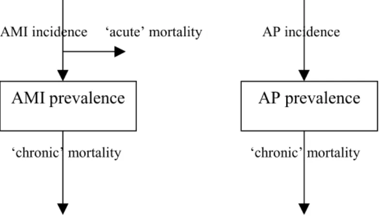
(14) Page 14 of 117. RIVM report 260751 001. infarction and the mortality during the years after surviving the first month. These two types are defined here as ‘acute mortality’ and ‘chronic mortality’ respectively.. 1.4. Basic model equations. The chronic disease model equations describe the changes in disease prevalence numbers over time, specified by gender and age. The prevalence numbers change due to disease incidence (inflow), remission and mortality (outflow) (see Figure 1).. time t. disease-free population mortality due to other causes. population with disease (prevalence) incidence. remission. mortality due to excess other causes. change. time t+1 disease-free population. population with disease (prevalence). Figure 1 The chronic diseases model structure. We describe here the several steps that result in the model equation of the change of the disease prevalence rate over age. The formal derivation is presented in Appendix 3. The disease prevalence number changes due to so-called inflow and outflow transitions. Inflows result in increasing prevalence numbers, outflows in decreasing numbers. For reason of simplicity we omit the changes due to birth and migration here. Then the transitions to be distinguished are disease incidence (inflow out of the state ‘disease-free’), remission (outflow to the state ‘disease-free’) and mortality (outflow to the state ‘deceased’). The disease incidence number is assumed proportional to the total population number here (see also note below). The disease remission number is assumed proportional to the disease prevalence number. The mortality rate for a patient is assumed to be the sum of a baseline mortality rate and an excess mortality rate. The baseline mortality rate is assumed to be the same for persons with and without the disease. Because the excess mortality results in both decreasing disease prevalence numbers and total population numbers, the relative decrease of the disease prevalence rate due to the excess mortality is smaller than the relative decrease of the prevalence number. The difference equals the proportion of the population having the disease. Because the baseline mortality is the same for both persons having the disease and without the disease, it has no affect on the proportion of the population having the disease. So.
(15) RIVM report 260751 001. Page 15 of 117. the model equation that describes the change of the disease prevalence rate over age due to incidence, remission and excess mortality is: d/dt previ = inci – remi previ – cfi previ (1-previ) with: previ d/dt inci remi cfi. (1). the disease i (point) prevalence rate over age interval the instantaneous (continuous-time) change the disease i incidence rate the disease i remission rate the excess mortality rate for patients with disease i. The disease-specific excess mortality on the population level is described by: morti = cfi previ. (2). with: morti: the disease-specific excess mortality rate on the population level, i.e. with the total population number instead of the prevalence number in the denominator. The incidence rate in model equation (1) refers to the total population, not to the disease-free population. When using incidence rates that refer to the disease-free population a factor ‘1-previ’ has to be added. In Appendix 3.1 it is shown that competing death risks, for example due to joint risk factors, result in more complex model equations. In Appendix 3.4 it is shown how the calculated excess mortality rates can be used to calculate disease duration times and mortality hazard ratios. The main assumptions that underlie the model equations are the following. Equation (1) describes the change of the prevalence rate over age for any cohort. It is assumed that the prevalence rates are constant over time (see Figure 2). However, it is possible to adjust for time trends. All cause-specific mortality rates are assumed independent conditional on gender and age (see also Appendix 3). For example, COPD incidence rates are assumed equal for persons with and without coronary heart disease. Only ‘chronic’ cause-specific mortality has been described in formula (1), i.e. the mortality is defined proportional to the disease prevalence. ‘Acute’ mortality is defined proportional to the disease incidence, and has been included in the model equation for diseases with acute events, i.e. coronary heart disease and stroke. Disease incidence and prevalence data are available specified by five-year age classes. We have found large fluctuations over age in these data, mainly due to small sample sizes. While the estimated mortality and remission rates are very sensitive to these fluctuations, we have smoothed the data and interpolated them over age, before estimating the model parameters. Smoothing is made by using a penalty matrix including weighting coefficients (see Appendix 4)..
(16) Page 16 of 117. RIVM report 260751 001. In Figure 2 the aspects of time and age are presented. The chronic disease model equations describe the change of the prevalence rate for any cohort (the diagonal line), the data are from cross-sectional studies (the vertical line). When the situation is stable, i.e. no time trends, the prevalence rate at the start and end of the year are equal. In case of time trends, we have to adjust the prevalence at the end of the year for the trend over the year. Time trends in the Netherlands can be assessed from the study with the longest registration period, i.e. CMR Nijmegen. Trend values are based on the time period until 1994 that shows a stable trend.. age. a+1 prev(a+1,t). a. prev(a+1,t+1). prev(a,t). t. t+1. time. Figure 2 Time and age in the model equations (Lexis-diagram) Note: a: age, t: time point, prev: disease prevalence.. Using the excess mortality rates it is possible to calculate mortality hazard rate ratios for patients, i.e. the ratio of the mortality rate for any patient to the mortality rate for a person without that disease. The formulas used are presented in Appendix 3.4.. 1.5. Data sources. The data are derived from different sources. -. Disease incidence and prevalence rates come from several sources that have been established for general chronic conditions (such as registration projects in general practice) or for specific diseases (such as the Netherlands Cancer Registry). In the framework of VTV-1997 a panel of experts has evaluated all disease-specific data from these sources (Ruwaard&Kramers, 1998). The main data sources used are: Dutch Survey of General Practice (National Study, NS), account: Netherlands Institute of Primary Health Care (NIVEL) Continuous Morbidity Registration Nijmegen (CMR), account: Medical Department of the University of Nijmegen (KUN) Amsterdam Transition Project (Trans), account: Medical Department of the University of Amsterdam Registration Network Family Practices (RNH), account: Medical Department of University Maastricht Dutch Sentinel Practice Network (CMR Peilstations, Peil), account: NIVEL.
(17) RIVM report 260751 001. Page 17 of 117. -. Netherlands Cancer Registry (NKR), account: Association of Comprehensive Cancer Centers (VvIK) Eindhoven Cancer Registry (IKZ), account: Comprehensive Cancer Center South; note: NKR is nation-wide, but IKZ is over much longer time period Monitoring Risk Factors and Health Status in The Netherlands (MORGEN), account: National Institute on Public Health and the Environment (RIVM) Rotterdam Elderly Study (ERGO), account: Erasmus University Rotterdam Cause-specific mortality rates from Statistics Netherlands (CBS) Disease remission, cause-specific case-fatality rates, mortality rate ratios and relative risks from literature on the specific diseases The characteristics of the main data sources are given in Table 1. Table 1 Characteristics of the data sources used. Registration projects in general practice:. Disease variables. Region. Period. Size. National Study NIVEL (NS). incidence4. national. 1987/1988 3 months. 330.000 persons 85.000 pers-years. CMR Nijmegen. incidence2,3,4,5,6 region prevalence2,3,4,5,6 Nijmegen. since 1971 continuous. 12.000 persons. Transition Project (Trans). incidence2,4,6 prevalence2. multi-regional. 1985-1995. 93.000 pers-years. RNH Limburg. incidence2,3,4,6 prevelence2,3,4,6. region Limburg. since 1988 continuous. 78.000 persons. CMR Peilstations (Peil). incidence. national. since 1970 continuous. 140.000 persons (1995). Statistics Netherlands (CBS). mortality1,2,3,4,5,6 national. continuous. national. ERGO Rotterdam. prevalence6. Rotterdam. since 1990 continuous. 8000 persons. Cancer Registration South-Netherlands (IKZ). incidence prevalence1 mortality1. region Eindhoven. since 1955 continuous. 1 million persons. National Cancer Registration (NKR). incidence1 mortality. national. since 1989 continuous. national. Health Care Information (SIG). incidence. national. since 1963 continuous. national. Other data sources:. Notes: data used for lung cancer (1), asthma and COPD (2), CHD and CHF (3), diabetes mellitus (4), dementia (5) and stroke (6)..
(18) Page 18 of 117. RIVM report 260751 001. More information on the diseases can be found in VTV-1997 (Ruwaard&Kramers, 1998), and in disease-specific literature (see References).. 1.6. Data selection criteria. The main criteria for selecting the sources of disease incidence and prevalence data to be used in the analyses are: 1 2. 3. 4. The observation period is around year 1994. In absence of indications for time trends, also data from the National Study are used. Ideally one would like to have data from a national and representative system which would continually monitor all disorders in the population on the basis of unambiguous diagnosis criteria. Since such a system is not available, data from various sources have been used, for example from epidemiological surveys and registrations in general practice. The former data involve the total population and are only available for a limited number of disorders. The latter data generally involve persons with serious complaints and therefore visiting the general practitioner. Because we do not want to use data from many different sources, and we also want to assess the effects of diseases in terms of health care use, we mainly employ registrations in general practices. In the framework of VTV-1997 (Ruwaard&Kramers, 1998; Gijsen et al., 1997) the validity of all data from the registrations has already been assessed by evaluating the degree of representativeness, continuity, completeness and freedom from ambiguity of each source. Where possible, we have used also data from epidemiological surveys. In general only sources that provide both disease incidence and prevalence data are selected. As both figures are used within one model equation they have to be compatible. Exceptions to this rule are allowed when the data do not differ much after visual comparison. For registrations in general practice it is necessary that even patients who only rarely visit the practice are registered as prevalent cases.. Note that these criteria may result in different data source selections for different diseases. As a consequence of criteria 1 and 3 the National Study (NS) has rarely been selected. CMR Nijmegen has mostly been selected, often the Transition Project and RNH Limburg as well. For chronic diseases with long disease duration and low frequency of GP-visits, the Transition Project often provides an underestimation of prevalence figures and for that reason has not been used (criterion 4). In the case of lung cancer we have used data from cancer registration projects. Incidence and prevalence data are presented in graphical form and in tabular form. All rates are per 1000 persons (per year). They are specified by gender, age (in tabular form by 5-year age classes with rest class 85+), and source. All incidence and prevalence data used in the.
(19) RIVM report 260751 001. Page 19 of 117. model calculations are smoothed using the penalty smoothing method (see Appendix 4). The smoothing penalty coefficient used has been specified for each disease separately. The agestandardised incidence and prevalence rates have been calculated for each selected source using the non-smoothed five-year age class data, or the calculated mean values respectively, using 1994 Dutch population numbers. Data from general practices include acute mortality and cases that are not hospitalised. Only for some diseases with high hospital admission rates, such as myocardial infarctions, health care data have been analysed.. 1.7. Reader. In separate chapters the results of the analyses have been provided for the selected disease categories. Each chapter presents a short epidemiological introduction to the disease, and an overview of the data sources available and those that have been selected according to the selection criteria that have been presented in §1.6. Based on the incidence and prevalence figures used, mortality rates are calculated using the chronic diseases model equations. Because the calculated mortality rates are often unstable for lower ages, they are generally only presented for the higher ages. The calculated mortality rates are compared to those from national statistics. For lung cancer also prevalence rates are calculated based on given incidence and survival rates. For asthma remission rates are calculated as well, given incidence, prevalence and mortality rates. The calculated mortality rates and relative risks are compared to data from the literature. Concluding remarks are presented in a final paragraph. The gender- and age-specific incidence and prevalence figures are presented in appendices. In the appendices data and methods are described in more detail. In Appendix 2 the genderand age-specific incidence and prevalence figures are presented for the diseases selected. In Appendix 3 the chronic disease model equations are derived. The original equation is given in terms of changing disease prevalence numbers specified by gender, age and other covariables. It is shown how these equations can be simplified under specific assumptions and by describing rates instead of numbers, and how the equation can be interpreted in terms of time trends instead of changes over age. In Appendix 4 the data smoothing method used is described. In Appendix 5 a simple model is presented that explains the differences between the incidence and prevalence rate ratios found in population surveys compared to those found in registrations in general practice. In Appendix 6 the symbols and abbreviations used are summarised. At the end of the report a list of references is presented, general and specified by disease..
(20) Page 20 of 117. RIVM report 260751 001.
(21) RIVM report 260751 001. Page 21 of 117. 2.. Lung cancer. 2.1. Introduction. Lung cancer (ICD 162) is the type of cancer with the highest mortality in the Netherlands and other Western countries. Lung tumours are divided in small-cell type (SCLC, approx. 20%) and other (NSCLC, approx. 80%). Most lung cancer patients already have metastases in the local or regional glands the moment they are diagnosed. For most new cases diagnosed the disease progression is such that only palliative treatment has become possible. In case of limited or localised stage cure may be possible. Lung cancer incidence in the Netherlands has decreased for men and increased for women in the Netherlands over the last years, reflecting different past trends in smoking prevalence trends among men and women.. 2.2. Data sources. The lung cancer data used in the analyses are from: incidence. prevalence. survival. NKR. IKZ. IKZ. Only one (national) source is available for incidence data and one (regional) source for prevalence data.. 2.3. Data on incidence and prevalence. The 1994 lung cancer incidence and prevalence data are presented in Table 40 and Figure 23 (Appendix 2). Incidence and prevalence rates are higher for males than for females. The age at which rates are highest is lower for females than for males. A possible explanation for these differences is the increasing smoking prevalence for females during the last decades. Rates have decreased for males and increased for females, except for the highest ages. The national NKR and regional IKZ data can be compared on disease incidence. The IKZincidence rates for males are larger than the NKR figures, for females they are smaller. The incidence rate ratios IKZ/NKR are 1.17 (males) and 0.89 (females) respectively for age 50+. We have not applied these ratios to adjust the IKZ regional prevalence data, because we have don’t know whether the rate ratios found for the incidence are also valid for the prevalence. Besides the differences of the rate ratios between men and women are substantial, and we cannot explain them..
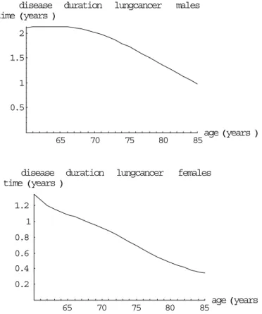
(22) Page 22 of 117. 2.4. RIVM report 260751 001. Estimated mortality rates. Lung cancer excess mortality rates can be calculated from incidence and prevalence data using the equations given in §1.4. They were calculated assuming no remission and prevalence trends of 0.7 % (men) and 2.3% (women) per year. These trends were based on prevalence trends found in IKZ/SOOZ (period 1989-1993). The excess mortality rates are presented in Figure 3 and Table 2 (only for males). Based on the calculated excess mortality rates also the disease duration times can be calculated, see Figure 4.. excess rate. mortality. rates lungcancer. males. 0.7 0.6 0.5 0.4 0.3 0.2 0.1 50. mortality rate. 60. 70. age Hyears L. 80. rates lungcancer. males. calculated. 0.008. registered. 0.006 0.004 0.002 50. 60. excess mortality rate 1. 70. 80. age Hyears L. rates lungcancer. females. 0.8 0.6 0.4 0.2 50. 60. 70. 80. age Hyears L.
(23) RIVM report 260751 001. mortality rate. Page 23 of 117. rates lungcancer. females. 0.0008. calculated. 0.0006. registered. 0.0004 0.0002 50. 60. 70. 80. age Hyears L. Figure 3 The calculated excess mortality and cause-specific population mortality rates, the mortality rates are also compared to empirical data disease duration time Hyears L. lungcancer. males. 2 1.5 1 0.5. 65. 70. disease duration time Hyears L. 75. 80. lungcancer. 85. age Hyears L. females. 1.2 1 0.8 0.6 0.4 0.2 65. 70. 75. 80. 85. age Hyears L. Figure 4 The calculated lung cancer disease duration times for males and females The calculated excess mortality rates are much higher for females than for males. This results in shorter disease duration times and calculated population mortality rates that are higher than the empirical ones. The main explanation is that the time trend adjustment applied does not work well enough for females. Therefore we have not presented the results for females in Table 2. The estimated lung cancer mortality rates are smaller than the empirical rates for the highest ages, for males and females (Figure 3). This is very surprising. We did not find an explanation..
(24) Page 24 of 117. RIVM report 260751 001. Table 2 The calculated lung cancer excess mortality rates for males age calculated males. 40-44. 45-49. 50-54. 55-59. 60-64. 65-69. 70-74. 75-79. 80-84. 85-89. .56. .45. .39. .37. .36. .36. .36. .40. .46. .53. Note: unit: /patient/year.. Table 3 Lung cancer mortality rates from the literature Berrino et al, 1995 .53 (all ages,M+F) IKZ, 1997 observed survival fraction age 15-59 60-74 75+ from survival data over 5 year .18 .13 .032 10 year .14 .05 .042. calculated 1-year excess mortality 15-59 60-74 75+ .29 .19. .34 .25. .39 .29. Note: The 1-year survival fractions were calculated from the n-year survival data using the formula: ‘1-year survival fraction’ = ‘n-year survival fraction^(1/n)’. The excess mortality rates are calculated as: calculated 1-year mortality rate – population mortality rate (Statistics Netherlands).. The 10-year survival is relatively better than the 5-year survival because conditional on survival after 5 years the 5-years rest prognosis is better than the 5-years prognosis on the time of onset. In the 1993 chronic diseases model version smaller lung cancer mortality rate values have been used. One reason is the different time discretisation method used: in the 1993 model version it was assumed that the all new cases ‘occur’ at the beginning of each year, whereas we have assumed occurrence halfway each year.. 2.5. Estimated prevalence rates. For lung cancer reliable mortality data are available. Therefore we can also estimate lung cancer prevalence rates given incidence and mortality rates. The prevalence rates are calculated using equation (1) and the mortality rates using equation (2) (see §1.4). The estimated prevalence and mortality rates are compared to prevalence and mortality data respectively (see Table 4). The 1st column describes the age-specification, the 2nd column the data values, and the 3rd to 6th column the calculated values for four different excess mortality data figures. The 1st block compares results on prevalence for males, the 2nd block those for females, in the 3rd and 4th block results are given on mortality. The four mortality data figures used are: 3-year survival probability = 0.1, 5-year survival probability = 0.1, and relative 5year survival fractions and 10-year fractions (Coebergh, 1995) respectively..
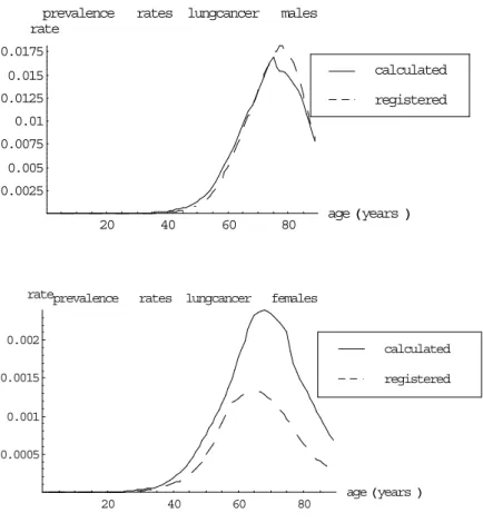
(25) RIVM report 260751 001. Page 25 of 117. Table 4 Calculated lung cancer prevalence and population mortality rates, compared to data age. mortality data for lung cancer patients used 3-year=0.1 5-year=0.1 5-year relat. age data results calculated prevalence rates (*.001) males (block 1) 40 .1 .2 .2 .2 50 .8 .9 1.1 1.3 60 5.0 4.0 5.3 6.0 70 13.2 9.9 13.7 14.3 80 17.5 15.1 22.8 18.0 females (block 2) 40 .1 .1 .2 .2 50 .5 .5 .6 .7 60 1.2 1.1 1.5 1.8 70 1.2 1.6 2.3 2.5 80 .6 1.3 2.0 1.7 calculated population mortality rates males (*.001) (block 3) 40 .1 .1 .1 .1 50 .4 .5 .4 .4 60 2.1 2.2 2.0 2.0 70 5.0 5.2 5.1 5.1 80 8.8 8.1 8.4 8.8 females (*.0001) (block 4) 40 .6 .6 .6 .5 50 2.5 2.5 2.3 2.2 60 5.8 6.0 5.7 5.7 70 7.5 8.4 8.5 8.5 80 7.1 6.9 7.3 7.7. 10-year relat.. .3 1.7 7.8 19.2 27.6 .2 1.0 2.5 3.6 2.8. .0 .3 1.7 4.9 9.3 .4 1.8 5.3 8.6 8.5. Notes: prevalence rates calculated using life-table method; population mortality rates calculated by multiplying the calculated prevalence rates by the given patient mortality rate.. Table 4 shows that for males the 5-year relative survival data result in prevalence and mortality rates that fit best to the data. The calculated prevalence rates based on these survival fractions are also presented in graphics (see Figure 5). For females the 5-year relative survival data does not result in prevalence rates that fit well to data. The main explanation is that lung cancer incidence rates have increased for females over the last years, and so the trend of the prevalence rate follows the trend of the incidence rate with a delay time (see also §2.4)..
(26) Page 26 of 117. RIVM report 260751 001. prevalence rate. rates lungcancer. males. 0.0175 0.015. calculated. 0.0125. registered. 0.01 0.0075 0.005 0.0025 20. rateprevalence. 40. 60. rates lungcancer. age Hyears L. 80. females. 0.002. calculated. 0.0015. registered. 0.001 0.0005. 20. 40. 60. 80. age Hyears L. Figure 5 Calculated prevalence rates using 5-year relative survival data, for males and females. 2.6. Concluding remarks. New lung cancer cases are registered by the National Cancer Registration (NKR). This national registration has become active only recently. For lung cancer prevalence, we have only figures from a regional registration (IKZ) that has been active since 1955. The regional incidence rates for males are smaller than the national figures, and for females are larger. We have not yet adjusted the prevalence rates, because the relevance of this result is still unclear. Moreover, future prevalence rates are not very sensitive to the initial prevalence figures because of the short disease duration time. For excess mortality we also used data from the IKZ-registration. The rates have also been estimated assuming no remission and for given trends in lung cancer prevalence. For males the estimated excess mortality rates fit well to the data on relative 5-years survival. Moreover, using the latter data results in prevalence and mortality figures that fit well to the data, especially for males. For females the excess mortality rates could not be estimated well. One important reason is probably time trends. We have decided to use the 5-year relative survival data from IKZ (1997) to calculate the lung cancer excess mortality rates..
(27) RIVM report 260751 001. Page 27 of 117. 3. Chronic non-specific lung diseases: asthma and chronic obstructive pulmonary disease 3.1. Introduction. Chronic aspecific respiratory diseases (ICD 490-496) is a group of diseases of the bronchial tubes and alveoli. Based on insight in the pathophysiology two subgroups are distinguished: asthma (493) and chronic obstructive pulmonary disease (COPD). Asthma is mainly but not exclusively found in children, COPD mainly in the elderly. Asthma prevalence rates in the Netherlands have increased in the last decade. COPD prevalence has not changed for men, but has increased for women, probably as a result of changing smoking behaviour. Mortality due to both diseases is neglectable in lower ages. For the elderly mortality due to asthma is still very small, but for COPD mortality rates increase with age, especially for women. Modelling asthma and COPD differs substantially from modelling lung cancer for several reasons. (1) We have to compare data and make a selection, as several data sources are available. (2) For asthma two types of ‘outflow’ are possible: remission and mortality. Both parameters cannot be identified uniquely using only incidence and prevalence figures. (3) There are significant trends in disease incidence and prevalence that have to be taken into account. (4) There is a relation between asthma and COPD. They can be interpreted as ‘competing’ diseases because the differences in symptoms are small.. 3.2. Data sources. The asthma and COPD data used in the analyses are from: incidence. prevalence. National Study CMR Nijmegen Transition Project RNH Limburg. CMR Nijmegen Transition Project (asthma) RNH Limburg (COPD). No data are available from epidemiological sources. CMR Nijmegen provides reliable data on both incidence and prevalence. The Transition Project provides low COPD prevalence figures, maybe as a result of a short time window (1year) of registration. For RNH Limburg it is supposed that the coding rules for asthma are too strict (‘once asthma, always asthma’) and yields prevalence figures that are too large. Therefore they have not been used in our analyses. The data sources on incidence and prevalence selected have been presented in bold..
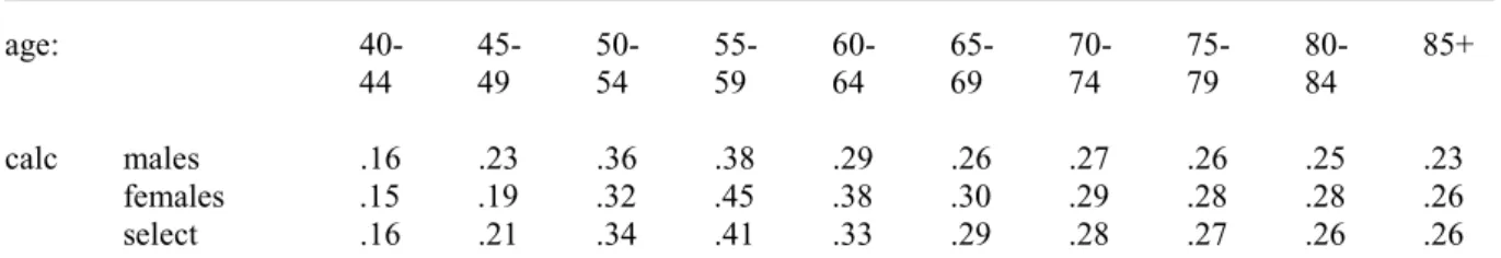
(28) Page 28 of 117. 3.3. RIVM report 260751 001. Data on incidence and prevalence. In Table 41 and Figures 25 & 26 (Appendix 2) we present the 1994 incidence and prevalence data that have been used in our analyses. Asthma incidence and prevalence rates are highest for the lower ages. However, asthma incidence rates seem to increase again slightly for the higher ages, especially for males (for a possible explanation, see §3.5). According to CMR Nijmegen asthma prevalence rates seem to have increased in the period 1981-1994 for the lower ages. COPD incidence and prevalence rates increase over age. They are larger for males than for females. The prevalence rates seem to have increased in the period 1984-1994 for elderly women according to CMR Nijmegen. The results for COPD can be explained by smoking behaviour, since smoking is an important risk factor.. 3.4. Estimated remission and mortality rates. Asthma remission and COPD excess mortality rates can be calculated from incidence and prevalence data using the equations given in §1.4. Based on CMR Nijmegen time series data (1984-1994) we assume the annual asthma prevalence trend to be 8% (males) and 11% (females) per year respectively, and for COPD 0% (males) and 7% (females) respectively. The calculated rates are presented in Table 5 and Figures 6&7 respectively. Based on data from literature (see Table 6) we have assumed that the mortality rate for asthma patients is 1.5 times the population mortality rate. For COPD it is assumed that there is no remission. Based on the calculated mortality rates also the disease duration times can be calculated, see Figures 8&9. For females no reliable COPD excess mortality rates could be calculated assuming an annual prevalence trend of 7%. Assuming no trend an excess mortality rate of 0.07 was found, that was almost constant over age. One possible explanation for the bad results for women is the time trend of COPD prevalence rates that is hard to deal with. Because in the literature no clear differences between men and women have been found, we assume that the mortality rates found for males also are valid for females. The excess mortality rate values are presented in Table 5..
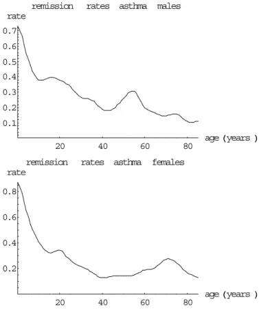
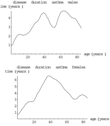
(29) RIVM report 260751 001. remission. Page 29 of 117. rates asthma males. rate 0.7 0.6 0.5 0.4 0.3 0.2 0.1 20. 40. remission rate. 60. rates asthma. 80. age Hyears L. females. 0.8 0.6 0.4 0.2. 20. 40. 60. 80. age Hyears L. Figure 6 The calculated asthma remission rates for males and females excess mortality rate 0.175. rates COPD males. 0.15 0.125 0.1 0.075 0.05 0.025 50. mortality. 60. 70. age Hyears L. 80. rates COPD males. rate 0.025 calculated 0.02 registered 0.015 0.01 0.005 50. 60. 70. 80. age Hyears L. Figure 7 The calculated COPD excess mortality and cause-specific population mortality rates for males, the mortality rates are also compared to empirical data (CBS).
(30) Page 30 of 117. RIVM report 260751 001. disease time Hyears L. duration. asthma. 40. 60. males. 4 3 2 1 20 disease time Hyears L. duration. 80. asthma. age Hyears L. females. 6 5 4 3 2 1 20. 40. 60. 80. age Hyears L. Figure 8 The calculated asthma duration times for males and females. disease time Hyears L 25. duration. COPD males. 20 15 10 5 50. 60. 70. 80. age Hyears L. Figure 9 The calculated COPD duration times for males.
(31) RIVM report 260751 001. Page 31 of 117. Table 5 The calculated asthma and COPD remission and excess mortality rates respectively. asthma remission males females select males age: (M+F) 0-4 .65 .77 .71 5-9 .45 .51 .48 10-14 .38 .36 .37 15-19 .39 .33 .36 20-24 .37 .31 .35 25-29 .30 .24 .27 30-34 .26 .20 .23 35-39 .22 .15 .18 40-44 .18 .13 .16 45-49 .22 .14 .18 50-54 .29 .14 .21 55-59 .27 .17 .21 60-64 .18 .19 .20 65-69 .15 .24 .20 70-74 .15 .27 .20 75-79 .14 .21 .18 80-84 .11 .15 .13 85+ .11 .12 .11. COPD excess mortality. .10 .06 .06 .05 .04 .03 .03 .03 .024 .014 .013 .014 .017 .03 .04 .07 .11 .15. Notes: unit:/person/year; select: mean over males and females for COPD for females no excess mortality rates could be calculated.. Calculated asthma remission rates decrease over age, with some irregularities for the higher ages. They are unstable for the lowest ages, and we have found differences between male and females for the higher ages. Because all differences are small, we have concluded that asthma remission rates are equal for men and women, with constant values for ages above 35 years (see Table 5).. 3.5. Other data from literature on remission and mortality. We have presented data from literature on mortality for asthma (Table 6), for COPD (Table 8) and on remission for asthma (Table 7). Mortality hazard ratios, relative risks and standardised mortality ratios are similar parameters to describe increasing risks, and so have been presented in one table. However, they differ when hazard ratios change over age..
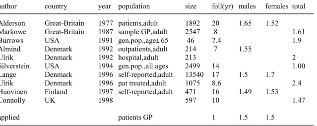
(32) Page 32 of 117. RIVM report 260751 001. Table 6 Mortality hazard ratios, relative risks and standardised mortality ratios for asthma patients, together with the applied model value author. country. year population. size. foll(yr) males. females total. Alderson Markowe Burrows Almind Ulrik Silverstein Lange Ulrik Huovinen Connolly. Great-Britain Great-Britain USA Denmark Denmark USA Denmark Denmark Finland UK. 1977 1987 1991 1992 1992 1994 1996 1996 1997 1998. 1892 2547 46 214 213 2499 13540 1075 471 597. 20 8 7.4 7. 1.52. patients,adult sample GP,adult gen.pop.,age– 65 outpatients,adult hospital,adult gen.pop.,all ages self-reported,adult pat treated,adult self-reported,adult. applied. patients GP. 1.65. 1.61 1.9 1.55 2 1.00. 14 17 8.6 16 10. 1.5. 1.7. 1.49. 1.53. 2.4 1.47. 1. 1.5. 1.5. Notes: foll: follow-up. Table 7 Proportions of remission for asthma author. country. year. population. size. follow-up(yr). Blair Schachter Burrows Aberg Gerritsen Burrows Panhuysen Barbee Norrman. Great-Britain Lebanon USA Sweden Netherlands USA USA USA Sweden. 1977 1984 1987 1990 1990 1991 1997 1998 1998. patients,child general,age– 7 general,all patients,age 14 patients,child general,age– 65 patients,adult general,child students. 417 73. 20 6 9 3-13. calculated. proportion (%). 52 68 35/65/30/15/5/15/30/301 74/55/44/252 50 7.5 19 25 11 retrospective 50 1 5.7. 1335 46 181 review 990. general. 1. 55/38/34/23/22/22/22/221. Notes: (1): proportions for age classes 0-10 .. 70-79, (2): proportions for age classes at onset: 0-2,2-4,4-7,7-11.. Table 8 COPD mortality rates author. country year. population. Tager. USA. hospital,adult. 1991. size. follow-up(yr). rate(/yr). <=17. .058. The asthma relative mortality risks found do not differ much, except for the one Danish study. A large part of the asthma excess mortality described has been found to be due to COPD. The asthma remission proportions (see Table 7) have been presented in their original form and have not been standardised to 1-year figures. The reason is that standardisation is only possible with extra data or extra assumptions on asthma incidence. That’s also why direct comparison with the calculated remission rates is difficult..
(33) RIVM report 260751 001. 3.6. Page 33 of 117. Concluding remarks. For asthma and COPD several data sources are available. According to our selection criteria CMR Nijmegen, Transition Project (not for COPD prevalence) and RNH Limburg (only for COPD) have been selected. For asthma the remission rates for males and females are almost equal after adjustment for the trend, except for higher ages. Because all differences are small, we have concluded that the asthma remission rates are equal for both sexes for all ages. The remission rates found are relatively large and result in small disease duration times compared to the literature. We have found several possible explanations. (1) Remission is defined here in terms of ‘inactivating’ the registration, and so may be different from the definition of remission used in literature. (2) We have presented 1-year remission rates, whereas in literature remission fractions are presented mainly over larger follow-up periods. Remission rates over different time periods cannot be compared easily. One reason is that part of the remission may relapse to asthma again after a longer time period. For COPD the excess mortality rates are different for males and females, especially for high ages. However, for females there have been large changes in COPD incidence and prevalence over the past years. So although we have adjusted for time trends, the results may still be biased. Therefore we have concluded that the calculated rates are unreliable for females and that the excess mortality rates for females are equal to those for males. There is a strong statistical correlation between the calculated excess mortality and remission rates for asthma. Assuming no excess mortality the calculated remission rates increase again for the higher ages, applying the mortality hazard ratio of 1.5 based on literature the rates decrease over age. According to literature a large part of the excess mortality among asthma patients is due to COPD..
(34) Page 34 of 117. RIVM report 260751 001.
(35) RIVM report 260751 001. Page 35 of 117. 4. Coronary heart diseases and congestive heart failure 4.1. Introduction. Coronary heart diseases (CHD, ICD 410-414) are the most common type of cardiovascular diseases. They are characterised by the narrowing of the coronary arteries. The most important types of CHD are acute myocardial infarction (AMI, ICD 410) and angina pectoris (AP, ICD 413). One example of ‘other acute and subacute CHD types’ (ICD 411) is unstable AP. An old infarction (ICD 412) is an AMI without symptoms that is confirmed afterwards. It could be a ‘silent infarction’. To the other forms of CHD (ICD 414) belong diseases such as chronic atherosclerosis. Time trends in the Netherlands suggest a change from the acute types of CHD to the chronic types. This change may result in a future increase of the incidence of AP and congestive heartfailure (heart decompensation, CHF, ICD 402,404,428,429.1 and 429.4) according to Barendregt&Bonneux (1998). Congestive heartfailure is characterised by a deficient blood circulation due to an insufficient pumping function of the heart, leading to shortness of breath, tiredness and oedema. The main risk factors for heartfailure are CHD and hypertension. In our analyses we distinguish only two manifestations of CHD, AMI (ICD 410) and other forms of CHD, mainly stable and unstable AP (ICD 411-414). Both CHD manifestations and CHF are strongly interrelated. For example, persons with AP have relatively high risks of an AMI, persons surviving an AMI are registered in some registrations in general practice as belonging to the group of CHD-patients, persons having AP or AMI have high risks of developing CHF, and persons having CHF have high mortality risks for AP and AMI.. 4.2. Data sources. The CHD and CHF data used in the analyses are: incidence. prevalence. National Study NIVEL CMR Nijmegen CMR Nijmegen Transition Project RNH Limburg RNH Limburg CMR Peilstations ERGO Rotterdam Health Care Information (SIG).
(36) Page 36 of 117. RIVM report 260751 001. Prevalence figures from epidemiological studies are available (e.g. ERGO), but only for myocardial infarction (life-prevalence; assessment by anamnesis) and CHF (pointprevalence; assessment by anamnesis and examination). We do not use them, because no matching incidence figures are available. Persons having an AMI are generally hospitalised, contrary to persons with AP. So only for the former patients admission data for inpatient care can be useful. Data from the National Medical Register can be used. For AMI, AP as well as CHF data from registrations in general practice are available. In these registration projects the denomination of the terms is complicated. Registrations differ in their classification system and registration practice. That’s why the morbidity figures are not at once comparable. However, in general the registrations distinguish AMI, a chronic form of CHD, and heart failure. But some registrations do not use a specific code for AMI, and register AMI as an incident CHD. In some registrations patients who survive an AMI are generally registered twice in one year, once as AMI incident, and once as incident for (a chronic form of) CHD. So the total CHD patient numbers are less than the sum of AMI and CHD cases. Other registrations distinguish suspected from confirmed myocardial infarction. Some projects register only first infarctions, other projects register all infarctions including recurrent ones. For selecting the appropriate registrations and codes in this report we use the decisions made for the Dutch PHSF-report (Gijsen et al., 1997; Maas et al., 1997). CHF is more or less registered on the same way in all registrations. A summary of the characteristics of the data from the registration projects in general practice about CHD is presented below: Incidence: National Study. AMI. K75.1. confirmed infarctions including recurrent ones. AP. K76.1. stable and unstable AP; a patient can have recurrent episodes. CMR Nijmegen AMI AP Trans 10 Project CHD. 2110 2120 K76. Trans 5 Project. AMI AP. K75 K74. RNH Limburg. AMI. K75. AP. K74. only first occurring infarctions excluding recurrent AMIs stable and unstable AP episode; a patient can have recurrent episodes clinical relevant CHD: AMI or AP or old AMI; corrected for double counts, so only first occurring events in a period of 4/5 years only one infarction per patient is registered stable and unstable AP; in a period of 4/5 years only the new AP-episode Per patient is registered not consistently registered because AMI is not a chronic problem; if registered, it is only for a short time period; after becoming inactive, AMI can become active again; so recurrent AMIs are included in the figures stable and unstable AP; after some period this code often becomes inactive and code K76 becomes active clinical problem often following AMI or AP confirmed infarctions, including recurrent ones. CHD CMR Peilstations AMI. K76 --.
(37) RIVM report 260751 001. Page 37 of 117. Prevalence: National Study CMR Nijmegen AMI AP Trans 10 Project CHD. 2110 2120 K76. Trans 5 Project. AMI AP. K75 K74. RNH Limburg. AMI AP. K75 K74. CHD. K76. no data life-prevalence: ever had one or more myocardial infarctions stable and unstable AP AMI or AP or old AMI with clinical significance; corrected for double Counts no meaning; duration of an AMI is short stable and unstable AP; it describes the number of patients with one or more episodes of AP in a period of 4/5 years no meaning; duration of an AMI is short stable and unstable AP; after some period this code becomes often inactive and K76 active clinical problem; interpreted as a status after an AMI, with AP or after AP; no complaints, with medication or regularly monitored. We have decided not to use data from the following sources: the National Study, CMR Peilstations and the 5-year database of the Transition Project because they provide only reliable disease incidence data; the 10-year database of the Transition Project because they provide only reliable incidence data and because the subgroup of patients with other forms of CHD is hard to attribute to the CHD subgroups we have distinguished. Finally we selected data from CMR Nijmegen and RNH Limburg, presented in bold.. 4.3. Data on incidence and prevalence for CHD. In Table 42 and Figures 27, 28 & 29 (Appendix 2) we present the 1994 data on incidence and prevalence and the selected values after smoothing. Unlike CMR Nijmegen, RNH Limburg distinguishes CHD apart from AMI and AP. Both AMI and AP patients can ‘flow’ to the group of CHD patients. According to Gijsen et al. (1997) patients who survive an AMI the AMI-code is made ‘inactive’ and the CHD-code (K76) is made ‘active’ soon after the disease onset. So we have decided to take the AMI and AP figures for the incidence rates, and to add the CHD figures to the AMI and AP prevalence rates. Because we have no explicit information on the disease history of CHD patients, we have decided to add the CHD prevalence numbers to the AMI and AP prevalence numbers proportional to the AMI and AP incidence numbers, i.e. AP : AMI = 1:1 (males), 3:1 (females). The AMI-prevalence is defined as lifetime prevalence. AMI incidence and prevalence rates increase over age, with decreasing prevalence rates for the highest ages. The rates are larger for males. CHD incidence and prevalence rates also increase over age, with decreasing incidence rates for the highest ages. The rates are slightly higher for males. After redistributing the CHD numbers for RNH over the AMI and AP numbers, the CMR and RNH data on incidence and prevalence do not differ much, except for incidence of AP. This may be due to different coding rules and/or inclusion criteria..
(38) Page 38 of 117. 4.4. RIVM report 260751 001. Data on incidence and prevalence for CHF. In Figure 29 and Table 43 we present the 1994 data on CHF incidence and prevalence and the selected rate values after smoothing. CHF incidence figures from CMR and RNH are similar. Prevalence figures are larger for CMR for higher ages.. 4.5. Estimated mortality rates for CHD. Excess mortality rates are estimated for AMI and AP-patients using the chronic disease model equation given in §1.4. We have assumed no remission. For AMI we have assumed an annual change of the prevalence rate of –2% for men and –3% for women, for AP of +3% and –1% respectively. These trends are based on CMR Nijmegen time series over 1987-1993. For AMI we have included ‘acute’ mortality. The differences between calculating the AMI and AP excess mortality rates become more clear from Figure 10. The AMI incidence numbers in CMR Nijmegen exclude recurrent infarctions, the RNH Limburg include these infarctions. We have found some information on the proportion of all infarctions that are recurrent. According to Vrieze (1994) 9% of all hospitalisations of AMI patients are recurrent ones within one year, and 13% of all incidence numbers (including fatal infarctions and infarctions not admitted to a hospital) are recurrent in the same year (1988 data). According to Weinstein (1987) 22% of all patients with a history of AMI are relapsed cases, and the probability of a recurrent infarction for all AMI patients including those with congestive heart failure is 0.078. Based on Weinstein (1987), Barendregt&Bonneux (1998) state that the probability of a recurrent infarction in the same year (excluding those with CHF) is 0.062. In our current CHD model version it is assumed that AMI patients have a 8fold risk of an infarction compared to CHD-free persons. Short calculation (15.0 million persons, 0.2 million AMI patients, 8-fold risk) results in the proportion of infarctions being recurrent is 0.11. Taking into account the increased life expectancy of CHD patients, we assume that nowadays 15% of all infarctions is recurrent. Persons having an AMI have a large case fatality during the first days. We call this the ‘acute’ mortality, that is proportional to the AMI incidence. Case fatality can be defined for two types of cases, the infarctions in the general population and the patients that are hospitalised. The case fatality rates for the former group are larger than the latter, because many new cases die before they reach the hospital. According to Jansen (1994) the clinical fatality after an AMI is 0.10/case. This result is confirmed by other literature. The fatality of cases in the general population is much higher. Based on the literature we have selected the value 0.40/case. The fatal infarctions are included in both the CMR Nijmegen and RNH Limburg data. To adjust for the recurrent infarctions we have multiplied the RNH Limburg AMI incidence rates with 0.85..
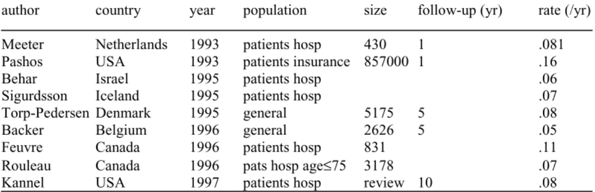
(39) RIVM report 260751 001. AMI incidence. Page 39 of 117. ‘acute’ mortality. AP incidence. AMI prevalence. AP prevalence. ‘chronic’ mortality. ‘chronic’ mortality. Figure 10 The CHD disease model used to calculate excess mortality rates Notes: AMI incidence is from total population including those persons with AP; likewise AP incidence includes those with a history of AMI.. The resulting mortality rates are presented in Table 9, and Figures 11&12. For AMI the excess mortality rates relate to the ‘chronic’ mortality only, the cause-specific population mortality rates include the ‘acute’ mortality. Table 9 The calculated AMI and AP excess mortality rates age. AMI male. female select. AP male. female select. 45-49 50-54 55-59 60-64 65-69 70-74 75-79 80-84 85+. .02 .03 .03 .04 .06 .09 .14 .20 .26. .19 .10 .07 .07 .09 .12 .16 .22 .27. .096 .072 .052 .035 .029 .032 .037 .043 .043. .087 .074 .069 .065 .063 .062 .060. .06 .06 .06 .06 .08 .10 .15 .21 .26. Notes: select: mean over male and female. .050 .050 .050 .050 .050 .050 .050 .050 .050.
(40) Page 40 of 117. RIVM report 260751 001. excess mortality rate. rates infarction. males. 0.25 0.2 0.15 0.1 0.05 65. mortality rate 0.035 0.03 0.025 0.02 0.015 0.01 0.005. 70. 75. 80. rates infarction. 85. age Hyears L. males calculated registered. 50. 60. excess mortality rate. 70. age Hyears L. 80. rates infarction. females. 0.25 0.2 0.15 0.1 0.05 65. mortality rate. rates. 70. 75. 80. infarction. 85. age Hyears L. females. 0.015 0.0125 0.01 0.0075 0.005 0.0025. calculated registered. 50. 60. 70. 80. age H years L. Figure 11 The calculated excess mortality (‘chronic’) and cause-specific population mortality (sum of ‘acute’ and ‘chronic’) rates for AMI, the mortality rates also compared to empirical data.
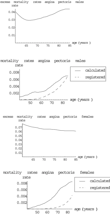
(41) RIVM report 260751 001. excess. mortality rate. Page 41 of 117. rates angina. pectoris. males. 0.04 0.03 0.02 0.01 65. 70. 75. 80. 85. age Hyears L. mortality rates angina pectoris rate. males calculated. 0.008. registered. 0.006 0.004 0.002 50. excess. 60. mortality rate. 70. age Hyears L. 80. rates angina. pectoris. females. 0.07 0.06 0.05 0.04 0.03 0.02 0.01 65. 70. mortality. 75. rates angina rate 0.008. 80. age Hyears L 85. pectoris. females calculated registered. 0.006 0.004 0.002 50. 60. 70. 80. age Hyears L. Figure 12 The calculated excess mortality and mortality rates for AP, the mortality rates also compared to empirical data.
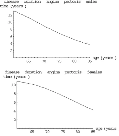
(42) Page 42 of 117. RIVM report 260751 001. disease duration time Hyears L. infarction. males. 10 8 6 4 2 65. 70. 75. disease duration time Hyears L. 80. infarction. 85. age Hyears L. females. 8 6 4 2 65. 70. 75. 80. 85. age Hyears L. Figure 13 The AMI disease duration times for males and females disease duration time Hyears L. angina. pectoris. males. 12 10 8 6 4 2 65. 70. disease duration time Hyears L. 75. 80. angina pectoris. 85. age Hyears L. females. 10 8 6 4 2 65. 70. 75. 80. 85. age Hyears L. Figure 14 The AP disease duration times for males and females.
(43) RIVM report 260751 001. Page 43 of 117. For lower ages the calculated excess mortality rates are unstable due to the small incidence and prevalence rates for both AMI and AP types. The gender differences between the calculated excess mortality rates are small for AMI. Following the literature we have concluded that these differences are by chance and selected the mean value for both genders. For AP the calculated excess mortality rates are almost constant over age, but are clearly different for men and women. The calculated excess mortality rates are sensitive to the trend of the prevalence rates assumed. When we assume increasing prevalence rates in time for women, the calculated excess mortality rates decrease. So the higher excess mortality rates found for women could be explained by underestimating the prevalence time trend. There is not much literature on the prognosis of AP patients, and no indication of significant gender differences. Therefore we have selected the mean values over both genders resulting in constant values over age, and thus implicitly concluded that the time trends assumed are wrong.. 4.6. Estimated mortality rates for CHF. The estimated mortality rates are presented in Table 10 and Figure 15. We have assumed no remission and prevalence trends of 2% (males) and –3% (females) respectively.. excess mortality rate 0.4. rates heartfailure. males. 0.3 0.2 0.1. 50. 60. mortality rate 0.035. 70. 80. rates heartfailure. age Hyears L. males. 0.03. calculated. 0.025. registered. 0.02 0.015 0.01 0.005 50. 60. 70. 80. age Hyears L.
(44) Page 44 of 117. RIVM report 260751 001. excess mortality rate. rates heartfailure. females. 0.4 0.3 0.2 0.1 50. 60. mortality rate. 70. 80. rates heartfailure. age Hyears L. females. 0.035. calculated. 0.03. registered. 0.025 0.02 0.015 0.01 0.005 50. 60. 70. 80. age Hyears L. Figure 15 The calculated excess mortality and cause-specific population mortality rates for CHF, the population mortality rates also compared to empirical data. Table 10 The estimated excess mortality rates for CHF, together with the selected model parameter values age:. calc. males females select. 4044. 4549. 5054. 5559. 6064. 6569. 7074. 7579. 8084. 85+. .16 .15 .16. .23 .19 .21. .36 .32 .34. .38 .45 .41. .29 .38 .33. .26 .30 .29. .27 .29 .28. .26 .28 .27. .25 .28 .26. .23 .26 .26. Notes: calc: calculated gender-specific rate values, select: mean over men and women. For higher ages the excess mortality rate slowly decreases, and is for males slightly lower than for females. The estimated mortality is almost three times as high as the mortality registered by Statistics Netherlands. One important reason for this large difference is that many persons with CHF die with primary cause of death CHD. Because the differences between men and women are very small, we have concluded that both are equal. The final excess mortality rate values are the means of the calculated gender-specific ones. Because for lower ages the calculated rates are unstable, we have assumed that the rates are constant until age 45..
(45) RIVM report 260751 001. 4.7. Page 45 of 117. Combining CHD and CHF. The model that is used to describe coronary heart diseases (CHD) and congestive heartfailure (CHF) simultaneously is based on the model of CHD presented in §4.5 (see Figure 10). Because of the overlap in the data, we can only describe overlapping disease-specific prevalence numbers so far. Our resulting model combining CHD and CHF is presented in Figure 16. ‘acute’ mortality. incidence. AP prevalence. mortality. AMI prevalence. CHF prevalence. Figure 16 Model of CHD and CHF simultaneously Notes: incidence of AP is from CHD-free population and from AMI prevalence after stabilisation; incidence of 1st AMI from CHD-free population, from AP prevalence and from CHF prevalence respectively; incidence of CHF from CHD- and CHF-free population, from AP and AMI prevalence respectively; mortality described includes excess mortality.. Because we describe overlapping disease prevalence numbers, we do not need to estimate rates for all transitions described in Figure 16. For example, the AMI incidence includes incidence from CHD-free population and persons with AP and CHF. For the same reason adding CHF to the CHD model does not affect the AMI and AP excess mortality rates estimated before. We need more information to disentangle the AMI, AP and CHF prevalence numbers. For example, Barendregt&Bonneux (1998) suggest that the ratios of the CHF incidence rates for CVD-free persons, persons with AP and those with history of AMI are 1:5:50. Given the total incidence rates, we can estimate the state-specific CHF incidence rates: age free of CHD and CHF with AP with AMI. 50 .0004 .002 .02. 60 .0007 .003 .03. 70 .001 .006 .06. 80 .0025 .012 .12. 85 .0036 .018 .18.
Afbeelding
+7
GERELATEERDE DOCUMENTEN
High Rate of Missed Lateral Meniscus Posterior Root Tears on Preoperative Magnetic Resonance Imaging
(A) Coronal T2-weighted magnetic resonance image of the left knee of a 16-year-old girl shows a “clearly evident” lateral meniscus posterior root tear (circled), as diag- nosed
Both national role conceptions and leadership styles have some influence on the decision- making process and can affect foreign policy change, but the model I designed is too
Taking into consideration Pyongyang’s numerous provocations during the periods of coercion (under Kim Young-sam) and engagement (under Kim Dae-jung and Roh Moo- hyun), this study
Patients provide informed consent for: (1) reuse of clinical data; (2) biobanking of primary tumor tissue; (3) collection of blood samples; (4) to be informed about relevant
We examined adherence to the eight The World Cancer Research Foundation/ American Institute for Cancer Research (WCRF/AICR) recommendations on diet, physical activity, and body
BEPS actiepunt 7 : het tegengaan van het op kunstmatige wijze vermijden van vaste vertegenwoordigers
Indien de ‘significant people functions’, dat wil zeggen de controle functies, in de woonstaat worden uitgeoefend, dienen deze slechts aan de fictieve vaste inrichting
Several plasmonic structures incorporating rare-earth ions doped potassium double tungstate gain material to provide gain and eventually to compensate the
To summarise: Tables 1 and 2 show that the count of publications rose by 60%; that the universities remain the dominant locus of scientific publication;
