Guidelines of care for the management of primary
cutaneous melanoma
Work Group: Christopher K. Bichakjian, MD,aAllan C. Halpern, MD (Co-chair),b Timothy M. Johnson, MD
(Co-Chair),aAntoinette Foote Hood, MD,cJames M. Grichnik, MD, PhD,dSusan M. Swetter, MD,e,f Hensin Tsao, MD, PhD,gVictoria Holloway Barbosa, MD,hTsu-Yi Chuang, MD, MPH,i,jMadeleine Duvic, MD,k
Vincent C. Ho, MD,lArthur J. Sober, MD,gKarl R. Beutner, MD, PhD,m,nReva Bhushan, PhD,o
and Wendy Smith Begolka, MSo
Ann Arbor, Michigan; New York, New York; Norfolk, Virginia; Miami, Florida; Palo Alto, Los Angeles, Palm Springs, San Francisco, and Fairfield, California; Boston, Massachusetts; Chicago and Schaumburg,
Illinois; Houston, Texas; and Vancouver, British Columbia, Canada
The incidence of primary cutaneous melanoma has been increasing dramatically for several decades. Melanoma accounts for the majority of skin cancererelated deaths, but treatment is nearly always curative with early detection of disease. In this update of the guidelines of care, we will discuss the treatment of patients with primary cutaneous melanoma. We will discuss biopsy techniques of a lesion clinically suspicious for melanoma and offer recommendations for the histopathologic interpretation of cutaneous melanoma. We will offer recommendations for the use of laboratory and imaging tests in the initial workup of patients with newly diagnosed melanoma and for follow-up of asymptomatic patients. With regard to treatment of primary cutaneous melanoma, we will provide recommendations for surgical margins and briefly discuss nonsurgical treatments. Finally, we will discuss the value and limitations of sentinel lymph node biopsy and offer recommendations for its use in patients with primary cutaneous melanoma. ( J Am Acad Dermatol10.1016/j.jaad.2011.04.031.)
Key words: biopsy; follow-up; melanoma; pathology report; sentinel lymph node biopsy; surgical margins.
DISCLAIMER
Adherence to these guidelines will not ensure successful treatment in every situation. Furthermore, these guidelines should not be interpreted as setting a standard of care, or be deemed inclusive of all proper methods of care nor exclusive of other methods of care reasonably directed to obtaining the same results. The ultimate judgment regarding the propriety of any specific therapy must be made by the physician and the patient in light of all the circumstances presented by the individual patient,
and the known variability and biological behavior of the disease. This guideline reflects the best available data at the time the guideline was prepared.
From the Department of Dermatology, University of Michigan Health System and Comprehensive Cancer Centera; Depart-ment of Dermatology, Memorial Sloan-Kettering Cancer Center, New Yorkb; Department of Dermatology, Eastern Virginia Medical Schoolc; Department of Dermatology, University of Miami Health System, University of Miami Hospital and Clinics, Sylvester Comprehensive Cancer Center, Melanoma Programd; Department of Dermatology, Stanford University Medical Centere and Department of Veterans Affairs Palo Alto Health
Care Systemf; Department of Dermatology, Massachusetts
General Hospital and Harvard Medical Schoolg; Department of
Dermatology, Rush University Medical Center, Chicagoh;
Uni-versity of Southern California, Los Angeles,iand Desert Oasis
Healthcare, Palm Springsj; Department of Dermatology,
University of Texas MD Anderson Cancer Centerk; Division of
Dermatology, University of British Columbial; Department of
Dermatology, University of California, San Franciscom, and
Solano Dermatology, Fairfieldn; and American Academy of
Dermatology, Schaumburg.o
Funding sources: None.
The authors’ conflict of interest/disclosure statements appear at the end of the article.
Accepted for publication April 20, 2011.
Reprint requests: Reva Bhushan, PhD, American Academy of Dermatology, 930 E Woodfield Rd, Schaumburg, IL 60173. E-mail:rbhushan@aad.org.
Published online August 24, 2011. 0190-9622/$36.00
Ó 2011 by the American Academy of Dermatology, Inc. doi:10.1016/j.jaad.2011.04.031
Abbreviations used:
AAD: American Academy of Dermatology AJCC: American Joint Committee on Cancer LND: lymph node dissection
PET: positron emission tomography SLN: sentinel lymph node
SLNB: sentinel lymph node biopsy
The results of future studies may require revisions to the recommendations in this guideline to reflect new data.
SCOPE
This guideline addresses the treatment of patients with primary cutaneous melanoma (including those in the nail unit), who may also have clinical or histologic evidence of regional disease, from the perspective of the US dermatologist. The guideline does not address primary melanoma of the mucous membranes. A discussion of adjuvant therapies for patients with high-risk melanoma (stage$ IIB), such as interferon and radiation therapy, falls outside the scope of this guideline. Consultation with a physi-cian or multidisciplinary group with specific exper-tise in melanoma, such as a medical oncologist, surgical oncologist, radiation oncologist, or derma-tologist specializing in melanoma, should be con-sidered for patients with high-risk melanoma. Finally, as an extensive discussion beyond the man-agement of melanoma falls outside the scope of the current guidelines, the expert work group recom-mends that separate guidelines be developed on screening and surveillance for early detection, clin-ical diagnosis of primary cutaneous melanoma, and the molecular assessment of borderline/indetermi-nate melanocytic lesions.
METHOD
A work group of recognized melanoma experts was convened to determine the audience and scope of the guideline, and identify important clinical questions in the management of primary cutaneous
melanoma (Table I). Work group members
com-pleted a disclosure of commercial support that was updated throughout guideline development.
An evidence-based model was used and evi-dence was obtained using a search of the PubMed database spanning the years 2000 through 2010 for clinical questions addressed in the previous version of this guideline published in 2001, and 1960 to 2010 for all newly identified clinical questions. Only English-language publications were reviewed. Published guidelines on melanoma were also evaluated.1-3
The available evidence was evaluated using a uni-fied system called the Strength of Recommendation Taxonomy developed by editors of the US family medicine and primary care journals (ie, American Family Physician, Family Medicine, Journal of Family Practice, and BMJ USA). This strategy was supported by a decision of the Clinical Guidelines Task Force in 2005 with some minor modifications for a consistent approach to rating the strength of the
evidence of scientific studies.4Evidence was graded using a 3-point scale based on the quality of metho-dology as follows:
I. Good-quality patient-oriented evidence (ie, evi-dence measuring outcomes that matter to patients: morbidity, mortality, symptom improvement, cost reduction, and quality of life).
II. Limited-quality patient-oriented evidence. III. Other evidence including consensus guidelines,
opinion, case studies, or disease-oriented evi-dence (ie, evievi-dence measuring intermediate, physiologic, or surrogate end points that may or may not reflect improvements in patient outcomes).
Clinical recommendations were developed on the best available evidence tabled in the guideline. These are ranked as follows:
A. Recommendation based on consistent and good-quality patient-oriented evidence.
B. Recommendation based on inconsistent or limited-quality patient-oriented evidence. C. Recommendation based on consensus, opinion,
case studies, or disease-oriented evidence. In those situations where documented evidence-based data are not available, we have used expert opinion to generate our clinical recommendations. This guideline has been developed in accordance with the American Academy of Dermatology (AAD)/AAD Association ‘‘Administrative Regulations Table I. Clinical questions used to structure
evidence review for management of primary cutaneous melanoma
What is the standard grading system for melanoma? What clinical and histologic information is useful in the
pathology report?
What are standard biopsy techniques?
What are recommended surgical margins stratified by grading system?
What is the effectiveness of sentinel node biopsy?*
What diagnostic laboratory and imaging tests are useful in asymptomatic patients with primary cutaneous melanoma?
What is the effectiveness of imiquimod?*
What is the effectiveness of cryotherapy?*
What is the effectiveness of radiation?*
What is effective for follow-up of asymptomatic patients with primary cutaneous melanoma to detect metasta-ses and/or additional primary melanomas?
How long should asymptomatic patients be followed up? What diagnostic and imaging tests are effective in
follow-up of asymptomatic patients?
for Evidence-based Clinical Practice Guidelines’’ (version approved March 2009), which include the opportunity for review and comment by the entire AAD membership and final review and approval by the AAD Board of Directors.
DEFINITION
Primary cutaneous melanoma is defined as any primary melanoma lesion, regardless of tumor thick-ness, in patients without clinical or histologic evi-dence of regional or distant metastatic disease (stage 0-IIC).
INTRODUCTION
Since the last publication of the guidelines of care for primary cutaneous melanoma by the American Academy of Dermatology in 2001, the most signifi-cant changes in the management of primary mela-noma are a result of the acknowledgment of the dermal mitotic rate as an important prognostic parameter.5,6This change is reflected in the recently published seventh edition of the American Joint Committee on Cancer (AJCC) staging system for
melanoma, effective January 1, 2010 (Tables II
and III).7 In the new staging system, mitotic rate has replaced Clark level of invasion as the second factor predicting melanoma survival in addition to tumor (Breslow) thickness for tumors less than or equal to 1 mm in thickness.
Although nonsurgical treatments of primary mel-anoma, in particular lentigo maligna, have been increasingly used in recent years, their efficacy has not been established. Suggestions on the use of these alternative treatments have been included in the current guidelines.
A significant amount of data has become avail-able on the use of sentinel lymph node (SLN) biopsy (SLNB) for melanoma since the previous guidelines. Although the procedure is not performed by der-matologists in the United States, the decision by a patient with melanoma to proceed with this staging procedure is frequently based on the advice from their dermatologist. Recommendations for the use of SLNB for primary melanoma are therefore in-cluded in the current guidelines. However, with regard to treatment recommendations, the current guidelines continue to be limited to the manage-ment of primary cutaneous melanoma. For patients with regional or distant metastases, we refer physi-cians to clinical practice guidelines, such as those developed by the National Comprehensive Cancer Network.1
BIOPSY
The first step for a definitive diagnosis of cancer is a biopsy that may occur by removing part of the lesion (incisional biopsy) or the entire lesion (ex-cisional biopsy). For a lesion clinically suspicious for cutaneous melanoma, one should ideally per-form a narrow excisional biopsy that encompasses the entire breadth of the lesion with clinically negative margins to a depth sufficient to ensure that the lesion is not transected.8-18 It has been suggested that 1- to 3-mm margins are required to clear the subclinical component of most atypical melanocytic lesions.1,3,19This can be accomplished in a number of ways including elliptical or punch excision with sutures, or shave removal to a depth below the anticipated plane of the lesion. The latter is commonly used when the suspicion of mela-noma is low, the lesion lends itself to complete removal by this technique, or in the setting of a macular lesion suspicious for lentigo maligna where a broad biopsy specimen may aid in histo-logic assessment.11,13
Clinically clear but narrow lateral margins on excisional biopsy, oriented along the longitudinal axis on extremities, will permit optimal subsequent wide local excision and, if indicated, SLNB. Incisional biopsy, with a variety of techniques noted above, of the clinically or dermatoscopically most atypical portion of the lesion, is an acceptable option in certain circumstances, such as a facial or acral location, low clinical suspicion or uncertainty of diagnosis, or a very large lesion, although the selected area may not always correlate with the
deepest Breslow depth.9 If an incisional biopsy
specimen is inadequate to make a histologic diag-nosis or to accurately microstage the lesion for treatment planning, a repeat biopsy should be performed.9
When a biopsy is performed of a suspicious nail lesion (melanonychia striata, diffuse pigmentation, or amelanotic changes, eg, ulceration), the nail matrix should be sampled. Because of the com-plexity of nail anatomy and the fact that melanoma arises in the nail matrix, suspicious nail lesions are best evaluated and sampled by a physician skilled in the biopsy of the nail apparatus. For suspicious subungual lesions, the nail plate should be suffi-ciently removed to expose the underlying lesion and an excisional or incisional biopsy should be performed based on the size of the lesion. Recommendations for the use of a biopsy for primary cutaneous melanoma are summarized in Table IV. The strength of these recommendations is shown inTable V.
PATHOLOGY REPORT
When a biopsy is performed of a lesion clinically suspicious for primary cutaneous melanoma, the expert work group recommends that the following
pertinent information is provided to the pathologist (Table VI; strength of recommendations shown in Table V). For identification purposes, the age and gender of the patient and the anatomic location of Table II. 2010 American Joint Committee on Cancer TNM definitions
Primary tumor (T)
TX Primary tumor cannot be assessed (eg, curettaged or severely regressed melanoma) T0 No evidence of primary tumor
Tis Melanoma in situ
T1 Melanomas # 1.0 mm in thickness
T2 Melanomas 1.01-2.0 mm
T3 Melanomas 2.01-4.0 mm
T4 Melanomas[4.0 mm
Note: a and b subcategories of T are assigned based on ulceration and No. of mitoses per mm2as shown below:
T classification Thickness, mm Ulceration status/mitoses
T1 # 1.0 a: Without ulceration and\1 mitosis/mm2
b: With ulceration or $ 1 mitosis/mm2
T2 1.01-2.0 a: Without ulceration b: With ulceration T3 2.01-4.0 a: Without ulceration b: With ulceration T4 [4.0 a: Without ulceration b: With ulceration Regional lymph nodes (N)
NX Patients in whom regional lymph nodes cannot be assessed (eg, previously removed for another reason) N0 No regional metastases detected
N1-3 Regional metastases based on No. of metastatic nodes and presence or absence of intralymphatic metastases (in transit or satellite)
Note: N1-3 and a-c subcategories assigned as shown below:
N classification No. of metastatic nodes Nodal metastatic mass
N1 1 Node a: Micrometastasis*
b: Macrometastasisy
N2 2-3 Nodes a: Micrometastasis*
b: Macrometastasisy
c: In transit met(s)/satellite(s)without metastatic nodes N3 $ 4 Metastatic nodes, or matted nodes, or in
transit met(s)/satellite(s)with metastatic node(s)
Distant metastasis (M) M0 No detectable evidence of distant metastases
M1a Metastases to skin, subcutaneous, or distant lymph nodes M1b Metastases to lung
M1c Metastases to all other visceral sites or distant metastases to any site combined with elevated serum LDH Note: Serum LDH is incorporated into M category as shown below:
M classification Site Serum LDH
M1a Distant skin, subcutaneous or nodal mets Normal
M1b Lung metastases Normal
M1c All other visceral metastases Any distant metastasis
Normal Elevated
LDH, Lactate dehydrogenase.
Used with permission of American Joint Committee on Cancer (AJCC), Chicago, IL. Original source for this material is AJCC Cancer Staging Manual, Seventh Edition (2010) published by Springer Science and Business Media LLC,www.springer.com.
*Micrometastases are diagnosed after sentinel lymph node biopsy and completion lymphadenectomy (if performed).
yMacrometastases are defined as clinically detectable nodal metastases confirmed by therapeutic lymphadenectomy or when nodal
the lesion must be included.20-34The clinical infor-mation in the pathology report should contain the type of surgical procedure performed (ie, biopsy
intenteexcisional or incisional) and size of the
lesion. Additional optional, but desirable, clinical information include ABCDE criteria, dermatoscopic features, a clinical photograph, and the presence or absence of macroscopic satellitosis.
The pathology of melanocytic tumors should be read by a physician experienced in the interpretation of pigmented lesions. The list of histologic features to be included in the pathology report is based on their prognostic value (Table VII; strength of recommen-dations shown inTable V). Definitions of histologic features are listed in Table VIII. There is strong evidence to support that 3 histologic features are the most important characteristics of the primary tumor to predict outcome.6,20-23,25,26,30-32,34-40(1) Maximum tumor (Breslow) thickness is measured from the
granular layer of the overlying epidermis or base of a superficial ulceration to the deepest malignant cells invading dermis to the nearest 0.1 mm, not including
deeper adventitial extension. Microsatellitosis
should not be included in this measurement, but commented on separately. (2) Presence or absence of microscopic ulceration, which is defined as tumor-induced full-thickness loss of epidermis with subja-cent dermal tumor and reactive dermal changes.41 (3) Mitotic rate, measured as the number of dermal mitoses per mm2(with 1 mm2approximately equal to 4.5 high-power [340] microscopic fields, starting in the field with most mitoses), was included as a prognostic value in the 2010 AJCC staging system to upstage patients with melanoma less than or equal to 1 mm in thickness from IA to IB, replacing Clark
level.6 Regardless of tumor thickness, a dermal
mitotic rate greater than or equal to 1 mitosis/mm2 is independently associated with a worse disease-specific survival. It is essential that these 3 charac-teristics are included in the pathology report. The anatomic (Clark) level of invasion is only included in the 2010 AJCC staging system for staging tumors less than or equal to 1 mm in thickness when mitotic rate cannot be assessed and is considered optional for tumors larger than 1 mm. An additional essential element of the pathology report is the status of the peripheral and deep margins (positive or negative) of the excision. The presence or absence of tumor at the surgical margin indicates whether the entire lesion was available for histologic evaluation and provides guidance for further management. It should be emphasized that for the management of primary cutaneous melanoma, treatment recommendations are based on the clinical measurement of surgical margins around the tumor and not on histologically measured clear peripheral margins.1The presence of microsatellites upstages the tumor to N2c (stage IIIB) Table III. 2010 American Joint Committee on
Cancer anatomic stage/prognostic groups
Clinical staging* Pathologic stagingy Stage 0 Tis N0 M0 0 Tis N0 M0 Stage IA T1a N0 M0 IA T1a N0 M0 Stage IB T1b N0 M0 IB T1b N0 M0
T2a N0 M0 T2a N0 M0
Stage IIA T2b N0 M0 IIA T2b N0 M0
T3a N0 M0 T3a N0 M0
Stage IIB T3b N0 M0 IIB T3b N0 M0
T4a N0 M0 T4a N0 M0
Stage IIC T4b N0 M0 IIC T4b N0 M0 Stage III Any T $ N1 M0 IIIA T1-4a N1a M0 T1-4a N2a M0 IIIB T1-4b N1a M0 T1-4b N2a M0 T1-4a N1b M0 T1-4a N2b M0 T1-4a N2c M0 IIIC T1-4b N1b M0 T1-4b N2b M0 T1-4b N2c M0 Any T N3 M0 Stage IV Any T Any N M1 IV Any T Any N M1
Used with permission of American Joint Committee on Cancer (AJCC), Chicago, IL. Original source for this material is AJCC Cancer Staging Manual, Seventh Edition (2010) published by Springer Science and Business Media LLC,www.springer.com.
*Clinical staging includes microstaging of primary melanoma and clinical/radiologic evaluation for metastases. By convention, it should be used after complete excision of primary melanoma with clinical assessment for regional and distant metastases.
yPathologic staging includes microstaging of primary melanoma
and pathologic information about regional lymph nodes after partial or complete lymphadenectomy. Patients with pathologic stage 0 or IA are the exception; they do not require pathologic evaluation of their lymph nodes.
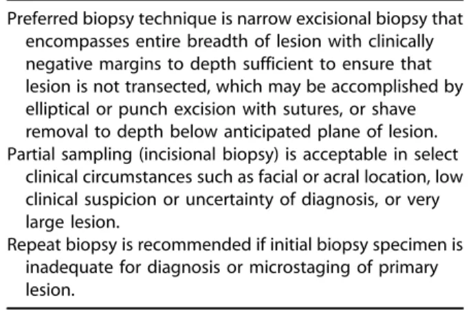
Table IV. Recommendations for biopsy
Preferred biopsy technique is narrow excisional biopsy that encompasses entire breadth of lesion with clinically negative margins to depth sufficient to ensure that lesion is not transected, which may be accomplished by elliptical or punch excision with sutures, or shave removal to depth below anticipated plane of lesion. Partial sampling (incisional biopsy) is acceptable in select
clinical circumstances such as facial or acral location, low clinical suspicion or uncertainty of diagnosis, or very large lesion.
Repeat biopsy is recommended if initial biopsy specimen is inadequate for diagnosis or microstaging of primary lesion.
melanoma according to the 2010 AJCC staging crite-ria and therefore must also be reported.27,37,42-44
There is evidence that several other histologic features of a primary melanoma provide prognostic value, including the presence or absence of a vertical growth phase, tumor-infiltrating lymphocytes,
der-mal regression, and angiolymphatic
inva-sion.21,23,27,33,35,37,38,45-48Although not essential, the expert work group recommends that these histologic characteristics are included as optional elements of
the pathology report. The prognostic value of the histologic subtype of melanoma has not been
established, with some notable exceptions.31 The
lentigo maligna pattern is associated with broader Table V. Strength of recommendations for management of primary cutaneous melanoma
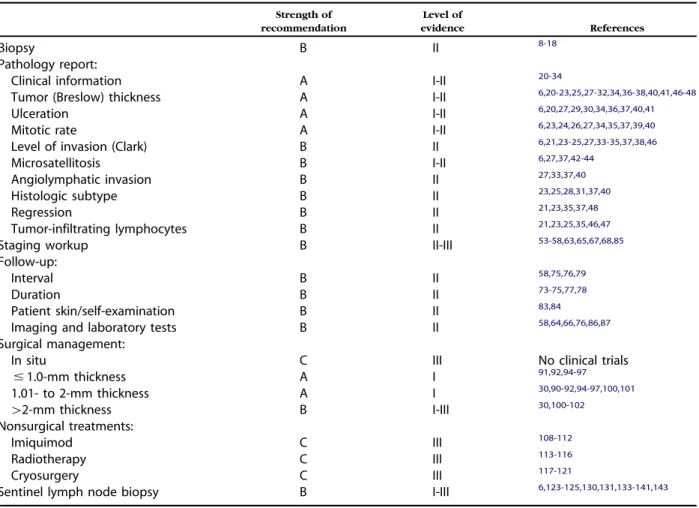
Strength of recommendation Level of evidence References Biopsy B II 8-18 Pathology report:
Clinical information A I-II 20-34
Tumor (Breslow) thickness A I-II 6,20-23,25,27-32,34,36-38,40,41,46-48
Ulceration A I-II 6,20,27,29,30,34,36,37,40,41
Mitotic rate A I-II 6,23,24,26,27,34,35,37,39,40
Level of invasion (Clark) B II 6,21,23-25,27,33-35,37,38,46
Microsatellitosis B I-II 6,27,37,42-44
Angiolymphatic invasion B II 27,33,37,40
Histologic subtype B II 23,25,28,31,37,40
Regression B II 21,23,35,37,48
Tumor-infiltrating lymphocytes B II 21,23,25,35,46,47
Staging workup B II-III 53-58,63,65,67,68,85
Follow-up:
Interval B II 58,75,76,79
Duration B II 73-75,77,78
Patient skin/self-examination B II 83,84
Imaging and laboratory tests B II 58,64,66,76,86,87 Surgical management:
In situ C III No clinical trials
# 1.0-mm thickness A I 91,92,94-97 1.01- to 2-mm thickness A I 30,90-92,94-97,100,101 [2-mm thickness B I-III 30,100-102 Nonsurgical treatments: Imiquimod C III 108-112 Radiotherapy C III 113-116 Cryosurgery C III 117-121
Sentinel lymph node biopsy B I-III 6,123-125,130,131,133-141,143
Table VI. Recommended clinical information to be provided to pathologist Essential Strongly recommended Optional Age of patient Biopsy technique (excisional or incisional) Clinical description and level of clinical suspicion
Gender Size of lesion Dermatoscopic features Anatomic
location
Photograph
Macroscopic satellitosis
Table VII. Recommended histologic features of primary melanoma to be included in pathology report
Essential Optional
Tumor (Breslow) thickness, mm
Angiolymphatic invasion Ulceration Histologic subtype Dermal mitotic rate,
mitoses/mm2
Neurotropism Peripheral and deep
margin status (positive or negative)
Regression
Anatomic level of invasion (Clark level)*
T-stage classification
Microsatellitosis Tumor infiltrating lymphocytes Vertical growth phase
*Essential for staging only in tumors# 1 mmin thickness when mitotic rate cannot be assessed; optional for tumors[1 mm in thickness.
superficial subclinical extension, often requiring wider surgical margins to clear histologically.49,50 In addition, there is some evidence to support that primary melanomas with a purely desmoplastic histologic subtype have a lower risk of nodal and distant metastases, but potentially higher risk of local recurrence.51,52 The expert work group rec-ommends that histologic subtype be included in the optional list of elements of the pathology report. Although the prognostic value of neurotropism is uncertain, its presence or absence provides valuable information that may alter future management of the primary tumor and is therefore included as an additional optional histologic characteristic to be reported.27,42-44
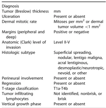
Finally, reporting microscopic features in a syn-optic report provides a level of completeness and standardization, which is strongly encouraged by the expert work group (Table IX).
STAGING WORKUP AND FOLLOW-UP
Initial workup for patients with newly diagnosed cutaneous melanoma
After the diagnosis of melanoma has been histo-logically confirmed, a thorough history and physical examination comprise the cornerstone of the initial diagnostic workup of a patient with newly diag-nosed cutaneous melanoma. For invasive disease, a
detailed patient history should include a focused review of systems with particular attention to con-stitutional, neurologic, respiratory, hepatic, gastro-intestinal, musculoskeletal, skin, and lymphatic signs or symptoms. The physical examination should include a total body skin examination and palpation of both regional and distant lymph node basins. Any abnormal finding should direct the need for further studies to detect regional and distant metastases.
In asymptomatic patients with localized cutane-ous melanoma of any thickness, baseline blood tests and imaging studies are generally not recommended and should only be performed as clinically indicated for suspicious signs and symptoms. Currently avail-able treatment for patients with asymptomatic stage IV disease is not associated with better outcomes than intervention when the patient becomes symp-tomatic. Furthermore, screening blood tests, includ-ing serum lactate dehydrogenase, are insensitive for the detection of metastatic disease.53 The use of routine imaging studies is limited by a very low yield and the frequent occurrence of false-positive find-ings.54,55Ample evidence exists that a routine chest x-ray is a cost-inefficient test for the detection of metastatic disease with a consistent relatively high false-positive rate.53,56-59 Although cross-sectional imaging by computed tomography is equally limited by a low detection rate of occult metastases and relatively high false-positive rate, the increased dose of ionizing radiation associated with repeated tests also needs to be considered.57,60. Positron emission tomography (PET) has gained increasing popularity in recent years, particularly for detection of distant Table VIII. Definitions of histologic features
Clark levels
Level I Tumor confined to epidermis Level II Tumor present in papillary
dermis
Level III Tumor fills papillary dermis Level IV Tumor present in reticular
dermis
Level V Tumor present in subcutis Vertical growth phase Presence of $ 1 clusters of
dermal tumor cells larger than largest epidermal tumor cluster and/or presence of any dermal mitotic activity Tumor regression Apparent loss of dermal tumor
with associated nonlamellar fibrosis, mononuclear cell inflammation, and vascular proliferation or ectasia Microsatellitosis Nest(s) of tumor cells ([0.05
mm in diameter) located in reticular dermis, subcutis, or vessels, separated from invasive component of tumor by $ 0.3 mm of normal tissue
Table IX. Synoptic melanoma report
Diagnosis
Tumor (Breslow) thickness mm
Ulceration Present or absent
Dermal mitotic rate Mitoses per mm2or dermal tumor volume\1 mm2 Margins (peripheral and
deep)
Positive or negative Anatomic (Clark) level of
invasion
Level II-V
Histologic subtype Superficial spreading, nodular, lentigo maligna, acral lentiginous,
desmoplastic/neurotropic, nevoid, or other
Perineural involvement Present or absent Regression Present or absent T-stage classification T1a-T4b
Tumor infiltrating lymphocytes
Not identified, nonbrisk, or brisk
metastatic disease. However, both PET and ultra-sound have been found to have a low sensitivity for the detection of occult regional nodal metastases in
comparison with SLNB.61-66 The impact of
false-positive findings, whether by PET, computed tomography, chest x-ray, or lactate dehydrogenase, that lead to unnecessary invasive procedures and substantial patient anxiety, should not be underestimated.
In patients with micrometastatic nodal disease detected by SLNB, the yield of screening computed tomography or PET scans remains low, ranging from 0.5% to 3.7%, with the majority of true positive findings detected in patients with thick, ulcerated primary melanomas or large tumor burden in the SLN(s).55,67-69 The detection rate of occult distant metastases is somewhat higher in patients presenting with clinically detectable nodal disease.70-72 The detection of widespread metastatic disease may alter patient treatment and obviate the need for extensive surgery. After diagnosis, consultation with a medical oncologist or other melanoma specialist should be considered for patients at higher risk of disease relapse based on disease stage, tumor thickness, ulceration, SLN status, or a combination of these. Follow-up of asymptomatic patients with cutaneous melanoma
The primary goal for follow-up of patients with a history of cutaneous melanoma is early detection of surgically resectable recurrent disease and addi-tional primary melanoma.73-79 It is generally ac-cepted that early detection of asymptomatic distant metastatic disease does not affect overall survival.
80-82
No strong evidence exists to support a specific follow-up interval for patients with melanoma at any stage. The expert work group recommends at least annual follow-up, ranging from every 3 to 12 months based on the risk for recurrence and new primary melanoma. Additional factors that may influence the follow-up interval include disease stage, a history of multiple primary melanomas, the presence of clinically atypical nevi, a family history of melanoma, patient anxiety, and the patient’s awareness and ability to detect early signs and symptoms of disease. All patients with a history of cutaneous melanoma should be educated about monthly self-examinations of their skin.83,84 If ap-propriate, patients can be instructed in monthly self-examination for the detection of regional lymph node enlargement.
Clinical examination remains the most important means of detecting local, regional, or distant disease. A comprehensive history and physical examination with emphasis on the skin and lymph nodes form the
most important components of follow-up for pa-tients with melanoma after surgical resection. A low threshold should exist for sign- or symptom-directed workup to detect metastatic disease. However, rou-tine surveillance laboratory tests or imaging studies are generally not useful and not recommended in asymptomatic patients.85-87 Serum lactate dehydro-genase is almost never the sole indicator of meta-static disease, but is a highly significant predictor of survival in patients with stage IV disease.88,89 Although surveillance imaging studies can be con-sidered in patients at higher risk for recurrence (stage IIB and above), the yield remains low in asymptom-atic patients with a relatively high false-positivity rate and is not recommended beyond 5 years. Recommendations for staging workup and follow-up for primary cutaneous melanoma are summarized inTable Xand the strength of recommendations is shown inTable V.
SURGICAL MANAGEMENT
The primary treatment modality for cutaneous melanoma is surgical excision. After the diagnosis of melanoma has been histologically confirmed and the primary lesion has been adequately microstaged, a wider and frequently deeper excision is needed to ensure complete removal. It is recognized that mel-anoma cells may extend subclinically several milli-meters to several centimilli-meters beyond the clinically visible lesion. Recommended surgical margins are based partly on prospective randomized controlled trials and partly on consensus opinion when no prospective data exist.1The primary goal of surgical excision of melanoma of any thickness is to achieve histologically negative margins and prevent local Table X. Recommendations for staging workup and follow-up
Baseline laboratory tests and imaging studies are generally not recommended in asymptomatic patients with newly diagnosed primary melanoma of any thickness. No clear data regarding follow-up interval exist, but at
least annual history and physical examination with attention to skin and lymph nodes is recommended. Regular clinical follow-up and interval patient
self-examination of skin and regional lymph nodes are most important means of detecting recurrent disease or new primary melanoma; findings from history and physical examination should direct need for further studies to detect local, regional, and distant metastasis. Surveillance laboratory tests and imaging studies in
asymptomatic patients with melanoma have low yield for detection of metastatic disease and are associated with relatively high false-positive rates.
recurrence because of persistent disease. It is essen-tial to recognize that surgical margin recommenda-tions are based on studies in which margins were clinically measured around the primary tumor and may not correlate with histologically measured tumor-free margins. Nonsurgical treatment modali-ties are discussed under adjunctive therapies.
Specific surgical margin recommendations for primary invasive melanoma are based on 3 concepts (Table XI): (1) wide excision is associated with a reduced risk of local recurrence; (2) there is no convincing evidence in thin melanomas to confirm an improvement in survival or local recurrence rate with excision margins exceeding 1 cm; and (3) no convincing evidence exists for primary melanoma of any thickness, that excision with greater than 2-cm margins offers any benefit in terms of overall survival or local recurrence.90The expert work group there-fore recommends that surgical margins for invasive melanoma should be at least 1 cm and no more than 2 cm clinically measured around the primary tumor. Although very limited data exist on excision of primary invasive melanoma less than or equal to 1.0 mm in thickness, wide excision with 1 cm margin
is recommended.91-93 Based on available evidence
and consensus opinion, the expert work group recommends that primary melanomas 1.01 to 2.0 mm in thickness are widely excised with 1- to 2-cm margins. However, clear evidence is not available and final surgical margins may vary based on tumor location and functional or cosmetic consider-ations.90,94-97 In melanomas thicker than 2.0 mm, one study found that narrow excision with 1-cm margin was associated with a somewhat higher combined local, regional, and nodal recurrence rate than wider excision with 3-cm margin.98There was, however, no significant difference in true local recurrence rate, melanoma-specific, or overall sur-vival. Several other studies have found slightly higher local recurrence rates for melanomas thicker than 1 mm treated with narrow excision with a 1-cm margin versus a wider excision with a 3-cm mar-gin.91,94For thick primary melanomas, most studies have failed to show a benefit with surgical margins
greater than 2 cm.99-102It is therefore recommended that primary melanomas greater than 2.0 mm in thickness are widely excised with 2-cm margins. Although the recommended depth of a therapeutic excision for invasive primary cutaneous melanoma has always been to the level of muscle fascia, no unequivocal evidence exists to support that this is necessary in every circumstance such as anatomic locations with an increased adipose layer. The expert work group recommends that, whenever possible, excision is performed to the level of muscle fascia, or at least deep adipose tissue depending on the loca-tion of the tumor.
No prospectively controlled data on excision margins for melanoma in situ are available. Based on consensus opinion, wide excision with 0.5- to
1.0-cm margin has been recommended.1However,
be-cause of the characteristic, potentially extensive, subclinical extension of melanoma in situ, lentigo maligna type, alternative surgical approaches may be considered. Particularly for larger lesions on the head and neck, greater than 0.5-cm margins may be necessary to achieve negative margins.49,50,103 Examination with a Wood lamp may be helpful in the presurgical assessment of subclinical extension. Contralateral sampling by punch biopsy may help distinguish true atypical junctional melanocytic hy-perplasia from background actinic damage. Careful histologic evaluation of surgical margins is strongly recommended. Various techniques to achieve com-plete histologic margin control have been described including, but not limited to permanent section total peripheral margin control and Mohs micrographic surgery.49,50,104-106No prospective randomized data are available to make specific recommendations. However, referral for more exhaustive histologic margin assessment may be considered when exten-sive subclinical extension is suspected. For both in situ and invasive melanoma, permanent paraffin sections rather than frozen sections are considered the gold standard for histologic evaluation of surgical margins of melanocytic lesions.107Recommendations for surgical management are summarized inTable XII and the strength of recommendations is shown in Table V.
NONSURGICAL TREATMENTS FOR
LENTIGO MALIGNA
For all stages of primary cutaneous melanoma, from in situ disease to a deeply invasive tumor, surgical excision remains the standard of care. However, for the treatment of lentigo maligna alter-native therapies may be considered when surgery is not a reasonable option because of patient comor-bidities or preferences. The limitations of all Table XI. Surgical margin recommendations for
primary cutaneous melanoma
Tumor thickness
Clinically measured surgical margin*
In situ 0.5-1.0 cm
# 1.0 mm 1 cm
1.01-2.0 mm 1-2 cm
[2.0 mm 2 cm
nonsurgical treatment modalities must clearly be discussed with patients when considering any alter-native therapies, including the risk of missing and undertreating invasive melanoma by not microstag-ing the primary lesion; higher local recurrence rates because of a lack of margin control; and the absence of long-term, randomized, controlled comparative studies.
The off-label use of topical imiquimod has been proposed as an alternative treatment to surgery, and an adjunctive modality after surgical excision.108-112 Studies are limited by highly variable treatment regimens and lack of long-term follow-up with an average of approximately 18 months. Histologic verification after treatment has shown persistent disease in approximately 25% of treated patients and progression to invasive melanoma has been noted. As an adjunctive modality after surgical exci-sion, the efficacy of topical imiquimod has not been established. High cost of treatment, an appropriate low threshold for subsequent biopsy to exclude residual or recurrent disease, and the risk of a severe inflammatory reaction should be taken into account when considering imiquimod.
Primary radiation therapy for lentigo maligna, with or without prior excision of a nodular compo-nent of lentigo maligna melanoma, may be consid-ered when complete surgical excision is not a realistic option.113-116 Reported clinical recurrence rates after radiation therapy range from 0% to 14%. Histologic confirmation of tumor clearance after radiation therapy has not been well documented. Cryosurgery for lentigo maligna has not been ade-quately studied, but also represents an alternative option. A clinical clearance rate of 60% or higher after cryotherapy has been documented, but data are insufficient to determine a histologic clearance rate.117-121
When surgery for lentigo maligna is not possible, observation may also be acceptable. Although it is reasonable to assume that therapy aimed at decreas-ing tumor burden may improve outcome, none of the above-mentioned alternative treatment modali-ties have been shown to be superior to observation. Recommendations for nonsurgical treatments are summarized in Table XIII and the strength of rec-ommendations is shown inTable V.
SLNB
Lymphatic mapping by lymphoscintigraphy and intraoperative injection of radioisotope and/or blue dye is used to identify the lymph node immediately
downstream from the primary tumor.122-125
Histologic examination of the first (sentinel) lymph node(s) identified with this technique has been demonstrated to identify the presence or absence of metastatic cells in the entire lymph node basin with a high degree of accuracy.126This procedure is considered the most sensitive and specific staging test for the detection of micrometastatic melanoma in regional lymph nodes.127-129 This procedure is not without controversy, but is widely accepted as a component of the treatment of a subset of patients with melanoma, and is incorporated in AJCC staging. The available evidence supports SLN status as the most important prognostic factor for disease-specific survival of patients with melanoma greater than 1 mm in thickness.126,130,131Whether early detection of occult nodal disease provides greater regional control has not been definitively shown, but avail-able evidence suggests a lower rate of postoperative complications in patients who underwent comple-tion lymph node disseccomple-tion (LND) for micrometa-static disease detected by SLNB, compared with those who underwent therapeutic LND for clinically
palpable disease.132 The current data from the
prospective, randomized Multicenter Selective
Lymphadenectomy Trial-I comparing SLNB with observation, showed no significant difference in overall survival.133Subgroup analysis of all patients with nodal metastases revealed higher 5-year Table XII. Recommendations for surgical
management
Treatment of choice for primary cutaneous melanoma of any thickness is surgical excision with histologically negative margins.
Surgical margins for invasive melanoma should be at least 1 cm and no more than 2 cm clinically measured around primary tumor; clinically measured surgical margins do not need to correlate with histologically negative margins.
For melanoma in situ, wide excision with 0.5- to 1.0-cm margins is recommended; lentigo maligna histologic subtype may require[0.5-cm margins to achieve histologically negative margins, because of characteristically broad subclinical extension.
Table XIII. Recommendations for nonsurgical treatments
Nonsurgical therapy for primary cutaneous melanoma should only be considered under select clinical circumstances, when surgical excision is not feasible. Alternatives to surgery include topical imiquimod,
radiation therapy, cryosurgery, and observation. Efficacy of nonsurgical therapies for lentigo maligna has
survival in patients who underwent completion LND for a positive SLNB, compared with those who underwent delayed therapeutic LND once palpable nodal disease developed. Whether completion LND is necessary for all patients with a positive SLNB is currently being investigated in Multicenter Selective Lymphadenectomy Trial-II.134-136
The overall rate of SLN positivity among patients with intermediate depth melanoma is approximately 15% to 20%, which decreases significantly when tumor thickness is less than 1 mm.137-141Because of a significant risk of regional micrometastatic disease with a tumor thickness greater than 1 mm, SLNB should be considered. For lesions less than 1 mm, various negative prognostic attributes have been used to identify a subset of patients whose risk of micrometastasis justifies SLNB. More caution is ap-propriate when considering this technique for these patients at lower risk.
Patients with T1a melanoma, or T1b melanoma less than or equal to 0.5 mm in tumor thickness, have a very low risk of nodal micrometastasis and ap-proximately equal 5-year survival around 97%.6 A noteworthy category is composed of patients with T1b melanoma greater than or equal to 0.76 mm in tumor thickness, in whom the risk of occult nodal disease increases to approximately 10%.6Data from the 2010 AJCC staging system demonstrate that SLN positivity correlates with mitotic rate as a continuous variable, and an increasing mitotic rate should there-fore be viewed with greater concern for nodal metastasis. Hence, a melanoma 0.5 to 0.75 mm in thickness with 2 or more mitoses/mm2, particularly in the presence of additional adverse parameters, may have a risk of nodal micrometastasis that could
justify SLNB. Conversely, a melanoma 0.76 to 1.00 mm in tumor thickness with a mitotic rate of exactly 1 mitosis/mm2still has a relatively low risk of nodal metastasis. In patients with stage I or II melanoma, additional adverse factors outside the AJCC staging system with prognostic significance with regard to SLN positivity include angiolymphatic invasion, pos-itive deep margin, and younger age.45 Contrary to previously held beliefs that overall survival in pa-tients with melanoma greater than or equal to 4 mm in tumor thickness (T4) is determined by high rates of distant metastasis irrespective of nodal status, SLNB remains a strong independent predictor of outcome in these patients.130,131,142,143
It is advisable to discuss with all patients given the diagnosis of primary cutaneous invasive melanoma whether SLNB is indicated. If a patient is a candidate for the procedure, the value, cost, complications, and limitations should be discussed in detail. When appropriate, the patient should be referred to a team of physicians with experience in the surgical, radio-logic, and pathological aspects of SLNB. The deci-sion not to proceed with SLNB may be based on significant comorbidities, patient preference, or other factors. Recommendations for the use of SLNB are summarized inTable XIVand the strength of recommendations is shown inTable V.
GAPS IN RESEARCH
In review of the currently available highest level evidence, the expert work group acknowledges that although much is known about the management of primary cutaneous melanoma, much has yet to be learned. Significant gaps in research were identified, including but not limited to the standardization of the interpretation of mitotic rate; placebo-controlled trials for the treatment of lentigo maligna; the use and value of dermatoscopy and other imaging mo-dalities; the clinical and prognostic significance of the use of biomarkers and mutational analysis; and the use of SLNB. Because of these and other gaps in knowledge, the recommendations provided by the expert work group are occasionally based on con-sensus expert opinion, rather than high-level
evi-dence as indicated in Table V. Management of
primary cutaneous melanoma should therefore al-ways be tailored to meet individual patients’ needs.
Disclosure: Allan C. Halpern, MD, served on the Advisory Board for DermTech and Roche receiving other financial benefits, was a consultant with Canfield Scientific receiving other financial benefits, and was an investigator with Lucid, Inc receiving no compensation. James M. Grichnik, MD, PhD, served as founder of Digital Derm Inc receiving stock and was consultant for Genentech, MELA Science, Inc and Spectral Image, In. receiving
Table XIV. Recommendations for sentinel lymph node biopsy
Status of SLN is most important prognostic indicator for disease-specific survival in patients with primary cutaneous melanoma; impact of SLNB on overall survival remains unclear.
SLNB is not recommended for patients with melanoma in situ or T1a melanoma.
SLNB should be considered in patients with melanoma [1 mm in tumor thickness.
In patients with T1b melanoma, 0.76-1.00 mm in tumor thickness, SLNB should be discussed; in T1b melanoma, with tumor thickness# 0.75 mm, SLNB should generally not be considered, unless other adverse parameters in addition to ulceration or increased mitotic rate are present, such as angiolymphatic invasion, positive deep margin, or young age.
honoraria. Hensin Tsao, MD, PhD, served as consultant for Genentech, Quest Diagnostics, SciBASE, and Metamark receiving honoraria. Victoria Holloway Barbosa, MD, served as founder of Dermal Insights Inc receiving stocks, and as consultant for L’Oreal USA receiving other benefits, and served another role with Pierre Fabre receiving other benefits. Madeleine Duvic, MD, served as investigator and on the advisory board for Allos and BioCryst receiving grants and honoraria, and as investigator and consultant for Celgeene, Kyowa Hakko Kirin Pharma, and Merck receiving grants and honoraria, serving as consulting for Dermatex, Hoffman-La Roche, Millennium Pharmaceuticals, Vertex, and Upside Endeavers, LLC, receiving honoraria; serving as consultant, investigator, and speaker for Eisai receiving grants and honoraria, serving as investigator for Eli Lilly, Genmab, Hannah Biosciences, NAAF, Hobartis, OrthoBiotech MSK, Pfizer, Sloan Kettering, Spectrum, Therakos, Topotarget, and Yapon Therapeutics receiving grants and also as investi-gator for NIH receiving salary; served as a speaker for P4 Healthcare and Peer Direct receiving honoraria, and, lastly, served on advisory board for Quintiles Pharma and Seattle Genetics receiving honoraria. Vincent C. Ho, MD, served on the advisory board and as an investigator and speaker for Abbott, Janssen Ortho and Schering, receiving grants and honoraria, served on advisory board and as investiga-tor for Amgen receiving grants and honoraria, served on the advisory board for Astellas and Basilea receiving honoraria, and served as investigator for Centocor, Novartis and Pfizer receiving grants. Arthur J. Sober, MD served as a consultant for MelaScience receiving other benefits. Karl R. Beutner, MD, PhD, Chair Clinical Research Committee, served as a consultant of Anacor receiving stock, stock options, and honoraria. Christopher K. Bichakjian, MD, Timothy M. Johnson, MD, Antoinette Foote Hood, MD, Susan M. Swetter, MD, Tsu-Yi Chuang, MD, MPH, Reva Bhushan, MA, PhD, and Wendy Smith Begolka, MS, had no relevant conflicts of interest to disclose.
REFERENCES
1. Coit DG, Andtbacka R, Bichakjian CK, Dilawari RA, Dimaio D, Guild V, et al. Melanoma. J Natl Compr Canc Netw 2009;7: 250-75.
2. Marsden JR, Newton-Bishop JA, Burrows L, Cook M, Corrie PG, Cox NH, et al. Revised UK guidelines for the management of cutaneous melanoma 2010. Br J Dermatol 2010;163: 238-56.
3. Australian Cancer Network, New Zealand Guidelines Group, Cancer Council Australia, New Zealand. Ministry of Health. Clinical practice guidelines for the management of melanoma in Australia and New Zealand: evidence-based best practice guidelines. Sydney, N.S.W.: Australian Cancer Network; 2008. 4. Ebell MH, Siwek J, Weiss BD, Woolf SH, Susman JL, Ewigman
B, et al. Simplifying the language of evidence to improve patient care: strength of recommendation taxonomy (SORT); a patient-centered approach to grading evidence in medical literature. J Fam Pract 2004;53:111-20.
5. Sober AJ, Chuang TY, Duvic M, Farmer ER, Grichnik JM, Halpern AC, et al. Guidelines of care for primary cutaneous melanoma. J Am Acad Dermatol 2001;45:579-86.
6. Balch CM, Gershenwald JE, Soong SJ, Thompson JF, Atkins MB, Byrd DR, et al. Final version of 2009 AJCC melanoma staging and classification. J Clin Oncol 2009;27:6199-206. 7. Balch CM, Gershenwald JE, Atkins MB, Buzaid AC, Cascinelli N,
Cochran AJ, et al. Chapter 31. Melanoma of the skin. In: Edge SB, Byrd DR, Compton CC, Fritz AG, Greene FL, Trotti A, editors. AJCC cancer staging manual. New York: Springer; 2009. pp. 325-44.
8. Stell VH, Norton HJ, Smith KS, Salo JC, White RL Jr. Method of biopsy and incidence of positive margins in primary mela-noma. Ann Surg Oncol 2007;14:893-8.
9. Karimipour DJ, Schwartz JL, Wang TS, Bichakjian CK, Orringer JS, King AL, et al. Microstaging accuracy after subtotal incisional biopsy of cutaneous melanoma. J Am Acad Dermatol 2005;52:798-802.
10. Austin JR, Byers RM, Brown WD, Wolf P. Influence of biopsy on the prognosis of cutaneous melanoma of the head and neck. Head Neck 1996;18:107-17.
11. Ng JC, Swain S, Dowling JP, Wolfe R, Simpson P, Kelly JW. The impact of partial biopsy on histopathologic diagnosis of cutaneous melanoma: experience of an Australian tertiary referral service. Arch Dermatol 2010;146:234-9.
12. Pariser RJ, Divers A, Nassar A. The relationship between biopsy technique and uncertainty in the histopathologic diagnosis of melanoma. Dermatol Online J 1999;5:4. 13. Ng PC, Barzilai DA, Ismail SA, Averitte RL Jr, Gilliam AC.
Evaluating invasive cutaneous melanoma: is the initial biopsy representative of the final depth? J Am Acad Dermatol 2003; 48:420-4.
14. Armour K, Mann S, Lee S. Dysplastic nevi: to shave, or not to shave? A retrospective study of the use of the shave biopsy technique in the initial management of dysplastic nevi. Australas J Dermatol 2005;46:70-5.
15. Bong JL, Herd RM, Hunter JA. Incisional biopsy and mela-noma prognosis. J Am Acad Dermatol 2002;46:690-4. 16. Lederman JS, Sober AJ. Does biopsy type influence survival in
clinical stage I cutaneous melanoma? J Am Acad Dermatol 1985;13:983-7.
17. Lees VC, Briggs JC. Effect of initial biopsy procedure on prognosis in stage 1 invasive cutaneous malignant mela-noma: review of 1086 patients. Br J Surg 1991;78:1108-10. 18. Martin RC II, Scoggins CR, Ross MI, Reintgen DS, Noyes RD,
Edwards MJ, et al. Is incisional biopsy of melanoma harmful? Am J Surg 2005;190:913-7.
19. Rigel DS, Friedman R, Kopf AW. Surgical margins for the removal of dysplastic nevi. J Dermatol Surg Oncol 1985;11:745. 20. Corona R, Scio M, Mele A, Ferranti G, Mostaccioli S, Macchini V, et al. Survival and prognostic factors in patients with localized cutaneous melanoma observed between 1980 and 1991 at the Istituto Dermopatico dell’Immacolata in Rome, Italy. Eur J Cancer 1994;30A:333-8.
21. Mansson-Brahme E, Carstensen J, Erhardt K, Lagerlof B, Ringborg U, Rutqvist LE. Prognostic factors in thin cutaneous malignant melanoma. Cancer 1994;73:2324-32.
22. Schuchter L, Schultz DJ, Synnestvedt M, Trock BJ, Guerry D, Elder DE, et al. A prognostic model for predicting 10-year survival in patients with primary melanoma: the pigmented lesion group. Ann Intern Med 1996;125:369-75.
23. Halpern AC, Schuchter LM. Prognostic models in melanoma. Semin Oncol 1997;24:S2-7.
24. Gimotty PA, Guerry D, Ming ME, Elenitsas R, Xu X, Czerniecki B, et al. Thin primary cutaneous malignant melanoma: a prognostic tree for 10-year metastasis is more accurate than American Joint Committee on Cancer staging. J Clin Oncol 2004;22:3668-76.
25. Massi D, Franchi A, Borgognoni L, Reali UM, Santucci M. Thin cutaneous malignant melanomas (\ or =1.5 mm): identifi-cation of risk factors indicative of progression. Cancer 1999; 85:1067-76.
26. Francken AB, Shaw HM, Thompson JF, Soong SJ, Accortt NA, Azzola MF, et al. The prognostic importance of tumor mitotic rate confirmed in 1317 patients with primary cutaneous melanoma and long follow-up. Ann Surg Oncol 2004;11: 426-33.
27. Nagore E, Oliver V, Botella-Estrada R, Moreno-Picot S, Insa A, Fortea JM. Prognostic factors in localized invasive cutaneous melanoma: high value of mitotic rate, vascular invasion and microscopic satellitosis. Melanoma Res 2005;15:169-77. 28. Leiter U, Buettner PG, Eigentler TK, Garbe C. Prognostic
factors of thin cutaneous melanoma: an analysis of the central malignant melanoma registry of the German Derma-tological Society. J Clin Oncol 2004;22:3660-7.
29. Cochran AJ, Elashoff D, Morton DL, Elashoff R. Individualized prognosis for melanoma patients. Hum Pathol 2000;31: 327-31.
30. Balch CM, Soong S, Ross MI, Urist MM, Karakousis CP, Temple WJ, et al. Long-term results of a multi-institutional random-ized trial comparing prognostic factors and surgical results for intermediate thickness melanomas (1.0 to 4.0 mm): intergroup melanoma surgical trial. Ann Surg Oncol 2000;7: 87-97.
31. Levi F, Randimbison L, La Vecchia C, Te VC, Franceschi S. Prognostic factors for cutaneous malignant melanoma in Vaud, Switzerland. Int J Cancer 1998;78:315-9.
32. Sahin S, Rao B, Kopf AW, Lee E, Rigel DS, Nossa R, et al. Predicting ten-year survival of patients with primary cutane-ous melanoma: corroboration of a prognostic model. Cancer 1997;80:1426-31.
33. Straume O, Akslen LA. Independent prognostic importance of vascular invasion in nodular melanomas. Cancer 1996;78: 1211-9.
34. Eldh J, Boeryd B, Peterson LE. Prognostic factors in cutaneous malignant melanoma in stage I: a clinical, morphological and multivariate analysis. Scand J Plast Reconstr Surg 1978;12: 243-55.
35. Clark WH Jr, Elder DE, Guerry DT, Braitman LE, Trock BJ, Schultz D, et al. Model predicting survival in stage I mela-noma based on tumor progression. J Natl Cancer Inst 1989; 81:1893-904.
36. Barnhill RL, Katzen J, Spatz A, Fine J, Berwick M. The importance of mitotic rate as a prognostic factor for localized cutaneous melanoma. J Cutan Pathol 2005;32:268-73. 37. Barnhill RL, Fine JA, Roush GC, Berwick M. Predicting five-year
outcome for patients with cutaneous melanoma in a population-based study. Cancer 1996;78:427-32.
38. Marghoob AA, Koenig K, Bittencourt FV, Kopf AW, Bart RS. Breslow thickness and Clark level in melanoma: support for including level in pathology reports and in American Joint Committee on Cancer Staging. Cancer 2000;88:589-95. 39. Azzola MF, Shaw HM, Thompson JF, Soong SJ, Scolyer RA,
Watson GF, et al. Tumor mitotic rate is a more powerful prognostic indicator than ulceration in patients with primary cutaneous melanoma: an analysis of 3661 patients from a single center. Cancer 2003;97:1488-98.
40. Massi D, Borgognoni L, Franchi A, Martini L, Reali UM, Santucci M. Thick cutaneous malignant melanoma: a reap-praisal of prognostic factors. Melanoma Res 2000;10:153-64. 41. Eigentler TK, Buettner PG, Leiter U, Garbe C. Impact of ulceration in stages I to III cutaneous melanoma as staged by the American Joint Committee on Cancer Staging System: an
analysis of the German central malignant melanoma registry. J Clin Oncol 2004;22:4376-83.
42. Kimsey TF, Cohen T, Patel A, Busam KJ, Brady MS. Micro-scopic satellitosis in patients with primary cutaneous mela-noma: implications for nodal basin staging. Ann Surg Oncol 2009;16:1176-83.
43. Shaikh L, Sagebiel RW, Ferreira CM, Nosrati M, Miller JR III, Kashani-Sabet M. The role of microsatellites as a prognostic factor in primary malignant melanoma. Arch Dermatol 2005; 141:739-42.
44. Rao UN, Ibrahim J, Flaherty LE, Richards J, Kirkwood JM. Implications of microscopic satellites of the primary and extracapsular lymph node spread in patients with high-risk melanoma: pathologic corollary of Eastern Cooperative On-cology Group Trial E1690. J Clin Oncol 2002;20:2053-7. 45. Paek SC, Griffith KA, Johnson TM, Sondak VK, Wong SL,
Chang AE, et al. The impact of factors beyond Breslow depth on predicting sentinel lymph node positivity in melanoma. Cancer 2007;109:100-8.
46. Thorn M, Bergstrom R, Hedblad M, Lagerlof B, Ringborg U, Adami HO. Predictors of late mortality in cutaneous malig-nant melanomaea population-based study in Sweden. Int J Cancer 1996;67:38-44.
47. Clemente CG, Mihm MC Jr, Bufalino R, Zurrida S, Collini P, Cascinelli N. Prognostic value of tumor infiltrating lympho-cytes in the vertical growth phase of primary cutaneous melanoma. Cancer 1996;77:1303-10.
48. Taran JM, Heenan PJ. Clinical and histologic features of level 2 cutaneous malignant melanoma associated with metasta-sis. Cancer 2001;91:1822-5.
49. Anderson KW, Baker SR, Lowe L, Su L, Johnson TM. Treatment of head and neck melanoma, lentigo maligna subtype: a practical surgical technique. Arch Facial Plast Surg 2001;3:202-6. 50. Demirci H, Johnson TM, Frueh BR, Musch DC, Fullen DR,
Nelson CC. Management of periocular cutaneous melanoma with a staged excision technique and permanent sections the square procedure. Ophthalmology 2008;115: 2295-300e3. 51. Hawkins WG, Busam KJ, Ben-Porat L, Panageas KS, Coit DG, Gyorki DE, et al. Desmoplastic melanoma: a pathologically and clinically distinct form of cutaneous melanoma. Ann Surg Oncol 2005;12:207-13.
52. Pawlik TM, Ross MI, Prieto VG, Ballo MT, Johnson MM, Mansfield PF, et al. Assessment of the role of sentinel lymph node biopsy for primary cutaneous desmoplastic melanoma. Cancer 2006;106:900-6.
53. Wang TS, Johnson TM, Cascade PN, Redman BG, Sondak VK, Schwartz JL. Evaluation of staging chest radiographs and serum lactate dehydrogenase for localized melanoma. J Am Acad Dermatol 2004;51:399-405.
54. Fogarty GB, Tartaguia C. The utility of magnetic resonance imaging in the detection of brain metastases in the staging of cutaneous melanoma. Clin Oncol (R Coll Radiol) 2006;18:360-2. 55. Miranda EP, Gertner M, Wall J, Grace E, Kashani-Sabet M, Allen R, et al. Routine imaging of asymptomatic melanoma patients with metastasis to sentinel lymph nodes rarely identifies systemic disease. Arch Surg 2004;139:831-7. 56. Hafner J, Schmid MH, Kempf W, Burg G, Kunzi W,
Meuli-Simmen C, et al. Baseline staging in cutaneous malignant melanoma. Br J Dermatol 2004;150:677-86.
57. Yancovitz M, Finelt N, Warycha MA, Christos PJ, Mazumdar M, Shapiro RL, et al. Role of radiologic imaging at the time of initial diagnosis of stage T1b-T3b melanoma. Cancer 2007; 110:1107-14.
58. Hofmann U, Szedlak M, Rittgen W, Jung EG, Schadendorf D. Primary staging and follow-up in melanoma
patientsemonocenter evaluation of methods, costs and patient survival. Br J Cancer 2002;87:151-7.
59. Terhune MH, Swanson N, Johnson TM. Use of chest radiog-raphy in the initial evaluation of patients with localized melanoma. Arch Dermatol 1998;134:569-72.
60. Brenner DJ, Hall EJ. Computed tomographyean increasing source of radiation exposure. N Engl J Med 2007;357:2277-84. 61. Singh B, Ezziddin S, Palmedo H, Reinhardt M, Strunk H, Tuting T, et al. Preoperative 18F-FDG-PET/CT imaging and sentinel node biopsy in the detection of regional lymph node metas-tases in malignant melanoma. Melanoma Res 2008;18:346-52. 62. Starritt EC, Uren RF, Scolyer RA, Quinn MJ, Thompson JF. Ultrasound examination of sentinel nodes in the initial assessment of patients with primary cutaneous melanoma. Ann Surg Oncol 2005;12:18-23.
63. Ho Shon IA, Chung DK, Saw RP, Thompson JF. Imaging in cutaneous melanoma. Nucl Med Commun 2008;29:847-76. 64. Bafounta ML, Beauchet A, Chagnon S, Saiag P.
Ultrasonog-raphy or palpation for detection of melanoma nodal inva-sion: a meta-analysis. Lancet Oncol 2004;5:673-80.
65. Krug B, Crott R, Lonneux M, Baurain JF, Pirson AS. Vander Borght T. Role of PET in the initial staging of cutaneous malignant melanoma: systematic review. Radiology 2008;249:836-44. 66. Machet L, Nemeth-Normand F, Giraudeau B, Perrinaud A,
Tiguemounine J, Ayoub J, et al. Is ultrasound lymph node examination superior to clinical examination in melanoma follow-up? A monocenter cohort study of 373 patients. Br J Dermatol 2005;152:66-70.
67. Aloia TA, Gershenwald JE, Andtbacka RH, Johnson MM, Schacherer CW, Ng CS, et al. Utility of computed tomography and magnetic resonance imaging staging before completion lymphadenectomy in patients with sentinel lymph node-positive melanoma. J Clin Oncol 2006;24:2858-65.
68. Gold JS, Jaques DP, Busam KJ, Brady MS, Coit DG. Yield and predictors of radiologic studies for identifying distant me-tastases in melanoma patients with a positive sentinel lymph node biopsy. Ann Surg Oncol 2007;14:2133-40.
69. Pandalai PK, Dominguez FJ, Michaelson J, Tanabe KK. Clinical value of radiographic staging in patients diagnosed with AJCC stage III melanoma. Ann Surg Oncol 2011;18:506-13. 70. Buzaid AC, Tinoco L, Ross MI, Legha SS, Benjamin RS. Role of
computed tomography in the staging of patients with local-regional metastases of melanoma. J Clin Oncol 1995;13:2104-8. 71. Johnson TM, Fader DJ, Chang AE, Yahanda A, Smith JW II, Hamlet KR, et al. Computed tomography in staging of patients with melanoma metastatic to the regional nodes. Ann Surg Oncol 1997;4:396-402.
72. Kuvshinoff BW, Kurtz C, Coit DG. Computed tomography in evaluation of patients with stage III melanoma. Ann Surg Oncol 1997;4:252-8.
73. DiFronzo LA, Wanek LA, Elashoff R, Morton DL. Increased incidence of second primary melanoma in patients with a previous cutaneous melanoma. Ann Surg Oncol 1999;6: 705-11.
74. Ferrone CR, Ben Porat L, Panageas KS, Berwick M, Halpern AC, Patel A, et al. Clinicopathological features of and risk factors for multiple primary melanomas. JAMA 2005;294:1647-54. 75. Francken AB, Accortt NA, Shaw HM, Colman MH, Wiener M,
Soong SJ, et al. Follow-up schedules after treatment for malignant melanoma. Br J Surg 2008;95:1401-7.
76. Garbe C, Paul A, Kohler-Spath H, Ellwanger U, Stroebel W, Schwarz M, et al. Prospective evaluation of a follow-up schedule in cutaneous melanoma patients: recommenda-tions for an effective follow-up strategy. J Clin Oncol 2003;21: 520-9.
77. Goggins WB, Tsao H. A population-based analysis of risk factors for a second primary cutaneous melanoma among melanoma survivors. Cancer 2003;97:639-43.
78. McCaul KA, Fritschi L, Baade P, Coory M. The incidence of second primary invasive melanoma in Queensland, 1982-2003. Cancer Causes Control 2008;19:451-8.
79. Dalal KM, Patel A, Brady MS, Jaques DP, Coit DG. Patterns of first-recurrence and post-recurrence survival in patients with primary cutaneous melanoma after sentinel lymph node biopsy. Ann Surg Oncol 2007;14:1934-42.
80. Barth A, Wanek LA, Morton DL. Prognostic factors in 1,521 melanoma patients with distant metastases. J Am Coll Surg 1995;181:193-201.
81. Atkins MB, Lotze MT, Dutcher JP, Fisher RI, Weiss G, Margolin K, et al. High-dose recombinant interleukin 2 therapy for patients with metastatic melanoma: analysis of 270 patients treated between 1985 and 1993. J Clin Oncol 1999;17:2105-16. 82. Atkins MB, Hsu J, Lee S, Cohen GI, Flaherty LE, Sosman JA,
et al. Phase III trial comparing concurrent biochemotherapy with cisplatin, vinblastine, dacarbazine, interleukin-2, and interferon alfa-2b with cisplatin, vinblastine, and dacarbazine alone in patients with metastatic malignant melanoma (E3695): a trial coordinated by the Eastern Cooperative Oncology Group. J Clin Oncol 2008;26:5748-54.
83. Pollitt RA, Geller AC, Brooks DR, Johnson TM, Park ER, Swetter SM. Efficacy of skin self-examination practices for early melanoma detection. Cancer Epidemiol Biomarkers Prev 2009;18:3018-23.
84. Moore Dalal K, Zhou Q, Panageas KS, Brady MS, Jaques DP, Coit DG. Methods of detection of first recurrence in patients with stage I/II primary cutaneous melanoma after sentinel lymph node biopsy. Ann Surg Oncol 2008;15:2206-14. 85. Tsao H, Feldman M, Fullerton JE, Sober AJ, Rosenthal D,
Goggins W. Early detection of asymptomatic pulmonary melanoma metastases by routine chest radiographs is not associated with improved survival. Arch Dermatol 2004;140: 67-70.
86. Weiss M, Loprinzi CL, Creagan ET, Dalton RJ, Novotny P, O’Fallon JR. Utility of follow-up tests for detecting recurrent disease in patients with malignant melanomas. JAMA 1995; 274:1703-5.
87. Morton RL, Craig JC, Thompson JF. The role of surveillance chest X-rays in the follow-up of high-risk melanoma patients. Ann Surg Oncol 2009;16:571-7.
88. Balch CM, Soong SJ, Gershenwald JE, Thompson JF, Reintgen DS, Cascinelli N, et al. Prognostic factors analysis of 17,600 melanoma patients: validation of the American Joint Com-mittee on Cancer melanoma staging system. J Clin Oncol 2001;19:3622-34.
89. Neuman HB, Patel A, Ishill N, Hanlon C, Brady MS, Halpern AC, et al. A single-institution validation of the AJCC staging system for stage IV melanoma. Ann Surg Oncol 2008;15:2034-41. 90. Lens MB, Nathan P, Bataille V. Excision margins for primary
cutaneous melanoma: updated pooled analysis of random-ized controlled trials. Arch Surg 2007;142:885-93.
91. Veronesi U, Cascinelli N. Narrow excision (1-cm margin): a safe procedure for thin cutaneous melanoma. Arch Surg 1991;126:438-41.
92. Veronesi U, Cascinelli N, Adamus J, Balch C, Bandiera D, Barchuk A, et al. Thin stage I primary cutaneous malignant melanoma: comparison of excision with margins of 1 or 3 cm. N Engl J Med 1988;318:1159-62.
93. McKenna DB, Lee RJ, Prescott RJ, Doherty VR. A retrospective observational study of primary cutaneous malignant mela-noma patients treated with excision only compared with