RIVM report 630111001/2004
Inventory of biomarkers for oxidative stress
I.M. Kooter
This investigation has been performed by order and for the account of the DG-RIVM, within the framework of project S/630111/01/AB, ‘Reducing uncertainties in the causality between inhalation exposure and health’.
Abstract
This inventory was carried out under the project ‘Reducing uncertainties in the causality between inhalation exposure and health’ prepared in the framework of strategic research at the National Institute for Public Health and the Environment (RIVM). The aim of the project is to validate a model to reduce uncertainties in the causality between inhalation exposure and health effects. The underlying question here is whether health effects due to air pollution are facilitated by an oxidative stress mechanism. This biomarker inventory for oxidative stress is one of the products resulting from this project. Oxidative stress results either due to increased exposure to reactive oxygen species or to the presence of decreased antioxidant defences, resulting in damage to macromolecules. Biomarkers that could be of interest for studying the role of oxidative stress in air pollution-related health effects are carbonyl levels as a measure for oxidized proteins, thiobarbituric acid-reactive substances for total lipid oxidation and 8-hydroxy-deoxyguanosine for oxidative injury to DNA. In the antioxidant response, superoxide dismutase, heme oxygenase, metallothioneins, and thioredoxin reductase and glutathion could be valuable markers.
Voorwoord
Dit document is de rapportage ‘Inventory of biomarkers for oxidative stress’. Deze rapportage is geschreven als eerste product van het SOR project ‘Vermindering van onzekerheden in causale relaties tussen inhalatoire blootstelling en gezondheidseffecten’, S/630111/01/AB voor de opdrachtgever SOR, de DG-RIVM.
Dit SOR project wordt uitgevoerd in het speerpunt ‘Kwantitatieve Risicoanalyse’ onder het programma 1, ‘Kwantitatieve risicoschatting’. Dit programma heeft als doel de kwantitatieve onzekerheden binnen de risicoschatting te reduceren. De gezondheid van mens en milieu kan namelijk nadelig worden beïnvloed (risico’s) door een aantal externe factoren waaronder fysische, chemische en biologische agentia. Dit specifieke SOR project richt zich op reductie van onzekerheden in de causale relaties tussen inhalatoire blootstelling en
gezondheidseffecten met behulp van dierexperimenteel onderzoek.
Blootstelling aan algemene luchtverontreiniging (ozon, fijn stof, binnenhuislucht), maar ook aan emissies bij rampen en branden vormt een voortdurende risicofactor voor de gezondheid. Er zijn vaak grote onzekerheden over de causale relatie tussen deze (acute of chronische) blootstelling en de gezondheidseffecten. Uit epidemiologische studies blijkt echter dat associaties tussen luchtverontreiniging en gezondheidseffecten het meest duidelijk zijn voor bepaalde risico groepen binnen de bevolking, zoals mensen met astma, ‘chronic obstructive pulmonary disease’ (COPD) of cardiovasculaire aandoeningen, alsmede ouderen, jonge kinderen enzovoort. Om het experimenteel onderzoek op het gebied van gezondheidseffecten ten gevolge van luchtverontreiniging te bespoedigen en beter inzicht te verkrijgen in het mechanisme wordt er veelal gebruik gemaakt van diermodellen die tevens representatief zijn voor deze risicogroepen.
Een groeiende risicogroep binnen de samenleving zijn bijvoorbeeld de ouderen. Leeftijdspecifieke analyses laten statistisch significante associaties zien tussen
luchtverontreiniging en sterfte in voornamelijk de ouderen boven de 65 jaar. Ouderen hebben meestal een verzwakte homeostase (de onderling op elkaar afgestemde processen die voor het leven noodzakelijke toestanden constant houden) waardoor ze minder goed in staat zijn adequaat te reageren op oxidatieve stress. Juist oxidatieve stress lijkt een cruciale rol te spelen bij het veroorzaken van gezondheidseffecten ten gevolge van luchtverontreiniging. Echter gedegen inzicht in het werkingsmechanisme ontbreekt.
De centrale hypothese binnen het huidige project is derhalve:
Oxidatieve stress speelt een cruciale rol bij het veroorzaken van de gezondheidseffecten ten gevolge van blootstelling aan luchtverontreiniging.
De producten van dit project als geheel zullen inzicht geven in de rol welke oxidatieve stress vervult als mechanisme in de gezondheidseffecten ten gevolge van algemene
bijvoorbeeld rampen en branden. De beoogde resultaten van het project liggen dan ook duidelijk op het gebied van verdieping en verbreding van onze kennis op het gebied van externe agentia in de inhalatie toxicologie.
In deze rapportage wordt uiteengezet wat oxidatieve stress is, hoe het ontstaat en wat de kenmerken er van zijn, resulterend in een lijst van zogenaamde biomarkers. Deze
inventarisatie is de basis en leidraad voor experimenteel onderzoek wat binnen dit project wordt uitgevoerd.
Contents
Samenvatting 6
Summary 7
1. Introduction 8
2. Radicals and ROS 9
2.1 Sources of ROS 9 2.2 Reactivity 10 3. Oxidative stress 12 3.1 Protein 12 3.2 Lipids 13 3.3 DNA 13 4. Antioxidant mechanisms 14 4.1 Antioxidant enzymes 14 4.2 Antioxidant proteins 15
4.3 Low molecular weight antioxidants 15
5. Signal transduction 17
6. Situation in the lung 18
6.1 Oxidative stress 18
6.2 Antioxidant 18
6.3 Signal transduction 19
7. Ozone and PM exposure 21
7.1 Ozone exposure 21
7.2 PM exposure 21
8. Conclusion biomarkers for oxidative stress 23
Samenvatting
Epidemiologische studies laten consistent associaties zien tussen cardiovasculaire aandoeningen, verminderde long functie, verhoogde ziekenhuis opnamen, sterfte, en
luchtverontreiniging. Er is behoefte aan beter inzicht in de causale relatie hiervan. Hiervoor is een grotere kennis van het mechanisme nodig, welke ten grondslag ligt aan deze
gezondheidseffecten. Enerzijds maakt dit een meer doelgerichte benadering mogelijk om de meest toxische componenten van lucht verontreiniging te verwijderen. Anderzijds zou het een verlaging kunnen bewerkstelligen van individuele gevoeligheid voor luchtverontreiniging. Recent is oxidatieve stress als mogelijk mechanisme voorgesteld welke ten grondslag ligt aan de toxische effecten van luchtverontreiniging.
Inventarisatie van parameters voor oxidatieve stress
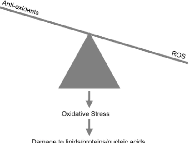
Oxidatieve stress kan ontstaan wanneer er een verhoging van reactieve zuurstof deeltjes, zogenaamde ROS, of een verlaging in de anti-oxiderende verdediging in een cel ontstaat (Figuur 1). Ten gevolge hiervan ontstaat er oxidatieve schade aan verschillende
macromoleculen (lipiden, eiwitten en DNA) en kunnen er redox gevoelige signaal routes geactiveerd worden. Door hun reactiviteit zijn ROS zelf zeer moeilijk te meten als indicator voor de ontstane oxidatieve stress. Als indirecte maat voor oxidatieve stress worden er daarom enerzijds de oxidatie producten gemeten en anderzijds de veranderingen in de antioxidant niveaus. Biomarkers welke van belang kunnen zijn in het onderzoek naar de rol van oxidatieve stress in luchtverontreiniging gerelateerde gezondheidseffecten zijn: carbonyl niveaus als maat voor de geoxideerde eiwitten, ‘thiobarbituric acid’ reactieve substanties als maat voor geoxideerde lipiden, en 8-hydroxy-deoxyguanosine als maat voor oxidatieve schade aan DNA. Wat betreft de antioxidant respons zijn, superoxide dismutase, heem oxygenase, metallothioneins, en thioredoxine reductase en glutathion waardevolle markers voor oxidatieve stress.
Anti-oxidant s
ROS
Oxidative Stress
Damage to lipids/proteins/nucleic acids
Figuur 1. Schematische weergave van oxidatieve stress welke resulteert in beschadiging van macromoleculen.
Summary
Epidemiological studies have shown a clear association between cardiovascular morbidity, decreased lung function, increased hospital admissions, mortality, and airborne concentration of photochemical and particulate pollutants.
A better appreciation of the mechanism underlying air pollution induced health problems would allow a more targeted approach to remove the most toxic components of air pollution, and could possibly provide a means to decrease individual sensitivity to air pollution. In recent research oxidative stress has been identified as a unifying feature underlying the toxic actions of the air pollutants that are causing concern.
Inventory of oxidative stress parameters
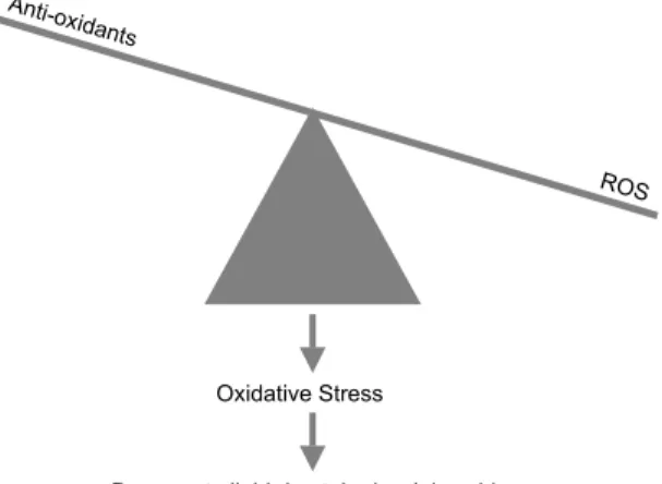
Oxidative stress results from either increased exposure to reactive oxidative species or the presence of decreased antioxidant defences resulting in oxidative damage to lipids, proteins, and DNA (Figure 1). Besides damaging of such macromolecules it also seem to trigger a
number of redox sensitive signalling pathways. Monitoring this process of oxidative stress is not easy since reactive oxygen species are very reactive particles and therefore difficult to measure. Therefore on one-hand measurements of products, macromolecules damaged by reactive oxygen species, is the most common technique to indirectly measure reactive oxygen species. On the other hand measurement of the primary defense against reactive oxygen species, the endogenous antioxidants, is a way to monitor the situation of oxidative stress. Changes in these antioxidants levels and its balance with reactive oxygen species might give us information about the state of oxidative stress. Biomarkers, which could be of interest for studying the role of oxidative stress in air pollution related health effects, are carbonyl levels as a level for oxidized proteins, thiobarbituric acid-reactive substances for total lipid
oxidation, and 8-hydroxy-deoxyguanosine for oxidative injury to DNA. In the antioxidant response, superoxide dismutase, heme oxygenase, metallothioneins, and thioredoxin reductase and glutathion are valuable markers.
Anti-ox idants
ROS
Oxidative Stress
Damage to lipids/proteins/nucleic acids
Figure 1. Schematic view of oxidative stress resulting in damage to macromolecules.
1.
Introduction
There is increasing concern over the adverse effects of air pollution on human health. Epidemiological studies have shown a clear association between cardiovascular morbidity, decreased lung function, increased hospital admissions, mortality, and airborne concentration of photochemical and particulate pollutants.
As with any toxic challenge the obvious solution is to remove, or at least decrease to an acceptable level, the source of trouble. It has been recognised that there is a need to improve our understanding of the impact of air pollution on biological systems as well. A better appreciation of the mechanism underlying air pollution induced health problems would allow a more targeted approach to remove the most toxic components of air pollution, and could possibly provide a means to decrease individual sensitivity to air pollution. Recent research has identified oxidative stress as a unifying feature underlying the toxic actions of the air pollutants that are causing concern. Oxidative stress, resulting from either increased exposure to oxidants or the presence of decreased antioxidant defences, seems to trigger a number of redox sensitive signalling pathways. There is now a strong body of evidence to indicate that the pulmonary inflammatory response that arises following exposure to a pollution episode, is mediated via oxidant signalling pathways. Moreover, it appears likely that an individual sensitivity to pollution is related, in part, to their pulmonary antioxidant defences 2.
Within RIVM’s strategic research programme launched project entitled ‘Vermindering van onzekerheden in causale relaties tussen inhalatoire blootstelling en gezondheidseffecten’ (S/630111/01/AB) the main hypothesis is that oxidative stress plays a crucial role in health effects caused by air pollution. This hypothesis is tested with use of sensitive models and with the help of new developing techniques, such as genomics. In order to have a good starting position an inventory of biomarkers will give some background about what oxidative stress is in general, what processes might occur in the lung and based on both which
2.
Radicals and ROS
To understand the way ‘oxidative stress’, as a mechanism, can play a role in the health effects due to air pollution some chemical background should be given concerning radicals and ROS, playing an important role in oxidative stress.
A free radical is by definition any chemical species that contains one or more unpaired electrons. Unpaired electrons alter the chemical reactivity of an atom or molecule by making it more reactive; molecules with an unpaired electron act like electron acceptors and ‘steal’ electrons from other molecules. When a molecule loses an electron, this is called oxidation. Free radicals are therefore called oxidising agents because they tend to make other molecules donate their electrons. Free radicals that contain oxygen are classified together as reactive oxygen species (ROS). Names as ‘active oxygen species’, ‘reactive oxygen mediators’ etcetera also appear in the literature. Well known ROS are superoxide radical anion (O2•-), hydroxyl radical (OH•), hydrogen peroxide (H2O2) (although not a free radical this is considered a ROS).
In the body, ROS are constantly produced and eliminated, since cells have endogenous defences against oxidants. However, problems occur when production of ROS exceeds elimination, either due to overproduction during trauma and detoxification, or due to damage to the natural antioxidant system. Such an imbalance in the amount of ROS produced, and ROS that is eliminated, is called oxidative stress. This can cause cellular damage to lipids, proteins and DNA, and subsequent death.
2.1
Sources of ROS
Sources of environmental ROS are ultraviolet light ionising radiation, (air) pollutants, smoking, ingestion of alcohol or other toxins that produce an excess of ROS during detoxification.
In the body the most important sources of ROS are enzymatic driven redox reaction involving oxygen. Oxygen, being the most abundant molecule in biological systems, exists as a
biradical and is therefore extremely reactive with other radicals. Oxygen itself is often the source of such radicals as partially reduced species are generated through normal metabolic processes, and some of these reactive species can escape 3. Continuous production of small amount of free radicals and ROS is therefore a basic feature of aerobic life. Within certain boundaries, the generation of ROS is even essential to maintain homeostasis. For example, ROS generation by phagocytic cells constitutes an essential host defence mechanism
necessary to combat infection. Likewise, cytosolic ROS produced in response to stimulation by growth factors are involved in regulating the proliferative response. Under certain
situation of metabolic stress, even mitochondrial-derived oxidants seem to function as signalling molecules. However a more extreme rise in intracellular oxidant levels has two
potentially important effects: damage to various cell components and triggering of the activation of specific signalling pathways 4.
The most abundant source of O2•-, being the major factor in oxygen toxicity 5, in the body is the mitochondrial electron transport chain, were the synthesis of adenosine triphosphate (ATP) is coupled to the reduction of O2. O2•- can be formed at Complex I, by the donation of an electron to oxygen from nicotinamide adenine dinucleotide (NADH) dehydrogenase. O2•- and/or H2O2 may also be formed following the auto-oxidation of reduced ubiquinone at Complex II (succinate-coenzyme Q) and/or Complex III (coenzymeQH2-cytochrome c reductases) sites. Making the generation of ROS predominantly a function of metabolic rate. Another important metabolic way were ROS are produced is by the nicotinamide adenine dinucleotide phosphate (NADPH) oxidase of the antibacterial function on phagocytic cells. Production of O2•- is an important part of the bacterial killing mechanism of neutrophils or polymorphonuclear leukocytes (PMNs). In this process, called phagocytosis invading
microorganisms are enclosed in phagosomes. During the respiratory burst the PMNs become activated, O2 consumption increases and a membrane-associated NADPH oxidase reduces oxygen to superoxide anion (O2•-) 6, which subsequently dismutates to form H2O2 7. However the expanding family of NADPH oxidases include as well the nitric oxide synthase (NOS) family, illustrating the apparent purposeful and deliberate use of oxidant generation in normal cellular signalling and homeostasis.
2.2
Reactivity
Among the ROS, the HO• is the most reactive and can attack almost every molecule in the body. Because it is so reactive, it does not diffuse very far from its point of synthesis and its half-life therefore is diffusion limited at about a nanosecond. It is produced via the Haber-Weiss reaction:
O2•- + H2O2 → OH- + OH• + O2
in the following manner:
Fe2+ + H2O2 → Fe3+ + OH• + OH- (Fenton reaction) Fe3+ + O2•- → Fe2+ + O2
As described previously O2•- is produced from the electron transport chain and during oxidative burst by neutrophils and phagocytes using NADPH oxidase. It is not lipid soluble and therefore cannot diffuse very far from its point of synthesis, it has a half live of about a microsecond.
H2O2 is a mild oxidising agent, is relatively stable, and easily moves through cell membranes and affects sites far from its origin. It is generated from superoxide dismutase (an enzyme that eliminates O2•-), when ascorbic acid reduces O2•- and it is produced in large amounts
during beta-oxidation of long-chain fatty acids. However, it is believed that H2O2 and O2 •-produce their most potent effects by forming the hydroxyl radical (OH•-) via the Haber-Weiss reaction (see previous).
3.
Oxidative stress
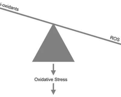
When cellular production of ROS overwhelms its antioxidant capacity, damage to molecules (and accumulation thereof) and disruption in cellular homeostasis may ensue (Figure 1). Free radicals, especially OH•, which as described is a highly reactive species interacting with almost anything in its immediate vicinity, can react with macromolecules such as proteins and carbohydrates, lipids (particularly polyunsaturated fatty acids) and DNA.
3.1
Protein
Proteins are vulnerable to oxidation by ROS at different levels. Protein oxidation results in an increase of protein carbonyl levels and glutamine synthetase activity 8. The protein carbonyls can be assayed by the stable hydrazone derivatives formed with 2,4-dinitrophenyl hydrazine9. When ROS attacks proteins formed carbonyls can react with nitrogen species and then with tyrosine to form nitrotyrosine 10,11,12. In a similar way bromotyrosine can be formed 13,14. Oxidation of proteins and carbohydrates may result in fragmentation at mainly proline and histidine residues and cross-linking, both with subsequent loss of function. Metal binding sites of proteins make them more susceptible to damage through pro-oxidant effects of metals. Reactive sulfhydryl groups such as cysteine and methionine residues undergo reversible modification. Protein repair systems do not appear to exist since it is apparently more efficient to either prevent oxidation or to simply destroy the modified species.
Anti-oxidants
ROS
Oxidative Stress
Damage to lipids/proteins/nucleic acids
Figure 1. Schematic view of oxidative stress resulting in damage to macromolecules.
3.2
Lipids
Peroxidation of lipid in membranes severely impairs membrane function. Lipid peroxides produce an irreversible impairment of membrane fluidity and elasticity, which can cause rupture of the cell. Lipid oxidation is self-propagating in cellular membranes. Products of lipid peroxidation are easily measured and an increased level of lipid peroxidation is the evidence most frequently cited in support of the involvement of oxidative stress in tissues. Malondialdehyde (MDA) is produced during lipid peroxidation 8 and can be measured using GC-MS 15. It is a known mutagen, reacting with proteins and amino acids. A peroxidation sequence in a membrane or polyunsaturated fatty acid (PUFA) is initiated by the attack of any free radical with sufficient reactivity to remove a hydrogen atom from a methylene group of an unsaturated fatty acid. This carbon-centred lipid radical then quickly reacts with
molecular oxygen to produce a hydroperoxyl radical. The peroxyl can then propagate the oxidising chain reaction by taking electrons from other susceptible PUFA, forming another lipid free radical and a molecule of lipid hydroperoxide.
ROS react with lipids to liberate ethane 16 and isoprostanes 17,18. Isoprostanes are considered to be a marker of oxidative stress and many disease states 19. They may in fact mediate the effects of free radicals and reactive oxygen species 20 and have the ability to affect signalling pathways including those that regulate the apoptotic form of cell death 3. For example,the non-enzymatic peroxidation of arrachidonic acid (normally enzymatically metabolised by cyclo-oxygenase (COX) to form a wide variety of prostanoids (prostaglandins and
thromboxanes)) by free radicals and reactive oxygen species produces isoprostanes.
Overall lipid peroxidation is the most common index of oxidative stress and can be estimated by the determination of non-specific thiobarbituric acid-reactive substances (TBARS) as described by Rhoden et al. 21, as well commercially available (Oxitek, Brussels, Belgium). However it has been suggested that this assay might overestimate lipid peroxidation when compared with a more specific gas chromatography-mass spectrometry (GC-MS assay)8.
3.3
DNA
In humans, oxidative damage to DNA has been estimated at 10000 hits per cell per day. ROS cause modifications in bases, which leads to genetic defects if the modifications are not repaired. The DNA base guanine is particularly sensitive to oxidation making the detection of 8-hydroxy-deoxyguanosine a reasonable biomarker for oxidative injury 22. Besides this also thymmidine glycol is used to measure DNA damage due to ROS degradation of bases. ROS can cause cross-linking of DNA with DNA or protein. This occurs less often but is more difficult to repair.
4.
Antioxidant mechanisms
As a basic defence mechanism towards ROS, removal of O2, scavenging of ROS and precursors, inhibition of ROS, chelation of metal ions, and upregulation of endogenous antioxidant defences occurs.
The burden of ROS production is largely counteracted by a complex antioxidant defence system. An antioxidant is defined as any substance that, when present at low concentrations compared with those of an oxidisable substrate, significantly delays or prevents oxidation of that substrate 23.
Classification of antioxidants can be based on their way of action, enzymatic and non-enzymatic scavengers, or for example on their site of action, in intra-cellular and extra-cellular. Intra-cellular scavengers include glutathione peroxidase, glutathione, catalase, superoxide dismutase. Extra-cellular antioxidants include vitamin E, vitamin C, carotenoids, uric acid, plasma proteins such as metal chelators and albumin 24.
It seems likely that especially in diseased persons, protective antioxidant mechanisms are reduced leading to an imbalance between free radical production and removal, resulting in accumulation of end products of free radical action and slow progressive tissue damage 24.
4.1
Antioxidant enzymes
Superoxide dismutase
Superoxide dismutase (SOD) converts superoxide to hydrogen peroxide in the following way:
2O2•- + 2 H+ → H2O2 + O2
Two different forms of SOD exist, a CuZn SOD which is found in cytoplasma and extracellular fluid, and MnSOD found in mitochondria.
Catalase
Catalase is a heme-containing enzyme located in the peroxisomes, which detoxifies H2O2: 2 H2O2 → 2 H2O + O2
Glutathion peroxidase
Glutathion peroxidase is a selenium (Se) containing enzyme found in the cytosol and mitochondria. It catalyses the following reaction:
2GSH + H2O2 → H2O + GSSG
Thioredoxin and glutaredoxin systems
Thioredoxin (Trx) and glutaredoxin (Grx) catalyse thiol-disulfide exchange reactions required for many functions including general regulation of prtein function by thiol redox control. Trx is reduced to the dithiol form by NADPH and thioredoxin reductase (together
called the thioredoxin system). Grx in contrast is reduced by the ubiquitos glutathione (GSH) and GSSG in turn is reduced by NADPH and glutathione reductase (together called the glutaredoxin system). The thioredoxin and glutaredoxin systems induced by a variety of stress conditions including viral infection, ischemic insult, exposure to UV light, X-ray irradiation, and hydrogen peroxide exposure 25. In vitro and in vivo studies have demonstrated that these systems have a protective effect against ROS-induced cellular damage 26,27.
Hemeoxygenase
The stress-responsive and antioxidant enzyme heme oxygenase (HO-1) is known to be induced by various oxidative agents. For example, HO-1 mRNA showed the most marked response to organic extracts of diesel exhaust particles (OE-DEP) in rat heart microvessel endothelial (RHMVE) cells 28.
Glutamine synthetase
Glutamine synthetase is a key enzyme in cellular nitrogen regulation facilitating the uptake of excitatory neurotransmitter glutamate and is among the enzymes that are rapidly induced by stress hormone glucocorticoids. A decreased uptake of glutamate due to decreased glutamine synthetase activity could result in neurotoxic effects of abnormally prolonged N-methyl-D-aspartate receptor activation and generation of oxidants 29. Therefore, an increase indicates a stress response.
4.2
Antioxidant proteins
Among antioxidant proteins we can distinguish iron and copper sequestering proteins (such as ceruloplasmin, albumin, transferrin) as well proteins that bind free heme (such as
haptoblobin, hemopexin). A special group form the metallothioneins (MTs) which are ubiquitous low molecular weight proteins of extremely high metal and sulphur content. They are thought to play a role in the protection from a variety of stress conditions, and have been recognised as potentially having antioxidant activity.
4.3
Low molecular weight antioxidants
Glutathione
Glutathione (GSH) is abundant in cytoplasm, nuclei, and mitochondria and is the major water-soluble antioxidant in these cell compartments (it is present in millimolar
concentrations within cells). The sulphur atom easily loses a single electron and reacts directly with OH•
GSH + OH• → GS• + H2O GS• + GS• → GSSG
Glutathione is the major redox buffer of several eukaryotic cell types and an important factor in the cellular defence against oxidative stress. This is due to its own antioxidant capacity and
because this thiol is involved in the recycling of other antioxidants. Glutathione homeostasis is regulated by several enzymes, such as glutathione reductase (GR), glutathione peroxidase (GPX), and glutathione transferase (GST). It has been suggested that the capacity of the GSH redox cycle, rather than the intracellular levels of reduced GSH, determines the resistance to oxidative stress at least in some cell types. The ratio between reduced GSH and total GSH is a good indicator of oxidative stress (reduction with increased oxidative stress). As well down regulation of GPX/GR ratio with increased oxidative stress 30.
Tocopherol
Tocopherol (vitamin E) is lipid soluble and resides almost exclusively on or at the surface of cell membranes. Vitamin E has a chromanol ring which is a hydroxylated aromatic ring, and several isoprene side chains.
Ascorbic acid
Ascorbic acid (vitamin C) is the most versatile and effective water-soluble antioxidant. It efficiently scavenges OH•, ROO•, singlet oxygen and reactive peroxides. It prevents lipid peroxidation by scavenging peroxyl radicals in the aqueous phase and regenerating vitamin E. It is known that high levels of ascorbic acid in neutrophils protect host tissue during oxidative burst. It is recycled to reduced state by GSH and NADPH.
Other low molecular weights antioxidants are carotenoids, uric acid, lipoic acid and melatonin.
5.
Signal transduction
Besides ROS being part of an oxidative stress situation, as mentioned briefly in the introduction, recent studies have also implicated ROS that are generated by specialised plasma membrane oxidase in normal physiological signalling by growth factors and cytokines 31.
ROS are able to act directly on signal transduction pathways, or indirectly through the
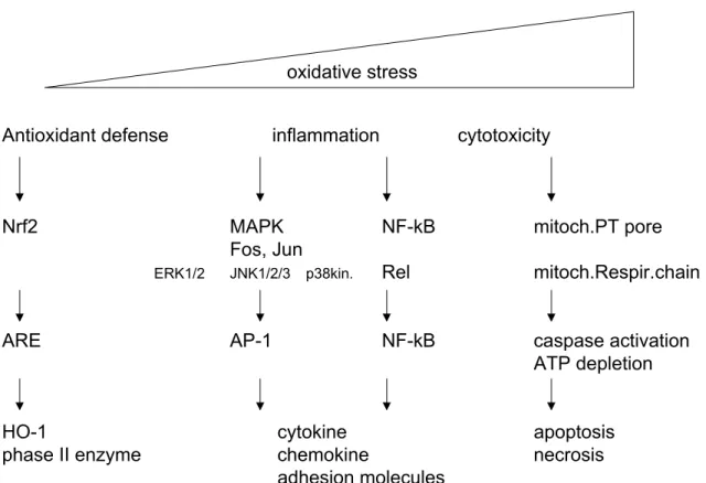
generation of bioactive mediators. ROS modulate quite a number of, so-called redox sensitive transcription factors, such as activator protein-1 (AP-1) and nuclear factor-kappa B (NF-κB) (Figure 2). Whereas ROS are also able to affect activity of calcium signalling, antioxidant enzymes, ion transporters, various cell growth related genes and, various kinases (c-jun N terminal kinase (JNK), extracellular regulated kinase (ERK) and the mitogen activated protein kinase (MAP kinase) family) 32,33. It seems that also controlling the thiol status of a cell is critical in altering the signalling pathway.
oxidative stress
Antioxidant defense inflammation cytotoxicity
Nrf2 MAPK NF-kB mitoch.PT pore
Fos, Jun
ERK1/2 JNK1/2/3 p38kin. Rel mitoch.Respir.chain
ARE AP-1 NF-kB caspase activation
ATP depletion
HO-1 cytokine apoptosis
phase II enzyme chemokine necrosis
adhesion molecules
Figure 2. Oxidative stress model in response to PM (adapted from Li et al.1). Different levels of oxidative stress can be distinguished. At a lower level of oxidative stress antioxidant enzymes are induced to restore cellular redox homeostasis. At an intermediate level an inflammation response is induced, via activation of MAPK and NFκB pathways. And at the highest level cytotoxicity results in cellular
6.
Situation in the lung
6.1
Oxidative stress
As stated above in its simples form, oxidative stress is a potentially harmful process, that occurs when there is an excess of oxidants, a decrease in antioxidant defences, or a
combination of these events (Figure 1). As a consequence, free radicals attack and oxidise other cell components such as lipids, proteins, and nucleic acids. Signals can consequently lead to the activation of transcription factors and increased expression of pro-inflammatory chemokines and cytokines (such as TNFα, IL-6 etc.). These signalling molecules lead to the up regulation of adhesion molecules. Consequently immune cells (neutrophils, eosinophils, and macrophages) move into the lung tissue. NADPH oxidase on these cells is activated, and using O2, produces O2•-. Removal of this reactive oxygen species by SOD results in
generation of H2O2, which can easily pass across cell membranes. It might activate
intracellular signalling pathways or generate other ROS (HO• in the presence of metals, HOCl by myeloperoxidase which is abundantly present in neutrophils). This highly organised series of events can therefore lead to a second wave of oxidative stress in the lung (6-18 hours after the initial exposure). In the absence of any invading organisms to kill, the generated and released free radicals attack local tissue components and cause cell injury.
Reactive oxygen intermediates are thought to be primary mediators of macrophage-induced tissue injury, and they have been implicated in the pathogenesis of xenobiotic induced lung toxicity 34. Thus following inhalation of irritants, pulmonary levels of reactive oxygen intermediates increase in the lung 34,35. Moreover, antioxidants have been shown to avoid tissue injury in several of these models.
6.2
Antioxidant
The lung has a well-developed antioxidant system. First of all the conducting airways are protected by an epithelial lining fluid (ELF) containing low molecular weight antioxidants such as uric acid, ascorbic acid, and GSH, as well as antioxidant proteins such as albumin and lactoferrin. Ascorbic acid is known to be positively related to pulmonary function.
Tocopherols are believed to be the first line of defence against membrane oxidation. The lung cells by them self possess relatively high levels of antioxidants such as SOD, catalase, and GSH to prevent or limit oxidative damage 36,37. However, during states of increased oxidative stress antioxidants become depleted, exacerbating tissue injury 34. For the lung SOD is generally considered one of the first lines of antioxidant defence 38. As described previously this enzyme converts O2•- into H2O2, which is then removed by catalase. Antioxidant enzymes are not uniformly distributed throughout the cells of the lung, which implies that some cell types may be relatively unprotected 39. MnSOD protein is primarily expressed in the mitochondria of type II alveolar cells, with lung fibroblasts containing very
little and type I alveolar cells and endothelial cells containing no detectable immunoreactive protein.
GSH
Special attention deserves GSH, being one of the key components of the well-developed antioxidant system of the lung 40. GSH, being a vital intra- and extracellular protective antioxidant, has been shown to be critical in protecting airspace epithelium from
oxidative/free radical mediated injury and inflammation 41. It protects cells from the toxic effects of oxidants by maintaining the reduced state of the sulfhydryl groups of a number of sulfhydryl-dependent enzymes 42. Induction of oxidative stress is evident by decreased levels of the antioxidant GSH in the lungs 43, and epithelial cells in vitro. Acute inhalation of oxidising agents such as ozone or NO2 depletes the lungs of GSH 44. Depletion of GSH, affecting the GSH/GSSG ratio, influences a variety of cellular signalling processes, such as activation of the transcription factors AP-1 and nuclear factor kappa B (NF-κB) (Figure 2). It as well can increase permeability of lung epithelium, which can lead to passage of particles into the interstitium 41. Alterations in the alveolar and lung GSH metabolism are widely recognised as a central feature of many inflammatory lung diseases.
The intracellular GSH redox homeostasis is strictly regulated to govern cell metabolism and protect cells against oxidative stress 45. The synthesis of glutathione requires two enzymes and three amino acids, glycine, cysteine and glutamic acid (with cysteine being the rate-limiting substrate). The tripeptide GSH is formed by the consecutive actions of γ-GCS and glutathione synthetase. Gamma-glutamylcysteine synthetase (γ -GCS) is the rate-limiting enzyme in GSH synthesis. γ-GCS consists of a catalytic heavy subunit (γ-GCS-HS) and a regulatory light subunit (γ-GCS-LS). The promoter regions of both corresponding genes contain putative AP-1 and antioxidant response element (ARE) response elements necessary for expression in response to diverse stimuli, such as oxidants, phenolic antioxidants,
inflammatory, and anti-inflammatory agents in lung cells.
6.3
Signal transduction
The development of an oxidant/antioxidant imbalance in lung inflammation may activate redox-sensitive transcription factors such as NF-κB, and AP-1 46,47. Transcription factors, are DNA-binding proteins, that interact with specific sequence motifs in the promoter region of the gene, controlling the expression of these. In particular NF-κB, a multisubunit
transcription factor, plays a central role in the regulation of the expression of numerous genes involved in the inflammatory and immune response (resulting in upregulation of cytokines and chemokines) 48 as well as protective antioxidant genes, such as γ-GCS. The critical balance between the induction of proinflammatory mediators and antioxidant genes in response to oxidative stress at the site of inflammation is not known 49. Cellular oxidative processes seem also to activate stress kinases (JNK, MAPK, p38).
Exogenous and endogenous antioxidants are effective in blocking activation of NF-κB and preventing the consequences of proinflammatory gene expression. Phase II enzymes either directly or indirectly play a major in vivo role in minimising oxidative stress by scavenging peroxides, peroxide breakdown products and dicarbonyls and in regeneration of lipid peroxidation chain-breaker, vitamin E 50.
As an antioxidant defence, induction of these genes, via mainly the ARE pathway, is a first step trying to restore cellular redox homeostasis (Figure 2). However during a prolonged situation of oxidative stress activation of MAPK and NF-κB cascade induces
proinflammatory responses. Expression of many proinflammatory mediators such as cytokines (IL-1b, TNFα, IL-6), chemokines (IL-8 and MIP-2), adhesion molecules
(VCMAM-1, ICAM-1, and E-selectin), and enzymes (iNOS) are regulated by NF-κB 51,52. Aside from NF-κB, other transcription factors such as activator protein-1, interferon
regulatory factors, and signal transducers and activators of transcription are also involved in the induction of proinflammatory cytokines 53. At the lowest levels of oxidative stress cytoprotective pathways are induced resulting in expression of antioxidant enzymes, such HO-1 and catalase 54. Induction of HO-1 expression is dependent on the ARE in the promoter of that gene, which is transcriptionally activated by a basic leucine zipper transcription factor, NF-E2-related factor-2 (Nrf2). In a next step (second tier of oxidative stress responses) the p38MAPK and Jun kinase cascades are activated resulting in activation of NF-κB and AP-1. Whereas under extreme high levels of oxidative stress the final tier of response, mediated by mitochondrial perturbation, leads to cytotoxic effects (see Figure 2).
7.
Ozone and PM exposure
Since ozone is a well-known and long-time studied oxidative air-pollution it is used within S/630111/01/AB as a model compound for generating oxidative stress by exposure.
7.1
Ozone exposure
Inhalation of ozone primarily affects the terminal bronchioles and the alveoli. The
bronchiolar ciliated cells and the epithelial alveolar type I cells are damaged, and are later replaced with proliferating Clara cells in the airways and epithelial type II cells in the alveoli, respectively.
It is believed that ozone operates via two main mechanisms of action. First by the oxidation of polyunsaturated fatty acids under the formation of acid peroxides and dialdehydes such as malonaldehyde 55. Second by oxidation of sulfhydryl groups and amino acids of proteins. Exposure to ozone is associated with increase in cellular and biochemical markers of inflammation in bronchoalveolar lavage fluid (BALF) and induced sputum 56,57. Ozone exposure causes an airway influx and activation of neutrophils (PMNs) 56, which induces a respiratory burst resulting in overproduction of ROS and H2O2 58. ROS generation is associated with lipoperoxidation of airway cell membranes and subsequent release of a number of molecules resulting from the cleavage of arachidonic acid, including
malondialdehyde (MDA) 59, 8-isoprostane 60, and the PMN chemotactic mediator leukotrine B-4 (LTB4) 61.
In exhaled breath condensate (EBC) a sudden increase in 8-isoprostane levels, as well as H2O2, and a late increase (after 18 h) in LTB-4 and TBARS levels is observed 62.
Biochemical studies have demonstrated that ozone exposure stimulates the induction of the glutathione peroxidase system. The induction of this system is thought to be a protective mechanism. Another activated protective mechanism is the induction of SOD, following exposure of animals to ozone 63,64. Whereas Fakhrzadeh et al. have shown that SOD overexpressing mice are resistant to ozone-induced tissue injury and increases in NO and TNFα, it is believed that macrophages and inflammatory mediators contribute to ozone-induced lung inflammation and toxicity 65. Since ozone increases NO production by alveolar macrophages and alveolar type II epithelial cells it is believed that NO has an adverse role in ozone-induced lung injury 66.
7.2
PM exposure
Currently, three theories of how particles induce oxidative stress have been suggested. Firstly, particles contain several soluble transition metals on the surface which induce generation of reactive oxygen species (ROS) in biological systems 67,68,69. The second
mechanism involves the organic fraction of particles, which consists of several redox-active quinones, existing or generated during metabolism of several polycyclic aromatic
hydrocarbons (PAH). Quinones produce ROS during their metabolism by redox cycling, unless they are catalysed by the phase II enzyme NAD(P)H:quinone oxidoreductase-1 (NQO1). The third mechanism involves reactions by alveolar macrophages (AM) and neutrophils. These are phagocytes known to ingest and remove inhaled particles from the lungs 70. This leads to activation of these cells causing release of several cytokines as well as ROS through a ‘respiratory burst’ 71. It is unclear which components of ambient particles are responsible for phagocyte activation.
Long term exposure to high particle levels increase risk of cancer, respiratory diseases and arteriosclerosis, whereas short-term exposure-peaks cause exacerbation of bronchitis, asthma and other respiratory diseases as well as changes in heart rate variability 72,73,74. Low
concentrations exposure assessment and establishing relationships with biological effects may be difficult. By means of monitors of individual exposure to air pollutants and of biomarkers of internal dose, biologically effective dose and early biologically effect as well as markers of individual susceptibility, such relationships may be documented.
Transition metals such as iron and copper being part of PM are capable of redox cycling and can induce Fenton reaction, resulting in the production of reactive oxygen and nitrogen species 75. It has been shown that equal masses of PM can induce disparate lung injuries, suggesting that particle components may be relevant in assessing health effects after their exposure. In the case of the Utah Valey study, specifically metals can participate in the biologic effects of PM collected.
In a recent experiment rats were exposed to PM and subsequently gene expression in the lung tissue was analysed. Strong induction of the following genes was observed, metallothionein, heme oxygenase, thioredoxin reductase, glutathione reductase and SOD as well as phase II enzymes such as P450.
As shown in Figure 2, at the lowest levels of oxidative stress cytoprotective pathways are induced resulting in expression of antioxidant enzymes, such HO-1 and catalase and phase II enzymes 54. Induction of HO-1 expression is dependent on the ARE in the promoter of that gene, which is transcriptionally activated by a basic leucine zipper transcription factor, Nrf2. PAH, organic component carried on the particle surface, can induce oxidative stress
indirectly, through biotransformation by cytochrome P450 and dihydrodiol dehydrogenase to generate redox active quinones that act as catalysts for free radical production.
8.
Conclusion biomarkers for oxidative stress
It is clear from the previous that oxidative stress is a very complicated process, which can be described as an imbalance between ROS and antioxidants resulting in damage to lipids, proteins and nucleic acids. Monitoring this process of oxidative stress is not easy since ROS are very reactive particles and therefore difficult to measure. Therefore measurements of products, macromolecules damaged by reactive oxygen species, is the most common technique to indirectly measure oxidative stress. These products, often called footprints, are not specific for individual reactive oxygen species. Many oxidative footprints are thought to be the result of nonenzymatic reactions between reactive oxygen species and organic
molecules, such as proteins, lipids, or DNA 76.
Another way to monitor the situation of oxidative stress is measurement of the primary defence against ROS, the endogenous antioxidants. Changes in these antioxidants levels and its balance with ROS might give us information about the state of oxidative stress (Figure 1). It might well be that certain risk groups in the population are more susceptible for air
pollution due to an off balance between ROS and antioxidants.
Based on the previous given overview on oxidative stress, consisting of ROS, oxidation products and the response to it, the following list of biomarkers is created (Table 1). This list of biomarkers is valuable to investigate oxidative stress as a mechanism in health effects due to air pollution.
Table 1. Biomarkers of oxidative stress
biomarker reference
Oxidation products
Protein Carbonyl content 9
Lipids MDA 59
Plasma isoprostane, 8-isoprostane 19, 60
Overall lipid peroxidation, TBARS 21 DNA 8-hydroxy-2’-deoxyguanosin Antioxidant Enzymes SOD 77 Catalase Hemeoxygenase 77 Metallothioneins 77 Thioredoxin system 77 Glutaredoxin system 77 LMW antioxidants GSH
It has been shown that inhalation exposure to concentrated ambient particles (CAPs) increases two-fold the steady-state concentration of ROS in the rat lung and heart 68. These
CAPs dependent increases in ROS are sufficient to cause accumulation of oxidised lipids and proteins and about a two-fold increase in the levels of TBARS and protein carbonyls was measured 21. Therefore both oxidation products are valuable markers for oxidative stress in the lung. The oxidation product of the DNA base guanine, 8-hydroxy-deoxyguanosine, is a reasonable biomarker for oxidative injury 22.
In the antioxidant response to oxidative stress, SOD and catalase are two important enzymes since they directly catalyse the reaction of the ROS, O2•- and H2O2. The response of these and other enzymes is very well followed by expression of their mRNA (Southern blotting, gene expression arrays, real time PCR). A recent gene expression study performed by our group with spontaneous hypertensive rats exposed to particulate matter showed increases of SOD, glutathion reductase, heme oxygenase, metallothioneins, and thioredoxin reductase 77.
In respect to the LMW antioxidants the intra- and extracellular protective antioxidant GSH is probably the most important antioxidant in the lung 40. It protects cells from the toxic effects of oxidants by maintaining the reduced state of the sulfhydryl groups of a number of
sulfhydryl-dependent enzymes 42. Changes in the levels of GSH or GSH/(total glutathion) ratio, therefore is a very important biomarker of oxidative stress.
References
(1) Li, N.; Hao, M.; Phalen, R. F.; Hinds, W. C.; Nel, A. E. Particulate Air Pollutants and Asthma. A Paradigm for the Role of Oxidative Stress in PM-Induced Adverse Health Effects. Clin. Immunol. 2003, 109, 250-265.
(2) Kelly, F. J. Oxidative Stress: Its Role in Air Pollution and Adverse Health Effects. Occup. Environ. Med. 2003, 60, 612-616.
(3) Kehrer, J. P. The Haber-Weiss Reaction and Mechanisms of Toxicity. Toxicology 2000, 149, 43-50.
(4) Finkel, T.; Holbrook, N. J. Oxidants, Oxidative Stress and the Biology of Ageing. Nature 2000, 408, 239-247.
(5) Fridovich, I. Superoxide Dismutases. Adv. Enzymol. Relat Areas Mol. Biol. 1974, 41, 35-97.
(6) Babior, B. M.; Kipnes, R. S.; Curnutte, J. T. Biological Defense Mechanisms. The Production by Leukocytes of Superoxide, a Potential Bactericidal Agent. J. Clin. Invest 1973, 52, 741-744.
(7) Weening, R. S.; Wever, R.; Roos, D. Quantitative Aspects of the Production of Superoxide Radicals by Phagocytizing Human Granulocytes. J. Lab Clin. Med. 1975, 85, 245-252.
(8) Liu, J.; Yeo, H. C.; Overvik-Douki, E.; Hagen, T.; Doniger, S. J.; Chyu, D. W.; Brooks, G. A.; Ames, B. N.; Chu, D. W. Chronically and Acutely Exercised Rats: Biomarkers of Oxidative Stress and Endogenous Antioxidants. J. Appl. Physiol 2000, 89, 21-28.
(9) Levine, R. L.; Williams, J. A.; Stadtman, E. R.; Shacter, E. Carbonyl Assays for Determination of Oxidatively Modified Proteins. Methods Enzymol. 1994, 233, 346-357.
(10) Hanazawa, T.; Kharitonov, S. A.; Barnes, P. J. Increased Nitrotyrosine in Exhaled Breath Condensate of Patients With Asthma. Am. J. Respir. Crit Care Med. 2000, 162, 1273-1276.
(11) Kaminsky, D. A.; Mitchell, J.; Carroll, N.; James, A.; Soultanakis, R.; Janssen, Y. Nitrotyrosine Formation in the Airways and Lung Parenchyma of Patients With Asthma. J. Allergy Clin. Immunol. 1999, 104, 747-754.
(12) Paredi, P.; Kharitonov, S. A.; Hanazawa, T.; Barnes, P. J. Local Vasodilator Response to Mobile Phones. Laryngoscope 2001, 111, 159-162.
(13) Heinecke, J. W. Eosinophil-Dependent Bromination in the Pathogenesis of Asthma. J. Clin. Invest 2000, 105, 1331-1332.
(14) Wu, W.; Samoszuk, M. K.; Comhair, S. A.; Thomassen, M. J.; Farver, C. F.; Dweik, R. A.; Kavuru, M. S.; Erzurum, S. C.; Hazen, S. L. Eosinophils Generate Brominating Oxidants in Allergen-Induced Asthma. J. Clin. Invest 2000, 105, 1455-1463.
(15) Liu, J.; Yeo, H. C.; Doniger, S. J.; Ames, B. N. Assay of Aldehydes From Lipid Peroxidation: Gas Chromatography-Mass Spectrometry Compared to Thiobarbituric Acid. Anal. Biochem. 1997, 245, 161-166.
(16) Paredi, P.; Kharitonov, S. A.; Barnes, P. J. Elevation of Exhaled Ethane Concentration in Asthma. Am. J. Respir. Crit Care Med. 2000, 162, 1450-1454.
(17) Dworski, R.; Roberts, L. J.; Murray, J. J.; Morrow, J. D.; Hartert, T. V.; Sheller, J. R. Assessment of Oxidant Stress in Allergic Asthma by Measurement of the Major Urinary Metabolite of F2-Isoprostane, 15-F2t-IsoP (8-Iso-PGF2alpha). Clin. Exp. Allergy 2001, 31, 387-390.
(18) Montuschi, P.; Corradi, M.; Ciabattoni, G.; Nightingale, J.; Kharitonov, S. A.; Barnes, P. J. Increased 8-Isoprostane, a Marker of Oxidative Stress, in Exhaled Condensate of Asthma Patients. Am. J. Respir. Crit Care Med. 1999, 160, 216-220.
(19) Devlin, R. B. Identification of Subpopulations That Are Sensitive to Ozone Exposure: Use of End Points Currently Available and Potential Use of Laboratory- Based End Points Under Development. Environ. Health Perspect. 1993, 101 Suppl 4, 225-230. (20) Janssen, L. J. Isoprostanes: Generation, Pharmacology, and Roles in Free-Radical-
Mediated Effects in the Lung. Pulm. Pharmacol. Ther. 2000, 13, 149-155.
(21) Rhoden, C. R.; Lawrence, J.; Godleski, J. J.; Gonzalez-Flecha, B. N-Acetylcysteine Prevents Lung Inflammation After Short-Term Inhalation Exposure to Concentrated Ambient Particles. Toxicol. Sci. 2004, 79, 296-303.
(22) Tsuboi, H.; Kouda, K.; Takeuchi, H.; Takigawa, M.; Masamoto, Y.; Takeuchi, M.; Ochi, H. 8-Hydroxydeoxyguanosine in Urine As an Index of Oxidative Damage to DNA in the Evaluation of Atopic Dermatitis. Br. J. Dermatol. 1998, 138, 1033-1035. (23) Halliwell, B. Reactive Oxygen Species in Living Systems: Source, Biochemistry, and
Role in Human Disease. Am. J. Med. 1991, 91, 14S-22S.
(24) Bunker, V. W. Free Radicals, Antioxidants and Ageing. Med. Lab Sci. 1992, 49, 299-312.
(25) Nakamura, H.; Nakamura, K.; Yodoi, J. Redox Regulation of Cellular Activation. Annu. Rev. Immunol. 1997, 15, 351-369.
(26) Fukuse, T.; Hirata, T.; Yokomise, H.; Hasegawa, S.; Inui, K.; Mitsui, A.; Hirakawa, T.; Hitomi, S.; Yodoi, J.; Wada, H. Attenuation of Ischaemia Reperfusion Injury by Human Thioredoxin. Thorax 1995, 50, 387-391.
(27) Sasada, T.; Iwata, S.; Sato, N.; Kitaoka, Y.; Hirota, K.; Nakamura, K.; Nishiyama, A.; Taniguchi, Y.; Takabayashi, A.; Yodoi, J. Redox Control of Resistance to
Cis-Diamminedichloroplatinum (II) (CDDP): Protective Effect of Human Thioredoxin Against CDDP-Induced Cytotoxicity. J. Clin. Invest 1996, 97, 2268-2276.
(28) Hirano, S.; Furuyama, A.; Koike, E.; Kobayashi, T. Oxidative-Stress Potency of Organic Extracts of Diesel Exhaust and Urban Fine Particles in Rat Heart Microvessel Endothelial Cells. Toxicology 2003, 187, 161-170.
(29) Smith, C. D.; Carney, J. M.; Starke-Reed, P. E.; Oliver, C. N.; Stadtman, E. R.; Floyd, R. A.; Markesbery, W. R. Excess Brain Protein Oxidation and Enzyme Dysfunction in Normal Aging and in Alzheimer Disease. Proc. Natl. Acad. Sci. U. S. A 1991, 88, 10540-10543.
(30) Glosli, H.; Tronstad, K. J.; Wergedal, H.; Muller, F.; Svardal, A.; Aukrust, P.; Berge, R. K.; Prydz, H. Human TNF-Alpha in Transgenic Mice Induces Differential Changes in Redox Status and Glutathione-Regulating Enzymes. FASEB J. 2002, 16, 1450-1452.
(31) Thannickal, V. J.; Fanburg, B. L. Reactive Oxygen Species in Cell Signaling. Am. J. Physiol Lung Cell Mol. Physiol 2000, 279, L1005-L1028.
(32) Adler, V.; Yin, Z.; Tew, K. D.; Ronai, Z. Role of Redox Potential and Reactive Oxygen Species in Stress Signaling. Oncogene 1999, 18, 6104-6111.
(33) Sen, C. K. Redox Signaling and the Emerging Therapeutic Potential of Thiol Antioxidants. Biochem. Pharmacol. 1998, 55, 1747-1758.
(34) Comhair, S. A.; Erzurum, S. C. Antioxidant Responses to Oxidant-Mediated Lung Diseases. Am. J. Physiol Lung Cell Mol. Physiol 2002, 283, L246-L255.
(35) Laskin, D. L.; Pendino, K. J. Macrophages and Inflammatory Mediators in Tissue Injury. Annu. Rev. Pharmacol. Toxicol. 1995, 35, 655-677.
(36) Muzykantov, V. R. Delivery of Antioxidant Enzyme Proteins to the Lung. Antioxid. Redox. Signal. 2001, 3, 39-62.
(37) Davis, J. M. Superoxide Dismutase: a Role in the Prevention of Chronic Lung Disease. Biol. Neonate 1998, 74 Suppl 1, 29-34.
(38) Noor, R.; Mittal, S.; Iqbal, J. Superoxide Dismutase--Applications and Relevance to Human Diseases. Med. Sci. Monit. 2002, 8, RA210-RA215.
(39) Holley, J. A.; Janssen, Y. M.; Mossman, B. T.; Taatjes, D. J. Increased Manganese Superoxide Dismutase Protein in Type II Epithelial Cells of Rat Lungs After Inhalation of Crocidolite Asbestos or Cristobalite Silica. Am. J. Pathol. 1992, 141, 475-485.
(40) Rahman, I.; MacNee, W. Oxidative Stress and Regulation of Glutathione in Lung Inflammation. Eur. Respir. J. 2000, 16, 534-554.
(41) Li, X. Y.; Donaldson, K.; Rahman, I.; MacNee, W. An Investigation of the Role of Glutathione in Increased Epithelial Permeability Induced by Cigarette Smoke in Vivo and in Vitro. Am. J. Respir. Crit Care Med. 1994, 149, 1518-1525.
(42) Puglia, C. D.; Powell, S. R. Inhibition of Cellular Antioxidants: a Possible Mechanism of Toxic Cell Injury. Environ. Health Perspect. 1984, 57, 307-311.
(43) Li, X. Y.; Gilmour, P. S.; Donaldson, K.; MacNee, W. Free Radical Activity and Pro-Inflammatory Effects of Particulate Air Pollution (PM10) in Vivo and in Vitro. Thorax 1996, 51, 1216-1222.
(44) Sagai, M.; Ichinose, T.; Oda, H.; Kubota, K. Studies on Biochemical Effects of Nitrogen Dioxide. II. Changes of the Protective Systems in Rat Lungs and of Lipid Peroxidation by Acute Exposure. J. Toxicol. Environ. Health 1982, 9, 153-164. (45) Rahman, I.; MacNee, W. Regulation of Redox Glutathione Levels and Gene
Transcription in Lung Inflammation: Therapeutic Approaches. Free Radic. Biol. Med. 2000, 28, 1405-1420.
(46) Gius, D.; Botero, A.; Shah, S.; Curry, H. A. Intracellular Oxidation/Reduction Status in the Regulation of Transcription Factors NF-KappaB and AP-1. Toxicol. Lett. 1999, 106, 93-106.
(47) Rahman, I.; MacNee, W. Role of Transcription Factors in Inflammatory Lung Diseases. Thorax 1998, 53, 601-612.
(48) Baeuerle, P. A.; Henkel, T. Function and Activation of NF-Kappa B in the Immune System. Annu. Rev. Immunol. 1994, 12, 141-179.
(49) Rahman, I. Inflammation and the Regulation of Glutathione Level in Lung Epithelial Cells. Antioxid. Redox. Signal. 1999, 1, 425-447.
(50) Christman, J. W.; Blackwell, T. S.; Juurlink, B. H. Redox Regulation of Nuclear Factor Kappa B: Therapeutic Potential for Attenuating Inflammatory Responses. Brain Pathol. 2000, 10, 153-162.
(51) Christman, J. W.; Lancaster, L. H.; Blackwell, T. S. Nuclear Factor Kappa B: a Pivotal Role in the Systemic Inflammatory Response Syndrome and New Target for Therapy. Intensive Care Med. 1998, 24, 1131-1138.
(52) Barnes, P. J.; Karin, M. Nuclear Factor-KappaB: a Pivotal Transcription Factor in Chronic Inflammatory Diseases. N. Engl. J. Med. 1997, 336, 1066-1071.
(53) Julkunen, I.; Sareneva, T.; Pirhonen, J.; Ronni, T.; Melen, K.; Matikainen, S.
Molecular Pathogenesis of Influenza A Virus Infection and Virus-Induced Regulation of Cytokine Gene Expression. Cytokine Growth Factor Rev. 2001, 12, 171-180. (54) Xiao, G. G.; Wang, M.; Li, N.; Loo, J. A.; Nel, A. E. Use of Proteomics to
Demonstrate a Hierarchical Oxidative Stress Response to Diesel Exhaust Particle Chemicals in a Macrophage Cell Line. J. Biol. Chem. 2003, 278, 50781-50790. (55) Victorin, K. Review of the Genotoxicity of Ozone. Mutat. Res. 1992, 277, 221-238. (56) Vagaggini, B.; Taccola, M.; Conti, I.; Carnevali, S.; Cianchetti, S.; Bartoli, M. L.;
Bacci, E.; Dente, F. L.; Di Franco, A.; Giannini, D.; Paggiaro, P. L. Budesonide Reduces Neutrophilic but Not Functional Airway Response to Ozone in Mild Asthmatics. Am. J. Respir. Crit Care Med. 2001, 164, 2172-2176.
(57) Samet, J. M.; Hatch, G. E.; Horstman, D.; Steck-Scott, S.; Arab, L.; Bromberg, P. A.; Levine, M.; McDonnell, W. F.; Devlin, R. B. Effect of Antioxidant Supplementation on Ozone-Induced Lung Injury in Human Subjects. Am. J. Respir. Crit Care Med. 2001, 164, 819-825.
(58) Fievez, L.; Kirschvink, N.; Dogne, S.; Jaspar, F.; Merville, M. P.; Bours, V.; Lekeux, P.; Bureau, F. Impaired Accumulation of Granulocytes in the Lung During Ozone Adaptation. Free Radic. Biol. Med. 2001, 31, 633-641.
(59) Pryor, W. A.; Church, D. F. Aldehydes, Hydrogen Peroxide, and Organic Radicals As Mediators of Ozone Toxicity. Free Radic. Biol. Med. 1991, 11, 41-46.
(60) Long, N. C.; Suh, J.; Morrow, J. D.; Schiestl, R. H.; Murthy, G. G.; Brain, J. D.; Frei, B. Ozone Causes Lipid Peroxidation but Little Antioxidant Depletion in Exercising and Nonexercising Hamsters. J. Appl. Physiol 2001, 91, 1694-1700.
(61) McBride, D. E.; Koenig, J. Q.; Luchtel, D. L.; Williams, P. V.; Henderson, W. R., Jr. Inflammatory Effects of Ozone in the Upper Airways of Subjects With Asthma. Am. J. Respir. Crit Care Med. 1994, 149, 1192-1197.
(62) Corradi, M.; Alinovi, R.; Goldoni, M.; Vettori, M.; Folesani, G.; Mozzoni, P.; Cavazzini, S.; Bergamaschi, E.; Rossi, L.; Mutti, A. Biomarkers of Oxidative Stress After Controlled Human Exposure to Ozone. Toxicol. Lett. 2002, 134, 219-225. (63) Rivas-Arancibia, S.; Vazquez-Sandoval, R.; Gonzalez-Kladiano, D.; Schneider-Rivas,
S.; Lechuga-Guerrero, A. Effects of Ozone Exposure in Rats on Memory and Levels of Brain and Pulmonary Superoxide Dismutase. Environ. Res. 1998, 76, 33-39. (64) Plopper, C. G.; Duan, X.; Buckpitt, A. R.; Pinkerton, K. E. Dose-Dependent
Tolerance to Ozone. IV. Site-Specific Elevation in Antioxidant Enzymes in the Lungs of Rats Exposed for 90 Days or 20 Months. Toxicol. Appl. Pharmacol. 1994, 127, 124-131.
(65) Fakhrzadeh, L.; Laskin, J. D.; Gardner, C. R.; Laskin, D. L. Superoxide Dismutase-Overexpressing Mice Are Resistant to Ozone-Induced Tissue Injury and Increases in Nitric Oxide and Tumor Necrosis Factor-Alpha. Am. J. Respir. Cell Mol. Biol. 2004, 30, 280-287.
(66) Zeidler, P. C.; Castranova, V. Role of Nitric Oxide in Pathological Responses of the Lung to Exposure to Environmental/Occupational Agents. Redox. Rep. 2004, 9, 7-18. (67) Ghio, A. J.; Richards, J. H.; Carter, J. D.; Madden, M. C. Accumulation of Iron in the
Rat Lung After Tracheal Instillation of Diesel Particles. Toxicol. Pathol. 2000, 28, 619-627.
(68) Gurgueira, S. A.; Lawrence, J.; Coull, B.; Murthy, G. G.; Gonzalez-Flecha, B. Rapid Increases in the Steady-State Concentration of Reactive Oxygen Species in the Lungs and Heart After Particulate Air Pollution Inhalation. Environ. Health Perspect. 2002, 110, 749-755.
(69) Han, J. Y.; Takeshita, K.; Utsumi, H. Noninvasive Detection of Hydroxyl Radical Generation in Lung by Diesel Exhaust Particles. Free Radic. Biol. Med. 2001, 30, 516-525.
(70) Stringer, B.; Imrich, A.; Kobzik, L. Flow Cytometric Assay of Lung Macrophage Uptake of Environmental Particulates. Cytometry 1995, 20, 23-32.
(71) Goldsmith, C. A.; Imrich, A.; Danaee, H.; Ning, Y. Y.; Kobzik, L. Analysis of Air Pollution Particulate-Mediated Oxidant Stress in Alveolar Macrophages. J. Toxicol. Environ. Health A 1998, 54, 529-545.
(72) Brunekreef, B.; Holgate, S. T. Air Pollution and Health. Lancet 2002, 360, 1233-1242.
(73) Dockery, D. W.; Pope, C. A., III; Xu, X.; Spengler, J. D.; Ware, J. H.; Fay, M. E.; Ferris, B. G., Jr.; Speizer, F. E. An Association Between Air Pollution and Mortality in Six U.S. Cities. N. Engl. J. Med. 1993, 329, 1753-1759.
(74) Peters, A.; Dockery, D. W.; Muller, J. E.; Mittleman, M. A. Increased Particulate Air Pollution and the Triggering of Myocardial Infarction. Circulation 2001, 103, 2810-2815.
(75) Jimenez, L. A.; Thompson, J.; Brown, D. A.; Rahman, I.; Antonicelli, F.; Duffin, R.; Drost, E. M.; Hay, R. T.; Donaldson, K.; MacNee, W. Activation of NF-KappaB by PM(10) Occurs Via an Iron-Mediated Mechanism in the Absence of IkappaB Degradation. Toxicol. Appl. Pharmacol. 2000, 166, 101-110.
(76) Bowler, R. P.; Crapo, J. D. Oxidative Stress in Allergic Respiratory Diseases. J. Allergy Clin. Immunol. 2002, 110, 349-356.
(77) Kooter, I. M.; Pennings, J. L. A.; Gerlofs-Nijland, M. E.; Cassee, F. R. Gene
Expression Pattern in Spontaneously Hypertensive Rats Exposed to Urban Particulate Matter (EHC-93). Inhalation Toxicology 2004.