Behaviour of Clostridium perfringens in
simulated gastrointestinal conditions
An interim report
Report 330371001/2008
RIVM Report 330371001/2008
Behaviour of Clostridium perfringens in simulated
gastrointestinal conditions
An interim report
L. M. Wijnands
A. van der Meij - Florijn E.H.M. Delfgou - van Asch M. E. Mensink
R. de Jonge F. M. van Leusden Contact:
L. M. Wijnands
Cib / Laboratory for Zoonoses and Environmental Microbiology (LZO) lucas.wijnands@rivm.nl
This investigation has been performed by order and for the account of the Food and Consumer Product Safety Authority, within the framework of Project 330371, Quantitative aspects of exposure assessment and hazard characterization of Clostridium perfringens
© RIVM 2008
Parts of this publication may be reproduced, provided acknowledgement is given to the 'National Institute for Public Health and the Environment', along with the title and year of publication.
Abstract
Behaviour of Clostridium perfringens in simulated gastrointestinal conditions An interim report
The RIVM is investigating how people become ill from eating food contaminated with the bacterium Clostridium perfringens. For the purpose of this research, conditions in the gastrointestinal tract are being mimicked.
The overall aim of the investigations is to estimate the number of disease cases occurring in the Netherlands each year. Currently, such estimates are hard to make because the disease symptoms, mainly watery diarrhoea, are relatively mild and short-lasting. As a consequence, few patients seek medical attention and disease cases are poorly registered.
In order to make solid estimates, data on certain factors, such as the pathogenic mechanism and the prevalence of the bacterium in food are necessary. Although the stomach, due to its acidity, presents an obstacle for many bacteria, C. perfringens cells appear to survive simulated gastric conditions easily. This is an interim report for ongoing investigations. This research is being hampered by difficulties in producing the toxin that is responsible for the complaints. In addition, there are many differences in how the various C. perfringens strains react. Moreover, the prevalence of the bacterium in Dutch food commodities needs to be investigated more closely. Finally, all investigations should be integrated so that the risk for disease can be determined and, with that, the annual number of disease cases.
Rapport in het kort
Gedrag van Clostridium perfringens in nagebootste maagdarm condities Een tussenrapportage
Het RIVM onderzoekt hoe mensen ziek worden als zij voedsel eten dat besmet is met de bacterie Clostridium perfringens. Dit wordt gedaan met experimenten die de omstandigheden in maag en darmen nabootsen.
Doel van het onderzoek is om een schatting te maken van het aantal ziektegevallen in Nederland per jaar. Momenteel is dat moeilijk omdat de klachten (voornamelijk waterige diarree) kortdurend en mild zijn. Daardoor gaan patiënten niet snel naar een arts en worden gevallen van de ziekte niet snel geregistreerd.
Voor een gedegen schatting van het aantal ziektegevallen is kennis nodig over de manier waarop de ziekte ontstaat en hoe vaak en in welke mate de bacterie in voedsel voorkomt. Voor veel bacteriën vormt de maag een moeilijk te nemen obstakel, maar C. perfringens lijkt de maag gemakkelijk te passeren.
Dit rapport is een tussenrapportage. Het onderzoek wordt bemoeilijkt doordat de productie van de gifstof, die de klachten veroorzaakt, een proces is dat moeilijk is na te bootsen en door veel factoren wordt beïnvloed. Ook bestaan er veel stammen van deze bacterie die op verschillende manieren reageren. Daarnaast moet nog worden onderzocht hoe vaak en in welke mate de bacterie in voedsel voorkomt. Nader onderzoek is daarom nodig.
Trefwoorden: Clostridium perfringens, voedselgerelateerde ziekte, nagebootste maagdarm-omstandigheden
Contents
Summary 6 1 Introduction 7 1.1 General 7 1.2 Background 7 1.3 Aim 8 1.4 Research concept 81.4.1 Rationale for experiments with spores 9
1.4.2 Rationale for experiments with vegetative cells 9
1.4.3 Rationale for experiments with purified enterotoxin 10
2 Materials and methods 11
2.1 Strains 11
2.2 Media 11
2.3 Storage of strains 13
2.4 Standard incubation conditions for C. perfringens 13
2.5 Production of vegetative cells 13
2.6 Production of spores 13
2.7 Production of enterotoxin 14
2.8 Growth and maintenance of Caco-2 cells 14
2.9 Growth and maintenance of VERO cells 14
2.10 Cytotoxicity test 14
2.11 Latex agglutination test 15
2.12 Behaviour of spores in simulated gastric conditions 15
2.13 Germination of spores 15
2.14 Behaviour of vegetative cells at pH-values representing gastric conditions 16 2.15 Behaviour of vegetative cells in simulated intestinal conditions 16
2.16 Adhesion capacity to differentiated Caco-2 cells 16
2.17 Determination of enterotoxin production and concentration 16
2.18 Stability of Cpe in simulated gastrointestinal fluids 17
3 Result 21
3.1 Spores in simulated gastric conditions. 18
3.2 Behaviour of spores in simulated intestinal conditions. 18
3.3 Behaviour of vegetative cells at pH-values representing gastric conditions 19 3.4 Behaviour of vegetative cells in simulated intestinal conditions 19
3.5 Adhesion capacity to differentiated Caco-2 cells 20
3.6 Enterotoxin production and quantification 20
3.7 Stability of Cpe in simulated gastrointestinal fluids 21
4 Conclusions and discussion 22
5 Future work 25
Figures and Tables 26
Summary
Clostridium perfringens is an ubiquitary spore forming Gram positive bacterium that may also be present in the intestines of warm blooded animals including humans. The bacterium may also be present in/on food commodities and may thus lead to food borne disease. Due to the short lasting and relatively mild symptoms patients do not seek medical attention very quickly, and thus the number of disease cases is hard to estimate.
We chose for the risk assessment approach to estimate the number of disease cases in the Netherlands by investigating the behaviour of various C. perfringens strains in simulated gastrointestinal conditions and combining these results with data of the prevalence of C. perfringens in Dutch food commodities. In this report results of experiments regarding the behaviour of the bacterium in simulated gastrointestinal conditions are described. Both the gastric passage and the production of the toxin, responsible for the onset of disease symptoms, in the small intestine have been studied.
The gastric conditions hardly influence the survival and viability of C. perfringens vegetative cells and spores. The production of the toxin in the small intestine depends on various variables. The presence of bile, the number of cells, and the availability of nutrients are some of the variables that influence toxin production.
This report is an interim report. Many experiments, especially those in simulated intestinal conditions, still have to be carried out with a larger number of strains to better describe the diversity of C. perfringens. Such experiments will be carried out with “extreme” strains, such as fast growing strains versus slow growing strains, high toxin-producing strains versus low toxin-producing strains.
To complete the risk assessment, detailed data on prevalence of C. perfringens in food commodities have to be collected. Not only total numbers have to be recorded, but also the distribution of spores and vegetative cells in the food commodities and the toxin producing potential have to be recorded.
1
Introduction
1.1
General
Clostridium perfringens is a Gram positive non-motile rod-shaped bacterium that can cause several diseases. Within the context of this project the enterotoxin mediated food borne disease, also known as type A food poisoning, is the most important. The symptoms of food borne disease are diarrhoea and severe abdominal pain. Nausea is less common, but fever and vomiting are unusual 8. The enterotoxin is also known as Cpe (Clostridium perfringens enterotoxin) 11.
C. perfringens is ubiquitous throughout the environment, and is among other places commonly encountered in human and animal faeces [103 – 106 colony forming units (CFU) per g].
The pathogenic mechanism of C. perfringens food borne disease is as follows. Food contaminated with spores/vegetative cells is ingested. After passage of the stomach C. perfringens grows in the small intestine and sporulates. During sporulation Cpe, produced intracellular during growth, is released and induces the diarrhoeal symptoms. Symptoms occur 8 – 22 hours after consumption of the contaminated food. The minimal dose able to cause disease is estimated 106 - 107 CFU per gram food 811.
The cpe gene is located on the chromosome of food poisoning isolates; while cpe is located on a plasmid in non-food borne GI disease isolates 1.
Cpe is transformed into a more active toxin by trypsin and chymotrypsin, after which it binds to receptors on the brush-border membrane of epithelial cells. The receptor is the 22 kDa claudin protein, located in tight junctions of many cells 5.
In the Sensor study, performed between December 1998 and December 1999, CPE was detected in 7 cases at a total of 310 cases, i.e. 2.3% 3. The Health Council of the Netherlands estimated the annual number of Clostridium perfringens related food borne infections to be 10,000 – 50,000 6. Probably
many outbreaks go unreported because the implicated foods or patients faeces are not tested routinely for C. perfringens or its toxin.
The Dutch tolerance level is 105 CFU per gram food.
1.2
Background
C. perfringens may be present in the gut of warm blooded animals, including man. It may be shed in the environment as spores, which may re-colonize other animals. Also as a contaminant of meat man may come into contact with the micro organism. Contamination does not imply infection and disease. According to the current knowledge high numbers of C. perfringens cells are necessary to induce disease 10. However, cell counts in food stuffs suspected for their involvement in food borne disease suggest that lower numbers of C. perfringens may be necessary for the onset of disease (not published results by the Dutch Food and Consumer Product Safety Authority).
1.3
Aim
The overall aim of these researches is: Determine the influence of factors such as pH, bile salts, enzymes and nutrients on the survival/growth of spores and vegetative cells of Clostridium perfringens in the gastro-intestinal tract using simulated conditions. Heterogeneity among C. perfringens will be overcome by using various strains originating from different foodstuffs.
This overall aim has been subdivided into the following research questions:
1. Investigate the behaviour of spores, vegetative cells and enterotoxin (Cpe) in conditions simulating the gastric passage and their behaviour in simulated small intestinal conditions. Also investigate the production of Cpe, the influence of food matrix components, and the influence of varying bile and pancreatic juice concentrations in the simulated intestinal conditions.
2.
Investigate the interaction of C. perfringens cells with the small intestinal epithelium, mimicked by differentiated Caco-2 cells. Set-up/initiation of a more dynamic system to simulate the gastro-intestinal passage and to monitor the interaction between epithelium and C. perfringens. The static system is based on a system described for Bacillus cereus 13.1.4
Research concept
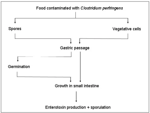
For the investigations described in this report the following concept of events was followed. Food is contaminated with C. perfringens. Often the micro organism is found in composite food commodities such as soup and prepared food that only needs reheating. The micro organism is ingested, passes the stomach which is considered the first hurdle in the human body, enters the small intestine where it sporulates and releases enterotoxin Cpe. Various stages, which have been deemed important for the pathogenesis, have been selected for further research (see Figure 1).
These stages include the passage of the stomach of vegetative cells and spores, sporulation and/or growth in small intestinal conditions, enterotoxin producing capacity and sporulation in small intestinal conditions, and interaction with the epithelium of the small intestine. Most experiments were carried out in conditions simulating the stomach, the small intestine or the small intestinal epithelium.
Investigation of the behaviour of C. perfringens in simulated gastric and intestinal conditions includes the influence on survival, growth and sporulation in the presence of food simulating compounds like glucose and beef-extract.
For investigations involving spores we assumed that spores were present in the food that was to be ingested. These spores should have to pass the stomach, and germinate and grow in the small intestine. For investigations involving vegetative cells we assumed these to be present in the food that was to be ingested. These cells should have to pass the stomach and grow in the small intestine.
We assumed preformed enterotoxin to be of no importance for the onset of disease since enterotoxin Cpe is a protein which we assume to be inactivated in gastric conditions if produced in food. Only enterotoxin produced during sporulation in the small intestine is assumed to take part in the pathogenesis.
An important fact was that most food involved in food borne incidents was contaminated with lower numbers of C. perfringens than assumed necessary for the onset of disease. This means that growth should take place in order to reach numbers that can cause disease. This was studied separately.
Some of the investigations described here were carried out with one or two strains, some with more different strains. All these strains have been involved in food borne incidents and isolated by the Food and Consumer Product Safety Authority region South.
1.4.1 Rationale for experiments with spores
Food may be contaminated with spores and vegetative cells from C. perfringens. Therefore, spores might take part in the onset of diarrhoeal disease. The stages between ingestion and the onset of disease starting with spores is therefore subject of research as well. When spores enter the gastrointestinal tract the first hurdle is the gastric passage. The next step in the process is germination in the small intestine and subsequent growth. Gastric passage, germination and growth all occur in the presence of food components, and should therefore be studied as such as well. The food components were mimicked by for example glucose or beef extract. At this stage in the investigations no food commodities, that have been found to contain C. perfringens cells in the past, were used.
1.4.2 Rationale for experiments with vegetative cells
The experiments involving vegetative cells have been carried out according to the following sequence of events.
Vegetative cells are ingested and reach their first hurdle, the stomach. Therefore, the behaviour of vegetative cells in gastric conditions, either in simulated gastric fluid or in artificial culture media with low pH values was investigated.
After reaching the small intestine the vegetative cells have to sporulate in order to release enterotoxin. Experiments investigating what conditions favour or depress sporulation and enterotoxin production have been carried out.
It is generally accepted that high numbers of C. perfringens (≥ 106/g food) are necessary to cause disease. However, a small inventory on numbers of C. perfringens that were found in food commodities suspected for having caused disease learned that these foods were contaminated with numbers < 106/g food. With the results of our investigations we try to give an explanation for this discrepancy. Within these investigations also the adhesive capacity of vegetative cells to differentiated Caco-2 cells, representing epithelial cells of the small intestine, was determined. Adhesion might be an attributive virulence factor of C. perfringens.
In vitro sporulation of C. perfringens is a process, for which various recipes exist 2 4 12. The overall conclusion of all these investigations is that no uniform method exists for sporulation of C. perfringens. Since most of these investigations have been carried out with synthetic media, and not in relation to the onset of food borne disease, i.e. in (simulated) intestinal conditions, we investigated the influence of bile and various food constituents on growth and sporulation in simulated intestinal conditions.
1.4.3 Rationale for experiments with purified enterotoxin
The enterotoxin Cpe is the actual causative agent of diarrhoeal symptoms. Earlier research with B. cereus enterotoxins showed that these were very sensitive for proteolytic activity as found in the small intestine 13. This made adhesion and, as a consequence, production of enterotoxins in close proximity to their target cells a prerequisite for the onset of diarrhoeal symptoms. In analogy, the stability of enterotoxin Cpe in simulated intestinal conditions was studied.
In order to compare the enterotoxin production capacity of various C. perfringens strains, the cytotoxic potential of culture supernatants of a number of strains was related to the reaction of the supernatants in the latex-test for Cpe and the number of cells in the culture from which the enterotoxin extract was prepared.
2
Materials and methods
2.1
Strains
The strains used for these investigations are described in Table 1. The strains were isolated from the indicated food commodities using SCA medium. Identity was confirmed by testing of the isolates for motility, nitrate reduction, lactose fermentation, and hydrolysis of gelatine. Isolation and identity testing were carried out by the Food and Consumer Product Safety Authority (VWA) region south.
2.2
Media
SC-agar (Oxoid cat. no. CM0587)
Prepare agar base according to the manufacturer’s instructions. Cool down to 50 °C and add TSC supplement as prescribed by the manufacturer (just before use).
Luria Bertani glucose broth (LBG 7.0):
Dissolve in 500 ml distilled water: 5.0 g Tryptone (Oxoid), 5.0 g sodium chloride (Merck), 2.5 g Yeast Extract (Oxoid), 10.47 g MOPS (Roche) and 2.0 g glucose (Merck). Set the pH at 7.0 using 1 M sodium hydroxide, and autoclave at 110 °C for 30 minutes. Store at 4 °C. Occasionally Luria Bertani broth (LB 7.0) was used. Preparation was carried out as described above except for the addition of glucose.
Duncan and Strong broth (DS), according to 4:
Dissolve in 1000 ml distilled water: 4.0 g Yeast extract (Oxoid), 15.0 g proteose peptone (Oxoid), 4.0 g soluble starch (Difco), 1.0 g sodium thioglycollate, (Sigma) and 10.0 g disodiumhydrogenphosphate (Merck). Autoclave (15 minutes, 121 °C) and store at room temperature.
Duncan and Strong broth with bile (DS-bile):
Add to 50 ml sterilized DS 20 +/- 2 mg bile (Sigma B3883). Thioglycollate medium:
Both thioglycollate medium (with agar, Oxoid CM173) and thioglycollate broth – alternative (without agar Oxoid CM391) were used.
Mineral medium with glucose:
Dissolve in 900 ml distilled water for the basic solution: 1.0 g ammonium sulphate (Merck), 10.5 g dipotassium hydrogen phosphate (Merck), 4.5 g potassium dihydrogen phosphate (Merck), 0.2 g magnesium chloride (Merck), and 38 mg ethylenediaminetetra acetic acid (EDTA, Acros).
Set the pH at 6.8 using 1 M sodium hydroxide and/or 1 M hydrochloride, and autoclave during 15 minutes at 121 °C.
Dissolve 0.45 g iron (II) sulphate (Riedel-de Haan) in 100 ml distilled water, and sterilize by using a 0.22 µm filter. Dissolve 5 g yeast extract (Difco) in 100 ml distilled water, and sterilize by using a 0.22 µm filter. Dissolve 4 g glucose (Merck) in 10 ml distilled water and sterilize by using a 0.22 µm filter.
Prepare the final mineral medium by mixing 900 ml basic solution, 100 ml yeast extract solution, 10 ml glucose solution and 200 µl iron (II) sulphate solution.
Simulated gastric fluid:
Mix for the inorganic part: 250 ml distilled water, 15.7 ml sodium chloride solution
(175.5 g l-1), 3.0 ml sodium dihydrogen phosphate solution (88.8 g l-1), 9.2 ml potassium chloride solution (89.6 g l-1), 18 ml freshly prepared calcium chloride solution (22.2 g l-1) and
10 ml ammonium chloride solution (30.6 g l-1). Set the pH to the desires value using 1M hydrochloride and adjust the volume to 500 ml using distilled water.
Mix for the organic part: 250 ml distilled water, 10.0 ml glucose solution (65 g l-1), 10 ml glucuronic acid solution (2g l-1), 3.4 ml urea solution (25 g l-1) and 10 ml glucosamine solution
(33 g l-1). Adjust the volume to 500 ml with distilled water.
Mix the inorganic part with the organic part, add 1.0 g bovine serum albumin and filter sterilize using a 0.22 µm filter. Add 3.0 g mucine and 1.0 g pepsin.
Simulated intestinal fluid:
Mix for the basic intestinal fluid 250 ml distilled water, 20 ml sodium chloride solution (175.3 g l-1), 20 ml sodium hydrogen carbonate solution (84.7 g l-1), 5 ml potassium di-hydrogen phosphate solution (8 g l-1), 3.15 ml potassium chloride solution (35.8 g l-1), 5 ml
magnesium chloride solution (5 g l-1), 2 ml urea solution (25 g l-1), 4.5 ml freshly prepared calcium chloride solution (29.8 g l-1) and 0.5 g bovine serum albumin (Roche, Fraktion V). Adjust the volume to 500 ml with distilled water. Add per 500 ml 0.25 g lipase and 1.5 g pancreatin (Merck).
Mix for the bile solution 200 ml distilled water, 7.5 ml sodium chloride solution (175.3 g l-1), 17.1 ml sodium hydrogen carbonate solution (84.7 g l-1), 2.5 ml urea solution (25 g l-1), 2.5 ml freshly prepared calcium chloride solution (29.8 g l-1) and 0.45 g bovine serum albumin (Roche, Fraktion V). Adjust the pH to 8.0 using either 1 M hydrochloride or sodium hydroxide. Filter sterilize using a 0.22 µm filter and add 1.5 g bile (Sigma B3883). Stir vigorously.
Prepare the complete simulated intestinal fluid by mixing just before use 3 parts basic simulated intestinal fluid with 1 part bile solution.
Caco-2 cell culture medium (DMEM 10%):
Dulbecco’s Modified Eagle Medium (DMEM, Gibco, cat. no. 424300) was supplemented with 10% foetal calf serum (Integro B.V, the Netherlands), 2 mM glutamine (Gibco, cat. no. 25030), 1% MEM non essential amino acids (Gibco, cat. no. 11140) and 10 μg ml-1 gentamicin (Gibco, cat. no 15710). After sterilization with a 0.22 µm filter, the medium was stored at 4 °C; the shelf life of the prepared medium was three months.
VERO cell culture medium (M199 1%):
Medium 199 (Gibco, cat. no. 22340) was supplemented with 1% foetal calf serum, 1% penicillin-streptomycin solution (Gibco, cat. no. 15070). After sterilization with a 0.22 µm filter, the medium was stored at 4 °C; the shelf life of the prepared medium was three months.
2.3
Storage of strains
Upon receipt the strains, as indicated in Table 1, were grown in thioglycollate medium and incubated overnight at 37 °C in jars containing Anaerogen sachets (Oxoid) to create an anaerobic environment. After incubation the haemolytic properties of the strains were checked on Columbia agar with horse blood (Oxoid). From each strain 300 µl of the thioglycollate culture was mixed with 100 µl sterile glycerol in a sterilized vial containing glass beads. The vials were stored at -70 °C.
2.4
Standard incubation conditions for C. perfringens
All incubations for growth of C. perfringens were carried out at 37 °C in anaerobic conditions. The anaerobic environment was established using a Mark II Anoxomat (Mart Microbiology, Drachten, The Netherlands). The final concentrations of the various gasses were: 0.2% O2, 99.8% N2, 0% CO2 and
0% H2.
2.5
Production of vegetative cells
The necessary number of tubes containing 10 ml LBG 7.0 each was prepared (usually 2 tubes per strain). The tubes were placed in a water bath at 100 °C during 10 minutes. A glass pearl of the intended strain was taken from the -70 °C storage and incubated in the cooled LBG7.0. Incubation was started preferably before 09.00 a.m. At the end of the day per tube 100 µl of initial LBG 7.0-culture was transferred to a new tube containing 10 ml heated LBG 7.0 (10 minutes in water bath at 100 °C) and incubated overnight.
2.6
Production of spores
The necessary number of tubes containing 10 ml LBG 7.0 each was prepared (usually 2 tubes per strain). The tubes were placed in a water bath at 100 °C during 10 minutes. A glass pearl of the intended strain was taken from the -70 °C storage and incubated in the cooled LBG7.0. Incubation was started preferably before 09.00 a.m. At the end of the day 100 μl of initial LBG 7.0-culture was transferred to a new tube containing 10 ml heated LBG 7.0. After overnight culture at 37 °C, 100 – 150 μl.LBG7.0 culture was transferred to a small erlenmeyer flask containing 50 ml Duncan and Strong medium supplemented with 20 ± 2 mg ox bile (Sigma B3883). After overnight incubation the culture was centrifuged, and the supernatant either discarded or used for further purification as enterotoxin extract. The spore pellet was washed three times with phosphate buffered saline before resuspending in 25 ml phosphate buffered saline. This suspension was placed in a water bath at 80 °C during
10 minutes to inactivate remaining vegetative cells. After determination of the concentration of spores, the suspension was diluted with phosphate buffered saline to obtain an end concentration of approximately 1x10⁷ spores per ml. After aliquotting the suspension in 1 ml portions in 1.5 ml vials, the spores were stored at -70 °C.
2.7
Production of enterotoxin
Enterotoxin was produced essentially as described by Mahony et al. (1989) 9. C. perfringens strains were, after pre-culture in LBG 7.0, grown anaerobically overnight in DS-bile at 37 °C. Cells and culture fluid were separated by centrifugation (10 minutes at 6000xg), and the supernatant was concentrated by adding ammonium sulphate (28 g 100 ml-1) and overnight stirring at 4 °C. The precipitate was collected by centrifugation, suspended in distilled water (approximately 1/40th of the original volume) and dialyzed against three batches of distilled water. Any floccular material that appeared during dialysis was removed by centrifugation, and the remaining supernatant was stored at 4 °C.
2.8
Growth and maintenance of Caco-2 cells
Human colorectal adenocarcinoma cells (Caco-2 cells, obtained from the American Type Culture Collection, HTB-37, ATCC, USA) were cultured in DMEM 10%. Cells were collected every 7th day by washing the monolayer twice with 0.022% (w/v) disodium-ethylenediamine tetra acetic acid (di-Na-EDTA, Acros, cat. no. 14785) in phosphate buffered physiological salt solution (PBS, 0.07M, pH 7.2) and incubating the cells during 10 minutes in 0.05 g/l trypsin (Gibco, cat. no. 25050-014) in 0.022% di-Na-EDTA in PBS. Cells were seeded at a concentration of 1x10⁶ cells in 10 ml DMEM 10% in 75 cm2 culture bottles (Costar cat. no. 3376). The bottles were incubated at 37ºC in a gas atmosphere with 5% carbon dioxide (CO2). The culture medium was refreshed every 4th day after passage of the cells.
Differentiated Caco-2 cells were prepared by seeding cells from passage 25 to 45 in 12-well plates at a concentration of 1.6x10⁵ cells/ml in DMEM 10%. Each well contained 1 ml of this suspension. Plates
were incubated at 37ºC in a gas atmosphere with 5% carbon dioxide for 14-16 days before use in adhesion experiments to allow the Caco-2 cells to differentiate. The medium in the wells was replaced by fresh medium three times a week.
2.9
Growth and maintenance of VERO cells
VERO cells (kidney cells from the African Green Monkey) were cultured in M199 1%. Every third or fourth day the cells were harvested with the culture medium after washing the cells with 0.022% EDTA in phosphate buffered saline and detachment from the bottle with 0.05 g l-1 trypsin. After counting and centrifugation the cell pellet was resuspended in M 199 1% to obtain a cell concentration of 1x10⁶ ml-1. Bottles (75 cm2) were seeded with 2.5 ml 1x10⁶ cell ml-1 and 13 ml M199 1% was added. Thereafter the bottles were incubated at 37 °C and 5% CO2.
2.10
Cytotoxicity test
The samples of interest were serially diluted in 96-wells plates (100 μl per well) starting with an 1:8 dilution and twofold following dilutions. M 199 1% was used to dilute the samples. Subsequently, VERO cells were added at a concentration of 1x10⁵ cell ml-1, 100 μl per well. The plates were
incubated overnight at 37 °C and 5% CO2. The supernatant of the wells was discarded and replaced
with MTT solution at 5 mg ml-1, 50 μl per well. The plates were incubated with this solution for
3 hours at 37 °C and 5% CO2. Finally, the MTT solution was replaced with 50 μl dimethyl sulfoxide
per well, and the optical densities at 570 nm were determined. For comparison of samples the dilution giving 50% inhibition of cell viability was determined.
2.11
Latex agglutination test
The PET-RPLA (Oxoid) test was carried out according to the manufacturer’s instructions.
2.12
Behaviour of spores in simulated gastric conditions
Spores of strains Cp 5 and Cp 8 were washed twice with sterile phosphate buffered saline. After the last centrifuge step the spores were resuspended in simulated gastric fluid pH2.5. After regular intervals (every 30 minutes during two hours) an aliquot was taken from the suspensions and the total number of C. perfringens cells and the number of C. perfringens spores were determined by spread plating decimal dilutions in peptone-physiological salt-solution on Tryptone Soy Agar. The number of spores was determined after heating the sample during 10 minutes at 80 °C, cooling the sample on melting ice, and making the decimal dilutions thereafter. After overnight incubation the numbers of spores and the total cell counts were determined.
2.13
Germination of spores
Spores from C. perfringens strain Cp 8 were incubated various media with and without supplements to determine the germination potential of the media/supplement combinations. The various combinations are shown in Table 2. At regular intervals samples were screened microscopically to assess the ratio of spores and vegetative cells in the preparations, and/or samples were investigated for the total number of CFUs and for the number of spores.
The concentrations of the supplements were as follows: L-alanine as single supplement at concentrations between 50 and 200 mM, L-alanine in combination with inosine at 50 mM and 5 mM, respectively; Tryptone (Oxoid), beef-extract (Oxoid), proteose peptone (Difco), glucose (Merck) and Bacto-peptone Difco), each at 4 g l-1 (which resembles the glucose concentration in LBG); bile (Sigma) 20 ± 2 mg per 50 ml.
Besides the influence of growth medium supplements on the germination potential, we investigated the influence of short term exposure to acidic conditions and short term heat exposure on the germination potential of spores from C. perfringens strain Cp 8. For the former experiments spores were exposed to simulated gastric fluid pH 2.5 during 1 hour. Subsequently the suspension was centrifuged and the spores were resuspended in simulated intestinal fluid. After 1 hour and after 24 hours of incubation total and spore counts were performed.
For the latter experiments spores from C. perfringens were exposed to 80 °C in a water bath during 10 minutes. Thereafter samples were investigated at regular intervals to assess the numbers of vegetative cells and spores.
2.14
Behaviour of vegetative cells at pH-values representing gastric
conditions
Vegetative cells of several stains, produced during overnight culture in LBG 7.0, were subjected to several conditions: exposure to simulated gastric fluid of pH 3.0, 3.5 and 4.5, exposure to LBG pH 3.0, 2.5, 2.3, 2.2, 2.0 and 1.8. At pH 2.5 the influence of several food constituents, such as salt (sodium chloride) or glucose, or materials mimicking food constituents, such as beef extract, Tryptone and peptone, was addressed as well.
2.15
Behaviour of vegetative cells in simulated intestinal conditions
Vegetative cells of C. perfringens Cp 8 were subjected to simulated intestinal conditions during 24 hours at starting concentrations of approximately 104, 105, 106 and 107 CFU/ml. Two factors were monitored: growth of the micro organism and the onset of sporulation. Sporulation capacity of Cp 8 was also monitored in LBG 7.0 and mineral medium. Since vegetative cells are ingested with food, food remnants are present in the small intestine together with the vegetative cells. Beef extract, mimicking the presence of meat remnants, was added to investigate its influence on sporulation and growth.
2.16
Adhesion capacity to differentiated Caco-2 cells
C. perfringens Cp 8 was grown in LBG 7.0 as described earlier. The Cp 8 cells were washed with ECM (= DMEM 10% minus foetal calf serum and gentamicin) in 4x 1 ml portions and resuspended in 4x 1 ml ECM. Differentiated Caco-2 cells were washed three times with ECM, and after the last washing step the cells were placed in ECM (1 ml per well). Cp8 in ECM was added, 40 µl per well. The plates were centrifuged 1 minute at 175xg, and incubated during 1 hour at 37 °C and 5% CO2.
After incubation sets of three wells were washed once, twice etc. before lysis of the Caco-2 cells with 1% Triton X-100 in phosphate buffered saline. Each Triton-lysate was investigated for the total number of C. perfringens by spread plating ten-fold serial dilutions on Tryptone Soy Agar, and overnight anaerobic incubation at 37 °C.
2.17
Determination of enterotoxin production and concentration
Two methods were used to detect and (semi-)quantify enterotoxin production. The PET-RPLA kit (Oxoid), a latex agglutination assay, and the VERO cytotoxicity test, both described earlier, were used
for this purpose. The enterotoxin production of various strains was correlated to the number of micro organisms at the moment of harvest.
2.18
Stability of Cpe in simulated gastrointestinal fluids
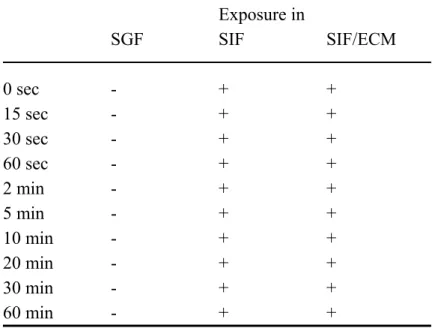
Purified enterotoxin from strain Cp 8 was mixed with simulated gastric fluid, with simulated intestinal fluid and with simulated intestinal fluid mixed 1:1 with ECM (= DMEM 10% without foetal calf serum and gentamicin). The times of exposure to each of the three media were 0, 15, 30 and 60 seconds and 2, 5, 10, 20, 30, and 60 minutes. All incubations were at 37 °C in a water bath. After incubation at 37 °C for the indicated time the sample was cooled down in melting ice until further evaluation.
After all samples had been exposed to the three types of simulated gastrointestinal fluid, the activity of the enterotoxin was tested in the cytotoxicity test with VERO cells.
3
Results
3.1
Spores in simulated gastric conditions.
The cumulative results of two separate experiments are shown in Figure 2.
From these results can be read that the spores are not affected by the 2-hour incubation at pH 2.5 in simulated gastric fluid. No significant differences were found between the counts at the different sampling times.
3.2
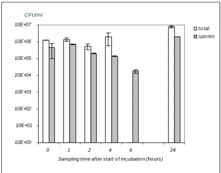
Behaviour of spores in simulated intestinal conditions.
The germination potential of C. perfringens spores in simulated intestinal fluid was investigated for spores from strain Cp 8. The results of these experiments are accumulated in Figure 3. The experiment was repeated since at T=6 no total count could be given due to contamination of the plates. In the repetition of the experiment no counts of total cells or spores were carried out: the experiment was followed by microscopic evaluation of samples at comparable times as in the first experiment. The reason for this difference in evaluation was the fact that within a limited number of days more experiments could be performed as no cultures were to be evaluated.
The microscopic evaluation did not show an increase in vegetative cells, nor a decrease of spores. This means that spontaneous sporulation in simulated intestinal fluid did not occur.
Addition to simulated intestinal fluid of various concentrations of L-alanine, a combination of
L-alanine and inosine, or glucose (all known inducers of germination for B. cereus 7) did not result
either in sporulation in simulated fluid.
Simulated intestinal fluid represents a poor medium, in which no germination was observed. In order to investigate whether this lack of sporulation was due to the medium, similar experiments were carried out in a rich medium, Tryptone Soy Broth (TSB). From Figure 4 can be concluded that only after 6 hours of incubation a substantial decrease in spore count was observed, but due to contamination of the plates for the total counts this decrease in spore count could not be linked to an increase in total count. However, such decrease in spore count was not observed in the repetition of the experiment with microscopic evaluation. Moreover, the addition of bile has a negative influence on the germination potential of the spores. No significant decrease in spore count is observed during the six hours of incubation.
Comparative experiments in LB 7.0 (another rich medium, but more defined than TSB) without and with the various additions of L-alanine, L-alanine and inosine, and glucose resulted in a visual increase of vegetative cells after 1 hour incubation at 37 °C (data not shown)
The addition of Tryptone, beef-extract, proteose peptone, and Bacto peptone to simulated intestinal fluid resulted in a slightly higher degree of germination compared to simulated intestinal fluid without extra supplements. The increased germination, however, did not match the germination potential in LB 2.0.
Neither pre-exposure to low pH (2.5) before germination assessment in simulated intestinal fluid, nor pre-exposure heat treatment increased the germination potential of spores from C. perfringens strain Cp 8 in simulated intestinal fluid (data not shown).
3.3
Behaviour of vegetative cells at pH-values representing gastric
conditions
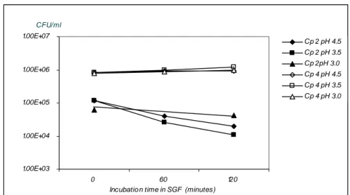
Vegetative cells from strains Cp 2 and Cp 4 were exposed to simulated gastric fluid pH 4.5, 3.5 and 3.0. The results are shown in Figure 5. Besides exposure to low pH in simulated gastric fluid, vegetative cells from strain Cp 4 were expose to low pH in LBG (pH 2.5, 2.0 and 1.5). The results of these experiments are show in Figure 6.
Since pH 2.0 appears to be a critical value in the survival of vegetative cells from C. perfringens, the survival of other C. perfringens strains at values around pH 2.0 was investigated as well. For these investigations strains 2 to 11 were used. The pH values used were pH 1.8, pH 2.0 and pH 2.2. These investigations, of which the results are shown in Figure 7, indicate differences in susceptibility for low pH conditions between the various C. perfringens strains. Some strains show a steep initial drop in numbers of cells directly after exposure to the low pH-conditions (Cp 2, 3, 5, 7, 8 and 9). After the initial drop in surviving numbers of cells the counts of strain Cp 5 are no longer affected at each of the pH values used in the experiments, while the number of surviving cells of Cp 2, 3, 7, 8 and 9 at each of the pH-values gradually decrease. The counts of strains Cp 10 and 11 remain similar at pH 2.0 during the time of the experiment while at pH 1.8 the counts markedly decrease, indicating the stability of these strains at pH 2.0
To investigate the influence of various food constituents LBG was supplemented with casein, peptone, Tryptone. Strain Cp 4 was exposed to these combinations during 2 hours. The results, shown in Figure 8 as a compilation of four separate experiments, indicate that beef and peptone have no protective influence, whereas Tryptone and caseine hydrolysate do protect Cp 4 against low pH conditions. The influence of glucose, a food constituent itself, was tested by testing the following combinations: preculture in LBG pH 7.0 > exposure in LBG pH 2.0, preculture in LBG pH 7.0 > exposure to LB pH 2.0 and preculture in LB pH 7.0 > LB pH 2.0. From the result shown in Figure 9 can be derived that glucose has some protective capacity against the low pH conditions.
Overall we conclude that stationary phase vegetative cells from C. perfringens are stable at low pH values until around pH 2.5. At lower values decay of the cells depends on the strain. In general, decrease in numbers may appear in the first few minutes of exposure to the low pH values after which stabilization occurs.
3.4
Behaviour of vegetative cells in simulated intestinal conditions
The effect of different initial total counts on growth and spore formation in simulated intestinal fluid was determined (Figure 10). When the incubation is started with approximately 107 CFU/ml, sporulation occurs after approxcimately 4 hours of incubation. When lower numbers of inoculation are applied sporulation is only seen after 24 hours of incubation. Unfortunately, the time between 6 and 24 hours of incubation was not covered. When starting with lower inoculation numbers than
107 CFU/ml, growth occurs but no detectable sporulation within the first 6 hours of incubation.
Comparison of a higher starting concentration, 108 CFU/ml, with the above used highest concentration
of 107 CFU/ml did not lead to a shorter sporulation time (data not shown). Experiments for investigation of the time span between 6 and 24 hours incubation showed that with a initial concentration of 105 CFU/ml sporulation started between 10 and 16 hours after start of the experiment. Comparison of sporulation capacity in simulated intestinal fluid (containing bile) and LBG 7.0 showed an earlier start of sporulation in SIF than in LBG 7.0. Whether sporulation in LBG 7.0 is started off earlier with the addition of bile remains to be investigated.
As simulated intestinal fluid is a quite poor medium and LBG 7.0 a quite rich medium the question arose whether similar effects as seen in simulated intestinal medium could be achieved in mineral medium, which as SIF consists of various salts mostly. However, similar 24-hour experiments as shown in Figure 10 carried out with mineral medium did not lead to results comparable with the experiment in simulated intestinal fluid (data not shown). Omission of glucose from the mineral medium did not lead to improvement of the sporulation capacity in this medium.
Beef-extract was used to mimic the presence of meat remnants in the simulated intestinal fluid. The growth potential, after starting with an initial concentration of approximately 104 CFU/ml, appears to increase with increasing beef extract concentration. This is reflected in the generation times between 4 and 8 hours of incubation as shown in Table 3.
The generation times were calculated as follows: generation time = log2 (t-t0) / logN – logN0
3.5
Adhesion capacity to differentiated Caco-2 cells
To determine whether C. perfringens adheres to differentiated Caco-2 cells, as a model for small intestinal epithelial cells, Caco-2 cells were exposed during 1 hour to cells. Subsequently the Caco-2 cells were washed several times. After each washing step a limited number of wells were treated with Triton to dissociate the Caco-2 cell monolayer and C. perfringens from the Caco-2 cells, after which the number of C. perfringens cells was determined. In this experiment we assume that in each washing step 90% of the supernatant was removed. Theoretically, if C. perfringens does not adhere, 90% of these cells would be removed in each washing step as well. In Figure 11 this is shown as the dotted line. As the numbers of recovered C. perfringens appear to follow the solid line in Figure 11, and more or less stabilizes after two washing steps, we conclude that C. perfringens indeed adheres to differentiated Caco-2 cells.
3.6
Enterotoxin production and quantification
Strains Cp 6, Cp 10 and Cp11 did not produce spore in DS-bile, and as enterotoxin production concurs with sporulation these strains were therefore assumed not to produce enterotoxin and are therefore not presented in Table 4. They were not further investigated with the latex test or the VERO cell-test. As can be derived from Table 4 Cp 2 and Cp 3 showed no reaction in the latex test or the VERO cell-test, and were therefore assigned non-producers for enterotoxin. Cp 4 produced only small amounts of enterotoxin as can be derived from the low latex titre and low enterotoxin concentration. The
differences in for strains Cp 5, 7, 8 and 9 are only illustrated by the results in the VERO-test in combination with the concentration of spores: Cp 9 produces the highest amount of enterotoxin per number of spores, while Cp 8 produces the least amount of enterotoxin per number of spores.
The minimal detectable concentration of enterotoxin in the latex test is approximately 2 ng ml-1. Based on this the enterotoxin concentration in the latex positive control is 64 ng ml-1. The provisional concentrations in the enterotoxin solutions from the various strains, based on the reaction of the positive control of the PET-RPLA kit, are also shown in Table 4. The reactions in the PET-RPLA kit should be repeated with more dilutions.
3.7
Stability of Cpe in simulated gastrointestinal fluids
Cytotoxicity testing of purified Cpe from C. perfringens strain Cp 8 exposed to simulated gastric fluid revealed that within seconds the activity of the enterotoxin had decreased to zero. However, in simulated intestinal fluid and simulated intestinal fluid mixed with ECM, Cpe remained active up to 1 hour, the longest incubation time applied. Moreover the VERO cell-test showed no visible decrease in activity of the Cpe with increasing incubation in simulated intestinal fluid.
4
Conclusions and discussion
From the results the following conclusions can be drawn:
• Vegetative cells survive simulated gastric conditions at pH values >2. • Spores survive simulated gastric conditions unharmed.
• Germination of spores in simulated intestinal conditions (poor medium) failed, while in Tryptone Soy broth (rich medium) ready germination was observed. This means that spores only contribute to the onset of disease symptoms if sufficient nutrients are present
• Bile inhibits germination.
• Bile enhances sporulation of some strains.
• At a starting concentration of 107 CFU/ml, no further growth seems to take place in simulated
intestinal conditions, while sporulation occurs after approximately 4 hours.
• At starting concentrations < 107 CFU/ml, growth appears to take place until a level of
approximately 107 CFU/ml.
• At starting concentrations < 107 CFU/ml, the addition of beef extract to simulated intestinal
fluid, mimicking the addition of meat components, decreases the generation time.
• Vegetative cells can adhere to differentiated Caco-2 cells as a model for small intestinal epithelial cells. This may extend the incubation time of the disease as vegetative cells may remain present in the small intestine for a longer period.
• Enterotoxin Cpe is not stable in simulated gastric fluid. Preformed enterotoxin plays no role in the onset of disease.
• Enterotoxin Cpe is stable in simulated intestinal fluid. • Enterotoxin production varies per strain.
The diarrhoeal syndrome by Clostridium perfringens is caused by enterotoxin Cpe produced upon sporulation of the micro organism in the small intestine. Investigation of events that lead from exposure to the onset of disease have been carried out using model systems for the gastrointestinal conditions, simulated gastric and intestinal fluids and differentiated Caco-2 cells, mimicking the small intestinal epithelium.
Both vegetative cells and spores survive simulated gastric condition, implying that both could take part in the pathogenesis of the diarrhoeal disease. Therefore, follow up investigations were carried out with spores and vegetative cells. In order to participate in the pathogenesis, spores must germinate before growth and sporulation in the small intestine. In our experiments, we were unable to induce (rapid) germination of spores. Not pre-exposure to acidic pH-conditions (mimicking exposure to gastric conditions), nor pre-heating of spores, nor the addition of known germinants, such as L-alanine and inosine, led to increased germination. We did observe in our experiments that bile can enhance sporulation, although not of all strains. This phenomenon, that a compound may enhance sporulation of a limited number of strains, has been observed before 2. Also we noticed that bile delayed growth of
vegetative cells. Possibly, bile does interfere with germination.
As far as vegetative cells are concerned, we observed that, at concentrations ≥ 107 CFU/ml, sporulation time is approximately 4 hours during exposure to simulated intestinal fluid. At starting concentrations
< 107 CFU/ml simulated intestinal fluid, first growth takes place before sporulation time is initiated. Since the incubation time of C. perfringens diarrhoeal disease is 8 – 24 hours, this implies that concentrations < 107 CFU/ml of C. perfringens may lead to the onset of the diarrhoeal syndrome, provided that the cells grow fast enough to reach concentrations ≥ 107 CFU/ml and sporulate within that time span. With this we assume that sporulation always takes place in the late logarithmic phase and that concentrations of ≥ 107 CFU/ml are necessary before sporulation can take place. We do not
take into account that lack of nutrients may induce sporulation at concentrations < 107 CFU/ml. This assumption implies that the growth rate of the contaminating strain is very important for the ability to sporulate within the previously mentioned incubation time.
C. perfringens is ingested with food, and reaches the small intestine with food components. Such food components may contribute to the growth and subsequent sporulation process. To mimic the presence of meat remnants, we added beef extract to the simulated intestinal fluid and noticed a decrease in generation time with increasing beef-extract concentration. This indicates that food components may well enhance growth and subsequent sporulation. Further investigations should be carried out to investigate what food components enhance growth and sporulation, and what do not. For such investigations an inventory on the type of products which are often contaminated with C. perfringens and an inventory on the type of products that are mostly related to food borne outbreaks should be used as a guide line.
Enterotoxin Cpe is stable in simulated intestinal conditions. The enterotoxin has its effect on the epithelial cells of the small intestine. Production of the enterotoxin in the lumen does not lead to decrease of toxicity of the enterotoxin, contrary to the enterotoxins of Bacillus cereus, which were very prone for proteolytic activity in simulated small intestinal conditions 13. For Bacillus cereus, adhesion to the epithelial cells was the method to circumvent the detrimental effect of proteolytic enzymes in the small intestine. Although C. perfringens cells are able to adhere to differentiated Caco-2 cells as a model for small epithelial cells, adhesion does not appear to be of vital importance for the onset of disease symptoms.
We studied the behaviour of vegetative cells from C. perfringens in simulated gastric and intestinal conditions separately. In none of the experiments vegetative cells were pre-exposed to conditions simulating the gastric passage or to conditions simulating food preparation before consumption, such as heating processes. Such experiments, mimicking the actual course of events from food preparation to consumption to the (possible) onset of diarrhoeal symptoms, are still to be carried out.
Most experiments were carried out with a very limited number of strains. From sporulation experiments and Cpe production data it is known that there is a large diversity in strains. The use of a larger number of strains for the experiments, and most preferably “extreme strains”, is recommended. Such a set of strains should include fast and slow growing strains, high and low Cpe producing strains, and good and poor sporulating strains.
When taking a closer look at the course of events from food preparation to the possible onset of diarrhoeal disease, it may be hard to imagine that spores would contribute to the onset of disease. The food commodities that are contaminated with C. perfringens nearly all require heating before consumption. When spores are present in these food commodities, they will most probably germinate upon heating. The number of spores at the time of consumption of the food will therefore be very small or even zero. Only vegetative cells will be ingested upon consumption of the contaminated food. As a
consequence, further investigations regarding the fate of spores in the gastrointestinal tract need not be carried out. What remains is investigation of the determination of the prevalence of spores in food commodities.
Our investigations indicate that spores are of minor importance for the onset of disease symptoms since we found hardly any germination in simulated intestinal conditions. Investigations of the Food and Consumer Product Safety Authority have shown that C. perfringens is found regularly in food commodities that still need heating before consumption Therefore it is important to investigate the ratio spore/vegetative cell not only in food commodities at retail level but also at the moment just prior to consumption, as the heating process may induce spores to germinate and grow in the food, thus altering the spore/vegetative cell ratio. The risk for disease may thus be better estimated.
5
Future work
Most of the work in conditions simulating the small intestinal environment as described in this report has been carried out with a very limited number of C. perfringens strains, sometimes one and sometimes two strains. Considering the probable diversity between different strains, which is reflected by the differences in Cpe producing potential, and considering the research question to provide a risk assessment for Clostridium perfringens in food in the Netherlands, it is advisable to investigate the behaviour of more strains. Since one of the most important parameters for the onset of disease is the ability to produce the enterotoxin Cpe, the most sensible manner is to investigate the behaviour of several strains with very high Cpe production potential and of several strains with very low production potential. Thus, the extremes with respect to Cpe production are investigated and can be incorporated into final calculations as worst and best case scenarios, respectively. A necessary condition is that the growth and sporulation potential of the strains is more or less equal in the media used for these investigations.
C. perfringens has been reported to have very short doubling times. All investigations for this project have been or will be carried out in simulated gastrointestinal conditions. Comparison of growth speed in artificial media and simulated intestinal media must give better insight in the usefulness of artificial media to predict behaviour of strains in simulated intestinal media.
Investigation of food commodities for the prevalence of C. perfringens must give insight in the presence of spores at the moment of consumption of the contaminated food. If spores are ingested as well, the ability of spores to take part in the onset of disease must be investigated more elaborate than described in this report.. An important issue in this respect is the germination capacity in simulated gastrointestinal media and the influence of food constituents in this process.
The results presented in this report derive from investigations carried out in either simulated gastric conditions or simulated intestinal conditions. To investigate the behaviour of C. perfringens in the different stages of the gastrointestinal tract and to mimic the flow of events in the human gastrointestinal tract as closely and realistic as possible, it is necessary to investigate the behaviour of C. perfringens strains in simulated intestinal conditions after exposure to simulated gastric conditions. Such investigations can be carried out batch wise or with continuous flow in a bioreactor. Also such investigations facilitate research of the possible influence of exposure to gastric conditions on the behaviour in intestinal conditions.
Figures and tables
Figure 1 Various stages of investigation of the gastrointestinal passage of Clostridium perfringens
.
1.0E+00 1.0E+01 1.0E+02 1.0E+03 1.0E+04 1.0E+05 1.0E+06
Cp 5 total Cp 5 spores Cp 8 total Cp 8 spores Strain number CFU/ml 0 min 30 min 60 min 90 min 120 min
Figure 2 Total counts and spore counts from C. perfringens strains Cp 5 and Cp 8 in samples taken from 2-hour incubation in simulated gastric fluid at pH 2.5.
1.0E+00 1.0E+01 1.0E+02 1.0E+03 1.0E+04 1.0E+05 1.0E+06 1.0E+07 0 1 2 4 6 24
Sampling time after start o f incubatio n (ho urs)
CFU/ml
to tal spo res
Figure 3 Germination of spores from C. perfringens strain Cp 8 in simulated intestinal fluid.
1.00E+00 1.00E+01 1.00E+02 1.00E+03 1.00E+04 1.00E+05 1.00E+06 1.00E+07 0 1 2 3 4 5 6 7
Sampling time after start of incubation (hours)
CFU/ml
To tal Spo res To tal with bile Spo res with bile
1.00E+03 1.00E+04 1.00E+05 1.00E+06 1.00E+07 0 60 120
Incubatio n time in SGF (minutes)
CFU/ml Cp 2 pH 4.5 Cp 2 pH 3.5 Cp 2pH 3.0 Cp 4 pH 4.5 Cp 4 pH 3.5 Cp 4 pH 3.0
Figure 5 Effect of simulated gastric fluid at pH 4.5, 3.5 and 3.0 on survival of vegetative cells from C. perfringens strains Cp 2 and Cp 4. 1.0E+00 1.0E+01 1.0E+02 1.0E+03 1.0E+04 1.0E+05 1.0E+06 0 1 5 10 15 30 45 60 90 120 Incubatio n time (min)
CFU/ml
pH 1.5 pH 2.0 pH 2.5
Cp 2 1 10 100 1000 10000 100000 1000000 0 20 40 60 80 100 120 140
Incubation time (min)
CFU/ml LBG-pH1,8 LBG-pH2,0 LBG-pH2,2 Cp 5 1 10 100 1000 10000 100000 1000000 0 20 40 60 80 100 120 140
Incubation time (min)
CFU/ml LBG-pH1,8 LBG-pH2,0 LBG-pH2,2 Cp 7 1 10 100 1000 10000 100000 1000000 0 20 40 60 80 100 120 140
Incubation time (min)
CFU/ml LBG-pH1,8 LBG-pH2,0 LBG-pH2,2 Cp 8 1 10 100 1000 10000 100000 1000000 0 20 40 60 80 100 120 140
Incubation time (min)
CFU/ml LBG-pH1,8 LBG-pH2,0 LBG-pH2,2 Cp 9 1 10 100 1000 10000 100000 1000000 0 20 40 60 80 100 120 140
Incubation time (min)
CFU/ml LBG-pH1,8 LBG-pH2,0 LBG-pH2,2 Cp 10 10 100 1000 10000 100000 1000000 0 20 40 60 80 100 120 140
Incubation time (min)
CFU/ml LBG-pH1,8 LBG-pH2,0 LBG-pH2,2 Cp 11 1 10 100 1000 10000 100000 1000000 0 20 40 60 80 100 120 140
Incubation time (min)
CFU/ml LBG-pH1,8 LBG-pH2,0 LBG-pH2,2 Cp 3 1 10 100 1000 10000 100000 1000000 0 20 40 60 80 100 120 140
Incubation time (min)
CFU/ml
LBG-pH1,8 LBG-pH2,0 LBG-pH2,2
1.0E+00 1.0E+01 1.0E+02 1.0E+03 1.0E+04 1.0E+05 1.0E+06 1.0E+07 0 30 60 90 120
Incubation time (minutes)
CFU/ml
pepto ne trypyto ne caseine hydro lysate beef
Figure 8 Influence of various food constituent mimicking supplements to LBG pH 2.0 on survival of vegetative cells from C. perfringens strain Cp 4.
1.0E+00 1.0E+01 1.0E+02 1.0E+03 1.0E+04 1.0E+05 1.0E+06 1.0E+07 0 30 60 90 120
Incubation time (minutes)
CFU/ml
LB G 7 > LB G 2 LB G 7 > LB 2 LB 7 > LB 2
1.0E+00 1.0E+01 1.0E+02 1.0E+03 1.0E+04 1.0E+05 1.0E+06 1.0E+07 1.0E+08 0 2 4 6 24 0 2 4 6 24
Incubation time (hours)
CF U/ m l 1.0E+04 1.0E+05 1.0E+06 1.0E+07
Figure 10 Influence of initial number of vegetative cells from C. perfringens strain Cp 8 on growth and sporulation in simulated intestinal fluid (left = total counts, right = spore counts). In the legend the initial total counts are mentioned.
1.00E+00 1.00E+01 1.00E+02 1.00E+03 1.00E+04 1.00E+05 1.00E+06 1.00E+07 1.00E+08 1 2 3 4 5 6 7 8 Washing step CFU/ml Trito n Theo retical
Figure 11. Effect of washing of differentiated Caco-2 cells after incubation with C. perfringens during 1 hour. Solid line is measured numbers of C. perfringens, dotted line represents the theoretical numbers.
Table 1
.
Origin of strainsRIVM-no Provided by Original number Sample ID* Food commodity
Cp 2 VWA (South)# C0009 38202693 Meat balls in spicy sauce
Cp 3 VWA (South) C0011 38088483 Meat in curry sauce
Cp 4 VWA (South) C0012 25938186 Salmon
Cp 5 VWA (South) C0031 37412686 Potato dish
Cp 6 VWA (South) C0045 37804088 Poultry dish
Cp 7 VWA (South) C0057 38520458 Fried rice
Cp 8 VWA (South) C0058 38793012 Poultry dish
Cp 9 VWA (South) C0059 38793004 Pasta dish
Cp 10 VWA (South) C0198 48186882 Peanut sauce
Cp 11 VWA (South) C0200 48186955 Egg dish
*= number from ISI-database of the Food and Consumer Product Safety Authority # = Food and Consumer Product Safety Authority section South.
Table 2
.
Combinations of media and supplements used for germination experiments with C. perfringens Cp8 no supplement L-alanine L-alanine + inosine glucose bileLBG X X X X
SIF X X X X
TSB X X
beef extract Tryptone proteose peptone Bacto peptone LBG
SIF X X X X
Table 3. Decrease in generation time with increasing beef extract concentration in simulated intestinal fluid
CFU/ml
log CFU/ml gen. time (hrs)*
T=4 T=8
T=4 T=8
0 1E4
1E4
4 4
-
4 1E4
2.5E4
4 4.4
3.0
8 1E4
1.2E5
4 5.08
1.11
16
1E4
2.2E5
4 5.34
0.90
* = gen. time (generation time) = log2 (t-t0) / logN – logN0
Table 4 Dilutions of the enterotoxin solutions from the various C. perfringens strains positive in the PET-RPLA test and the VERO cell-test and the calculated enterotoxin concentration
Enterotoxin Dilution causing
concentration# 50% inhibition in Cp spores Cp total
Strain Latex test (ng ml-1) VERO cell-test #/ml #/ml
Cp 2 < 1:2 < 2 < 1:8 Cp 3 < 1:2 < 2 < 1:8 Cp 4 1:128 128 n.d. Cp 5 > 1:2048 2,048 n.d. 1.7x10⁷ 1.5x10⁷ Cp 7 > 1:2048 2,048 1:5790 1.3x10⁷ 3.6x10⁶ Cp 8 > 1:2048 2,048 1:4460 2.5x10⁷ 5.5x10⁶ Cp 9 > 1:2048 2,048 1:6890 6.6x10⁶ 1.8x10⁶ Latex pc* 1:64 64 1:11
* = latex pc = positive control from the PET-RPLA kit
# = enterotoxin concentration based on positive control from the PET-RPLA kit (see text).
Table 5 Cytotoxicity of Cpe 8 against VERO cells after incubation in simulated gastric fluid (SGF), simulated intestinal fluid (SIF), and a mixture of ECM and simulated intestinal fluid (SIF/ECM)
Exposure
in
SGF
SIF
SIF/ECM
0
sec
- +
+
15
sec
- +
+
30
sec
- +
+
60
sec
- +
+
2
min
- +
+
5
min
- +
+
10
min
- +
+
20
min
- +
+
30
min
- +
+
60
min
- +
+
References
1. Cornillot E, Saint-Joanis B, Daube G et al. The enterotoxin gene (cpe) of Clostridium perfringens can be chromosomal or plasmid-borne. Mol Microbiol 1995; 15(4):639-47. 2. De Jong AEI, Beumer RR, Rombouts FM. Optimizing sporulation of Clostridium perfringens. J
Food Prot 2002; 65:1457-62.
3. De Wit MAS, Koopmans MPG, Kortbeek LM et al. Sensor, a population-based cohort study on gastroenteritis in the Netherlands: incidence and etiology. Am J Epidemiol 2001; 154:666-74.
4. Duncan CL, Strong DH. Improved medium for sporulation of Clostridium perfringens. Appl Microbiol 1968; 16(1):82-9.
5. Granum PE, Richardson M. Chymotrypsin treatment increases the activity of Clostridium perfringens enterotoxin. Toxicon 1991; 29(7):898-900.
6. Health Council of the Netherlands. Foodborne infections in the Netherlands. The Hague, the Netherlands: Health Council of the Netherlands, 2000; Publication no. 2000/09.
7. Hornstra LM, de Vries YP, de Vos W, Abee T. Characterization of the germinant receptors in Bacillus cereus ATCC 14579. Appl Environ Microbiol 2006; 72(1):44-53.
8. Labbe R. Clostridium perfringens. Doyle MP, Editor. Foodborne bacterial pathogens. New York: Marcel Dekker Inc., 1989: 191-234.
9. Mahony DE, Gilliat E, Dawson S, Stockdale E, Lee SH. Vero cell assay for rapid detection of Clostridium perfringens enterotoxin. Appl Environ Microbiol 1989; 55(9):2141-3.
10. McClane BA. An overview of Clostridium perfringens enterotoxin. Toxicon 1996; 34(11/12):1335-43.
11. McClane BA. Clostridium perfringens. Doyle MP, Beuchat LR, Montville TJ, Editors. Food Microbiology: fundamentals and frontiers. 1st edition. Washington DC: ASM Press, 1997: 305-26.
12. Sacks LE, Thompson PA. Clear, defined medium forthe sporulation of Clostridium perfringens. Appl Environ Microbiol 1978; 35(2405-410).
13. Wijnands LM, Zwietering MH, Van Leusden FM, Abee T. Caco-2 cell surface-associated growth of enterotoxic B. cereus is a prerequisite for initiation of cytotoxic effects in simulated intestinal fluid. Submitted for publication.
RIVM
National Institute for Public Health and the Environment P.O. Box 1