Evaluating the feasibility to perform
vagus nerve stimulation in a rodent
model for glioma-related epilepsy
Delphine Dhont
Student number: 01508685Promotor: Prof. Dr. R. Raedt
Co-Promotor: Prof. Dr. K. Vonck
Mentor: PhD C. Bouckaert
A dissertation submitted to Ghent University in partial fulfilment of the requirements for the degree of Master of Biomedical Sciences
2
PREFACE
Ever since I was a kid I wanted to be a doctor, but fate decided otherwise. Biomedical Sciences was the best thing that could ever happen to me. Especially in the last two years where I discovered neurological research and sciences. My motivation, curiosity and ambition to become a neurological researcher only grew.
First, I would like to thank my promotor Prof. Dr. Robrecht Raedt to give me the opportunity to investigate the LGG rodent model as a project during my master thesis. This would not have been possible without the guidance of Jeroen Verhoeven. Thank you Jeroen for learning me everything about cell cultures and taking care of my cells in the weekends.
Secondly, I would like to thank my mentor Charlotte Bouckaert. She fulfilled her role as mentor excellently and guided me through two fascinating master years. She gave me a lot of opportunities and trust in doing experiments independently . Thank you Charlotte for believing in me and being such an amazing mentor.
Further, I would like to thank the whole 4BRAIN research group and my colleague students. Thank you for the wonderful ambiance in the lab. In particularly, I would also like to thank Charlotte Germonpré, Marie Goossens, Latoya Stevens and Jana Desloovere for helping me out when Charlotte Bouckaert was not available.
Last but not least, a special thank you for my family, boyfriend and friends to support me and dealing with my stress during the last two years. Without you all it was not possible to have such two amazing master years.
3 TABLE OF CONT ENTS
Preface ... 2
Table of contents ... 3
Summary ... 6
Influence of Sars-CoV2 ... 7
1. VNS experiment ... 7
2. Low-grade glioma model ... 7
Introduction ... 8 1. Glioma-related epilepsy ... 8 Glioma tumours ... 8 Epileptogenesis of gliomas ... 9 1.2.1 Tumourocentric hypothesis ... 9 1.2.2 Epileptocentric hypothesis ... 9 Glutamatergic mechanisms ... 9 GABAergic mechanisms ...10
Role of inflammation in glioma growth ...11
Standard of care for high-grade gliomas ...11
Standard of care for low-grade gliomas ...12
Glioma-related epilepsy ...12
2. Vagus nerve stimulation ...13
Vagus nerve stimulation for medication-resistant epilepsy ...13
Anti-inflammatory effect of vagus nerve stimulation ...15
3. Glioblastoma-related epilepsy rodent model ...16
4. VNS as treatment for glioma-related epilepsy? A hypothesis ...16
5. Low-Grade glioma rodent model ...17
Res186: Pilocytic Astrocytoma grade I ...18
Res259: Diffuse astrocytoma grade II ...18
Markers for low-grade gliomas...18
Materials and methods ...20
1. Feasibility to perform VNS in a GB-related epilepsy rodent model ...20
Cell culture ...20
Animals ...20
Surgery ...20
1.3.1 VNS implantation ...21
1.3.2 Inoculation and implantation of EEG electrodes ...21
EEG monitoring and VNS stimulation ...23
4 Blood samples ...24 Histology ...24 2. Low-grade glioma ...25 Cell culture ...25 Animals ...25 2.2.1 Non-immunocompromised rats ...25
2.2.2 T cell deficient rats ...26
Histology ...26
2.3.1 Immunocytochemistry ...26
2.3.2 In vivo low-grade glioma model ...27
Results ...29
1. Feasibility of VNS in a glioblastoma-related epilepsy rodent model ...29
VNS stimulation and EEG analyses ...29
Histology ...29
Blood samples ...29
2. Low-grade glioma ...30
Immunocytochemistry ...30
In vivo low-grade glioma model ...31
2.2.1 Non-immunocompromised rats ...31
2.2.2 T cell deficient rat ...31
Discussion ...32
1. Feasibility of VNS as treatment for glioma-related epilepsy? ...32
2. Low-grade glioma rodent model ...33
References ...37
Poster ...44
Addendum ...45
1. Cell passage ...45
2. Low-Grade glioma experiments ...45
In vivo ...45
2.1.1 Coordinates of inoculation of RNu rat ...45
2.1.2 Weight follow-up of the animals ...46
3. Tumour volumes ...48
VNS rat 1 ...48
SHAM rat 1 ...49
VNS rat 2 ...50
Tumour volumes of all animals ...51
5 5. Abbreviations ...52
6
SUMMARY
Background. 50-90% of low-grade glioma (LGG) patients and 20-60% of high-grade glioma
patients have epileptic seizures. Additional treatment with anti-epileptic drugs can have severe side effects. 1. Immune cells of the tumour micro-environment express α7nAchR. Thereby, vagus nerve stimulation (VNS) could influence glioma progression via the cholinergic anti-inflammatory pathway. Feasibility to perform VNS in a glioblastoma-related epilepsy rat model was investigated. 2. A potential LGG model was investigated since LGG patients present more often with seizures.
Methods. 1. MaleF344/IcoCrl rats were implanted with a VNS electrode and EEG recording
electrodes, and F98 glioblastoma cells were inoculated. From day 7 to 14 post-inoculation brain activity was monitored. At day 10, VNS stimulation (30Hz, biphasic pulses (250µs/phase), 0.5sec ON/29.5sec OFF) was initiated in half of the animals at an intensity of 250μA and increased daily in steps of 250μA until day 13. 2. Grade I and II glioma cells were injected in three non-immunocompromised rats and one RNu rat and immunocytochemistry was performed on both cell lines.
Results. 1. Stimulated animals had tumour volumes of 11.66 and 8.18mm3 and one
non-stimulated animal had a tumour volume of 16.96mm³. Only one out of six animals had seizures.
2. Both cell lines stained negative for glial fibrillar acidic protein and no tumour growth was
obtained.
Discussion. 1. So far our data is still inconclusive regarding the effect of VNS on glioma
progression and seizures. Further experiments with longer EEG monitoring are needed. 2. Further studies should first investigate the characteristics of long cultured cells.
7 INFLUENCE OF SARS-COV2
1. VNS experiment
Normally, the blood samples that were taken from the animals at several time points in the study (mentioned in material and methods) would have been analysed for specific markers. This was not possible due to the Sars-CoV-2 outbreak. The results obtained from the analyses of the blood samples would have helped to estimate the effect of tumour growth, VNS and seizures on certain inflammatory parameters. Secondly, more VNS studies would have been conducted if there was no Sars-Cov-2 outbreak. Thereby, we possibly would have had significant results or a better idea about the effects of VNS on glioma progression and glioma-related seizures. Additionally, we could have adjusted our studies, for example longer EEG monitoring. Without these additional studies it was not possible to make a significant conclusion about the effects of VNS on glioma progression and glioma-related epilepsy in this master thesis.
The Sars-CoV-2 outbreak could also have influenced the quality of slicing as the slices of all animals were made in one day. This was needed to perform the cresyl violet staining of all animals in one day and thereby minimize the time in the lab.
2. Low-grade glioma model
For the in vitro experiments and the immunocytochemistry staining, the F98 cells could not be stained for vimentin. This would have given additional information in the comparison between the LGG cell lines and the F98 cell line and could have concluded the low-grade characteristics of the Res186 and Res259 cells.
For the in vivo experiments it was not possible to inject the RNu rat with 100 000 Res259 cells in 5μl PBS in the left and right entorhinal cortex. This would have given a better estimation about the possible tumour growth of the Res259 cells in a T cell deficient rat and could have been one step closer towards an LGG rodent model. Furthermore, an ex vivo MRI would have been carried out in the perfused RNu rat. The animal was perfused from the moment the government measures were in place and no further experiments could be done. The idea was to make an ex vivo scan before the deadline of this master thesis, to exclude tumour growth with certainty. It was not possible to perform an ex vivo MRI in time due to ongoing Sars-CoV-2 outbreak and government measures.
8
INTRODUCTION
1. Glioma-related epilepsy
Glioma tumours
Gliomas are the most prevalent primary intracranial tumours, representing 81% of all malignant brain tumours in human1. The overall age adjusted incidence of gliomas is 4.67-5.73 per 100
000 persons worldwide and these tumours are more prevalent in males1,2. Gliomas are derived
from supportive non-neuronal glial cells within the central nervous system (CNS). Non-neuronal glial cells include different cell types, each fulfilling their own function within the CNS. Firstly, astrocytes are responsible for neuronal-glial communication, synaptic signalling, maintenance of the blood-brain barrier (BBB) and have a trophic, metabolic and structural support for neurons3. Secondly, oligodendrocytes play a key role in the production of the myelin
sheath that insulates axons and is responsible for the fast information transduction of neurons4.
Thirdly, ependymal cells are involved in the production of cerebrospinal fluid (CSF) and are an essential component of the blood-brain and brain-CSF barrier5. Finally, microglia function as
the first form of active immune defence within the CNS6.
Furthermore, gliomas are classified according to their presumed non-neuronal glial cell of origin (“astrocytoma”, “oligodendroglioma”, “ependymoma” and “glioblastoma”) and according to their grade (low-grade vs. high-grade gliomas), which will determine their pathology and progression7. Low-grade gliomas (LGGs), World Health Organisation (WHO) grade I and II,
are not anaplastic meaning they will not lose their cellular differentiation8. Grade I gliomas
occur mainly in children and are together with grade II gliomas characterised by slow growth and progression but often possess a malignant transformation over time2. The prognosis and
overall survival of LGGs is partly defined by mutations in the isocitrate dehydrogenase (IDH) genes 1 and 2 which are recognised as an early marker for glioma genesis. Patients suffering from LGGs containing IDH gene mutations have an improved prognosis compared to patients without IDH mutations2. High-grade gliomas (HGGs), WHO grade III and IV, are anaplastic
meaning they will lose their cellular differentiation during disease progression9.
Therefore, HGGs have a worse prognosis compared to LGGs and grade IV glioma or glioblastoma (GB) possesses the worst prognosis and survival2. Furthermore,
epigenetic silencing of the methyl-guanine methyl transferase (MGMT) gene promotor due to methylation is a crucial prognostic factor in HGGs and LGGs for overall and progression free survival2,10.
Methyl-guanine methyl transferase is an enzyme responsible for DNA repair and patients who are diagnosed with a methylated MGMT gene promotor will therefore have a better response to chemotherapy than patients with an unmethylated MGMT gene
promoter. Additionally, the prognosis of LGG and especially these with oligodendroglial components is strongly influenced by the presence of the loss of heterozygosity (LOH) or co-deletion of 1p/19q2,11. The co-deletion or LOH of 1p/19q is established by the chromosomal
translocation between chromosome 1 and 192. Patients which are diagnosed with LGG or
oligodendrogliomas containing this co-deletion or LOH have a prolonged survival without treatment and a higher sensitivity for radio- and chemotherapy2,11. Finally, the expected
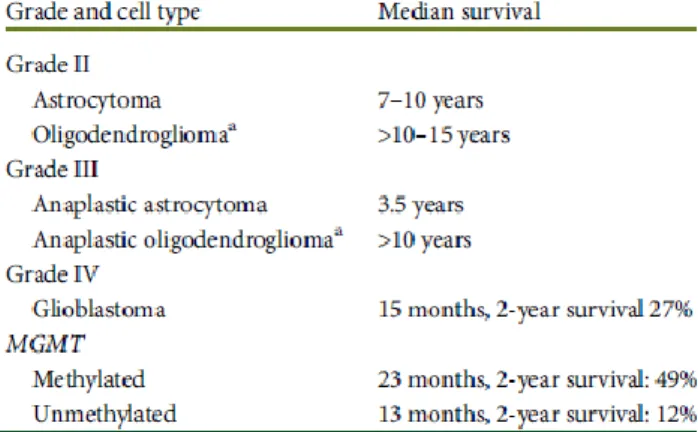
survival of glioma patients based on grade and origin is illustrated in Figure 1.
Figure 1. Expected survival of gliomas based on grade and origin and some molecular markers. Figure from Stupp et al., 2014.
9
Epileptogenesis of gliomas
The most common symptom in patients with gliomas is the manifestation of epileptic seizures12. Therefore, gliomas are responsible for the phenomenon of glioma-related epilepsy
according to the International League Against Epilepsy (ILAE)13. An epileptic seizure is defined
by the ILAE as a transient occurrence of signs and/or symptoms due to abnormal or synchronous neuronal activity in the brain. Epilepsy can be defined as a chronic disease characterised by recurrent seizures13. Patients with LGGs are more vulnerable to epileptic
seizures (50-90%) than patients with HGGs (20-60%)8. The lower vulnerability to seizures in
HGGs is probably due to their characteristics of rapid and invasive tumour growth which might preclude epileptogenesis and some patients will not survive long enough to develop these glioma-related seizures14.
The mechanism and origin of glioma-related epilepsy is not clear yet. Two epileptogenic approaches are considered for the moment.
1.2.1 Tumourocentric hypothesis
Gliomas are space-occupying brain tumours and can therefore contribute to epileptic activity15.
The mechanical effects of mass and oedema are responsible for microcirculation impairments by local hypoperfusion which is responsible for ischemic changes in the surrounding neocortex. LGGs, characterised by slow growth and invasion of the surrounding brain tissue, are responsible for isolation and deafferentation of the cortico-subcortical local and distant networks, resulting in epileptogenesis15. Conversely, HGGs are characterised by rapid and
invasive tumour growth and neoangiogenesis will occur in these gliomas, meaning new blood vessels will be formed. This process will be responsible for acute tissue damage such as haemorrhage and necrosis. Furthermore, the BBB of these newly formed blood vessels will become permeable to pro-epileptogenic molecules and immune cells. In the end, these processes might contribute to the epileptogenicity of gliomas.
1.2.2 Epileptocentric hypothesis
The epileptocentric hypothesis is based on dysregulation of the equilibrium between excitatory and inhibitory signals in the brain15. This hypothesis can be clarified by two different
assumptions namely the glutamatergic mechanisms or the GABAergic mechanisms.
Glutamatergic mechanisms
Firstly, gliomas express an upregulated amount of the Xc- cystine glutamate transporter, a sodium and chloride dependent antiporter of the anionic forms of cystine and glutamate (Glu)16. The mechanism of
action of this transporter is illustrated in Figure 216.
Although this transporter can exchange the molecules in both directions, the transport will only take place in one direction for each molecule. This one-way transport is due to the low intracellular concentration of cystine, caused by the immediate metabolization into cysteine after being transported into the neuron and the low extracellular concentration of Glu (2-9 μM) (which is) necessary for optimal neurotransmission. Therefore, cystine will be transported intracellularly for a 1:1 counter transport of glutamate extracellularly. Once cystine is transported into the cell it will be
immediately converted to cysteine, which is further used for the synthesis of antioxidant glutathione (GSH)15. GSH is the most important antioxidant within the brain. GSH will reduce
high levels of free radicals like reactive-oxygen species (ROS), which are produced by oxidative phosphorylation in the mitochondria during production of adenosine triphosphate
Figure 2. System Xc- is composed of the 4F2 heavy chain and the xCT light chain, which are linked by a disulfide bond. System Xc- imports cystine (CySS-) in exchange for glutamate (Glu). Figure from Lewerenz et al., 2013.
10 (ATP)17. This oxidative stress is responsible for oxidative modification of lipids, proteins and
DNA, resulting in cell death16. High concentrations of extracellular Glu can inhibit this Xc-
transporter resulting in the inhibition of the GSH synthesis18. Thereby excessive Glu
concentrations are responsible for oxidative glutamate toxicity. The excessive expression of this Xc- transporter will protect the glioma cells against their excessive Glu release and the related oxidative glutamate toxicity. Secondly, some gliomas lack the excitatory amino acid transporters (EAATs)15. These EAATs fulfil the key role in the rapid re-uptake of released Glu
in the synaptic cleft. There exist five different types of EAATs. Glioblastomas and astrocytomas lack EAAT1 and EAAT2 which are expressed by astrocytes for the immediate re-uptake of Glu16. The mechanism of action of these transporters is based on the co-transport of Na+ ions
and protons for each molecule of Glu that is transported into the cell and the counter transport of K+ ion extracellularly. Therefore, the EAATs are capable of transporting Glu against its high
concentration gradient. Taken together, gliomas can be responsible for the high extracellular concentration of Glu due to the lack of EAATs for Glu re-uptake and the overexpression of Xc- transporters. Thereby more excitatory signals will appear in the environment of a glioma resulting in an imbalance between the inhibitory and excitatory signals in the brain. Eventually, this phenomenon will give rise to epileptogenicity.
GABAergic mechanisms
The imbalance between inhibitory and excitatory signals is observed in multiple epileptic disorders15. Gliomas are characterised by the excessive expression of gamma-aminobutyric
acid (GABA) receptors, which contribute to the fast volume reduction of the cells. This process is required for proliferation and migration of gliomas. Furthermore, reduced GABAergic inhibitory pathways are observed in the peritumoral cortex15. This reflects a reduction in
inhibitory GABA interneurons and inhibitory synapses on pyramidal cells around the tumour. GABAa receptors are ion channels responsible for hyperpolarisation of neurons by influx of chloride ions after binding of GABA on the receptor19. The excessive expression of GABAa
receptors in glioma cells is responsible for crucial changes in the chloride homeostasis and partly engaged in the increased intracellular chloride concentration needed for fast volume reduction15. Increased intracellular chloride concentrations will create an outwardly driven force
for the movement of chloride followed by water20. When the outward flow of chloride is inhibited
in proliferating cells, there will be no condensation of chromatin into chromosomes. This process is needed for cell division. Therefore, the fast volume reduction due to the high intracellular chloride concentrations and the high chloride conductance of glioma cells will provide a signal to the glioma cell to continue cell division. This way, stimulation of the GABAa receptor can enhance glioma progression. Furthermore, the Na+/K+/2Cl- co-transporter 1
(NKCC1) is highly expressed in glioma cells and their peritumoral cortex15. These NKCC1
transporters are accountable for high intracellular chloride concentrations which are also typically expressed during neuronal development15,19. These high intracellular
chloride concentrations in the peritumoral neurons will influence the reversal potential of chloride, resulting in the disappearance of the GABAergic inhibitory effect19. Additionally, the
expression of the potassium-chloride (KCC2)
co-transporter is decreased in glioma cells and their peritumoral cortex. This co-transporter is
Figure 3. GABAergic role in epileptogenesis by the depolarising effect of GABA. Depolarisation is possible due to high intracellular Cl- concentration in
response to upregulation of NKCC1 co-transporters and downregulation of KCC2 co-transporters. Higher intracellular chloride concentrations will increase the reversal potential of chloride. Image created using BioRender.
11 in healthy circumstances accountable for low intracellular chloride concentrations and protection against neurotoxicity and overexcitability15,19. The overexpression of NKCC1 and
decreased expression of KCC2 transporters in glioma cells and peritumoral neurons are mediated by brain-derived neurotrophic factor (BDNF) release of glioma cells or activated microglia15. However, stimulation of GABAa receptors in the peritumoral neurons which have
increased expression of NKCC1 and decreased expression of KCC2 transporters will result in depolarisation of the cell and will stimulate epileptogenesis15. The GABAergic role during
epileptogenesis is illustrated in Figure 3.
Role of inflammation in glioma growth
Inflammation is crucial in cancer development and progression21. Even more, the role for
inflammation in tumorigenesis is now generally accepted and the inflammatory tumour microenvironment (TME) has become an essential component of all tumours. Tumour associated macrophages (TAMs) are by far the most important immune cells within the TME of GB and are present at all stages of tumour progression21,22. TAMs are proven to promote
GB growth, invasion and metastasis by the release of tumour necrosis factor α (TNF-α) and interleukin-6 (IL-6)22,23. Furthermore, the secreted TNF-α in TME plays a crucial role in cancer
progression21. In general, two different phenotypes of TAMs can be distinguished namely the
M1 and M2 phenotype.
Firstly, the M1 phenotype functions as potent killer of pathogens or tumour cells and is mainly activated after TLR-4 or IFN-γ binding21,22. The absence of M1 orientating signals in the TME
was already early linked to increased tumour growth in vitro and in vivo in experimental animal models23. But, the associated pathways of M1 TAM phenotype have recently been associated
with glioma growth for example by the release of IL-1β22. Secondly, the M2 phenotype is
generally associated with a pro-tumoral role and activated by IL-4, IL-10 and IL-13 exposure21,22. In the TME of GB, the TAMs mostly resemble the M2 phenotype which can
promote tumour progression by the release of growth factors, immunosuppressive molecules, chemokines and cytokines21. Inhibition of the nuclear factor ĸB (NF-ĸB) results in the inversion
of the pro-tumoral M2 phenotype into the anti-tumoral M1 phenotype. Thereby the maintenance of the M2 phenotype is suggested to be regulated by NF-ĸB21.
Standard of care for high-grade gliomas
High-grade gliomas such as GB are currently treated multidisciplinary24. Multidisciplinary
treatment includes the combination of maximal surgical resection of the tumour, radiotherapy (RT) and chemotherapy with temozolomide (TMZ). Firstly, new patients diagnosed with GB will undergo surgery but pre-operative issues such as medical condition, neuropsychological state of patient and the use of corticosteroids or anti-epileptic drugs (AEDs) should be considered. Corticosteroids control cerebral oedema and signs of intracranial hypertension. In this way the administration of corticosteroids will improve brain conditions for surgical resection of the GB24. The goal of surgical resection is the realisation of maximal safe resection,
obtaining tissue for pathological diagnosis, improve conditions for complementary treatment, delaying clinical worsening and the improvement of quality of life of the patient. Secondly, complementary treatment including TMZ and RT is applied after maximal safe resection of the tumour and followed by a maintenance treatment including six cycles of TMZ. Combined treatment of TMZ and RT after surgical resection has an improved effect on overall survival and progression free survival compared to RT alone24. Temozolomide is an oral
chemotherapeutic drug which induces DNA methylation and tumour cytotoxicity through blocking the cell cycle. Furthermore, the cytotoxicity of TMZ is apparent by the formation of O6-methylguanine DNA which is repaired by the enzyme MGMT. Additionally, the primary resistance mechanisms to TMZ in GB will be dependent on MGMT activity within the tumour. In the end, when tumours recur, treatment options include supportive care, reoperation, re-irradiation, systemic therapies and combined modality therapy24.
12
Standard of care for low-grade gliomas
Since patients who suffer from LGGs have a longer overall survival, considerations of treatment toxicity as consequence of radio-
and chemotherapy have to be taken into account25. In patients with LGG, the decision
for surgical resection is more complicated than in patients with HGG. Only if a patient shows clear signs due to the mass effect of the tumour or has uncontrolled seizures, surgical resection will be considered. Controversially, many LGGs are discovered by accident during imaging of the brain for other reasons such as headaches and traumas and are therefore asymptomatic. The decision for surgical resection of an asymptomatic LGG is much more complicated25. The same conclusion as in
surgical resection of HGGs can be made for
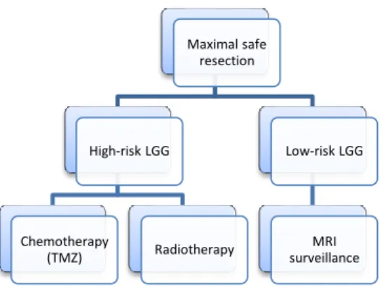
LGGs. The greater the extent of resection, the lower the chances of recurrence of the tumour and malignant transformation. Therefore, patients with LGGs are recommended to undergo the greatest degree of safe surgical resection. Furthermore, treatment after surgical resection is managed for each patient individually and risk-benefit ratio of RT and chemotherapy is considered. It is crucial that patients undergo long-term surveillance after surgical resection because the tumour can recur. Therefore, patients will undergo further treatment including RT, chemotherapy and/or regular magnetic resonance imaging (MRI) surveillance (Figure 4)25.
Radiotherapy is not without the risk of short- and long-term adverse effects including fatigue, cognitive decline and memory deficits. In general, combination therapy of RT and adjuvant TMZ chemotherapy also results in an improved overall survival. In the end, the prognosis of patients suffering from LGGs is affected by different factors and varies from two years to decades25. Therefore, quality of life and neurocognition of the patients will play a crucial role
in treatment decision making.
Glioma-related epilepsy
Glioma-related seizures are often resistant to AEDs or tumour resection2. The difficulties of a
successful treatment in these patients is defined by two factors26. Firstly, the number of patients
who develop medication-resistant epilepsy is approximately 30-40% of the LGG patients and 15-25% of the GB patients. Seizures within these patients cannot be controlled by administration of AEDs26,27. A well-considered reason for the development of
medication-resistant epilepsy is the overexpression of the ATP-binding cassette (ABC) transporter family proteins such as P-Glycoprotein (MDR-1), MRP1 and MRP5 in glioma patients26. These
transporter proteins function as an efflux pump in the endothelial cells of the BBB and will transport lipophilic drugs out of the brain parenchyma into the systemic circulation. Since most of the AEDs administered to glioma patients function as a substrate for these ABC transporter proteins, an insufficient concentration of AEDs remains within the brain parenchyma. However, levetiracetam and valproic acid are no substrates for these proteins and are therefore preferred to treat medication-resistant epilepsy in glioma patients26.
Secondly, there is a risk for drug-drug interactions during treatment which can result in insufficient control of seizures or tumour activity and drug toxicity in glioma patients26. On the
one hand, first generation AEDs (phenytoin, carbamazepine, phenobarbital, primidone and their derivates) are known as strong inducers of the hepatic metabolism, namely the cytochrome P450 (CYP) co-enzyme system responsible for the degradation of pharmaceuticals2,26. Therefore, administration of these AEDs in glioma patients also receiving
other treatments can result in insufficient plasma levels of these medications. Besides this, hepatic degradation of pharmaceuticals is also managed through the
UDP-Maximal safe resection High-risk LGG Chemotherapy (TMZ) Radiotherapy Low-risk LGG MRI surveillance
Figure 4. Standard of care for LGG. Patients are divided into two groups, high- and low-risk LGGs, based on clinical features such as age and extent of surgical resection.
13 glucuronosyltransferase (UGT) and the glucuronidation system26. Valproic acid is a well-known
UGT enzyme inhibitor and administration of this AED can therefore result in an increased, even toxic plasma concentration of pharmaceuticals metabolized by these enzymes. On the other hand, chemotherapeutic agents can be enzyme inducers, for example of the 3A4 co-enzyme, resulting in insufficient plasma concentrations of the administered AEDs, such as carbamazepine, and a higher risk for seizures and developing medication-resistant epilepsy. In conclusion, second and third generation AEDs (such as gabapentin, levetiracetam and topiramate) are preferable in glioma patients because they do not interfere with other administered therapeutics, especially when patients require an intensive medical treatment with several therapeutics26. A lot of AEDs can possibly interact with administered
chemotherapeutic agents in glioma patients, but not all of these agents are influenced by AEDs, for example TMZ (standard therapy in GB)2,26. However, adverse drug reactions can
still occur26. Temozolomide for example can induce severe thrombopenia and therefore an
AED which will lower the blood platelets levels such as valproic acid, is not recommended in these situations26.
Guidelines for the treatment of glioma-related seizures include that AEDs are only indicated in patients presenting with seizures and after tumour resection the administration of AEDs should be revised and only continued when seizures still occur2. Furthermore, antitumoral therapy can
also be effective in controlling seizures26. Surgical resection of the tumour can result in seizure
freedom or control of medication-resistant epilepsy, especially in LGG patients and children. In addition, RT can result in a decreased seizure frequency of 75% for at least 12 months and TMZ can reduce seizure frequency in 50-60% of GB patients.
Besides difficulties in treatment of glioma-related seizures due to drug-drug interactions and development of medication-resistant epilepsy, AEDs in glioma patients also have a negative impact on cognition and quality of life26. Glioma patients experience brain damage due to the
presence of a tumour, standard of care or previous surgical treatment. However, standard of care and surgical resection of the tumour are nowadays considered as safe. The administration of AEDs can still enhance the subtle noxious effects of these therapies which may result in substantial cognitive damage. In addition, AEDs themselves and the manifestation of seizures have several side-effects on the quality of life of glioma patients2. Taken all together, AEDs are
often not the best option to treat glioma-related epilepsy and the need for an alternative treatment is growing.
2. Vagus nerve stimulation
Vagus nerve stimulation for medication-resistant epilepsy
Vagus nerve stimulation (VNS) is a neurophysiological and neuromodulatory treatment developed in the late 1980s and currently broadly used in patients with medication-resistant epilepsy28,29. This therapy was initially only approved for
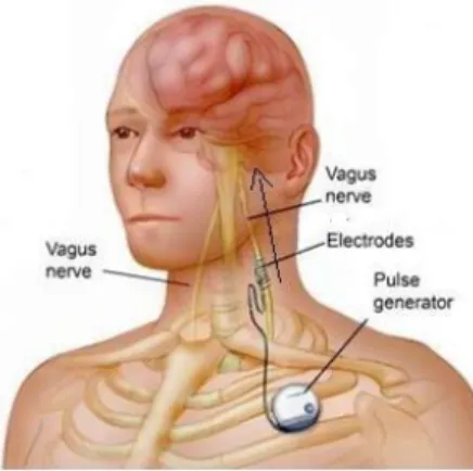
partial onset seizures in adults (>12 year) but is extended to several forms of epilepsy and is also administered to children29. In these patients, a bipolar spiral electrode is
wounded around the left cervical vagal nerve and subcutaneously connected to a pulse generator which delivers electrical pulses and is implanted in the sub-clavicular area (Figure 5)30. The right vagus nerve is
responsible for the innervation of the sinoatrial node and closely associated with the cardiac atrial function. Therefore the left vagus nerve is chosen to minimize the vagal effects on the heart29. The telemetric programming unit makes it
possible to programme and adjust the stimulation parameters including output current, pulse width, frequency and duty cycle30. VNS requires an invasive surgical procedure, but the
discomfort of the surgery and acute/chronic side effects are mild, mainly transient and will
Figure 5. Implanted VNS electrode in a patient. Figure from G. Bedi, 2014.
14 disappear a few weeks after surgery. Hoarseness, a tingling sensation in the throat and coughing are the most prevalent side effects. The efficacy of VNS within one year is estimated around 50% reduction in seizure frequency in at least 1/3 of the patients with medication-resistant epilepsy29,30. But more responders are observed over time, up to 60-70% after five
year29. Furthermore, long-term VNS in patients showed an improved quality of life in several
clinical studies.
The precise mechanism of action of VNS and how it suppresses epileptic seizures is still not completely understood29. The vagus nerve is the 10th cranial nerve
consisting of 80% afferent fibres and 20% efferent fibres26,28.
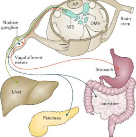
The afferent fibres originate in the nodose ganglion and primary project towards the nucleus tractus solitarii (NTS) (Figure 6) which projects towards several regions of the forebrain, brainstem and important structures in epileptogenesis such as the amygdala and thalamus28.
Besides these projections, the NTS has also direct connections towards the raphe nuclei, major source of serotonin, and indirect connections towards the locus coeruleus (LC) and A5 nuclei (a group of noradrenergic neurons in the brainstem), both noradrenergic sources. In literature, several research groups tried to identify potential anatomic brain structures which mediate the antiseizure effect of VNS in animals28. Increased activity of the amygdala, the
thalamus, the LC and the A5 nuclei was observed after VNS
in animals. VNS was responsible for the bilateral activation of all these brain structures, confirming the bilateral anticonvulsant effect despite the unilateral stimulation. The amygdala is a highly epileptogenic region that plays a crucial role in the generalization of seizures and the thalamus is also implicated in seizure regulation. Furthermore, the crucial role of the LC was demonstrated by the bilateral dissection of the LC which was accompanied by loss of the seizure supressing effect of VNS by minimizing the norepinephrine release in the brain. In patients, VNS is also able to suppress seizures in the ‘off’ mode suggesting an anti-epileptic effect. Even more, some literature suggests that seizures might be predicted early enough to trigger stimulation of the vagus nerve28. Furthermore, the anti-epileptic effect can also partly
be described by the observed increase of GABA in the CSF in patients receiving VNS28,29.
Figure 6. At the subdiaphragmatic level, vagal afferent neurons innervate the stomach, intestines, liver and pancreas and relay. Figure from Waise et al., 2018.
15
Anti-inflammatory effect of vagus nerve stimulation
The neuronal response mechanism of the anti-inflammatory pathway relies on the activation of the sensory afferent vagus nerve fibres which provide a signal to the brain that inflammation is occuring31.
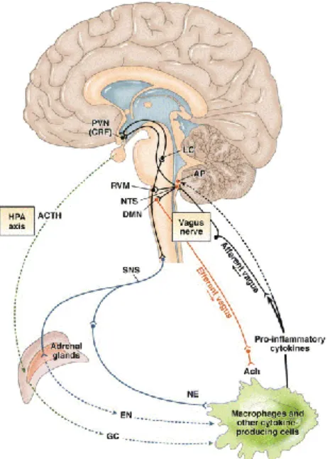
Thereby, the vagal afferent neuronal pathway is suggested to play a dominant role in mild to moderate peripheral inflammatory responses. Via the efferent vagus nerve fibres the anti-inflammatory pathway will be activated. The efferent vagus nerve neurons include the NTS which connects towards hypothalamic nuclei including the paraventricular nucleus (PVN). This PVN releases the corticotrophin releasing hormone (CRP) which in his turn activates the hypothalamic-pituitary-adrenal (HPA) axis by releasing the adrenocorticotropic hormone (ACTH) as illustrated in Figure 7. The latter forms the connection towards the humoral anti-inflammatory pathway and thereby the ascending link between the NTS and the PVN modulates the neurohormonal anti-inflammatory response31. Besides
this, the NTS possesses also indirect connections towards the LC via the rostral ventrolateral medulla (RVM) (Figure 7). The LC innervates in his turn higher brain structures such as the hypothalamus and the PVN in a noradrenergic manner (Figure 7). In addition, the PVN has also descending projections towards the NTS and the RVM. In conclusion, all these ascending and descending connections of the PVN result in an immunomodulatory role for neurons on the HPA axis31.
The cholinergic inflammatory pathway is suggested as the key player in the anti-inflammatory effect of VNS31. Acetylcholine (Ach) is a crucial neurotransmitter (NT) and
neuromodulator within the brain. This NT mediates the synaptic signalling in the ganglionic synapses of the sympathetic and parasympathetic neurons and is the main NT of vagal efferent neurons. Acetylcholine can bind on two different types of receptors, the muscarine receptor (metabotropic) and the nicotinic receptor (ionotropic)31. The RNA of these receptors has been
detected in a mixed population of lymphocytes and other (non-)immune cytokine producing cells. Furthermore, the α seven
subunit of the nicotinic receptor (α7nAchR) is found on macrophages. Binding of Ach on the α7nAchR of macrophages will result in a decreased production TNF-α and an effective suppression of 1β, 6 and IL-18 by post transcriptional mechanisms31. The direct role of
the vagus nerve in suppression of TNF-α levels was suggested via vagotomy in rats resulting in increased TNF-α levels in response to an intravenous injection of endotoxins. VNS resulted in significantly attenuated TNF-α levels after endotoxin
Figure 8. The cholinergic anti-inflammatory pathway. Pro-inflammatory cytokines activate the afferent vagus nerve fibres which (in)directly stimulate the NTS. The NTS will activate the vagal afferents in the dorsal motor nucleus and the vagal efferent fibres will activate the cholinergic anti-inflammatory effect. Figure from Garzoni et al., 2013.
Figure 7. Neuronal response mechanism of the anti-inflammatory pathway in response to systemic inflammation. NTS, nucleus tractus solitarii; DMN, dorsal
motor nucleus of the vagus; PVN, paraventricular nucleus; RVM, rostral ventrolateral medulla; LC, locus coeruleus; SNS, sympathetic nervous
system; ACTH, adrenocorticotropin hormone; GC, glucocorticoids; EN, epinephrine; NE, norepinephrine; ACh, acetylcholine. Figure from Bonaz and Bernstein, 2013.
16 introduction. TNF-α amplifies inflammation by the release of IL-1, HMGB1, nitric oxide, and reactive oxygen species. Based on these findings the vagus nerve has a crucial role in maintaining immunological homeostasis. In the end, the involvement of the efferent vagus nerve neurons in neuromodulation is supported by the protective role of the cholinergic anti-inflammatory pathway which is activated by the vagus nerve31. The activation of the cholinergic
anti-inflammatory pathway by the vagus nerve is illustrated in Figure 8. Additionally, the NTS may integrate the cholinergic anti-inflammatory pathway into other central immunomodulatory responses31. This way, VNS may present a novel approach to inhibit the release of
pro-inflammatory cytokines such as TNF-α, IL-1β, IL-6 and IL-18, and thereby protects the patient against pathological inflammation.
Furthermore, VNS also resulted in decreased levels of IL-1β and TNF-α and increased levels of IL-10 in the brain tissue and serum, after a traumatic brain injury in rabbits32. Altered levels
in brain tissue suggest that the anti-inflammatory effect of VNS is also valid within the brain itself. Additionally, electrical stimulation of the efferent vagal nerves in a haemorrhage shock model resulted in significantly decreased NF-ĸB activation21.
3. Glioblastoma-related epilepsy rodent model
Glioblastoma is a grade IV glioma and is responsible for the development of seizures in 30-60% of the patients26. Therefore, a GB-related epilepsy rodent model is desired to investigate
possible therapies against tumour growth and GB-associated seizures. There exist a lot of GB rodent models but almost studies did not investigate related seizures or these models are not reproducible33.
Recently, the 4BRAIN research group successfully established a GB-related epilepsy rat model33. Therefore, 20 000 F98 cells were inoculated in the right entorhinal cortex, susceptible
for the development of epileptic activity, of male F344/IcoCrl rats. The F98 cell line is a N-ethyl-N-nitrosourea-induced glioma cell line and isolated out of a female Fischer 344 rat on the 20st fetal day and has a doublings time in vitro of approximately 13 hours (ExPASy, Bioinformatics Resource Portal, cellosaurus)34. In all animals GB growth was confirmed using T2 weighted
MRI. In order to detect seizures in these animals, electrodes to record electroencephalography (EEG) were implanted after confirmation of tumour growth and video-EEG was continuously monitored until euthanasia. During a common 8-day period (day 13 to day 20 post-inoculation) all GB animals had seizures with a mean of 15 ± 11 seizures per animal and no seizures were detected in control animals. All animals were euthanized 21 days post-inoculation based on humane endpoints. Furthermore, immunohistochemical staining for glial filament acidic protein (GFAP) and vimentin, both astrocytic markers, confirmed the astrocytic origin of the tumour.
4. VNS as treatment for glioma-related epilepsy? A hypothesis
This master thesis partly investigated the feasibility to perform VNS as a therapy for glioma and related seizures. Treatment with VNS could provide serious advantages in glioma patients targeting both epileptic seizures, resulting from gliomas, and glioma progression. This hypothesis is based on two fundamental working mechanisms of VNS.
Firstly, VNS could influence the TME by the activation of the cholinergic anti-inflammatory pathway. Via this pathway VNS will be responsible for the suppression of TNF-α, IL-6, IL-1β and NF-ĸB31. Since research illustrated that the TME mainly consists out of innate and adaptive
immune cells and most of them express the α7nAchR, VNS might target the TME via his cholinergic anti-inflammatory pathway21. To begin, TNF-α and IL-6 are crucial cytokines for the
promotion of glioma growth and invasion22,23. Since VNS decreases the production of these
cytokines, VNS will thereby potentially inhibit glioma progression31. Furthermore, the TME of
gliomas consists out of M1 and M2 TAMs, with M2 being the main phenotype21. The M2 TAMs
are maintained by the expression of NF-ĸB and will promote glioma progression by the production of pro-tumoral substances. Since VNS results in significant decreased levels of NF-ĸB, VNS might potentially result in the conversion of M2 TAMs into M1 TAMs and might
17 suppress glioma progression. Moreover, VNS will result in decreased IL-1β production by the M1 TAMs and thereby inhibit the glioma growth associated pathways of M1 TAMs31,35.
Considering all the above, VNS could potentially influence the TME and more specific the TAMs by the activation of the cholinergic anti-inflammatory pathway and thereby result in the suppression of glioma progression. Secondly, VNS is an FDA-approved anti-epileptic treatment for medication-resistant epilepsy36. The effect of VNS in patients with glioma-related
epilepsy is similar to the effect in patients suffering from seizures without a history of brain tumours, resulting in a seizure reduction of at least 50%.
Controversially, the increased levels of GABA in patients and IL-10 within the brain of rabbits observed after receiving VNS, could question the potential therapeutic effect of VNS on glioma growth29,32. Firstly, gliomas are characterised by the excessive expression of GABAa
receptors15. These GABAa receptors are responsible for fast volume reduction of the glioma
cells, resulting in the proliferation and migration of glioma cells. Thereby, VNS could enhance glioma progression by his increased GABA levels observed in the CSF of VNS patients. Secondly, TAMs play a crucial role in the hypothesis that VNS could influence glioma progression as mentioned above. Since VNS is mentioned to enhance IL-10 levels in rabbits suffering from a traumatic brain injury, VNS could possibly activate the M2 TAMs22,32. These
M2 TAMs are known to enhance tumour progression by releasing pro tumoral substances21.
But VNS can also be responsible for the conversion of M2 TAMs into M1 TAMs by the inhibition of NF-ĸB. Therefore, the possible increased activation of M2 TAMs by releasing IL-10 can be counteracted by the increased inhibition of NF-ĸB.
In conclusion, VNS can form a potential treatment for glioma-related epilepsy by targeting tumour progression and epileptic seizures as hypothesized above. VNS could form a treatment without influencing the quality of life of the patients. This hypothesis was investigated as a first part of this master thesis by applying VNS in a GB-related epilepsy rodent model.
5. Low-Grade glioma rodent model
Since patients with LGGs are more vulnerable for the development of epileptic seizures (50-90%) and these patients have a prolonged overall survival, an LGG model is desirable for further research on alternative treatments2,8. Since LGG animal models barely exist and are mainly not reproducible or miss the host immune reactions against tumour presence by their immunocompromised character and this often results in an in vivo progression towards HGG, the establishment of an LGG rodent model was attempted as a second part of this master thesis37,38. Therefore, two different patient-derived paediatric LGG cell lines, Res186 and
Res259, were used.
In general, clinical manifestations in patients with LGGs can be divided into two different groups39. Firstly, generalizing symptoms develop due to increased intracranial pressure
resulting in headaches, vomiting, nausea and lethargy. Secondly, localizing symptoms manifest including seizures, focal neurological deficits and endocrinopathies depending in the localisation of the tumour.
18
Res186: Pilocytic Astrocytoma grade I
Res186 cells are isolated out of paediatric pilocytic astrocytoma (PA) in a 3 year old female and are classified as WHO grade I glioma cells. PA are responsible for approximately five percent of all gliomas and are most prevalent in children
and young adults40. These gliomas occur mainly in
children from 5 to 19 years old with a peak incidence from five to nine year39. PA occur in different areas within the
central nervous system including the cerebellum, optic pathway and hypothalamus39. These tumours are
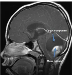
characterized by a well-circumscribed growth, low to moderate cellularity and a biphasic grow pattern. This biphasic growth pattern includes the presence of compacted bipolar cells with Rosenthal fibres, eosinophilic fibrillary aggregates, and areas containing multipolar cells, microcysts and eosinophilic granular bodies. PA often possess a large cystic component and an enhancing mural nodule (Figure 9). In addition, the Res186 cell line is defined by a doubling time of 46 hours when cultured in ideal circumstances (ExPASy, Bioinformatics Resource Portal, cellosaurus).
Res259: Diffuse astrocytoma grade II
The Res259 cell line is isolated out of 4 year old female suffering from a paediatric diffuse astrocytoma, this tumour isclassified as a grade II glioma by the WHO41. Diffuse astrocytomas
present themselves with headache, epileptic seizures and depending on the localisation of these tumours certain neurological deficits (UCSF Brain Tumour Center). Furthermore, diffuse astrocytomas are the second most prevalent gliomas after GB with a median survival of five to eight years (UCSF Brain Tumour Center)42. Additionally, these LGGs have a high recurrence
risk due to their diffuse infiltration pattern into the brain and inherent malignant potential to transform towards high-grade astrocytic tumours such as GB.
Diffuse astrocytomas show a pronounced heterogeneity which makes it challenging to grade these tumours based on their histopathological diagnosis42. In histology diffuse astrocytomas
are characterized by increased cellularity, little nuclear atypia, low mitotic activity, absence of necrosis and microvascular proliferation. Furthermore, formation of “secondary structures of Scherer” including perineuronal satellitosis, subpial growth and perivascular spread take place within these tumours. In addition, the Res259 cell line is characterised by a doubling time of 24 hours when cultured (ExPASy, Bioinformatics Resource Porta, cellosaurus).
Markers for low-grade gliomas
The above described LGG cell lines Res186 and Res259 can be characterized and distinguished from HGG gliomas such as F98 cells using immunocytochemistry staining. Three different markers can be used, glial fibrillar acidic protein (GFAP), vimentin and nestin. Firstly, GFAP is an intermediate filament which is responsible for the cytoarchitecture and the mechanical strength of astrocytes43. Furthermore, GFAP is a regularly used marker for reactive
astrogliosis, like seen at the tumour border of patients with GB and for mature astrocytes43,44.
GFAP expression has a significant effect on astrocyte properties, such as morphology, cell-growth and division44. In literature, GFAP expression was significantly higher in LGGs than in
grade IV gliomas and was negatively correlated with glioma grade. Even more, the expression of GFAP in grade IV gliomas can be less than in healthy brain tissue. Therefore, the expression of GFAP can be used as marker for the astrocytic origin of glioma cells and for the low-grade characteristics of the Res186 and Res259 cell lines during immunocytochemistry staining. Secondly, vimentin is an intermediate filament type III and typically expressed by astrocytes and cells who undergo epithelial to mesenchymal transition (EMT) which is associated with
Figure 9. Cerebellar pilocytic astrocytoma T2 weighted MRI. Image from Sievert and Fischer, 2009.
19 tumour invasiveness and motility45,46. Vimentin is a part of the cytoskeleton and responsible for
the maintenance of cellular integrity and the protection of the cells against mechanical stress45.
This together with the fact that vimentin is an EMT biomarker, suggests that vimentin expression is closely associated with the malignant processes of CNS tumours. In literature, vimentin expression was positively correlated with glioma grade. Therefore, vimentin can be used as an astrocytic marker and high expression patterns are expected in the HGG cell lines such as F98. Thirdly, nestin can be used as marker for HGG. Nestin is an intermediate filament type IV protein involved in the control of cell morphology, adhesion and proliferation and is therefore typically expressed in proliferating and migrating cells47. Therefore, nestin is usually
used as a stem cell marker. Furthermore, when differentiation starts, cells will exit the cell cycle and nestin expression will be downregulated and alternative intermediate filaments such as GFAP in glial precursor cells will be upregulated. In literature, nestin expression was observed in different brain tumours such as PA and GB47. Furthermore, high nestin expression was
observed in high malignant tumours (for example GB) when compared to less anaplastic glial tumours. Therefore, nestin can be used as a marker for HGG and a higher expression of nestin is expected in the F98 cell line when compared to the Res186 or Res259 cell lines during immunocytochemistry staining. In the end, the expected results of the three cell lines and the above markers are illustrated in Table1.
Marker Res186 (Grade I) Res259 (Grade II) F98 (Grade IV) GFAP High expression High expression Lower expression
vimentin Lower expression Lower expression High expression
nestin Lower expression Lower expression High expression
20
MATERIALS AND METHODS
1. Feasibility to perform VNS in a GB-related epilepsy rodent model
Cell culture
The F98 rat GB cells (ATTC) were cultured as monolayers in Dulbecco’s Modified Eagle Medium (DMEM) in T75 falcons. The DMEM medium (flaks of 500ml) was supplemented with 10% fetal calf serum (FCS), 1% penicillin-streptomycin and 1% of L-glutamine (all products were purchased from Invitrogen®). Fetal calf serum or fetal bovine serum is essential in cell
cultures because it contains a large amount of nutritional and macromolecular factors essential for cell growth and a smaller amount of antibodies than non-fetal serums48. Furthermore, 1%
penicillin-streptomycin was added to prevent bacterial contamination by their combined action against gram-negative and -positive bacteria. The concentration of penicillin is 10 000 units/ml and for streptomycin 10 000μg/ml. Also 1% of L-glutamine was added to participate in the formation of amino acids, protein synthesis and glucose production. Furthermore, the DMEM contains a Phenol red indicator which is used as a pH indicator. Thereby, the DMEM will turn yellow when the pH drops below 6.8 (acid) and pink when the pH exceeds 8.2 (base). Since acidic and basic environments are unfavourable conditions for cell culturing this Phenol red indicator was crucial in monitoring the cell cultures. The cell cultures were maintained in an incubator at 37°C and in 5% CO2 and were split when 80% confluency was reached (illustrated
in addendum section 1. Cell passage).
During the preparation of 20 000 F98 cells in 5µl PBS for inoculation, a Bürker chamber was used to count the correct number of cells. Firstly, a cell suspension was obtained via the first four steps of cell passage; taking off the DMEM medium, washing the cells with PBS, adding 2ml of trypsin and letting it incubate for 5 minutes and adding 2ml of DMEM as illustrated in the addendum section 1. Cell passage. Thereafter, 50μl of the cell suspension was taken out and 50μl of trypan blue was added in order to stain the death cells, meaning the cell suspension was ½ diluted. Then, the cells were counted three times in the Bürker chamber and the mean was calculated. For the number of cells in 1ml, the mean was multiplied by two (due to the dilution of ½) and 10 000 (the volume in one square is 100nl or 0.0001ml). Once the number of cells in 1ml was known, the correct volume of cell suspension was calculated in order to have 2 000 000 F98 cells. This volume of cells suspension was centrifuged, the liquid was taken off and the cells were diluted in 500µl phosphate buffered saline (PBS). Resulting in a final concentration of 20 000 F98 cells in 5µl PBS. The cells were prepared right before inoculation in order to minimize the amount of death cells injected. A counting error occurred in one animal and only 11 000 cells were inoculated (an estimation of the total amount of cells injected is illustrated in the addendum section 3.4 Tumour volumes of all animals).
Animals
Eight male F344/IcoCrl rats (Charles River®) of 10 weeks old were used during this study. The study was approved by the animal ethics committee of the Faculty of Medicine and Health Sciences of Ghent University (ECD 17/111). All animals were kept and handled according to the European guidelines (Directive 2010/63/EU) and housed under environmentally controlled conditions: 12h light/dark cycle, temperature between 21-24°C and humidity between 40-60%.
Surgery
All animals were anaesthetised with a mixture of medical oxygen (rate: 1l/min) and isoflurane (induction: 5%; maintenance: 2%). At the start of surgery all animals were weighted and blood samples from the tail were collected. The blood samples were collected by heating the tail using an infrared lamp in order to make the tail veins visible. Next, a winged intravenous catheter was put into the tail vein and the blood was captured in a sterile Eppendorf tube of 1.5ml. Maximum 0.6ml blood was collected from each animal. Thereafter, the animals were implanted with a VNS electrode around the left vagus nerve. After implantation of the VNS electrode, animals were immobilized in a stereotaxic frame, EEG registration electrodes were
21 implanted and 20 000 F98 cells in 5μl PBS were inoculated into the right entorhinal cortex. During surgery the body temperature was maintained at 37°C using a heating pad and this was controlled by rectal measurement every 30 minutes.
1.3.1 VNS implantation
Before implantation of a ‘custom-made’ VNS electrode (described by El Tahry et al. 49) which
consisted out of an anode(+) and a cathode(-), the impedance was tested and only VNS electrodes with an impedance between 2-5kΩ were used. At the start of the surgery, animals received a subcutaneous injection of Vetergesic® (0.1 ml from a concentration of
0.03mg/ml). Then the head and neck region (from the chin till the forelimbs) were shaved using soap (Hibiscrub®) and water. The
animals were placed on their back on a heating pad and a nose cone was used to deliver the mixture of medical oxygen (rate:1.2l/min) and isoflurane (2%). The shaved neck region was disinfected using iso-Betadine® before a medial
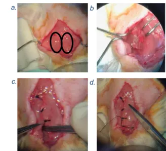
incision, ranging from the sternum to the chin, was made. Next, the skin was separated from the tissue and tissue was removed from the fused glands using forceps. The two fused glands (Figure 10 a.) were separated in order
to expose the triangle point of three muscles (m. sternocleidomastoid and the omohyoid muscles) located at the left. Thereafter, the left vagus nerve (recognized by its striped ladder pattern) was isolated from the carotid sheath and carotid artery. Once the left vagus nerve was properly isolated, the VNS electrode was wrapped around the vagus nerve with the anode placed caudally (Figure 10 b.) and saline was applied immediately. The muscles were placed back over the vagus nerve followed by the glands which were stitched together (Figure 10 c.). Then the leads of the VNS electrode were placed underneath the stitched glands and an additional stitch was added above the leads (Figure 10 d.). The remainder leads were tunnelled subcutaneously to the head. At the end of this procedure the neck region was sutured. 1.3.2 Inoculation and implantation of EEG electrodes
After the implantation of the VNS electrode, the animals were immobilized using a stereotaxic frame (Figure 11) and anaesthesia was lowered to 1.5% isoflurane and a medical oxygen rate of 0.8ml/min. Iso-Betadine® was applied to the shaved head and a
subcutaneous injection of adrenaline/xylocaine (0.1ml of the solution containing 20mg/ml lidocainehydrochloride, xylocaine 2% and adrenaline 1:200 000) was given right above the skull. Thereafter an incision was made to expose the skull. Firstly the skull was hatched to make it rougher, then acetone was applied to destroy the periost and make the sutures more visible, next green activator was applied for an optimal functioning of later applied Metabond® and lastly saline was applied in
order to rinse off the green activator (result illustrated in Figure 12). The coordinates of
Bregma and Lambda were defined to check whether the skull was positioned correctly and in order to determine the inoculation site and
Figure 11. Rat immobilized in a stereotaxic frame. a. b . c. d.
Figure 10. a. The two fused glands seen during surgery. b. The VNS electrode is wrapped caudally around the left vagus nerve. c. The glands are stitched together and the leads from the VNS electrode are partly put underneath the glands. d. An extra stitch is made between the glands.
.
Figure 12. Result of the skull after hatching, applying acetone, green activator and saline.
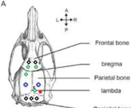
22 coordinates for the bipolar depth-electrodes as accurate as possible. This was followed by the implantation of scalp electrodes and anchor screws. Firstly, burr holes of 1.1mm were made for the type I screws; two traumatic scalp electrodes
consisting of type I screws were placed in the os frontale functioning as ground and spare ground, and three type I anchor screws were placed in the os occipitale (Figure 13). Secondly, burr holes of 0.9mm were made for the type II anchor screws and an atraumatic scalp electrode consisting of a type II srew. The atraumatic scalp electrode, to record surface EEG, was placed over the right parietal cortex, near the tumour border and medial-posterior from the right bipolar depth-electrode (Figure 13). Two type II anchor screws were placed in the os frontale and one in the os parietale (Figure 13). Traumatic and atraumatic scalp electrodes differ in shape of the extremity of the screw for attachment in the skull. An type II screw contains a flat end with screw thread covering the whole tip, whereas type I screws have rounded tips without screw thread.
Type I screws have a better fixation but will reach deeper through the skull when attaching them and therefore these screws are more likely to cause damage to the cortex. This will be prevented by using type II screws. Thus, type II screws were used above cortical areas, to avoid damage to the cortex and the risk of provoking seizures, since this trial investigated glioma-related seizures. After placements of the anchor screws and the scalp electrodes, Metabond® was applied around the screws in order to secure the attachment of the screws to
the skull and to secure the dental cement that was used at the end to make the head cap. Then a craniotomy for inoculation was made 8mm posterior and 4.5mm right to Bregma. Next, an insulin needle (BD 0.5 ml Insulin Syringe Microfine 0.33 mm (29G) x 12.7 mm) was filled with the F98 cell suspension and mounted on a pump for automatic injection (Stoelting Quintessential Stereotaxic Injector (QSITM), Stoelting Co.). The insulin needle was stereotactically guided to the right coordinates relative to bregma (anteroposterior (AP): -8.0mm; mediolateral (ML): +4.5mm). Then the insulin needle was inserted through the craniotomy to a depth of -4.1mm relative to dura in order to inoculate the cells in the right entorhinal cortex. 20 000 F98 cells in 5μl of PBS were injected over a time period of 10 minutes and the syringe was kept in place for five minutes post-inoculation and thereafter slowly removed. In this study, the inoculation day was referred to as day 0.
After the inoculation two craniotomies for the bipolar depth-electrodes (Figure 13) were made (AP: -5.0mm; ML: +/- 3.5mm relative to bregma). Bipolar depth-electrodes were custom-made and the tips were 0.9mm apart in order to simultaneously record activity in the dentate gyrus and the CA1 region of the ipsi- and contralateral hippocampus. Via a small incision in dura the depth-electrodes were lowered in the brain 3.5mm below dura. The electrodes were fixed to the skull and neighbouring anchor screws using UV-cement (Filtek Supreme XTE Plus Flowable 2 x 2 gr, 3M, Belgium). The leads of the VNS, scalp and depth-electrodes were assembled in a head cap on the skull of the rat using resin-based dental cement (Simplex Rapid fluid/powder, Kemdent, UK). At the end of the surgery the scalp was sutured, Metacam®
(1ml/kg) and 1ml of saline were administered subcutaneously. The animals were transferred to their cages and one DietGel® Recovery was given to each animal for fast recovery. Furthermore, animals were inspected daily.
Figure 13. Rat Skull. Brunner, Clément. (2016). Type I screws are black and type II screws green. The scalp electrodes are indicated with an E abbreviated from electrode. The bipolar depth-electrodes are presented in blue. The inoculation place is marked in red.
E E
23
EEG monitoring and VNS stimulation
Six animals were connected to the EEG setup at day 7. They were anaesthetized with a mixture of medical oxygen and isoflurane (induction: 5%; maintenance: 2%), a second blood sample was taken (as described in 1.3) and thereafter they were connected to the EEG setup. The EEG setup consisted out of a head stage carrying a 4-channel unity gain amplifier (based on a TL074 SMT Opamp, Texas Instruments, Dallas, TX, USA) that is connected via tethers to a 12-channel commutator (Plastics One, Roanoke, VA, USA) allowing the animals to move freely. The commutator is connected to a custom-made 4 channel high pass filter with a time constant of 1 second followed by a 4-channel 512 x amplifier (based on TL074 Opamp) and a NiDAQ to digitize the signal (USB-6259, National Instruments Device). Stimuli to the left vagus nerve were delivered via an external constant current stimulator.
For VNS stimulation the animals were ad random assigned to the SHAM or VNS group and only the VNS group received stimulation. Both groups consisted out of three animals; the animals belonging to the VNS group were the 1st, 2nd and the 3rd VNS rat and the animals
belonging to the SHAM group were the 1st, 2nd and 3rd SHAM rat. In the VNS group the
impedance of the electrode was measured before stimulation was started using an oscilloscope. Impedance should be below 10 kΩ. The VNS group was stimulated with the following parameters: 30Hz, biphasic pulses of 250µs/phase, 0.5sec ON/29.5sec OFF as illustrated in Figure 14. The VNS stimulation was gradually increased in steps of 250µA from 250μA at day 10 to 1mA at day 13. According to previous research, VNS can induce hypothermia 50. Therefore, the parameters above were chosen because they were not related
to any hypothermia in rats51.
Perfusion
All animals were transcardially perfused at day 14 and the brains were snap frozen in liquid nitrogen. The animals were first anaesthetised with a mixture of medical oxygen and isoflurane (induction: 5%; maintenance: 2%) and blood samples were taken (as described in 1.3). This was followed by an overdose of sodium pentobarbital (200mg/kg) which was injected intraperitonially. From the moment the animals stopped breathing they were transcardially perfused. The chest was opened and the beating heart was carefully isolated out of the pericardium. Firstly, 0.1ml of heparin was injected into the apex of the beating heart to avoid blood clotting. Secondly, a needle was placed through the apex into the aorta and a cut was given in the right atrium. Next, the veins were flushed with saline at room temperature for one
minute followed by ice cold PBS till the no more blood was present in the liquid coming out of the right atrium (Figure 15). After transcardial perfusion, the brains were isolated. Immediately thereafter the brains were put into liquid nitrogen and stored until histology.
Figure 15. Transcardial perfusion of a rat. 500 μs 250 μA 500 μs 250 μA Interpulse period (1/30Hz): 0.033s
Figure 14. VNS stimulation protocol.
0.5s ON 0.5s ON