Bacillus cereus associated food borne disease
quantitative aspects of exposure assessment
and hazard characterization
Promotoren
Prof. Dr. T. Abee
Persoonlijk hoogleraar bij de leerstoelgroep Levensmiddelenmicrobiologie Wageningen Universiteit
Prof. Dr. Ir. M. H. Zwietering
Hoogleraar Levensmiddelenmicrobiologie Wageningen Universiteit
Copromotor
Dr. R. R. Beumer
Universitair docent, leerstoelgroep Levensmiddelenmicrobiologie, Wageningen Universiteit
Promotiecommissie
Prof. Dr. P. E. Granum
Norges Veterinærhøgskole, Noorwegen Prof. Dr. M. Kleerebezem
Wageningen Universiteit Prof. Dr. S. Brul
Universiteit van Amsterdam Dr. Ir. M. H. J. Wells – Bennik NIZO Food Research, Nederland
Bacillus cereus associated food borne disease
quantitative aspects of exposure assessment
and hazard characterization
Lucas Maria Wijnands
Proefschrift
ter verkrijging van de graad van doctor op gezag van de rector magnificus
van Wageningen Universiteit Prof. Dr. M. J. Kropff in het openbaar te verdedigen
op maandag 2 juni 2008
L. M. Wijnands – Bacillus cereus associated food borne disease: quantitative aspects of exposure assessment and hazard characterization – 2008.
Thesis Wageningen University, Wageningen, The Netherlands – With
summary in Dutch – 175p.
Key words: Bacillus cereus / food borne disease / exposure assessment / hazard characterization.
aangegaan. Een uitdaging in de zin van “hoever kan ik zelf gaan, waar liggen mijn mogelijkheden, mijn grenzen”. Juist op het moment dat besloten was het onderzoek naar de gezondheids effecten veroorzaakt door mycotoxinen in voedsel te staken diende het Bacillus onderzoek zich aan. En kon een al langer levend en uitgesproken verlangen om “de uitdaging” aan te gaan omgezet worden in daden.
Meestal worden aan het eind van een proefschrift mensen bedankt die op enigerlei wijze van belang zijn geweest voor het tot stand komen van een dergelijk project. Daarin verschilt dit proefschrift niet van andere. Maar ik vind dat ik één uitzondering moet maken. Want de bijdrage van Frans van Leusden aan dit onderzoek is enorm geweest. Als collega en projectleider gedurende al mijn RIV(M) jaren. Maar ook als iemand die zich mijn uitdaging voor kon stellen. Op alle mogelijke manieren heeft hij zich ingezet om mijn uitdaging te laten lukken. Frans, ontzettend bedankt! Tijd nu voor volgende persoonlijke uitdagingen, voor volgende projecten. zoals me gaan bekwamen in de Spaanse taal, en me bezighouden met huis, tuin en keuken zaken, in de meest letterlijke zin van het woord. En, Lineke, laten we nu eindelijk dat kustpad maar eens gaan wandelen en al die andere uitgestelde goede voornemens waar gaan maken.
Contents
Abstract……… 9
1. Introduction………....………...………...11
2. Prevalence of potentially pathogenic Bacillus cereus in food commodities in the Netherlands………...33
3. Spores from mesophilic Bacillus cereus strains germinate better and grow faster in simulated gastro-intestinal conditions than spores from psychrotrophic strains………..51
4. Modelling the number of viable vegetative cells of Bacillus cereus passing through the stomach ………69
5. Germination of Bacillus cereus spores is induced by germinants from differentiated Caco-2 cells, a human cell line mimicking the epithelial cells of the small intestine…..87
6. Caco-2 cell surface-associated growth of enterotoxic Bacillus cereus is a prerequisite for initiation of cytotoxic effects in simulated intestinal fluid……….….95
7. Integration of results and conclusive remarks………....109
References………..133
Samenvatting………..165
List of publications……….169
Nawoord………...173
Abstract
Bacillus cereus is a versatile, spore-forming micro-organism capable of invoking two different types of food borne disease: a diarrhoeal syndrome, caused by enterotoxins produced during growth in the small intestine and an emetic syndrome, caused by the emetic toxin cereulide, produced in food prior to consumption. To enable a better risk assessment for the two syndromes, we focused investigations on exposure assessment and hazard characterization.
B. cereus exposure assessment includes investigations to obtain detailed information on the prevalence of potentially pathogenic B. cereus strains in Dutch food commodities. The data revealed that almost all of the strains found in the investigated food commodities posses the potential to produce one or more virulence factors, i.e. enterotoxins or cereulide. Also the concentration of B. cereus in the samples has been determined.
The hazard characterization focused on processes in the gastro-intestinal tract that lead to the diarrhoeal syndrome using model systems. Spores of B. cereus pass the gastric barrier, the first line of defence in humans, unaffected, while the number of vegetative cells able to survive simulated gastric conditions depends on the strain type, food type and the type of food acidification capacity of the stomach. It was shown that up to 26% of the vegetative cells can survive the gastric passage, indicating that they may contribute to the onset of diarrhoeal disease.
The enterotoxins, responsible for the onset of disease through the destruction of the small intestinal epithelium, were shown to be highly instable molecules in simulated intestinal conditions. We have shown that adhesion and subsequent production of enterotoxins in very close proximity of the epithelium are a prerequisite to circumvent degradation of the enterotoxins by proteolytic activity in the small intestine. The potential for adhesion was demonstrated for spores and vegetative cells at an efficiency rate of approximately 1%. In a model system the correlation between adhered B. cereus cells and the measure of destruction of differentiated Caco-2 cells, mimicking small intestinal epithelial cells, was proven.
B. cereus is also a micro-organism with a broad growth temperature range. The growth temperature profile of the strains is of importance for both their ability to grow in foods and to cause disease. We revealed that strains able to grow at temperatures below 10ºC
(psychrotrophic strains) seem to be less pathogenic than strains lacking this ability (mesophilic strains). This is not only due to their lower growth potential at 37ºC, but also due to their lower growth potential in conditions simulating the small intestine.
B. cereus spores may be very important for the onset of disease, but before they can grow and produce enterotoxins they first have to germinate. They contain receptors that need triggering by certain low molecular weight substances to set of germination. Such inducers may not only be present in the food, also differentiated Caco-2 cells were found to induce spores of some strains to germinate.
Through integration of the results of the investigations described in this thesis the annual number of cases of diarrhoeal disease in the Netherlands is estimated to range from 17,000 to 310,000. Data on the prevalence of B. cereus in Dutch food commodities together with literature and outbreak data were used to estimate the number of cases of emetic disease. These range from 630,000 to 3 million per year.
1
General
In 1955 Steinar Hauge inoculated sterilized vanilla powder with
104 cells Bacillus cereus per gram and prepared a vanilla sauce from it. He
left the sauce at room temperature for 24 hours, and subsequently consumed the sauce. Around 13 hours later he experienced severe abdominal pain and diarrhoea that lasted for 8 hours. It was the ultimate proof that Bacillus cereus was able to cause food borne disease (Hauge, 1955). The reason for this voluntary experiment were four outbreaks of food borne disease in Norway between 1947 and 1949, involving around 600 individuals. The food vehicle in these outbreaks was vanilla sauce,
prepared from corn starch, which appeared to be contaminated with 104 B.
cereus per gram. After preparation the vanilla sauce was stored at room temperature until it was served and eaten the next day. Before that date, the disorder in the taxonomy of B. cereus was the cause of slow recognition of the micro-organism as being able to lead to food borne disease. After the first description of the micro-organism (Frankland and Frankland, 1887), several cases of food borne disease caused by Bacillus-like organisms have been described (Lubenau, 1906; Seitz, 1913; Brekenfeld, 1926). Since the experiment of Hauge numerous food borne outbreaks attributed to B. cereus and associated with watery diarrhoea have been reported (Table 1.1).
In 1976 a novel toxigenic activity by B. cereus was described, based on monkey feeding studies (Melling et al. 1976). This was the first report on emetic properties of B. cereus, caused by an extra cellular heat stable compound with a molecular weight < 10,000 Dalton (Melling and Capel, 1978)
Bacillus cereus and gastro-intestinal disease
Although recognized as an agent causing food borne disease after the experiments by Hauge the mechanism of pathogenesis remained unknown for more than 20 years. The first description of a diarrhoeal toxin came in 1972 after partial purification by gel chromatography (Spira and Goepfert, 1972). It lasted until 1979 before clearly two toxic entities both produced by B. cereus were distinguished, each causing different symptoms (Turnbull et al. 1979). From that time on, B. cereus was recognized to be the cause of two types of disease, a diarrhoeal syndrome and an emetic syndrome.
Table 1.1 A selection of food borne outbreaks caused by Bacillus cereus
Year # persons affected Implicated food Reference Diarrhoeal
1990 4 cod fish (Van Netten et al. 1990) 1995 17 stew (Granum et al. 1995) 2000 300 chicken and
rice meal (Ripabelli et al. 2000) 2002 95 and 78 cake at
2 banquets (Ghelardi et al. 2002) 2002 25 mayonnaise (Gaulin et al. 2002) Emetic
1997 2 spaghetti
with pesto (Mahler et al. 1997) 2000 116 vegetarian
rice dish (Essen et al. 2000) 2000 7 vegetarian dish (Ripabelli et al. 2000) 2001 4 fried rice (Grein, 2001)
2003 2 pasta dish (Jääskeläinen et al. 2003b) 2005 6 pasta salad (Dierick et al. 2005)
The diarrhoeal syndrome and its virulence factors
The diarrhoeal syndrome is a typical example of a toxico-infection, caused by enterotoxins after the ingestion of food contaminated with Bacillus cereus. After passage of the stomach the vegetative cells and/or spores reach the small intestine. Spores germinate; vegetative cells grow and produce enterotoxins. Vegetative cells are believed to be of minor importance in the onset of the disease due to their inactivation in the stomach.
The enterotoxins affect the epithelial lining of the small intestine causing disturbance of the water – solute transport. Eventually this may lead to diarrhoea. The syndrome is characterized by abdominal cramps and watery diarrhoea within 8 – 16 hours after consumption of contaminated food. The symptoms generally last no more than 24 hours, although longer
duration times have been described (Kramer and Gilbert, 1989; Granum, 2007).
In the classification of Granum et al. (1995) B. cereus belongs to the group of micro-organisms causing disease through enterotoxin production without any direct interaction with the small intestinal epithelium. The disease symptoms are comparable to food borne disease caused by Clostridium perfringens, although the pathogenic mechanism is different. After all, the enterotoxin produced by C. perfringens is released during sporulation in the small intestine, whereas the enterotoxins of B. cereus are produced during growth in the small intestine (McClane, 1997; Granum, 2007).
The various steps in this pathogenic mechanism have not all been fully elucidated. According to earlier research intraluminal multiplication of micro-organisms was not involved in fluid accumulation in the ileum, and therefore B. cereus-induced diarrhoea was probably the result of intoxication rather than infection (Spira and Goepfert, 1972). After all, feeding of cell free preparations of B. cereus cultures induced diarrhoea in monkeys treated with 10% sodium bicarbonate. Also, a direct correlation was observed between the ability to cause fluid accumulation in rabbit ileal loops, altered skin capillary permeability and diarrhoea in the monkeys (Goepfert, 1974). Later however, it was found that crude or purified material was not able to elicit diarrhoea. This suggested that human diarrhoea was caused by toxins developed after growth in the intestine following ingestion of large amounts of organisms.
During the 1980’s and 1990’s research concerning the diarrhoeal syndrome increased due to the discovery and identification of enterotoxins. Improvement of molecular biological techniques has increased knowledge on the production and regulation of enterotoxins largely.
However, despite all the research dedicated to understand the diarrhoeal syndrome, hardly any data are known concerning the dose response relationship. Based on epidemiological data using numbers of B. cereus detected in food related to (possible) diarrhoeal outbreaks, the number of cells necessary to induce diarrhoeal symptoms has been
estimated to be 103 – 108 cells per gram food (Granum and Lund, 1997).
Apart from the experiment by Hauge (1955) one study with human volunteers has been described linking exposure level to disease complaints. In this study volunteers consumed naturally contaminated milk. The number of complaints by the volunteers was related to the number of B. cereus per consumption: the higher the number of B. cereus the more
complaints (see Table 1.2) (Langeveld et al. 1996). The results of the experiment, however, may be biased due to a concurrent flu epidemic.
Enterotoxins are responsible for the onset of the diarrhoeal syndrome. They damage the epithelial barrier causing fluid accumulation in ligated rabbit ileal loops and they alter the vascular permeability of guinea-pig skin. Also they show cytotoxicity towards VERO cells (African Green Monkey kidney cells) (Glatz et al. 1974; Gilbert and Kramer, 1984).
Table 1.2 Number of consumptions and number of complaints classified according to
number of Bacillus cereus cells ingested per consumption of contaminated milk (Langeveld et al. 1996)
Number of consumptions Complaints Number of B. cereus
cells ingested Total Yes (%) No (%)
< 106 132 5 (4) 127 (96)
106 - 107 32 2 (6) 30 (94)
107 - 108 26 2 (8) 24 (92)
> 108 69 9 (13) 60 (87)
Several enterotoxins produced by B. cereus have been described, namely haemolysin BL (HBL), non haemolytic enterotoxin (NHE), cytotoxin K, and enterotoxin FM. Of these HBL, NHE and cytotoxin K have been related to food borne disease (Table 1.3).
Haemolysin BL (HBL)
The first described enterotoxin of B. cereus is now called haemolysin BL (HBL). Previous names are diarrhoeagenic factor, fluid accumulation factor and vascular permeability factor (Shinagawa et al. 1991a; Shinagawa et al. 1991b; Sutherland and Limond, 1993). All names are somehow related to the effect of HBL observed in an in vivo and in vitro experiment (Kramer and Gilbert, 1989). HBL is a three component protein toxin, encoded by three polycistronic genes hblA, hblC and hblD.
B ac ill us c er eu s as so ci at ed fo od b or ne d is ea se - 1 6 - T ab le 1 .3 Sh or t d es cr ip tio ns o f t he B ac ill us c er eu s (e nt er o) -t ox in s (E nt er o) to xi n # su bu ni ts m ol . w ei gh t ( kD a) ge ne s de te ct io n m et ho ds ro le in fo od re fe re nc e of e ac h su bu ni t bo rn e di se as e H B L 1 3 38 hb lA E IA a , c yt ot ox ic ity te st pr ov en (B ee ch er e t a l. 1 99 40 hb lC B C E T -R PL A b 45 hb lD N H E 2 3 39 nh eA E IA , c yt ot ox ic ity te st pr ov en (L un d an d G ra nu m , 1 45 nh eB B C E T -R PL A 36 nh eC C yt ot ox in K 3 1 34 cy tK cy to to xi ci ty te st pr ov en (L un d et a l. 2 00 0) E nt er ot ox in F M 4 1 45 un kn ow n (S hi na ga w a, 1 99 0b ) C er eu lid e 1. 2 N R PS c cy to to xi ci ty te st , pr ov en (M ah le r et a l. 1 99 7) co m pl ex b oa r s pe rm te st , (D ie ri ck e t a l. 2 00 5) L C -M S d (E ss en e t a l. 2 00 0) a E IA = e nz ym e im m un o as sa y 1 (B ee ch er a nd L ee W on g, 1 99 4) b B C E T-R P LA = B ac ill us c er eu s en te ro to xi n – re ve rs e pa ss iv e la te x ag gl ut in at io n 2 (L un d an d G ra nu m , 1 99 6) c N R P S co m pl ex = n on r ib os om al p ep tid e sy nt he ta se c om pl ex 3 (L un d et a l. 2 00 0) d L C -M S = li qu id c hr om at og ra ph y – m as s sp ec tr om et ry 4 (A sa no e t a l. 1 99 7)
The genes encode the B, L1, and L2 components, respectively. The
function of a fourth gene in the operon, hblB, is unknown. The molecular
weight of the individual proteins is 38 kDa1 (B-component), 40 kDa (L
1
-component), and 45 kDa (L2-component). The primers, described initially
for the detection of the individual enterotoxin genes by Heinrichs et al. (1993) and Ryan et al. (1997), have been optimized to cope with polymorphisms in the gene (Guinebretiere et al. 2002).
Non haemolytic enterotoxin (NHE)
The first indication of another enterotoxigenic complex of B. cereus was found in 1996. Investigation of over 300 strains from various sources including strains from a number of outbreaks revealed that HBL could not have been the causative agent in some of the outbreak cases. The cytotoxic effects had to be assigned to a hitherto unknown enterotoxin (Granum et al. 1996). The new three component enterotoxin was discovered after a food borne outbreak in Norway (Lund and Granum, 1996). It appeared to lead to symptoms similar to those caused by HBL, but it lacked the haemolytic activity. Even though they contain several structural resemblances, the cytotoxic potential of the two three component enterotoxins, HBL and NHE, were different (Lund and Granum, 1997). Elucidation of the sequences of the genes provided sequences for primers for each of the three genes (Granum et al. 1999). Later comparison of gene sequences of various strains resulted in improved primers for the detection of the separate genes (Guinebretiere et al. 2002). Like HBL, translation of the NHE operon is regulated by the PlcR regulator (Granum, 2007).
Cytotoxin K
An outbreak of severe food poisoning in France, killing three individuals, led to the discovery of cytotoxin K (Lund et al. 2000). The toxin is a single protein with a molecular weight of approximately 34 kDa. Cytotoxin K causes more severe diarrhoea including necrotic enteritis (Lund et al. 2000). In potency the cytotoxin K, as discovered in this outbreak, appears to be the strongest form discovered so far. Later research revealed that the less potent cytotoxin K variants, also named cytotoxin K like enterotoxin or cytK-2, were approximately 89% amino acid homologous to the original cytotoxin K (cytK-1) and were 20% less toxic towards human intestinal Caco-2 cells and Vero-cells. This suggests that
cytK-2 may be less harmful than cytK-1 (Fagerlund et al. 2004). Typically, the outbreak reported by Lund et al. (2000) is the only one involving cytotoxin K. Recently, a PCR-method was developed to discriminate between the two CytK varieties (Guinebretiere et al. 2006). Two other strains were found to produce the cytK-1 during these investigations. Enterotoxin FM
Enterotoxin FM was discovered after cloning and expression of a gene. Toxicity of this gene product was compared to a previously described enterotoxin with approximately the same molecular weight (Shinagawa, 1990a). Enterotoxin FM has, up to now, not been related with outbreaks of diarrhoeal B. cereus syndrome.
Regulation of enterotoxin expression
Translation of the operons of HBL, NHE and CytK is regulated by a gene that also regulates phospholipase C expression, called the phospholipase C regulator (PlcR) gene (Agaisse et al. 1999). More detailed knowledge on the regulation by PlcR and the control of PlcR has been gained since (Lereclus et al. 2000; Okstad et al. 1999; Slamti and Lereclus, 2002; Slamti et al. 2004; Slamti and Lereclus, 2005).
Mode of action of the enterotoxins
The mode of action of the two three-component enterotoxins HBL and NHE is more or less similar (Lund and Granum, 1997). All three components of the HBL-complex are necessary for maximal enterotoxic activity, according to cytotoxicity experiments with VERO-cells (Powell, 1987; Rousset and Dubreuil, 2000; Belaiche, 2000; Black et al. 2005). The optimal ratio for HBL activity is 1:1:1 (Beecher et al. 1995). The latest model for the action of HBL suggests that all three components bind to the target cells and constitute a membrane attacking complex leading to lysis of the target cells (Beecher and Lee Wong, 1997).
Although all three components of the NHE-complex are necessary for maximal cytotoxic activity as well, the optimal ratio is 10:10:1 (NHE-A : NHE-B : NHE-C) (Lindback et al. 2004).
The mode of action of cytotoxin K is different. The amino-acid sequence of cytotoxin K suggests the toxin to be a -barrel channel-forming toxin such as -toxin from Clostridium perfringens and - and -haemolysin of Staphylococcus aureus (Hardy et al. 2001). Its symptoms include more severe epithelial lesions and bloody diarrhoea.
Detection of enterotoxins and enterotoxic activity
The definition for enterotoxic activity is: a bacterial protein that, following the release into the intestine, causes cramps, diarrhoea and nausea (Zaid et al. 1999). The oldest methods for the detection of enterotoxic activity are the vascular permeability assay and the rabbit ileal loop test (Glatz et al. 1974; Spira and Goepfert, 1972). Due to reduction in the use of experimental animals and the complexity of these tests they are no longer used routinely. These tests can be considered the standard for the detection of enterotoxins, including those produced by B. cereus.
Current methods employ cell lines; the most commonly used for the detection of cytotoxicity of diarrhoeal toxins are CHO (Chinese hamster ovary) cells and VERO (monkey kidney) cells. The CHO cell test is more sensitive than the commercially available immunological tests (Buchanan and Schultz, 1994). Yet, comparison of various cell lines for their efficacy on the detection of enterotoxic activity showed that VERO cells appeared to be the most suitable for detecting enterotoxic activity (Wegscheider, 2004). The specificity of the cell test cannot be guaranteed since other extracellular proteins produced by B. cereus, such as haemolysins and proteolytic enzymes, may also react in the test.
Two commercial kits are available for determining the presence of enterotoxin (components), the TECRA kit and the BCET-RPLA kit (Oxoid). The TECRA kit detects the 45kDa component from the NHE-complex. The BCET-RPLA (Bacillus cereus enterotoxin reverse passive
latex agglutination) assay detects the L2 component of the HBL-complex.
Though not commercially available, monoclonal antibodies have been developed against each of the three components of the HBL
enterotoxin (Dietrich et al. 1999). The L1 component is detected by a
sandwich ELISA, using two different monoclonal antibodies. The other two components, B and L2, are detected by an inhibition ELISA, each using one monoclonal antibody. The L2-test cross-reacts with the B component of the NHE enterotoxin. Recently monoclonal antibodies against each of the three components of the NHE-enterotoxin were described (Dietrich et al. 2005). No immunological detection method for detecting cytotoxin K has been developed yet, although the development and production of monoclonal antibodies against cytotoxin K was one of the aims of a European project on the prevention of Bacillus cereus (EU-Project QLK1-2001-00854).
The emetic syndrome and its virulence factor
The pathogenic mechanism of the emetic syndrome is different from that of the diarrhoeal syndrome. B. cereus can grow in food prior to consumption and may produce emetic toxin (cereulide) either before or after preparation of the food. Shortly after ingestion, and when sufficient toxin has been ingested, the symptoms arise. The emetic syndrome is a typical example of food intoxication. The main symptoms are nausea and vomiting, similar to the symptoms caused by Staphylococcus aureus enterotoxin (Granum and Lund, 1997).
Cereulide is a heat and pH stable circular dodecadepsipeptide consisting of three repeating units of four amino acids, each consisting of D-O-leucine, D-alanine, L-O-valine and L-valine (Agata et al. 1994; Agata et al. 1995b). The structure resembles the structure of the known potassium ionophore valinomycin (see Figure 1.1), which suggested an ionophoric nature for cereulide as well. Such ionophoric properties were indeed detected (Mikkola et al. 1999). Moreover, cereulide proved to possess even stronger ionophoric properties than valinomycin at physiological concentrations of K+ (Teplova et al. 2006). Synthetically assembled cereulide proved to have the same pathological effects (vacuolation of Hep-2 cells and emetic toxicity) as the natural compound (Isobe et al. 1995).
Figure 1.1 Comparison of the amino acid compositions of cereulide [D-Ala – D-O-Leu –
L-Val – L-O-Val]3 (left) and of valinomycin [D-Val – L-O-Ala – L-Val – D-O-Val]3 (right) (Teplova et al. 2006)
The production of cereulide by B. cereus is a multifaceted process. Recently, two research groups showed that cereulide was produced by a non ribosomal peptide synthetase (NRPS) complex (Toh et al. 2004; Horwood et al. 2004). After initial partial characterization of the complex, resulting in a PCR method highly specific for cereulide producing B. cereus strains (Ehling-Schultz et al. 2004), the entire cluster was identified (Ehling-Schulz et al. 2006).
Determination and confirmation of cereulide (-like) activity and identity Cereulide not only induces vomiting, but also vacuolization of cells. For this it passes the mitochondrial membrane and disrupts the energy production by mitochondria (Mikkola et al. 1999). This last property is used for the detection of cereulide-like activity of B. cereus strains and in food commodities, suspected for the presence of cereulide-like toxin (Andersson et al. 1998). Two major tests are known, the Hep-2 cell test (Finlay et al. 1999) and the boar sperm test (Andersson et al. 1998). None of the tests is specific for cereulide only: the former detects any molecule that can inhibit growth of Hep-2 cells, the latter detects any molecule that can inhibit sperm motility. Since its first description the boar sperm test has not only been transformed into a rapid test (Andersson et al. 2004), also the read-out of the test has been automated to provide more objective results (Rajkovic et al. 2006). The identity of the cereulide-like compound can be confirmed by a LC-MS method (Häggblom et al. 2002). Enterotoxins responsible for the diarrhoeal syndrome can be detected by serological/immunological methods (Dietrich et al. 1999; Dietrich et al. 2005; Shinagawa et al. 1991b). However, cereulide, can not detected by such methods. The molecule is too small in size to function as an immunogen itself. Nor can it be conjugated to known immunogens such as keyhole limpet hemocyanin, ovalbumin, bovine serum albumin or
tetanus toxoid to initiate the production of antibodies in test animals.
Bacillus cereus and non-gastro-intestinal disease
Besides food borne diseases B. cereus may cause local and systemic non food related infections including endocarditis and wound infections (Drobniewski, 1993). Also ocular infections, such as keratitis, endophthalmitis and panophthalmitis are reported but are not common (Kotiranta et al. 1999). Although the enterotoxin HBL was suggested to be involved as a virulence factor in these ocular infections, no increased
inflammatory responses in endophthalmitis could be found when
comparing a HBL+ strain with an HBL- strain (Callegan et al. 1999).
Systemic non food related infections, such as endocarditis and post-operative meningitis, may occur in immunologically compromised patients (Steen et al. 1992; Barrie et al. 1992; Berner et al. 1997). Drobniewski (1993) described the involvement of Bacillus cereus in ocular infections and in cases of systemic infections, mainly pneumonia, endocarditis and meningitis cases.
Microbiology of Bacillus cereus
Bacillus cereus is a Gram-positive, spore-forming, motile, facultative aerobic rod. The cell width and length are 1.0 – 1.2 m and 3 – 6 m, respectively. The spores are ellipsoidal to cylindrical in shape, and are produced centrally to terminally in the vegetative cell. Motility is lost during the early stages of sporulation, which may take place after 2 to 3 days on most media. B. cereus belongs to the B. cereus group, i.e. a group of Bacillus species which are closely related morphologically and molecularly. The other members of this group are B. thuringiensis, B. anthracis, B, mycoides, B. pseudomycoides (Nakamura, 1998) and B. weihenstephanensis (Lechner et al. 1998).
Based on genetic similarities some investigators state that B. thuringiensis, B. anthracis and B. cereus should be regarded as one species (Helgason et al. 2000b). This indicates the complexity of the taxonomy of the genus Bacillus. Even within the species B. cereus, a subgroup can be discerned: investigation of 100 strains from different origin (food commodities, outbreaks of B. cereus induced food borne disease and environment) learned that emetic strains showed distinct characteristics within the B. cereus group (Carlin et al. 2006). Moreover, emetic B. cereus strains appear to be more closely related to B. anthracis than not-emetic B. cereus strains (Apetroaie et al. 2005).The close relationship between B. cereus and B. anthracis has been underlined by the discovery that the cereulide synthetase cluster is located on a pXO1-like virulence plasmid, similar to the plasmid containing the toxin genes in B. anthracis (Ehling-Schulz et al. 2006).
Most species from the B. cereus group can be differentiated on the following criteria: colony morphology, haemolysis, motility, susceptibility for penicillin, parasporal crystal inclusion, and on various biochemical characteristics (see Table 1.4).
Table 1.4 Criteria for differentiating between four closely related Bacillus cereus group members
Parasporal Colony Susceptibility crystal Species morphology Haemolysis Motility to penicillin inclusion
B. cereus white + + - -
B. anthracis white - - + -
B. thuringiensis white/grey + + - +
B. mycoides rhizoid (+) - - -
From: (Granum, 2007)
Three of the species from the B. cereus group are involved in human and animal disease. B. cereus is known to cause food-borne disease as described earlier. B. anthracis is a mammalian pathogen through plasmid coded toxins. Crystal toxins produced by B. thuringiensis are pathogenic to species of insects. Little is known about the pathogenicity of B. mycoides and B. pseudomycoides. The recently discovered B. weihenstephanensis is a true psychrotrophic micro-organism (Lechner et al. 1998). Although it may possess the ability to produce toxins involved in food borne disease, it is mainly associated with food spoilage since, like other psychrotrophic strains, it growth potential at 37ºC is very poor (Larsen and Jorgensen, 1999).
Toxicity of related organisms
As stated before the members of the B. cereus group, are closely related. DNA-DNA hybridization studies on B. cereus, B. anthracis and B. thuringiensis have shown that these organisms share relatively high levels of chromosomal base sequence identity. Based on the 16S rRNA sequences, similarities of more than 99% were observed (Ash et al. 1991). The genes encoding the virulence factors of B. anthracis and B. thuringiensis are plasmid based. The genes encoding the B. cereus toxins that cause diarrhoea are located on the chromosome, and the genes encoding the cereulide synthetase complex are plasmid based. Since DNA-DNA hybridization and 16S rRNA sequence comparisons deal with the chromosomal DNA, it is highly likely that besides their “normal” virulence, B. thuringiensis and B. anthracis are potentially enterotoxigenic.
Indeed, enterotoxin encoding genes have been found in both B. thuringiensis and B. anthracis. The PlcR gene in B. anthracis is, however, not intact and PlcR is not produced. Therefore B. anthracis is not able to produce enterotoxins (Agaisse et al. 1999). B. thuringiensis on the other hand has been proven to produce enterotoxins (Damgaard, 1995; Lereclus et al. 2000), and such strains have even been isolated from food (Damgaard et al. 1996). No confirmed food borne illness cases caused by B. thuringiensis have been described yet. This, however, may also be due to the difficulties encountered in the discrimination between B. cereus and B. thuringiensis. The most reliable method for distinguishing the two species is the detection of the parasporal inclusions in B. thuringiensis. Other methods, such as DNA-based methods (Manzano et al. 2003; Chen and Tsen. H.Y., 2002; Te Giffel et al. 1997b; McMinn et al. 1996; Johnson, 1984) have been suggested, but have so far not proven adequate enough for distinguishing the two species. PCR detection of the toxin-gene carrying plasmid of B. thuringiensis can be used for discrimination between the two species (Ben-Dov et al. 1997).
Prevalence of Bacillus cereus in food commodities
Bacillus cereus is commonly isolated from soil, meat, and vegetable products. Similar to the distribution of food borne disease (diarrhoeal form associated with carbohydrate poor commodities and emetic form associated with carbohydrate rich commodities), there seems to be a distribution of types of strains found in soil and agricultural products. In rice paddies the prevalence of emetic strains is around 44% (Ueda and Kuwabara, 1993), whereas only three strains out of 177 (1.7 %) isolated from soil in Scotland used for the production of vegetables produced emetic toxin (Altayar and Sutherland, 2006).
Milk is regularly investigated for the presence and enterotoxin production abilities of B. cereus (Christiansson et al. 1989; Van Netten et al. 1990). Te Giffel et al. (1997a) investigated pasteurized low fat milk from refrigerators in Dutch households, and found B. cereus at low levels in 40% of the samples. Despite the worldwide prevalence of B. cereus in milk, surprisingly few food poisoning cases caused by B. cereus from milk have been reported.
The increasing popularity of cooked chilled foods may lead to problems with sporeforming bacteria such as B. cereus. The mild heat treatment used for REPFEDs (REfrigerated Processed Foods of Extended
Durability) may eliminate vegetative forms of micro-organisms, but not spores. Choma et al. (2000) found 20% of the vegetable purées pasteurized in their final package to be contaminated with low levels of B. cereus, < 10
CFU g-1. After 20 days at 10ºC 50% of the samples were positive for B.
cereus at levels between 104 and 106 CFU g-1. Del Torre et al. (2001) found
33% of the REPFED samples they investigated to be contaminated with B. cereus at levels < 102 CFU g-1. With increasing storage temperatures and
storage times the counts increased.
Rusul and Yaacob (1995) investigated a variety of foods, including noodles, spices, grains, vegetables and cooked foods, for the presence of B. cereus. Most samples were found to contain B. cereus at
levels between 102 – 106 CFU g-1. The toxin-producing capacity of the
strains was not investigated.
Incidence of B. cereus food borne disease
European literature contains numerous accounts of food poisoning caused by B. cereus-like spore-forming organisms since the beginning of
the 20th century. Lubenau (1906) described an outbreak involving patients
and staff in a sanatorium: 300 of the 400 people suffered from profuse diarrhoea, stomach cramps and vomiting. Although he called the spore forming organism, found in meatballs from the incriminated meal, Bacillus peptonificans, the description resembles that of Bacillus cereus. In 1913 Seitz reported the isolation of a cereus-like bacillus from the faeces of a man suffering from enteritis and profuse diarrhoea (Seitz, 1913).
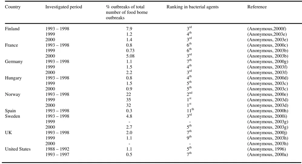
Nowadays many countries register food borne outbreaks and investigate the causative agents. In principle this should lead to better knowledge of the magnitude of the problem of food borne disease. However, since not all food borne diseases are notifiable, the magnitude of the problem is still hard to estimate including the role of B. cereus. Moreover, due to the relatively mild symptoms of B. cereus food borne disease, patients do not generally seek medical attention, thus no test is performed to identify the causative organism. From the available data it can be concluded that B. cereus is a micro-organism frequently causing food borne outbreaks. In The Netherlands it causes the highest number of bacterial food borne outbreaks (Table 1.5). Also in Norway, in 1999 and 2000, B. cereus was responsible for the highest number of bacterial food borne outbreaks (Table 1.6).
Table 1.5 Incidence of Bacillus cereus food borne outbreaks in the Netherlands
Investigated period % outbreaks of total Ranking in bacterial agents Reference number of food borne
outbreaks 1991 – 1994 19 1st (Simone et al. 1997) 1993 – 1998 2.4 1st (Anonymous, 2000b) 1999 – 2000 3.1 1st (Anonymous, 2003a) 2001 3.9 1st (Duynhoven et al. 2002) 2003 4.1 1st (Duynhoven et al. 2004) 2004 2.8 1st (Duynhoven et al. 2005) 2005 3.0 1st (Doorduyn et al. 2006) 2006 5.4 1st (Doorduyn et al. 2007)
Table 1.6 Incidence of Bacillus cereus food borne outbreaks in countries other than The Netherlands
Country Investigated period % outbreaks of total Ranking in bacterial agents Reference number of food borne
outbreaks Finland 1993 – 1998 7.9 3rd (Anonymous,2000f) 1999 1.2 4th (Anonymous,2003e) 2000 1.4 3rd (Anonymous, 2003e) France 1993 – 1998 0.8 6th (Anonymous, 2000c) 1999 0.73 6th (Anonymous, 2003b) 2000 5.08 3rd (Anonymous, 2003b) Germany 1993 – 1998 1.1 7th (Anonymous, 2000g) 1999 1.5 4th (Anonymous, 2003f) 2000 2.2 3rd (Anonymous, 2003f) Hungary 1993 – 1998 0.8 4th (Anonymous, 2000d) 1999 1.5 5th (Anonymous, 2003c) 2000 0.9 5th (Anonymous, 2003c)
Norway 1993 – 1998 22 2nd (Anonymous, 2000e)
1999 35 1st (Anonymous, 2003d)
2000 32 1st (Anonymous, 2003d)
Spain 1993 – 1998 0.3 11th (Anonymous, 2000h)
Sweden 1993 – 1998 4.8 3rd (Anonymous, 2000i)
1999 - - (Anonymous, 2003g)
2000 2.7 5th (Anonymous, 2003g)
UK 1993 – 1998 2.0 7th (Anonymous, 2000j)
1999 1.1 9th (Anonymous, 2003h)
2000 - - (Anonymous, 2003h)
United States 1988 – 1992 1.1 5th (Anonymous, 1996)
Objective of this thesis
Bacillus cereus, a spore forming micro-organism with ubiquitous distribution, is known to cause food borne disease. The short-lasting and relatively mild symptoms do not urge patients to seek medical assistance. Therefore, the real extent of the number of disease cases is hard to determine. Another factor is the lack of knowledge on the mechanisms by which disease is caused. It is known that for the diarrhoeal type B. cereus has to be ingested, has to grow in the small intestine and has to produce enterotoxins. For the emetic type of disease, the emetic toxin, cereulide, has to be produced in food prior to consumption The main objectives of this thesis were the closer investigation of processes in the gastro-intestinal tract that may contribute to the onset of or the protection against B. cereus diarrhoeal disease, and the closer investigation of factors involved in the production of emetic toxin. In combination with prevalence data the outcomes of these investigations should contribute to a better estimate on the incidence of disease.
The investigations can be categorised within the framework of risk assessment, with data on prevalence of the micro-organism falling within the scope of exposure assessment, and data on the pathogenic mechanism falling within the scope of hazard characterization.
The work described in this thesis was carried out within a project named: Quantitative research concerning Bacillus cereus within the framework of risk assessment, with an emphasis on exposure assessment and hazard characterization on the account of the former Food and Commodities Inspectorate, the present Food and Consumer Products Safety Authority.
Outline of this thesis
This thesis deals with two important issues in risk analysis, namely exposure assessment and hazard characterization.
Exposure assessment was addressed by studying the prevalence of potentially pathogenic Bacillus cereus strains in various food commodities. The strains, isolated from randomly sampled food commodities in the Netherlands, were investigated for the presence of genes encoding enterotoxins haemolysin BL (HBL), non haemolytic enterotoxin (NHE) and cytotoxin K (CytK), for the growth temperature profile of the strains (psychrotrophic and mesophilic) by discrimination of the 16S rDNA signature, and for the ability to produce cereulide-like toxin. The results of these characterizations are described in Chapter 2.
Investigations regarding the mechanisms leading to disease, in order to deal with the hazard characterization, focused on part 2 and 3 from the pathogenic mechanism of the diarrhoeal syndrome, presented as the current working model in Figure 1.2.
Figure 1.2 General pathogenesis of the diarrhoeal syndrome.
1. Food contaminated withBacillus cereus (spores and vegetative cells)
2. Gastric passage (only spores survive)
3. Small intestine
a) Germination of spores
b) Growth
c) Production of enterotoxins
d) Interaction of toxins with epithelium
4. Disease symptoms
1. Food contaminated withBacillus cereus (spores and vegetative cells)
2. Gastric passage (only spores survive)
3. Small intestine
a) Germination of spores
b) Growth
c) Production of enterotoxins
d) Interaction of toxins with epithelium
The investigations were carried out using simulations of the gastro-intestinal conditions namely simulated gastric and intestinal fluids to mimic conditions in the stomach and small intestine, and differentiated Caco-2 cells to mimic the epithelial cells of the small intestine.
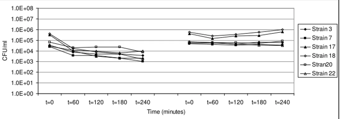
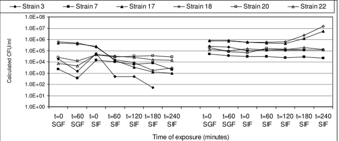
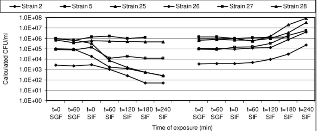
In Chapter 3 the influence of simulated gastro-intestinal fluids on the survival of spores, their germination and growth in simulated intestinal fluid has been investigated. Spores of a selection of strains were exposed to simulated gastric fluid to record their survival capacity, and were exposed to simulated intestinal fluid to investigate their germination and growth potential.
Spores of B. cereus are well equipped to withstand unfavourable conditions such as low pH and elevated temperatures, and can therefore take part in the pathogenesis of the diarrhoeal syndrome. Since the pH in the stomach may vary due to the buffering capacity of the food that functions as a carrier and because of reduced acidification capacity of the stomach such as generally found in elderly people, vegetative cells of B. cereus may survive the gastric passage. Their contribution to the pathogenesis of the diarrhoeal syndrome was determined. The results of these investigations are described in Chapter 4. Exposure to simulated gastric fluid of different acidic pH-values resulted in pH dependent
decimal reduction values (DpH). We used these DpH-values and models for
the course of pH in the stomach during the consumption of food to quantify the number of vegetative cells able to pass the stomach, which after arrival in the small intestine can take part in the pathogenic mechanism.
Upon arrival in the small intestine, spores and vegetative cells come into contact with intestinal fluid and the small intestinal epithelium. Investigations on the interaction between the epithelium and spores are described in Chapter 5. Based on the effect of Caco-2 cells on spore germination, it is suggested that small intestinal epithelial cells may initiate germination of spores. Based on the diversity in Caco-2 induced germination of the B. cereus strains included in this study, the in situ germination capacity may be an important determinant of B. cereus pathogenicity.
The actual diarrhoeal symptoms are caused by enterotoxins produced by B. cereus growing in the small intestine. The behaviour of NHE in simulated intestinal fluid and the interaction of B. cereus cells and
the enterotoxin with epithelial cells were studied more closely and the results are described in Chapter 6.
In Chapter 7, finally, the risks for diarrhoeal and emetic disease are estimated and discussed
2
Prevalence of potentially pathogenic Bacillus
cereus in food commodities in the Netherlands
L.M. Wijnands, J.B. Dufrenne, F.M. Rombouts, P.H. in ’t Veld, F.M. van Leusden
Published in:
Abstract
Randomly selected food commodities, categorized in product groups, were investigated for the presence and number of B. cereus. If positive, and when possible, five separate colonies were isolated and investigated for the presence of four virulence factors: presence of genes encoding three enterotoxins [haemolysin BL (HBL), non haemolytic enterotoxin (NHE), and cytotoxin K], and the ability to produce cereulide. In addition, the presence of psychrotrophic and mesophilic signatures was determined.
The genes for NHE are found in more than 97% of the isolates, those for HBL in approximately 66% of the isolates, and the gene for cytotoxin K in nearly 50% of the isolates. Significant associations between product groups and (combinations of) virulence factors were: the relatively low percentage of isolates from the “flavourings”-group containing genes encoding NHE and the higher than average occurrence of both the genes encoding HBL and NHE in the “pastry”-group. Cereulide was produced by 8.2 % of the isolates, but only in combination with the presence of genes for one or more other virulence factors.
Most isolates (89.9 %) were mesophilic, minorities of the isolates were psychrotrophic (4.4 %) or of intermediate signature (5.7 %). In the product group “milk and milk products” the incidence of strains with psychrotrophic or intermediate signatures is significantly higher than in the other product groups.
In the product groups “flavourings”, “milk and milk products”, “vegetable(s) and vegetable products”, “pastry”, and “ready-to-eat foods” a relatively high number of samples contain high numbers of B. cereus. Within the product group “ready-to-eat foods”, the products containing rice and pasta show a relatively high incidence of high numbers of B. cereus.
Introduction
Bacillus cereus is a ubiquitous spore forming Gram positive micro-organism, capable of causing two types of food borne disease. The emetic syndrome (an intoxication) is caused by cereulide, a pH and temperature stable cyclic peptide toxin (Agata et al. 1995b), and disease may arise after the ingestion of food contaminated with cereulide. The diarrhoeal syndrome (a toxico-infection) is caused by enterotoxins, which are produced by vegetative cells growing in the small intestine after consumption of food contaminated with spores and/or vegetative cells (Kramer and Gilbert, 1989). To date three enterotoxins, capable of inducing the diarrhoeal symptoms of the toxico-infection have been described (Granum, 2007). These are the three-component enterotoxin haemolysin BL (HBL), the three-component enterotoxin non haemolytic enterotoxin (NHE), and the single-component enterotoxin cytotoxin K. The symptoms caused by cytotoxin K are more severe than those caused by the other two enterotoxins: cytotoxin K not only causes watery diarrhoea but also necrosis. Based on the mode of action the (entero)-toxins ought to be regarded as virulence factors.
Another important feature of B. cereus is the ability to grow at a wide range of temperatures. However, grouping of strains according to growth temperature is confusing. Where one definition divides strains in those with a high temperature growth range (10-42ºC) and those with a low temperature growth range (4-37ºC), another definition divides strains upon their ability to grow below 7ºC. Psychrotrophs can grow below 7ºC; mesophiles cannot (Meer et al. 1991; von Stetten et al. 1998). In food microbiology mesophiles are characterized as strains with a temperature optimum around 37ºC, while the optimum for psychrotrophs lies around 25-30ºC (Adams and Moss, 2000).
Although the potential to cause disease is known, little to no details are known about the number of B. cereus cells necessary to cause either of the syndromes, let alone about the dose of (entero)-toxin necessary to induce disease. From outbreak data, the number of B. cereus,
able to cause disease, has been estimated to be higher than 103 cells per
gram food (Granum, 2007).
In order to improve the knowledge on exposure assessment with respect to both syndromes, more data on the prevalence of B. cereus in daily food commodities should be available. Previous investigations yielded data on B. cereus in milk (Larsen and Jorgensen, 1997; Te Giffel et
al. 1996; Te Giffel et al. 1996) and data on B. cereus in other food commodities such as refrigerated processed foods of extended durability (REPFED’s) (Choma et al. 2000; Del Torre et al. 2001) and chicken products (Smith et al. 2004). Choma et al. (2000) investigated REPFED samples for the presence of B. cereus and found low initial numbers (< 10
CFU.g-1). The growth characteristics of the isolates were determined during
storage of the products at various temperatures, and cytotoxicity of supernatants of the isolates was investigated. Instead of investigating the cereulide producing potential of the isolates, Choma et al. (2000) determined their starch degrading properties. This feature is linked to but not qualifying for the production of cereulide. Del Torre et al. (2001) found that 33% of REPFED samples were contaminated with low initial levels (<
102 CFU.g-1) of B. cereus but did not investigate the isolates for virulence
factors. Smith et al. (2004) detected B. cereus in 27 of 60 samples of chicken products after enrichment. Testing the isolates for the presence of enterotoxin coding genes by PCR, resulted in finding no isolates carrying the cytotoxin K gene and a high prevalence of the genes coding for the non haemolytic enterotoxin. Smith et al. (2004) did not study the cereulide producing ability of their isolates.
Since (entero-)toxins cause the symptoms of the emetic and diarrhoeal syndrome, an inventory on the prevalence of these virulence factors in food commodities would be preferable over an inventory on B. cereus itself. However, direct detection methods are not (yet) available for all enterotoxins and the methods available to detect cereulide have not yet been optimized for the direct detection [boar sperm test (Andersson et al. 1998) and HEp-2 cell test (Finlay et al. 1999)], or are too expensive to use for large scale investigations [LC-MS method (Häggblom et al. 2002)].
The aim of the investigations described in this paper is to make an inventory of the occurrence of potentially pathogenic B. cereus strains in retail food commodities in The Netherlands. Strains are considered potentially pathogenic when possessing the genes encoding one or more (entero-) toxins, and/or when able to produce cereulide in vitro. Also the growth temperature profile of each strain was determined, as earlier research has shown a relation between this trait and the importance for the onset of diarrhoeal disease.
Materials and Methods
Collection of samples and isolates
The Food and Consumer Product Safety Authority (VWA) collected food samples at retail level. The food samples were investigated for the presence and, in most cases, also for the level of Bacillus cereus by
plating decimal dilutions on MEYP2-agar. Occasionally only the 10-2, 10-3,
or the 10-4-dilution (the Dutch legal limit being 105 B. cereus ml-1 or g -1
for most foods) was investigated, and presence or absence was recorded. When B. cereus was present in a food sample, and if available, five typical colonies were selected and sent to the National Institute for Public Health and the Environment (RIVM) for further characterization of the isolates. Viable counts were available from most samples.
The isolates sent for further investigation did not cover all positive B. cereus samples: only some of the samples were used for extensive characterization of their isolates.
Classification of the samples
The VWA uses a classification for the food commodities which is based on the former “Kokswarenbesluit” (Anonymous, 1979). Accordingly, the collected food samples were classified in the following product groups which comprise food commodities used for regular meals: pastry, vegetable(s) and vegetable products, ready-to-eat foods, milk and milk products, oil(s) / fat(s) and oil and fat products, flavourings, fish and fish products, and meat and meat products. All samples from the milk and milk products group were from commodities that need storage at refrigerator temperatures according to the manufacturers.
Preparation and storage of isolates
All isolates were grown on Columbia agar with sheep blood (overnight at 30°C) for purity assessment. If pure, the isolate was subcultured on Tryptone Soy agar (TSA) in a 9 cm Petri dish for ten days at 30°C for cereulide production, and in 10 ml Brain Heart Infusion broth (BHI) for 1 day at 30°C for DNA extraction. Also, the isolate was stored at -70°C by adding 200 l BHI culture to a tube containing glass beads and 200 l glycerol.
Determination of ability to produce cereulide
The colonies were collected from the TSA plates with a cell scraper and suspended in 1 ml methanol in 1.5 ml micro centrifuge tubes (Eppendorf AG, Hamburg, Germany). After vigorous mixing on a vortex-mixer the tubes were placed on a blood tube rotator for 30 minutes for homogenization of the suspension. This suspension was centrifuged (5 min, 13,000xg), and the supernatant transferred to a glass tube. The supernatant was dried at ca. 60°C with a flow of nitrogen. The residue was resuspended in 1 ml cell culture medium and stored at 4°C until further investigation. Further investigation consisted of screening 5 two-fold dilutions (1:2 to 1: 32) of the extracts for the presence of cereulide using the previously described HEp-2 cell test (Finlay et al. 1999). The optical densities (OD’s) of the dilutions of the isolates were compared to OD’s of positive and negative controls (extracts from NCTC 11143 and NCTC11145, respectively). Resemblance of the sample OD’s with the OD’s of the negative or positive control, determined the sample to be either negative or positive. If no conclusion could be drawn from this test investigation of the extract was extended to dilutions starting at 1: 8 and ranging to 1: 16,392. Optical densities were plotted against the dilutions and compared with the positive and negative control. If showing the same trend in optical density as the positive control an extract was recorded as positive. In all other cases extracts were designated as negative. As positive results were not confirmed by LC-MS, all positive isolates are referred to as producing the cereulide-like compound.
Detection of genes for enterotoxins
After the overnight cultivation in BHI, DNA was collected from the B. cereus strain by using the Wizard Genomic DNA Purification Kit (Promega, Madison WI, USA). Purified DNA was stored at -20°C until further use.
The presence of enterotoxin genes was determined by various PCR protocols. For detection of the haemolysin BL (HBL)–genes, haemolytic enterotoxin (NHE)-genes and the cytotoxin K-gene previously described protocols were used (Guinebretiere et al. 2002; Heinrichs et al. 1993; Ryan et al. 1997). The reverse primer for the cytotoxin K PCR, as described by Guinebretiere et al. (2002), was replaced by a primer with the following sequence: TCC AAC CCA GTT (A T) (G,C), as the wrong sequence for this primer was mentioned in the publication (M.H.
Guinebretiére, personal communication). The three components of HBL are encoded by a single operon. Therefore, if any of the three genes encoding HBL was detected we assumed all three HBL genes were present. The same strategy was use for the detection of NHE genes.
Determination of growth temperature profile
The distinction between psychrotrophy and mesophily was determined by using a previously described PCR-method (von Stetten et al. 1998), detecting a difference in the 16S rDNA gene. After electrophoresis of the PCR products, mesophilic strains are recognized by a 250 base pair (bp) fragment and psychrotrophic strains by a 180 bp fragment. Intermediate strains possess both fragments upon electrophoresis. Earlier results show a good correlation between assessing growth temperature profile by culture at various temperatures and the PCR method (Prüss et al. 1999)
Results
Table 2.1 Distribution of samples and isolates of Bacillus cereus as determined on
MEYP-agar over the product groups which were investigated for virulence factors
Product group Samples Isolates Isolates per sample Oil(s) and fat(s) and oil
and fat products 1 5 5 Fish and fish products 6 15 2.5 Meat and meat products 6 24 4
Flavourings 22 92 4.2
Milk and milk products 17 80 4.7 Ready-to-eat foods 81 384 4.7 Vegetable(s)
and vegetable products 30 115 3.8
Pastry 19 81 4.3
For this study 796 isolates, originating from 182 samples, were investigated for the following factors: presence of genes encoding the enterotoxins, ability to produce cereulide, and growth temperature signature. The distribution of the samples and isolates over the product groups as defined in the “Kokswarenbesluit” (Anonymous, 1979) and employed by the VWA is shown in Table 2.1. These isolates, sent to the RIVM for further investigation, originated from approximately 10% of all the samples investigated by the VWA (the selection was random).
Table 2.2 Distribution of growth temperature signatures in numbers (and percentages) of isolates of Bacillus cereus in various food product groups. The total number of isolates per product group is shown in the bottom line. The total number of isolates (and percentages) per growth temperature signature is shown in the last column
Growth Oil(s) and fat(s) and Fish and Meat and Flavourings Milk and Ready-to-eat Vegetable(s) and Pastry Total # isolates (%) temperature oil and fat products fish products meat products milk products foods vegetable products per signature signature [# isolates (%)] [# isolates (%)] [# isolates (%)] [# isolates (%)] [# isolates (%)] [# isolates (%)] [# isolates (%)] [# isolates (%)]
Mesophilic 5 (100)a 9 (60) 19 (79.2) 91 (98.9) 58 (72.5) 356 (92.7) 105 (91.4) 73 (90.1) 716 (89.9)
Psychrotrophic 0 (0) 6 (40) 5 (20.8) 0 (0) 5 (6.3) 12 (3.1) 5 (4.3) 2 (2.5) 35 (4.4) Intermediate 0 (0) 0 0 1 (1.1) 17 (21.2) 16 (4.2) 5 (4.3) 6 (7.4) 45 (5.7)
Total # isolates 5 15 24 92 80 384 115 81 796
The distribution of the growth temperature signatures over the various product groups is shown in Table 2.2. The distribution is given in absolute numbers of isolates per profile per product group and in percentages of isolates per product group. In the last column the overall numbers of isolates per profile are shown with their corresponding percentages. The majority of the isolates is mesophilic (89.9%), and only minorities are either psychrotrophic or of intermediate signature (4.4% and 5.7% respectively). Remarkably, none of the strains from the “flavourings”-group have a psychrotrophic signature, although a large group of samples originates from this product group. Apart from one isolate (1.1%) which is of intermediate nature, all other isolates (98.9%) have mesophilic signatures and presumably do not grow at temperatures below 10°C (Adams and Moss, 2000).
In order to determine whether a significant number of samples from a certain product group is different from the overall results with respect to growth temperature profile, the mean percentages per growth temperature profile with the 95% confidence intervals were determined. This was accomplished by adding up the percentages of the different groups, dividing the total by the number of groups and determining the confidence interval of the mean value. Taking all product groups into account, the results are shown in Table 2.3, in columns “mean” and “interval”, respectively. The product groups that do not fall within the confidence interval, and that are therefore significantly different, are shown in Table 2.3 in columns “below interval” and “above interval”, respectively. As the product groups with small numbers of isolates might bias the result, the same calculations were performed with product groups with > 80 isolates only. Also similar calculations with product groups with >80 isolates were carried out excluding the “milk and milk products” group. Both calculations indicated that a significant number of isolates from the “milk and milk products” group has a psychrotrophic signature (data of the last calculations not shown).
Notably, all isolates able to produce the cereulide-like toxin are of mesophilic nature. This is in concurrence with results previously described by (Pielaat et al. 2005).


