A COMPARISON OF THE MUSCLE ELASTICITY
OF THE HAMSTRING MUSCLE COMPLEX
BETWEEN HEALTHY MALE AND FEMALE
SOCCER PLAYERS USING SHEAR-WAVE
ELASTOGRAPHY
Artoer Galimov, Nicholas Janssens, Tom Maes
Student number: 01403388, 01404981, 01408575
Promotor: Prof. Dr. Erik Witvrouw
Copromotors: Dr. Evi Wezenbeek, Drs. Dries Pieters
A dissertation submitted to Ghent University in partial fulfilment of the requirements for the degree of Master of Rehabilitation Science and Physiotherapy.
A COMPARISON OF THE MUSCLE ELASTICITY
OF THE HAMSTRING MUSCLE COMPLEX
BETWEEN HEALTHY MALE AND FEMALE
SOCCER PLAYERS USING SHEAR-WAVE
ELASTOGRAPHY
Artoer Galimov, Nicholas Janssens, Tom Maes
Student numbers: 01403388, 01404981, 01408575
Promotor: Prof. Dr. Erik Witvrouw
Copromotors: Dr. Evi Wezenbeek, Drs. Dries Pieters
A dissertation submitted to Ghent University in partial fulfilment of the requirements for the degree of Master of Rehabilitation Science and Physiotherapy.
Dankwoord
Met graagte schrijven wij een dankwoord voor alle personen die ervoor gezorgd hebben dat deze masterproef tot zijn geheel kon worden gebracht.
Ten eerste bedanken wij onze promotor prof. dr. Erik Witvrouw en copromotor dr. Evi Wezenbeek voor de mogelijkheid tot het schrijven van deze masterproef. Vervolgens willen wij graag onze copromotor drs. Dries Pieters hartelijk danken voor de begeleiding, bijsturing, advies en vele hulp gedurende het hele onderzoek. Zijn interesse en gedrevenheid omtrent dit onderwerp werd door ons ten zeerste op prijs gesteld.
Ook richten wij graag een woord van dank richting de faculteit Geneeskunde en Gezondheidswetenschappen, het UZ Gent en diens werknemers voor het aanbieden van al het vereiste materiaal en de geschikte lokalen tijdens het onderzoek.
Met plezier danken wij onze familieleden, vrienden en medestudenten
Revalidatiewetenschappen en kinesitherapie voor de tips, hulp en continue ondersteuning tijdens het hele proces.
Ten laatste bedanken wij elkaar voor de goede samenwerking en communicatie maar vooral de steun en het vertrouwen waarop gerekend kon worden.
Table of contents
1. Abstract: English ...7 2. Abstract: Dutch ...9 3. Introduction ... 11 4. Methods ... 14 4.1. Participants... 14 4.2. Test procedure ... 14 4.3. Outcome measurements ... 154.3.1. Pain-free passive knee extension test (PKET) ... 15
4.3.2. Isometric mid-range strength test (IMST) ... 15
4.3.3. Single leg hamstring bridge test (SLHB) ... 16
4.3.4. Shear-wave elastography (SWE) ... 16
4.4. Statistical analysis ... 17
5. Results ... 18
5.1. Participants characteristics ... 18
5.2. Comparison between male and female soccer players ... 18
5.3. Comparison of stiffness between the different hamstring muscles within both sexes... 20
5.4. Correlations ... 21
5.5. Influence of variables ... 22
6. Discussion ... 23
6.1. Summary of evidence ... 23
6.1.1. Male and female population: a comparison ... 23
6.1.2. Comparison between different hamstring muscles ... 25
6.1.3. Correlations ... 26
6.1.4. Influence of variables ... 27
6.2. Limitations and strengths ... 27
6.3. Clinical implications and perspectives ... 28
7. Conclusion ... 29
8. References ... 30
9. Abstract in lekentaal ... 34
List of tables and figures
Table 1: Participants characteristics .18
Table 2: Results Independent T-Test 18
Table 3: Results comparison within male population .20
Table 4: Results comparison within female population ...20
Table 5: Correlations SWE male and female population 22
Table 6: Correlations IMST, PKET and SLHB male and female population 22
Table 7: MANOVA results male and female population .22
Figure 1: Boxplots of muscle stiffness by sex for each hamstring muscle 19
Figure 2: B ff e f d ffe e c e b e e 21
List of abbreviations
SWE: Shear-Wave Elastography RTS: Return To SportsST: Semitendinosus SM: Semimembranosus BF: Biceps Femoris
BFlh: Biceps Femoris Long head BFsh: Biceps Femoris Short head TA: Tibialis Anterior
IMST: Isometric Mid-range Strength test PKET: Passive Knee Extension Test SLHB: Single-Leg Hamstring Bridge ROM: Range Of Motion
BMI: Body Mass Index
1. Abstract: English
Background: Soccer is a high-intensity sport with a high occurrence of hamstring injuries. As muscle stiffness affects the behavior of the hamstring muscles, it is important to gain insight into this subject. A recent non-invasive technique, shear-wave elasticity (SWE), is proven to be reliable to measure the muscle stiffness. Although it is proven to be of great value, few studies have investigated the difference in muscle stiffness using the SWE technique, between men and women. Additionally, not many researchers examined a possible correlation between the stiffness and the flexibility of the hamstring muscles.
Objectives: The main purpose of this study was to make a comparison of the passive
hamstring muscle stiffness, measured with the SWE, between healthy male and female soccer players. Secondly, the passive muscle stiffness was compared in-between all three hamstring muscles for both sexes. Subsequently, possible correlations between the SWE and the hamstring strength and flexibility were examined.
Study Design: Cohort Study
Methods: Twenty healthy male (n: 20, age: 22,75 ± 3,75) and thirteen healthy female (n: 13,
age: 22,71 ± 2,61) soccer players, with no history of hamstring injuries, were recruited for this study. Primarily, the passive stiffness of all three hamstring muscles was examined, with the participants placed in a passive neutral position (0° hip and knee flexion). Furthermore, the isometric hamstring strength, the hamstring flexibility and the functional hamstring strength were measured with an Isometric Mid-range Strength Test (IMST), a Passive Knee Extension Test (PKET) and a Single-Leg Hamstring Bridge (SLHB) respectively.
Results: A comparison of the SWE measurements between both sexes, showed no significant
difference for the stiffness of the Biceps Femoris Long Head (BFlh) muscle (p=0.556), the Semimembranosus (SM) muscle (p=0.200) and the Semitendinosus (ST) muscle (p=0.633). Within the male population, the BFlh had a significantly lower muscle stiffness than the ST (p<0.001) and the SM (p<0.001). Within the female population, the ST had a significantly higher muscle stiffness than the SM (p<0.05) and the BFlh (p<0.001). For both the male and female population a de a e c e a (0.40 0.69) a ee be ee e stiffness of the SM and the PKET.
Conclusion: Shear-wave elastography measurements of the hamstring muscles showed no
difference in passive muscle stiffness between both sexes. The BFlh showed the lowest passive muscle stiffness, whilst the ST proved to have the highest passive muscle stiffness for both sexes. For both male and female soccer players, only the stiffness of the SM showed a moderate correlation with the PKET.
2. Abstract: Dutch
Achtergrond: Voetbal is een sport met een hoge intensiteit, wat een hoge incidentie aan
hamstringblessures met zich meebrengt. Daar de spierstijfheid een invloed kan hebben op het gedrag van de hamstringspieren, is het belangrijk inzicht te krijgen in dit onderwerp. Een nieuwere techniek, shear-wave elastografie (SWE), wordt als betrouwbaar gezien voor het opmeten van de spierstijfheid. Ondanks dat SWE gezien wordt als een betrouwbare techniek, is er tot op heden nog weinig onderzoek uitgevoerd naar een mogelijk verschil in spierstijfheid tussen mannen en vrouwen, met gebruik van deze techniek. Aansluitend zijn er maar weinig studies die de correlatie onderzoeken tussen de stijfheid en flexibiliteit van de hamstrings.
Doelstellingen: Het belangrijkste doel van deze studie was het maken van een vergelijking
van de passieve spierstijfheid van hamstrings tussen gezonde mannelijke en vrouwelijke voetbalspelers. Verder werd de passieve spierstijfheid van de hamstrings onderling vergeleken voor beide geslachten. Vervolgens werden mogelijke correlaties tussen de SWE en de hamstrings kracht en lenigheid onderzocht.
Onderzoeksdesign: Cohorten Studie.
Methode: Twintig gezonde mannelijke (n: 20, leeftijd: 22,75 ± 3,75) en dertien vrouwelijke (n:
13, leeftijd: 22,71 ± 2,61) voetbalspelers, zonder hamstringblessures in de voorgeschiedenis, werden gerekruteerd voor deze studie. Allereerst werd de stijfheid (SWE) van de drie hamstringspieren van alle deelnemers in passieve neutrale positie (0° heup- en knieflexie) opgemeten. Aansluitend werd de isometrische kracht, de lenigheid en de functionele kracht opgemeten met respectievelijk de Isometric Mid-Range Strength Test (IMST), de Passive Knee Extension Test (PKET) en de Single-Leg Hamstring Bridge (SLHB).
Resultaten: In de vergelijking van de SWE-metingen tussen mannen en vrouwen werd er
geen significant verschil gevonden, voor de stijfheid van zowel de Biceps Femoris long head (BFlh) (p=0.556), als de Semimebranosus (SM) (p=0.200) en de Semitendinosus (ST) (p=0.633). Binnen de mannelijke populatie, vertoonde de BFlh significant minder stijfheid in vergelijking met de ST (p<0.001) en de SM (p>0.001). Binnen de vrouwelijke populatie, vertoonde de ST significant meer stijfheid in vergelijking met de SM (p<0.05) en de BFlh
(p<0.001). Voor zowel de mannen als de vrouwen werd er een matige (0.40 r 0.69) correlatie gezien tussen de stijfheid van de SM en de PKET.
Conclusie: Shear-wave elastografie metingen van de hamstrings toonden geen verschil
tussen beide geslachten. De BFlh bleek de minst passieve spierstijfheid te vertonen, terwijl de ST de meest passieve spierstijfheid bleek te hebben. Voor zowel mannelijke als vrouwelijke voetbalspelers, toonde alleen de stijfheid van de SM een matige correlatie met de PKET.
Sleutelwoorden: SWE, passieve spierstijfheid, hamstringspieren, geslachtsvergelijking,
3. Introduction
The hamstring muscles play a significant role in numerous static and dynamic movements such as walking, standing, jumping and running(1, 2). Subsequently, these muscles are important in sports and in particular in soccer. Worldwide, soccer is seen as one of the most common sports practiced by both men and women, regardless of the level of competition. As hamstrings are an important anatomical structure for human performance, this muscle complex has a high occurrence of strain injury in sports, in particular in soccer where it is the most common musculoskeletal injury subtype(3). Muscle injuries in soccer constitute 31% of all injuries, of which 37% affected the hamstring muscles(4). Besides a frequent occurrence of initial hamstring injuries, a high reinjury rate exists in sports. Approximately 50% of the athletes suffer a reinjury within the first 25 days after return to sports (RTS). Even worse, 25% of them have a reinjury in the first week after RTS. Furthermore, reinjury occurred almost every time at the same location as the initial injury(5).
Anatomically, this group of muscles consists of three main heads: the biceps femoris (BF), the semitendinosus (ST) muscle and the semimembranosus (SM) muscle. The BF consists of two heads: the long head (BFlh) and the short head (BFsh). Like the ST and the SM muscles, the BFlh is bi-articular which ensures they provide movement in the hip as well as movement in the knee, more specifically flexion of the knee and extension of the hip. The BFsh is mono-articular, which means it only provides knee flexion(1).
Due to a substantial loading during dynamic movements, such as high velocity running, the hamstring muscles are exposed to a considerable amount of stress at long muscle lengths(1). A previous study showed that during running the activity of the hamstring muscles reached its peak in the late toe-off phase and at initial foot contact, being an eccentric activity due to lengthening of the hamstring muscles(6). During this activity, the eccentric hamstring strength provides dynamic joint stability. Additionally, this eccentric strength minimizes the injury risk, given that poor eccentric strength is a cause of hamstring injury(7, 8).
Tests to measure the hamstring strength are listed in the ASPETAR hamstring protocol(9). Examples of such tests are isometric and eccentric strength tests. This hamstring protocol was considered useful to reassess the progression of a hamstring rehabilitation program and therefore, this protocol could be used to determine the implementation of load during different rehabilitation stages(9).
Another assessment to functionally objectify the hamstring strength is the Single Leg Hamstring Bridge (SLHB). In the study of Freckleton et al(10) this test has proven its contribution to clinically examine the risk of a hamstring injury. Furthermore, the SLHB might be used to evaluate the physical fitness of the athlete to return to sports and could contribute as an evaluation after a rehabilitation program for hamstring injury(10).
Produced strength during dynamic movement implies a great amount of stress on the hamstring muscle complex, which must be resisted and is determined by the flexibility of the hamstrings(1, 11). There are various tests available to objectify the flexibility of the hamstring complex. For instance, there is a wide range of tests useable in the ASPETAR hamstring protocol(9), such as the Passive Straight Leg Raise Test, the Pain-Free Passive Knee Extension Test (PKET) and the Maximal Hip Flexion Knee Extension test. These assessments, which are based on the range of motion (ROM) measurements, elongate numerous structures besides muscle tissue, such as joints, ligaments, blood vessels and nerves(12).
A more specific and objective technique to measure tissue stiffness is shear-wave ultrasound elastography (SWE)(13). This is a method of ultrasound imaging, which is based on the detection of through the tissue propagated shear waves. Such ultrasound technique presents tremendous use in evaluating the complex skeletal architecture of more superficial muscles and its mechanical properties. With the aid of an inversion algorithm, the waves are mapped into elastograms and as a result of this charting, the tissue stiffness can be determined by calibrating the shear modulus value(14). Thus, such ultrasound-based technology provides the opportunity to reliably measure the muscle shear elastic modulus and tissue properties in a non-invasive way(15). It measures muscle quality and implements a spatial representation of soft tissue stiffness(13). Tissue stiffness is expressed as the magnitude of stress divided by the strain induced by the stress (formula: stiffness = stress/strain). It is defined as the resistance to deformation and measured as the elastic modulus(16). While SWE is considered a reliable method to measure muscle tissue stiffness, only a few studies investigated the difference in muscle stiffness between sexes. Previous research, investigating the active stiffness of the m. tibialis anterior (TA) showed no difference between sexes(17). Nevertheless, a study investigating the BFlh of healthy male and female basketball players showed a higher passive muscle stiffness for the male population(18). This shows the evidence, considering a possible difference in tissue stiffness between the male and female, is still inconclusive.
Therefore, the main purpose was to compare the passive stiffness of all hamstring muscles between the male and the female soccer players, using SWE. This intersexual comparison
was also made for the PKET, isometric strength test (IMST) and SLHB. The second goal was to make a comparison of passive muscle stiffness between all three hamstring muscles within both sexes.
The third purpose was to find a possible correlation between the passive muscle stiffness of the hamstring muscle complex and the flexibility of the hamstring complex. Furthermore, all possible correlations between the different tests were analyzed. Finally, the influence of different variables on all outcome measurements was analyzed.
4. Methods
4.1. Participants
This study included 33 Belgian soccer players aged between 18 and 33 years. The participants consisted of 13 female players with a mean age of 22,71 years ± 2,61. Furthermore, 20 male players were included with a mean age of 22,75 years ± 3,75. The entire sample of this study had a mean age of 22,74 ± 3,29.
The researchers used multiple tools to gather participants. First word-of-mouth, social media and a written questionnaire were used. In the next phase, an online questionnaire was composed for possible participants who were screened on inclusion and exclusion criteria. In the last phase of recruitment, the help of friends and family was utilized.
In order to partake in this study, the participants had to meet the following criteria: the athletes had to be 18 years or older and had to participate in minimal two practices and one competitive game per week. Potential participants were excluded from this study if they had a history of hamstring injuries or recent severe trauma or surgery in the lower extremity within the previous six months.
The Ethical Committee UZ Ghent (Ghent University, Belgium) approved this study (approval number: EC/2019/0909).
4.2. Test procedure
The testing took place between February the 10th and February the 14th 2020 at UZ Ghent. During the testing, 50 minutes were provided for each participant.
The test battery used in this study consisted of five different stations. Firstly, every participant was asked to fill in a questionnaire while lying in a prone position on a treatment table for ten minutes in order to minimize the possible effects of previous activity. Information such as length, weight, BMI, medical history, current injuries, leg dominance, weekly hours of sport and level of competition were gathered from this questionnaire. Following these ten minutes, SWE measurements were executed bilaterally. These measurements lasted approximately 20
minutes. The three remaining tests were a flexibility test (PKET), an isometric strength test (IMST) and a functional hamstring strength test (SLHB), which were performed on both legs. To randomize the sequence of these three tests every participant had to pick one of three cards blindly, each with a different test written on it. After performing the first test, the participant had to blindly pick one of the two remaining sheets. This second test was then executed, followed by the last remaining test. There was one minute of rest between every test to reduce possible influences of the tests on each other.
4.3. Outcome measurements
4.3.1. Pain-free passive knee extension test (PKET)
The flexibility was measured with the PKET, a test proven to be reliable to evaluate hamstring tightness in healthy subjects(19). To ensure consistency, every PKET was performed by the same examiner using a digital ACUMAR inclinometer. By placing this inclinometer on a vertical wall, it was calibrated to ensure the vertical plane was seen as 90°. Every participant had to lay down barefooted in a supine position on a treatment table. Using the inclinometer, the examined leg was placed in 90° hip flexion. Once 90° hip flexion was reached, the inclinometer was placed on the tibia of the examined leg, just below the patella. Then, the leg was extended passively by the examiner until a maximum tolerable stretch in the hamstring muscle was reached. The knee angle of this position was used as the test result, expressed in degrees knee flexion. After testing the first leg, both hips were flexed maximally and circumduction in both directions (left and right) was executed to reduce a possible shift of the pelvis. Afterward, the second leg was tested using the same procedure. To randomize the side tested first, a coin was flipped where heads meant starting with the right leg and tails meant starting with the left leg.
4.3.2. Isometric mid-range strength test (IMST)
To measure the isometric muscle strength, the same examiner performed the IMST for every participant. This isometric hamstring test, measured with a hand-held dynamometer (HHD), was proven to be valuable and reliable in a previous study(20). The participant was asked to lie on the treatment table barefooted and in a prone position. The knee was flexed by the examiner until the heel was raised one-foot length above the table to obtain approximately 30° knee flexion. The HHD was placed against the posterior heel of the participant. Then, the participant
performed a maximal isometric knee flexion against the HHD. First, the participant had to build up its strength for three seconds followed by delivering a maximal isometric hamstring contraction for three seconds. This was repeated three times on both legs with the best repetition of each side, expressed in Nm, being used for further analysis. To randomize the side tested first, a coin was flipped again where heads meant starting with the right leg and tails meant starting with the left leg.
4.3.3. Single leg hamstring bridge test (SLHB)
To evaluate the functionality of the hamstring muscles, the SLHB was repeatedly performed by the same researcher for each participant. This test previously showed to be reliable(10). The participant was asked to lie supine, this time wearing shoes, with one heel on a box with a height of 60 cm. The tested leg was positioned in approximately 20° knee flexion by the use of a goniometer and the participant was instructed to push its heel into the box in order to lift the hip from the ground. The other leg was positioned stationary by the examiner in a vertical position with relaxed knee flexion. The purpose of this test was to perform as many repetitions as possible until failure or until compensations (e.g. loss of knee flexion, incomplete hip extension) occurred. To perform a repetition correctly, the participant had to touch the ground with its bottom without resting and then extend the hip to 0° flexion. Feedback was given during the test. A warning was given the first time any compensation occurred. When the correct posture was lost a second time, the test was terminated and the repetition maximum was noted. Afterward, the test was performed on the other leg. To randomize the side tested first, a coin was flipped where heads meant starting with the right leg and tails meant starting with the left leg.
4.3.4. Shear-wave elastography (SWE)
Shear-wave elastography is a technique to measure tissue stiffness. It uses ultrasound imaging, which is based on the detection of through the tissue propagated shear waves(14). T e e ff e a ea ed a Aixplorer ea a e e a a de ce (version Mach 30; Supersonic Imagine, Aix-en-Provence, France) coupled with a linear transducer array (5-18 MHz; SuperLinear L18-5; Vermon, Tours, France) in SWE mode (musculoskeletal pre-set, Penetrate Mode, Smoothing level 5, persistence OFF, scale 0-100 kPa). All elasticity measurements were performed by the same operator, who was trained extensively by experts in the field of ultrasound imaging. The participants were placed in a
prone position with 0° hip and knee flexion and the ankles in a relaxed position. This position had to be maintained throughout the measurement. Initially, the distance between the ischiatic tuber and the lateral femur epicondyle was measured and the midpoint of this distance was marked. At this location, the stiffness measurements were executed. The specific location of the measurement for each muscle was searched in B-mode US, with an axial orientation of the probe. After which the probe was placed in a longitudinal position, parallel to the muscle fiber direction, to perform the actual measurements in SWE-mode. Initially, the ST and the SM muscles were examined, followed by the BFlh. This procedure was implied for every participant. The SWE-mode showed a box that represented the level of stiffness using a color index. The SWE measured the shear wave velocity which was automatically converted into the shear modulus. The shear wave velocity is expressed in m/s, while the shear modulus is expressed in kPa. Because the shear modulus is the most common outcome measure to quantify the tissue stiffness, only the values expressed in kPa were used for further analysis. To ensure reliable results, the participants were asked to hold their breath during the actual measurement and to remain as still as possible during the entire test procedure. It is important to mention that whenever muscle stiffness of this present study was discussed, it concerned passive muscle stiffness.
4.4. Statistical analysis
The obtained data were analyzed using the IBM SPSS Statistics 25.0 (IBM Corporation, New York, USA). Initially, SPSS was used to calculate the averages and standard deviations for each variable. For the first goal, Independent T-tests were performed to compare the results between the male and female soccer players. This for the SWE measurements as well as for the PKET, IMST and SLHB. In addition, boxplots were executed in order to visualize possible differences. For the second research question, the Paired T-test was used to compare muscle stiffness between all three hamstring muscles within both sexes. The population was split by gender to examine the male and female participants separately. For the third goal, the Pearson correlation test was used to examine possible correlations between all tests. The population was divided by gender. Finally, Multivariate Analysis of Variance (MANOVA) was used to investigate the possible influence of multiple variables on the outcome of the SWE measurements. As before, the population was split by gender and once again the used variables were age, BMI, leg dominance and weekly hours of training.
5. Results
5.1. Participants characteristics
The participants characteristics are represented in Table 1.
Number Age (years)
Average + SD
BMI (kg/m2) Average + SD
Sports participation (hours/week) Average + SD
Male population 20 22,75 ± 3,75 22,93 ± 2,36 5,78 ± 0,83
Female population 13 22,71 ± 2,61 20,99 ± 1,07 6,12 ± 2,14
Table 1: Participants characteristics
5.2. Comparison between male and female soccer players
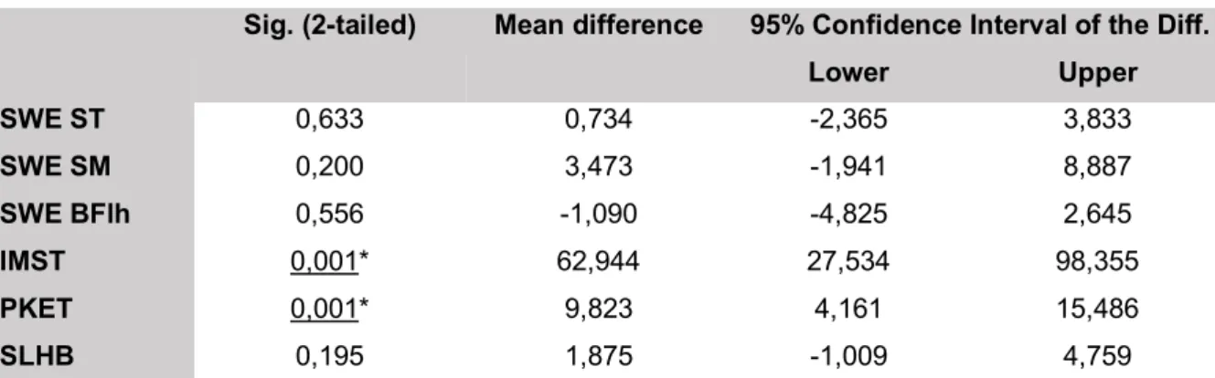
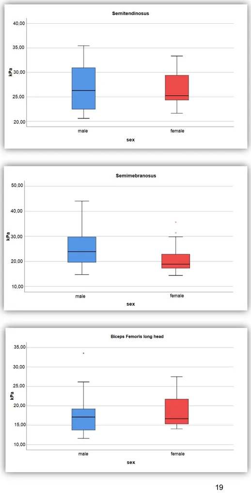
The SWE measurements of the ST, SM and BFlh showed no significant difference between the male and female soccer players, as seen in Table 2 and Figure 1.
For the IMST, male soccer players showed significantly more isometric strength (p<0.01). Furthermore, female soccer players showed significantly more hamstring flexibility (p<0.01), measured with the PKET, all seen in Table 2.
As seen in Table 2, The SLHB showed no significant difference between both sexes.
Sig. (2-tailed) Mean difference 95% Confidence Interval of the Diff.
Lower Upper SWE ST 0,633 0,734 -2,365 3,833 SWE SM 0,200 3,473 -1,941 8,887 SWE BFlh 0,556 -1,090 -4,825 2,645 IMST 0,001* 62,944 27,534 98,355 PKET 0,001* 9,823 4,161 15,486 SLHB 0,195 1,875 -1,009 4,759
Figure 1: Boxplots of muscle stiffness by sex for each hamstring muscle.
5.3. Comparison of stiffness between the different hamstring muscles
within both sexes
Within the male population, the BFlh showed significantly less stiffness compared to both the ST (p<0.001) and the SM (p<0.001). No significant difference was found between the ST and the SM, as seen in Table 3 and Figure 2.
Within the female population, the SWE measurements showed a significantly higher muscle stiffness for the ST compared to both the SM (p<0.05) and BFlh (p<0.001). However, no significant difference in muscle stiffness was found between the SM and the BFlh, as seen in Table 4 and Figure 2.
Sig. (2-tailed)
Mean diff. 95% Confidence Interval of the Diff.
Lower Upper
Semitendinosus & Semimembranosus 0,398 1,469 -2,106 5,045
Semitendinosus & Biceps Femoris (lh) 0,000** 9,295 6,600 11,990
Semimembranosus & Biceps Femoris (lh) 0,000** 8,697 4,932 12,463
Table 3: Results comparison within male population. (*significant, **highly significant)
Sig. (2-tailed)
Mean diff. 95% Confidence Interval of the Diff.
Lower Upper
Semitendinosus & Semimembranosus 0,023* 4,488 0,726 8,250
Semitendinosus & Biceps Femoris (lh) 0,000** 7,846 4,554 11,137
Semimembranosus & Biceps Femoris (lh) 0,126 3,417 -1,127 7,961
Figure 2: boxplot stiffness of different muscles within both sexes.
5.4. Correlations
W e a e a , a de a e c e a (0.40 0.69) a f d be ee e stiffness of the SM & the PKET, as seen in Table 5. Wea c e a (0.10 0.39) e e found between the stiffness of the ST & the IMST, the stiffness of the ST & the SLHB, the stiffness of the SM & the IMST, the stiffness of the BFlh & the IMST and the stiffness BFlh & the SLHB, as seen in Table 5. Furthermore, the male soccer players showed a moderate c e a (0.40 0.69) between the IMST & the SLHB, as seen in Table 6. All other correlations were negligible, as seen in Table 5 and Table 6.
W e fe a e a , a de a e c e a (0.40 0.69) a f d be ee e stiffness of the SM & the PKET. Weak corre a (0.10 0.39) e e f d be ee e stiffness of the ST & the SLHB, the stiffness of the BFlh & IMST and the stiffness of the BFlh & PKET, as seen in Table 5. M e e , ea c e a (0.10 0.39) e e f d be ee the IMST & the PKET, the IMST & the SLHB and the PKET & the SLHB, shown in Table 6. All other correlations were negligible, as seen in Table 5 and Table 6.
Male population Female population
IMST PKET SLHB IMST PKET SLHB
ST Pearson Corr. (r) 0,259* 0,053 0,206* 0,015 0,045 0,340* Sign. (2-tailed) 0,270 0,823 0,383 0,960 0,883 0,255 SM Pearson Corr. (r) 0,121* 0,497** - 0,083 0,038 0,487** -0,077 Sign. (2-tailed) 0,633 0,036 0,743 0,901 0,091 0,802 BFlh Pearson Corr. (r) 0,014 0,269* - 0,108* - 0,171* - 0,173* -0,019 Sign. (2-tailed) 0,953 0,251 0,652 0,595 0,591 0,954
Table 5: Correlations SWE male and female population. (*weak correlation, **moderate correlation)
Male population Female population
IMST PKET SLHB IMST PKET SLHB
IMST Pearson Corr. (r) - 0,081 0,550** - 0,215* 0,170*
Sign. (2-tailed) 0,735 0,012 0,480 0,580
PKET Pearson Corr. (r) - 0,081 - 0,052 -0,215* -0,246*
Sign. (2-tailed) 0,735 0,828 0,480 0,418
Table 6: Correlations IMST, PKET and SLHB male and female population. (*weak correlation, **moderate correlation)
5.5. Influence of variables
The following variables were examined for the male and female population separately: age, BMI a d ee f a .
W e a e a , e MANOVA ed a f ca f e ce ( <0.05) f ee hours of a e SWE ea e e . A e a d BMI d d a e a f ca influence on the stiffness of all three hamstring muscles, as seen in Table 7. Furthermore, the analysis of the variables within the female population showed no significant influe ce f ee
f a , BMI a d a e e c e ff e .
Male population Female population
Significance Significance
Weekly hours of training Wi k La bda 0,044* 0,862
BMI Wi k La bda 0,199 0,096
Age Wi k La bda 0,265 0,645
6. Discussion
6.1. Summary of evidence
Previous literature seems to lack information about the difference in muscle stiffness between male and female soccer players using SWE, more specifically the stiffness of hamstring muscles. To our knowledge, there are no studies comparing the stiffness of the hamstring muscles between both sexes, measured in a passive and neutral position (prone with 0° hip and knee flexion). Furthermore, no studies tried to find a correlation between the SWE and clinical tests, measuring the flexibility (PKET) and strength (IMST, SLHB). Therefore, this study tried to gain more insight in this subject. The present study found: (1) no significant difference in hamstring muscle stiffness between both sexes for all three hamstring muscles, (2) more isometric hamstring strength and less hamstring flexibility for the male population, (3) the BFlh measured the lowest stiffness for both male and female population, (4) a moderate correlation between the muscle stiffness of the SM and the flexibility of the hamstrings.
6.1.1. Male and female population: a comparison
Studies comparing the muscle stiffness between sexes, measured for different muscles of the upper and lower extremities, revealed inconsistent results(18, 22-24). A study, which compared the passive muscle stiffness of BFlh between male and female basketball players, showed a higher passive muscle stiffness for the BFlh of the male players(18). Unlike these findings, this present study showed no difference for the stiffness of the BFlh between male and female soccer players. Moreover, no differences were found for the stiffness of both SM and the ST comparing both sexes. Similar to these results, a study comparing male and female athletes with controls, including soccer players, did not found a significant difference in passive hamstring muscle stiffness between both sexes(1). A previous study investigating active and passive muscle stiffness, found comparable results for another muscle group, being the vastus lateralis (VL) and the vastus medialis obliquus (VMO), between healthy male and female volunteers(24). The different results, when comparing muscle stiffness between sexes, could be explained due to the fact that different muscle groups were tested. These previous studies were performed on the hamstring muscles(1, 18), the biceps brachii muscle(22), the quadriceps muscles(23, 24), the gastrocnemius and soleus muscles(23). Moreover, not all studies were performed within the same sports population, so it is possible that the sport being practiced, could have an influence on the results of previously mentioned studies. As seen
in Avrillon et al(1), some sports showed differences, while other sports showed no differences when comparing the passive muscle stiffness of the SM between athletes and controls. Despite age not having an influence on the results of this present study, the participants tested in the study of McPherson et al(18) were considerably younger than in this present study. Besides the age difference, the difference in population size between studies could possibly be an explanation for the varying results. For example, the studies of Avrillon et al(1) and McPherson et al(18), both performed on the hamstring muscles, have a considerably larger population.
As the hypothesis for this study stated, this present study demonstrated that women show a greater hamstring muscle flexibility compared to men, measured with the PKET. Similar results were found in previous literature, which measured the hamstring flexibility by use of the passive straight leg raise (25), the Sit-and-Reach test(26), modified Straight Leg Raise(27) and the Active Knee Extension Test(28).
This present study showed a significant difference in flexibility between both sexes, while no significant intersexual difference in muscle stiffness (SWE) was found. Magnusson et al(29) found a short-term effect of stretching which showed increasing joint mobility due to a decrease of the joint torque and muscle stiffness. In a previous study(30) which investigated the effect of stretching on muscle stiffness, the passive joint torque was defined as the resistance to stretch. Miyamoto et al(31) mentioned that joint torque is a result of different structures (muscles, tendons, ligaments) around one joint, rather than a measurement that shows the behavior of an individual muscle. This implies that the decrease in joint torque, showed in the study of Magnusson(30), could not only be explained by the decrease in muscle stiffness. Eby et al(22) mentioned that this difference in joint torque between males and females could be associated with the different structures around one joint, rather than the muscle stiffness. Furthermore, Eby et al(22) mentioned that previous research showed the passive joint torque to be lower for females than for males for several joints. In addition, Miyamoto et al(31) showed that females had greater flexibility compared to men and also mentioned that flexibility is influenced by different joint structures. Thus, this could possibly explain why this current study showed no differences in hamstring muscle stiffness between both sexes, while the female population showed greater hamstring flexibility than the male population.
In agreement with previous studies(32, 33), the male population proved to reach higher hamstring strength values compared to the female population. Similar to this study, Hedt Ca(33)
et al proved that professional soccer players have greater absolute knee flexion strength compared to their female counterparts. When absolute strength was converted to relative strength with respect to body weight, no difference was found between both sexes. These results could arguably be compared with the functional strength (SLHB) measured in this present study, as this could be considered as relative strength because the participants had to lift their body weight. Similar to the relative strength in Hedt et al(33), the SLHB showed no significant difference between male and female soccer players.
6.1.2. Comparison between different hamstring muscles
In the comparison of the muscle stiffness in-between all three hamstring muscles within both sexes, various results similar to previous literature were found(34). When compared to the BFlh, both the ST and SM showed a higher muscle stiffness in the male and female athletes. Based on these results, the BFlh could be defined as the hamstring muscle with the lowest muscle stiffness for both sexes in a passive condition. However only significantly in the male population. On the other hand, the ST displayed the highest muscle stiffness for both sexes, this time only significantly for the female population.
However, in the study of Nakamura et al(15) and Umegaki et al(34), the SM presented the highest passive muscle stiffness compared to the other hamstring muscles. Moreover, in both studies, a supine testing position was applied. On the contrary, the BFlh was found to be the muscle with the highest passive muscle stiffness in the study of Le Sant et al(35). This inconsistency in muscle stiffness could possibly be explained by differences in the testing position and the performed sport. Methodological differences between all studies are another essential factor, which could possibly explain the discrepancy in the outcome. Specifically, Le Sant et al(35) performed measurements during passive knee extension on the total stretch range of motion, while Umegaki et al(34) performed measurements at the static condition for a specific knee angle. On the contrary, in this present study the players were placed in a relaxed prone position, with 0° of hip and knee flexion.
Therefore, these results suggest dissimilar mechanical properties of the hamstring muscles occur during rest position, passive knee extension and in different hip joint positions. Thus, in a relaxed position, the BFlh was the muscle with the lowest stiffness and the ST was the muscle with the highest muscle stiffness for both sexes.
6.1.3. Correlations
An additional purpose of this study was trying to find a correlation between the hamstring muscle stiffness and the hamstring flexibility, measured by SWE and PKET respectively. Similar results were found for male and female players, both showing a moderate correlation between the PKET and the muscle stiffness of SM. A possible explanation for this correlation is that during stretching and in the course of passive tension conditions, the SM is the most impacted muscle of the hamstring muscle complex(34, 36). This could possibly mean that high muscle stiffness of the SM is accompanied by less flexibility. Additionally, both sexes showed a similar, negligible correlation between the stiffness of the ST and the PKET and a weak correlation between the stiffness of the BFlh and the PKET. However, this weak correlation was negative in the female population, which might mean that with increasing stiffness of the BFlh, the flexibility could decrease. Two main reasons could explain why these results could be interpreted as inconclusive. Firstly, there was no consistent positive relationship between the stiffness of all hamstring muscles and flexibility. Secondly, only the stiffness of the SM showed a moderate correlation with the PKET, while the stiffness of the BFlh and ST showed a negligible-to-weak relationship. Therefore, a flexibility test, such as the PKET, does not seem suitable to evaluate the muscle stiffness of the BFlh and the ST. A similar interrelation was investigated in the study of Miyamoto et al(37), in which the Passive Straight Leg Raise and the Sit-and-Reach had a weak correlation with the SWE results of all three hamstring muscles. A possible explanation for these findings might be that flexibility tests not only stretch the hamstring muscle but likewise elongate numerous structures, such as tendons, ligaments and articular structures(31).
Analyzing the correlation between the stiffness of the hamstring muscles and the isometric hamstring strength, showed varying and negligible-to-weak correlations for both male and female soccer players. Furthermore, similar negligible-to-weak correlations were found between the hamstring stiffness and the functional strength for both men and women. Therefore, in this study could be concluded that the muscle stiffness could not be estimated by analyzing the results of the IMST and the SLHB, for both the male and female population. The intertest relationship showed a moderate correlation between the SLHB and the IMST within the male population. Such a relationship might mean that the results on a functional strength test could be estimated with an analytical isometric strength test. However, a similar relationship was found for the female population, but this correlation was rather weak. Also, the IMST, SLHB and PKET related with eachother within the female population. An important
remark to mention is the negative correlation between the PKET and the IMST on the one hand and the PKET and the SLHB on the other hand. This could display an inverse relationship between hamstring strength and hamstrings flexibility. In current literature, a study investigated the relationship between passive knee extension, i.e. hamstring flexibility, and isokinetic extensor strength(38). A positive correlation was found between both tests. Despite these findings, there is a lack of studies that investigated a link among the flexibility and flexor strength of the hamstring muscle complex. However, a study of Wan et al(39) investigated such correlation and found no correlation between hamstrings flexibility and hamstring strength, while an inverse relationship was found in this current study. This disparity in outcome with this current study could possibly be explained by two reasons. The first is the diversity in the statistical analysis, as Wan et al(39) tried to find a correlation between the flexibility and strength for the whole population and not for the male and female athletes separately. The second reason is the implementation of a warm-up prior to the PKET, which could improve the score on the flexibility test(40) and consequently influence the statistical correlation between the hamstring flexibility and the hamstring strength.
6.1.4. Influence of variables
Besides finding a link between the muscle stiffness and the three tests, the possible influence of different variables on the hamstring stiffness was investigated. T e a ab e a e d d have an influence on the hamstring muscle stiffness for both sexes. Similar results were seen in a previous study, which showed no influence of age under 60 years on muscle stiffness(22). Be de a e , e a ab e BMI d d a e a f e ce c e ff e , a ee is present study and the study of Eby et al(22). However, within the male population, the results e ed a ee f a ad a f e ce e a c e ff e , but the strength of this relationship was weak.
6.2. Limitations and strengths
Some limitations could be mentioned in this study. The first limitation is that the number of participants in this study could be considered as a small sample size. Twenty male soccer players were included in this study. On the contrary, only 13 female soccer players were tested. Initially, twenty female soccer players were recruited for the testing. However, due to injury and personal issues, seven women dropped out of this study which resulted in a smaller number of female participants. The number of participants differing between both sexes could possibly explain the disparity of results and conclusions from other studies. Furthermore, the
participants filled in a questionnaire prior to the testing procedure, in which variables such as a e , BMI , a d ee f a , e e e ed. T e a ab e BMI a d ee
f a could not be measured during the testing, thus they were filled in rather subjectively. Therefore, these variables could have had an influence on the results.
In addition, the participants did not warm-up prior to the execution of the PKET and the IMST. However, each participant did perform one SLHB trial prior to the actual testing of this test. This study also contains some strengths. The tests performed in this study were based on scientific literature and therefore could be considered valid and reliable. During the testing procedures, the researchers checked each other for possible errors. Despite several limitations, this study is the first to investigate the comparison of passive hamstring muscle stiffness between male and female soccer players in a relaxed prone position.
6.3. Clinical implications and perspectives
Some implications for further research could be mentioned. This study found no difference in passive muscle stiffness of the BFlh, the ST and the SM between the male and female soccer players. Considering these findings, it could be interesting to investigate a possible difference between a soccer population and a non-soccer population or between soccer and other sports. Furthermore, this study established that absolute and relative strength could not be used to estimate the muscle stiffness of the ST, SM and BFlh. Meanwhile, this study established that the PKET might be able to give an estimation of the SM muscle stiffness. However, further research is needed concerning the relationship between the PKET and the SWE measurements of the hamstring muscles.
Secondly, the height and weight of the participants could be measured, by the researchers, before e e ced e. I a a , e a ab e BMI be e acc a e a d reliable to be included in the statistical analysis.
7. Conclusion
Throughout the course of this study, the researchers tried to identify similarities or differences for the passive muscle stiffness of the hamstring muscles between male and female soccer players. The obtained results revealed that there was no significant difference between both sexes. This was the case for either the stiffness of the ST, the SM as well as the BFlh. For the comparison of the stiffness in-between all three hamstring muscles, the BFlh showed the lowest muscle stiffness, whilst the ST was proven to be the muscle with the highest muscle stiffness. Considering correlations for both the male and female soccer players, the stiffness of the SM correlated moderately with the PKET. While the stiffness of the ST and BFlh showed negligible-to-weak correlations with the PKET. Therefore, the relationship between the stiffness of the hamstring muscles and the PKET is still inconclusive. Furthermore, male soccer players presented more strength, while female players showed more flexibility. The most important finding was that the passive muscle stiffness of all three hamstring muscles was similar for the male and female soccer players. However, further research with a larger population could be beneficial to confirm the findings in this study.
8. References
1. Avrillon S, Lacourpaille L, Hug F, Le Sant G, Frey A, Nordez A, et al. Hamstring muscle elasticity differs in specialized high-performance athletes. Scand J Med Sci Sports. 2020;30(1):83-91.
2. Vaughn JE, Cohen-Levy WB. Anatomy, Bony Pelvis and Lower Limb, Posterior Thigh Muscles. StatPearls. Treasure Island (FL): StatPearls PublishingStatPearls Publishing LLC.; 2020.
3. Mendes B, Firmino T, Oliveira R, Neto T, Infante J, Vaz JR, et al. Hamstring stiffness pattern during contraction in healthy individuals: analysis by ultrasound-based shear wave elastography. Eur J Appl Physiol. 2018;118(11):2403-15.
4. Ekstrand J, Hägglund M, Waldén M. Epidemiology of Muscle Injuries in Professional Football (Soccer):. https://doiorg/101177/0363546510395879. 2011.
5. Wangensteen A, Tol JL, Witvrouw E, Van Linschoten R, Almusa E, Hamilton B, et al. Hamstring Reinjuries Occur at the Same Location and Early After Return to Sport: A Descriptive Study of MRI-Confirmed Reinjuries. Am J Sports Med. 2016;44(8):2112-21.
6. Pinnington HC, Lloyd DG, Besier TF, Dawson B. Kinematic and electromyography analysis of submaximal differences running on a firm surface compared with soft, dry sand. Eur J Appl Physiol. 2005;94(3):242-53.
7. Alt T, Knicker AJ, Struder HK. Assessing thigh muscle balance of male athletes with special emphasis on eccentric hamstring strength. Phys Sportsmed. 2020:1-8.
8. Jonhagen S, Nemeth G, Eriksson E. Hamstring injuries in sprinters. The role of concentric and eccentric hamstring muscle strength and flexibility. Am J Sports Med. 1994;22(2):262-6.
9. Whiteley R, van Dyk N, Wangensteen A, Hansen C. Clinical implications from daily physiotherapy examination of 131 acute hamstring injuries and their association with running speed and rehabilitation progression. Br J Sports Med. 2018;52(5):303-10. 10. Freckleton G, Cook J, Pizzari T. The predictive validity of a single leg bridge test for
hamstring injuries in Australian Rules Football Players. Br J Sports Med. 2014;48(8):713-7.
11. Koulouris G, Connell D. Hamstring muscle complex: an imaging review. Radiographics. 2005;25(3):571-86.
12. Rabita G, Dupont L, Thevenon A, Lensel-Corbeil G, Perot C, Vanvelcenaher J. Quantitative assessment of the velocity-dependent increase in resistance to passive stretch in spastic plantarflexors. Clin Biomech (Bristol, Avon). 2005;20(7):745-53. 13. Flattres A, Aarab Y, Nougaret S, Garnier F, Larcher R, Amalric M, et al. Real-time shear
wave ultrasound elastography: a new tool for the evaluation of diaphragm and limb muscle stiffness in critically ill patients. Crit Care. 242020.
14. Eby SF, Song P, Chen S, Chen Q, Greenleaf JF, An KN. Validation of shear wave elastography in skeletal muscle. J Biomech. 2013;46(14):2381-7.
15. Nakamura M, Hasegawa S, Umegaki H, Nishishita S, Kobayashi T, Fujita K, et al. The difference in passive tension applied to the muscles composing the hamstrings - Comparison among muscles using ultrasound shear wave elastography. Man Ther. 2016;24:1-6.
16. Wells RG. Tissue mechanics and fibrosis. Biochim Biophys Acta. 2013;1832(7):884-90.
17. Souron R, Bordat F, Farabet A, Belli A, Feasson L, Nordez A, et al. Sex differences in active tibialis anterior stiffness evaluated using supersonic shear imaging. J Biomech. 2016;49(14):3534-7.
18. McPherson, A.L., Nagai, T., Schilaty, N.D. et al. High School Male Basketball Athletes Exhibit Greater Hamstring Muscle Stiffness Than Females as Assessed With Shear Wave Elastography. Skeletal radiology. 2020.
19. Gnat R, Kuszewski M, Koczar R, Dziewonska A. Reliability of the passive knee flexion and extension tests in healthy subjects. J Manipulative Physiol Ther. 2010;33(9):659-65.
20. Van der Made AD, Paget LDA, Altink JN, Reurink G, Six WR, Tol JL, Kerkhoffs GM. et al. Assessment of Isometric Knee Flexor Strength Using Hand-Held Dynamometry in High-Level Rugby Players Is Intertester Reliable. Clinical journal of sport medicine : official journal of the Canadian Academy of Sport Medicine. 2019.
21. Schober P, Boer C, Schwarte LA. Correlation Coefficients: Appropriate Use and Interpretation. Anesth Analg. 2018;126(5):1763-8.
22. Eby SF, Cloud BA, Brandenburg JE, Giambini H, Song P, Chen S, et al. Shear wave elastography of passive skeletal muscle stiffness: influences of sex and age throughout adulthood. Clin Biomech (Bristol, Avon). 2015;30(1):22-7.
23. Akagi R, Yamashita Y, Ueyasu Y. Age-Related Differences in Muscle Shear Moduli in the Lower Extremity. Ultrasound Med Biol. 2015;41(11):2906-12.
24. Botanlioglu H, Kantarci F, Kaynak G, Unal Y, Ertan S, Aydingoz O, et al. Shear wave elastography properties of vastus lateralis and vastus medialis obliquus muscles in normal subjects and female patients with patellofemoral pain syndrome. Skeletal Radiol. 2013;42(5):659-66.
25. Youdas JW, Krause DA, Hollman JH, Harmsen WS, Laskowski E. The Influence of Gender and Age on Hamstring Muscle Length in Healthy Adults. http://dxdoiorg/102519/jospt2005354246. 2005.
26. Lemmink KAPM, Kemper HCG, Greef MHG, Rispens P, Stevens M. The Validity of the Sit-and-Reach Test and the Modified Sit-and-Reach Test in Middle-Aged to Older Men and Women. http://dxdoiorg/101080/02701367200310609099. 2013.
27. Wang SS, Whitney SL, Burdett RG, Janosky JE. Lower extremity muscular flexibility in long distance runners. J Orthop Sports Phys Ther. 1993;17(2):102-7.
28. Blackburn JT, Padua DA, Riemann BL, Guskiewicz KM. The relationships between active extensibility, and passive and active stiffness of the knee flexors. J Electromyogr Kinesiol. 2004;14(6):683-91.
29. Magnusson SP, Simonsen EB, Aagaard P, Kjaer M., Team Denmark Test Center R, Copenhagen, Denmark. Biomechanical responses to repeated stretches in human hamstring muscle in vivo. The American Journal of Sports Medicine. 2020;24(5):622-8.
30. Passive properties of human skeletal muscle during stretch maneuvers - Magnusson - 1998 - Scandinavian Journal of Medicine & Science in Sports - Wiley Online Library. 2020.
31. Miyamoto N, Hirata K, Kanehisa H. Effects of hamstring stretching on passive muscle stiffness vary between hip flexion and knee extension maneuvers. Scand J Med Sci Sports. 2017;27(1):99-106.
32. Lephart SM, Ferris CM, Riemann BL, Myers JB, Fu FH. Gender differences in strength and lower extremity kinematics during landing. Clin Orthop Relat Res. 2002(401):162-9.
33. Hedt CA, Pearson JM, Lambert BS, McCulloch PC, Harris JD. Sex-Related Hip Strength Measures Among Professional Soccer Players. J Strength Cond Res. 2019. 34. Umegaki H, Ikezoe T, Nakamura M, Nishishita S, Kobayashi T, Fujita K, et al. Acute
effects of static stretching on the hamstrings using shear elastic modulus determined by ultrasound shear wave elastography: Differences in flexibility between hamstring muscle components. Man Ther. 2015;20(4):610-3.
35. Le Sant G, Ates F, Brasseur JL, Nordez A. Elastography Study of Hamstring Behaviors during Passive Stretching. PLoS One. 2015;10(9):e0139272.
36. Askling CM, Tengvar M, Saartok T, Thorstensson A. Acute First-Time Hamstring Strains During Slow-Speed Stretching: Clinical, Magnetic Resonance Imaging, and Recovery Characteristics. The American journal of sports medicine. 2007;35(10). 37. Miyamoto N, Hirata K, Kimura N, Miyamoto-Mikami E. Contributions of Hamstring
Stiffness to Straight-Leg-Raise and Sit-and-Reach Test Scores. Int J Sports Med. 2018;39(2):110-4.
38. Yildirim MS, Tuna F, Demirbag Kabayel D, Sut N. The Cut-off Values for the Diagnosis of Hamstring Shortness and Related Factors. Balkan Med J. 2018;35(5):388-93. 39. Wan X, Qu F, Garrett WE, Liu H, Yu B. Relationships among hamstring muscle optimal
length and hamstring flexibility and strength. J Sport Health Sci. 2017;6(3):275-82. 40. Aguilar AJ, DiStefano LJ, Brown CN, Herman DC, Guskiewicz KM, Padua DA. A
dynamic warm-up model increases quadriceps strength and hamstring flexibility. J Strength Cond Res. 2012;26(4):1130-41.
9. Abstract in lekentaal
Voetbal is een intensieve sport die een hoge incidentie aan hamstringblessures met zich meebrengt. Naast flexibiliteit speelt ook spierstijfheid een belangrijke rol, daar het een invloed kan hebben op het gedrag van de hamstringsspieren. Zowel kracht als flexibiliteit kunnen met klinische testen eenvoudig worden gemeten. Een nieuwere, betrouwbare techniek om spierstijfheid op te meten, is shear-wave elastografie (SWE). Tot op heden is er weinig onderzoek uitgevoerd waarin de spierstijfheid van de hamstrings tussen mannen en vrouwen werd vergeleken. Dit leidt tot het doel van deze studie: een vergelijking van passieve spierstijfheid van de hamstrings tussen mannelijke en vrouwelijke voetbalspelers met gebruik van SWE. Er werden in totaal 33 deelnemers gerekruteerd, bestaande uit 20 mannelijke en 13 vrouwelijke voetbalspelers. Deze voldeden allemaal aan de inclusiecriteria van deze studie. Na het uitvoeren van de metingen en na statistische analyses, werd er geen verschil in passieve spierstijfheid van de hamstrings gezien tussen mannen en vrouwen. Verder toonde de mannelijke populatie meer hamstrings kracht, maar minder hamstrings lenigheid in vergelijking met de vrouwelijke populatie. Ten slotte bleek voor zowel mannen als vrouwen de lange kop van de M. Biceps Femoris de minst stijve spier te zijn en de M. Semitendinosus de meest stijve spier.