RIVM report 340320006/2006
Immune effects of the probiotic Bifidobacterium breve
J. Ezendam and H. van Loveren
Contact: J. Ezendam
Laboratory for Toxicology, Pathology and Genetics (TOX) E-mail: Janine.Ezendam@rivm.nl
This report contains an erratum d.d. 17 July 2007 on the last page
This investigation has been performed by order and for the account of The Food and Consumer Product Safety Authority (VWA) within the framework of project V/340320: Gezondheidsbevorderende Voedingsmiddelen
HET RAPPORT IN HET KORT
Probioticum Bifidobacterium breve heeft gunstige effecten op immuunsysteem proefdieren
Bifidobacterium breve, een probiotische bacterie, heeft gunstige effecten op zowel allergieën
als auto-immuniteit – een afweerreactie op lichaamseigen bestanddelen – bij proefdieren. Probiotica worden in reclameboodschappen ook wel ‘goede bacteriën’ genoemd. Fabrikanten claimen een positief effect van probiotica op darmflora, weerstand en preventie van allergieën. De meeste van deze effecten zijn echter niet wetenschappelijk onderbouwd.
Het is bekend dat de effecten van probiotica afhangen van de soort probiotica die wordt toegepast. Eerder onderzoek naar het probioticum Lactobacillus casei Shirota leidde tot geringe verergering van allergie en auto-immuniteit bij proefdieren en toonde aan dat het gebruik van probiotica, afhankelijk van de stam, op een potentieel risico duidt.
In dit rapport worden de effecten van het probioticum Bifidobacterium breve op het
immuunsysteem beschreven. Om dit te onderzoeken zijn proefdiermodellen voor allergie en auto-immuniteit gebruikt. Toediening van Bifidobacterium breve leidde tot een vermindering van zowel allergische als auto-immuun reacties. Bifidobacterium breve heeft dus een positief effect op het immuunsysteem, dit in tegenstelling tot Lactobacillus casei Shirota.
Effecten van probiotica op het immuunsysteem zijn dus duidelijk afhankelijk van de soort probiotica die wordt toegepast. Om de invloed van beide probiotica op mensen te bepalen, zijn nieuwe studies nodig. Deze studies dienen zich te richten op zowel de werkzaamheid als de veiligheid van probiotica bij mensen.
ABSTRACT
Probiotic Bifidobacterium breve has beneficial effects on the immune system of experimental animals
Bifidobacterium breve, a probiotic, has beneficial effects on both allergy and autoimmunity –
an immune reaction against the body’s own constituents – in experimental animals. Probiotics are called ‘friendly bacteria’ in advertisements, in which manufacturers claim their beneficial effects on gut flora, resistance and allergies. However, most of the claimed effects have not been scientifically proven in human trials.
Effects of probiotics are known to be dependent on the strain of probiotics used. Previously performed research has demonstrated that Lactobacillus casei Shirota moderately stimulates both allergy and autoimmunity in experimental animals, implying that intake of probiotics, depending on the strain used, can be a hazard.
This report describes the effects of the probiotic Bifidobacterium breve on allergy and autoimmunity. Animal models for allergy and autoimmunity were used for this investigation. Administration of Bifidobacterium breve alleviated both allergic and autoimmune responses.
Bifidobacterium breve has a positive effect on the immune system, in contrast to
Lactobacillus casei Shirota. Effects of probiotics on the immune system are clearly
strain-dependent.
Trials in humans are necessary to be able to extrapolate these data to application in the human body. These studies should therefore focus on both efficacy and safety of probiotics in
humans.
CONTENTS
SUMMARY ...5
1 INTRODUCTION...6
2 MATERIALS AND METHODS ...8
2.1 Bacteria...8
2.2 Animals ...8
2.3 Experimental design respiratory allergy ...8
2.3.1 Bronchoalveolar lavage ...9
2.3.2 Culture of spleen cells ...9
2.3.3 Ovalbumin-specific IgE and IgG1 ELISA...10
2.3.4 Bioplex for cytokines...10
2.4 Experimental design experimental auto-immune encephalomyelitis ...11
2.4.1 Stimulation of spleen cells with myelin basic protein ...11
2.4.2 IFN-γ and IL-4 ELISA...12
2.5 Statistical analysis ...12
3 RESULTS...13
3.1 Effects of B. breve on respiratory allergy...13
3.1.1. Inflammatory response and cytokines in the lungs...13
3.1.2. OVA-specific serum IgE and IgG1 ...14
3.1.3 Cytokine production by spleen cells stimulated with ConA...15
3.1.4 Cytokine production by spleen cells stimulated OVA...18
3.2 Effects of B. breve on experimental autoimmune encephalomyelitis...19
3.2.1 Body weight...19
3.2.2 Clinical symptoms ...20
3.2.3 MBP-specific IFN-γ production ...23
4. DISCUSSION ...24
SUMMARY
Probiotics are promoted as being beneficial for health, for instance by affecting the immune system. Previously we have demonstrated that administration of the probiotic Lactobacillus
casei Shirota (LcS) during lactation aggravated allergic and autoimmune responses that were
induced at an adult age. Effects of probiotics are known to be strain-dependent. Therefore, we decided to further investigate the effects of administration of probiotics during lactation on the development of experimental allergy and autoimmunity by using a different probiotic:
Bifidobacterium breve (B. breve).
Administration started during lactation when mice or rats were two weeks old until the end of the experiment. Respiratory allergy or EAE were induced when the animals were six to seven weeks old.
In mice, B. breve modestly reduced the inflammatory lung response in males and females by decreasing the number of infiltrating eosinophils and lymphocytes. B. breve had no effect on allergen-specific serum IgE levels, but increased IgG1 in females. Cytokine profiles assessed after culturing spleen cells with the mitogen ConA showed that B. breve skewed the Th1/Th2 balance towards Th1 in females. However, allergen-specific cytokine production in females was not affected by B. breve. In males, a decrease in allergen-specific Th1 and Th2 cytokines was observed after administration of B. breve. This decrease was not associated with serum IgE levels. B. breve had beneficial effects on EAE in rats. In males a significant reduction of duration of disease was observed and rats also recovered faster after weight loss. A similar non-significant trend was observed in females.
These data show that the probiotic B. breve alleviates both allergic and autoimmune responses. This is in contrast with our previous studies with LcS, which stimulated both allergic and autoimmune responses. Our data demonstrate that immune effects of probiotics are strain-dependent. Studies in humans are warranted to evaluate the possible risks and benefits of consumption of probiotics for adults and children.
1 INTRODUCTION
Probiotics are non-pathogenic microorganisms that are promoted as being beneficial for health, in particular effects on the gut (Madsen, 2001; Penner et al., 2005) and the immune system (Matsuzaki and Chin, 2000; Ezendam and Van Loveren, 2006) are described. The number of products that contain probiotics is rising. These products can be found in the supermarket, where they are sold as dairy products, but also in pharmacies or on the internet, where probiotics are sold as food supplements. For infants, there are currently also infant formulas available that contain probiotics.
Conflicting data on beneficial effects of probiotics on allergy have been published. Alleviation of atopic dermatitis has been demonstrated in infants that received either
Lactobacillus GG (LGG) (Isolauri et al., 2000; Kalliomaki et al., 2001; Kalliomaki et al.,
2003; Viljanen et al., 2005) or a combination of L. rhamnosus 19070-2 and L. reuteri DSM 122460 (Rosenfeldt et al., 2003; Rosenfeldt et al., 2004). In contrast, recent studies showed no improvement of atopic eczema after treatment with either LGG or L. rhamnosus (Weston et al., 2005; Brouwer et al., 2006). In patients with asthma or rhinitis L. paracasei-33 improved parameters for quality of life (Peng and Hsu, 2005; Wang, 2006), whereas consumption of
L. acidophilus had no effect on quality of life or clinical parameters (Wheeler et al., 1997). L. rhamnosus could also not alleviate symptoms in patients with an allergy for birch-pollen
(Helin et al., 2002).
Mechanisms that can explain the observed beneficial effects include effects on the epithelial barrier of the gut (Petrof et al., 2004; Rosenfeldt et al., 2004), increased intestinal IgA production (Rautava et al., 2006), effects on dendritic cells (Christensen et al., 2002; Drakes et al., 2004), modulation of the Th1/Th2 balance (Mohamadzadeh et al., 2005), stimulation of cytokine production (Cross et al., 2002; He et al., 2002; Cross et al., 2004) and effects on regulatory T cells (Chapat et al., 2004; Smits et al., 2005).
It is important to note that effects of probiotics are strain dependent. In a mouse model for allergy it was shown that the cytokine profile that was induced by a probiotic strain predicted the effects on allergen-specific IgE in serum. Strains that produced high levels of IL-12 (Th1 cytokine) were able to reduce IgE, whereas low IL-12 producers were not able to do so. (Sashihara et al., 2006). Similar results were observed in a mouse model for experimental autoimmune encephalomyelitis (EAE). L. casei and L. murines improved the disease, while
L. reuteri aggravated EAE. These effects were in line with cytokine profiles: L. casei induced
immunoregulatory (Th3) cytokines and L. reuteri proinflammatory cytokines (Maassen et al., 1998). In previous studies it has been shown that a decrease of Th1 cytokines (IL-12, IFN-γ)
and an increase of Th2 (IL-4) and Th3 cytokines (IL-10, TGF-β) was associated with alleviation of this Th1-mediated autoimmune disease (Xu et al., 2000; Monteiro de Castro et al., 2004; Yang et al., 2004). Thus, the cytokine profile that is induced by a probiotic strain seems predictive for the effect on the immune response. Furthermore, dependent on the probiotic used beneficial or adverse immune effects on EAE can be induced (Maassen et al., 1998).
Adverse effects on EAE have been shown by us previously in a rat model for EAE (Baken et al., 2006;Ezendam et al., 2006;Ezendam and Van Loveren, 2006). Administration of L. casei strain Shirota (LcS) started during the lactation phase or when the rats were adults.
Independent of the timing of administration, i.e. lactation phase or adult exposure, LcS aggravated EAE (Baken et al., 2006; Ezendam et al., 2006; Ezendam and Van Loveren, 2006). Additionally, effects of LcS were studied in a mouse model for respiratory allergy. LcS administration started during lactation or when the mice were adults and in this model timing of LcS administration appeared to induce different effects. Early administration increased the influx of inflammatory cells in both females and males (Ezendam and Van Loveren, 2006), whereas this influx decreased when LcS was given to female adult mice (Ezendam et al., 2006).
To further investigate effects of probiotics on allergy and autoimmunity the probiotic
Bifidobacterium breve (B. breve) was administered from lactation phase onward and effects
were studied in the same allergy and autoimmunity models that were used in the experiments with LcS.
2 MATERIALS AND METHODS
2.1 Bacteria
Bifidobacterium breve (B. breve) was a kind gift of Numico Research (Wageningen, The
Netherlands). B. breve was cultured at Numico Research and delivered as a freeze-dried powder that was stored at -80°C until use. Suspensions for oral gavage were prepared on the day of gavage by dissolving the powder in saline at a concentration of 5*10^9 CFU per ml.
2.2 Animals
Pregnant BALB/c mice were obtained from our own breeding colony. Mice were bred specific pathogen free and kept under conventional conditions. Mice received Hope Farms chow pellets (Woerden, NL) and water ad libitum. The breeding colony of the animals was pre-screened/monitored for endogenous pathogenic viruses and bacteria and was found negative.
Pregnant specific pathogen-free Lewis rats (LEW/HanHsD) were obtained from Harlan (Horst, The Netherlands). Rats were fed Hope Farms chow pellets (Woerden, NL) and water
ad libitum.
The experimental setup of the studies was examined and agreed upon by the Ethical Committee on Experimental Animals.
2.3 Experimental design respiratory allergy
After birth, mice were divided over the mothers. Each nest contained the same amount of pups with an equal male/female ratio. Oral administration of B. breve started when the mice were two weeks old. Mice received 1*109 CFU B. breve or saline alone (controls) daily in a
volume of 200 μl, except for the first week when this dose was administered in 100 μl. At weaning (21 days after birth) mice were taken away from their mothers and housed in the experimental groups (Table 1).
Sensitization and challenge were performed as described earlier (Smit et al., 2003) with some minor modifications. Sensitization of the mice started when they were six weeks old (day 0). Mice were sensitized twice on day 0 and day 14 by two intraperitoneal injections with 10 mg ovalbumin (grade V, Sigma Aldrich, St. Louis, USA) adsorbed on to 2.25 mg aluminum hydroxide (AlumInject, Pierce, Rockford, IL, USA) in saline or with saline alone. Mice were
challenged on day 35, 38 and 41 by inhalation of ovalbumin or saline aerosols in a plexiglass exposure chamber for 20 minutes. Aerosols were generated by nebulizing a solution with 10 mg/ml ovalbumin in saline or saline alone using a nebulizer. At day 43 mice were sacrificed and blood was collected, clotted and serum was obtained for determination of ovalbumin specific immunoglobulines. Spleens were collected and single cell suspensions were prepared and cultured for cytokine measurements.
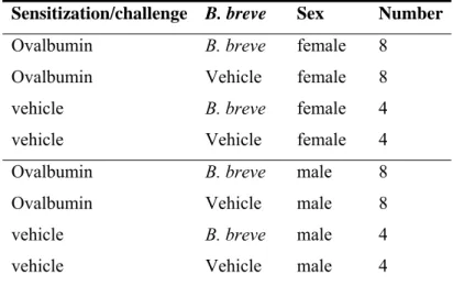
Table 1: Experimental groups respiratory allergy
Sensitization/challenge B. breve Sex Number
Ovalbumin B. breve female 8
Ovalbumin Vehicle female 8
vehicle B. breve female 4
vehicle Vehicle female 4
Ovalbumin B. breve male 8
Ovalbumin Vehicle male 8
vehicle B. breve male 4
vehicle Vehicle male 4
2.3.1 Bronchoalveolar lavage
Bronchoalveolar lavage (BAL) was performed by flushing the lungs with 1 ml sterile PBS. BAL fluid was centrifuged at 1200 rpm for 10 minutes. Cell pellets were used for
determination of total cell number and for cytospin preparations. Cytospins were stained with May-Grünwald Giemsa and on each preparation 400 cells were counted. Supernatants obtained for cytokine measurement were stored at -80°C.
2.3.2 Culture of spleen cells
Spleens were collected and single-cell suspensions were prepared under aseptic conditions by pressing the spleen through a sterile 70 μm nylon cell strainer. Cells were washed in 5% FCS medium (10 min., 4oC, 300 g) and resuspended in 10 ml standard medium with 10% FCS.
The concentration of the cell suspensions was adjusted to 2*106 cells/ml. Spleen cells
(100 μl/well; 2*105 cells) were stimulated with 5 μg/ml ConA (100 μl/well) for 48 hours or
with ovalbumin (100 μg/ml) for 96 hours. Supernatants were collected for cytokine measurements.
2.3.3 Ovalbumin-specific IgE and IgG1 ELISA
Specific ovalbumin IgE and IgG1 titers in sera were determined by ELISA. Incubations were followed by extensive washing on an automatic plate washer with PBS containing 0.1% Tween-20. To measure ovalbumin-specific IgE 96-wells plates (Nunc-Immuno Plate, Denmark) were coated overnight at 4oC with 2 μg/ml rat-anti-mouse IgE (rαm IgE, Zymed, 04-7000) diluted in sodium carbonate buffer (pH 9.6) and incubated overnight at 4°C. Plates were blocked by adding 0.05M Tris buffered saline with 1% BSA, pH 8 (Sigma) for 1 h at 37°C. Thereafter, serial dilutions of mouse serum samples and a pooled positive reference serum were incubated for 1 h at 37°C. All dilutions were done in blocking buffer plus 0.05% Tween-20. Then, wells were incubated for 1 h at 37°C with DIG-conjugated ovalbumin. The coupling of ovalbumin to DIG (molar mixture 1:10) was performed according to the
manufacturer's instructions (Roche Diagnostics). Then, wells were incubated with anti-DIG Fab fragments labeled with peroxidase (PO) (Roche Diagnostics) for 2 hours at 37°C. Plates were incubated with TMB substrate and the enzyme reaction was stopped with 2 M H2SO4
and absorbance was read at 450 nm.
To measure ovalbumin-specific IgG1 wells were coated overnight at 4°C with 10 μg/ml ovalbumin/ml PBS (grade V, Sigma). Blocking buffer was added and wells were incubated for 1 hour at 37°C. Thereafter, serial dilutions of mouse serum samples and a pooled positive reference serum were added to the wells and incubated for 2 hours at RT. Biotinylated rat-anti-mouse IgG1 (Zymed Laboratories, San Francisco, CA) was added and wells were incubated for 1.5 hour at RT, followed by incubation with poly-horseradish peroxidase labeled streptavidin for 45 minutes at RT. Then plates were incubated with TMB substrate and the enzyme reaction was stopped with 2 M H2SO4 and absorbance was read at 450 nm.
Extinction values of the positive reference serum were used to calculate the amount of IgG1 and IgE in the samples and extinction values were expressed as arbitrary units.
2.3.4 Bioplex for cytokines
Th1 and Th2 cytokines were measured in BAL fluid and in supernatants of spleen cells that were cultured with ConA or with ovalbumin. Cytokine levels were detected with a Bioplex 5-plex cytokine assay kit that could detect IL-4, IL-5, IL-10, IL-13 and IFN-γ (Biorad Life Science, Hercules, CA, USA) according to the manufacturer’s instructions. Cytokine
measurements were performed on a Luminex® (Biorad Life Science) and Luminex software was used to calculate the amount of cytokines (in pg/ml supernatant).
2.4
Experimental design experimental auto-immune
encephalomyelitis
After birth, rats were divided over the mothers. Each nest contained the same amount of pups with an equal male/female ratio. Oral administration of B. breve started when the rats were two weeks old. Rats received 1*109 CFU daily in a volume of 200 μl. Control rats received
200 μl saline daily. At weaning (21 days after birth) rats were taken away from their mothers and housed in the experimental groups (Table 2).
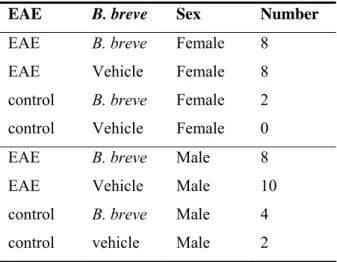
Table 2: Experimental groups EAE
EAE B. breve Sex Number
EAE B. breve Female 8
EAE Vehicle Female 8
control B. breve Female 2
control Vehicle Female 0 EAE B. breve Male 8
EAE Vehicle Male 10
control B. breve Male 4
control vehicle Male 2
Acute EAE was induced at the age of seven weeks as described previously (Hendriks et al., 2004). Rats were injected subcutaneously in the left ankle with 100 μl of an emulsion containing 20 μg guinea pig myelin basic protein (MBP; Sigma), 500 μg Mycobacterium
tuberculosis type H37RA (Difco, Detroit, MI), 50 μl complete Freund’s adjuvant (CFA, Difco) supplemented with saline (0.9% NaCl) to reach a volume 100 μl. After induction of EAE body weights were recorded daily. Also, neurological signs were scored daily and graded from 1 to 5; 0, no clinical signs; 0.5, loss of tonicity in distal half of tail; 1, flaccid tail; 1.5, unsteady gait; 2, partial hind limb paralysis; 2.5, complete hind limb paralysis; 3,
paralysis of the complete lower part of the body up to the diaphragm; 4, paraplegia; and 5, death due to EAE. Rats were sacrificed 26 days after induction of EAE. The spleen was removed and single-cell suspensions were prepared.
2.4.1 Stimulation of spleen cells with myelin basic protein
Single cell suspensions of spleens were prepared as described in 2.3.3. Spleen cells (100 μl/well; 2*105 cells) were stimulated with 0, 10 or 25 μg/ml MBP (100 μl/well) for
2.4.2 IFN-γ and IL-4 ELISA
IFN-γ was determined with a rat IFN-γ OptEIA set (BD Biosciences Pharmingen, San Diego, CA, USA) and IL-4 with a rat IL-4 cytoset (Biosource), both according to the manufacturer’s instructions.
2.5 Statistical analysis
In the respiratory allergy experiment statistical differences between the four experimental groups were determined by one-way ANOVA followed by Scheffe’s or Games-Howell post hoc test. Differences in ovalbumin-specific IgE and IgG1 levels were determined with a two-tailed Student’s t-test comparing the two experimental groups that were sensitized and challenged with ovalbumin.
In the EAE experiment mean body weights on all 26 days were calculated and significant differences between the experimental groups were determined with a two-tailed Student’s t-test. A one-tailed Mann-Whitney Test was used to determine if onset of disease, duration of symptoms and cumulative clinical score were significantly different between the experimental groups.
3 RESULTS
3.1 Effects of B. breve on respiratory allergy
3.1.1. Inflammatory response and cytokines in the lungs
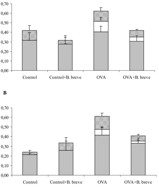
In both male and female mice that were sensitized and challenged with ovalbumin an increase of eosinophils and lymphocytes was found (Figure 1). In females and males, daily
administration of B. breve reduced the number of cells in sensitized mice by reducing the number of infiltrating lymphocytes and eosinophils (Figure 1A and B). These changes were not statistically significant.
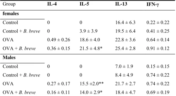
Several cytokines were assessed in the BAL fluid (Table 3). IL-10 could not be detected and levels of IFN-γ and IL-4 were very low. IL-5 increased after sensitization and challenge with ovalbumin in both males and females. In sensitized and challenged males IL-13 levels were higher compared to controls. In sensitized mice, treatment with B. breve did not affect the increased IL-5 and IL-13 levels.
Table 3: Effect of B. breve on cytokine levels in BAL fluid
Group IL-4 IL-5 IL-13 IFN-γ
females Control 0 0 16.4 ± 6.3 0.22 ± 0.22 Control + B. breve 0 3.9 ± 3.9 19.5 ± 6.4 0.41 ± 0.25 OVA 0.49 ± 0.26 18.6 ± 4.0 22.8 ± 3.6 0.64 ± 0.14 OVA + B. breve 0.36 ± 0.15 21.5 ± 4.8* 25.4 ± 2.8 0.91 ± 0.12 Males Control 0 0 7.0 ± 1.9 0.15 ± 0.15 Control + B. breve 0 0 8.4 ± 4.9 0.74 ± 0.22 OVA 0.27 ± 0.17 15.5 ±2.0** 21.7 ± 2.7 0.74 ± 0.22 OVA + B. breve 0.16 ± 0.11 14.0 ± 2.9* 18.4 ± 4.7 0.69 ± 0.19
Cytokines were assessed with a bioplex Th1/Th2 kit on a Luminex® and expressed as mean ± SE in pg/ml supernatant. Statistical significant from the control group: *p<0.05,
A 0,00 0,10 0,20 0,30 0,40 0,50 0,60 0,70
Control Control+B. breve OVA OVA+B. breve
N u m b er o f cel ls ( * 1 0 ^ 6 ) B 0,00 0,10 0,20 0,30 0,40 0,50 0,60 0,70
Control Control+B. breve OVA OVA+B. breve
N u m b er o f cel ls ( * 1 0 ^ 6 )
Figure 1: Number of cells in lung lavage fluid in female (A) and male (B) mice. Mice were sensitized and challenged with ovalbumin (OVA) or saline (control) and orally treated with B. breve or saline. Cell number is expressed in 106 cells. Grey bars represent the number of
macrophages, white bars the number of eosinophils and striped bars the number of lymphocytes.
3.1.2. OVA-specific serum IgE and IgG1
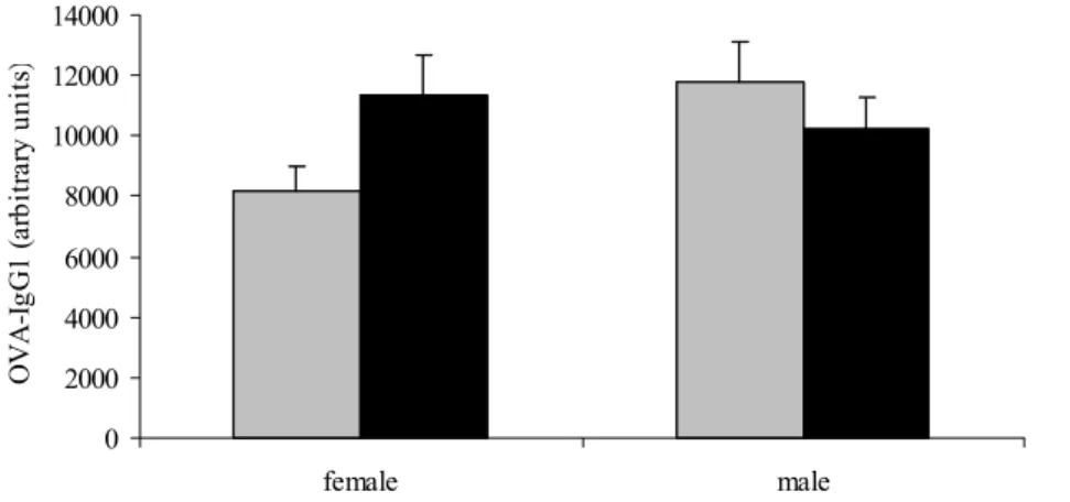
OVA-specific IgE (Figure 2) and IgG1 (Figure 3) levels were detected in serum of mice that were sensitized and challenged with ovalbumin. Mice that were sensitized and challenged with vehicle served as negative controls and serum of these mice did not contain ovalbumin-specific IgG1 and IgE (not shown). In both males and females, OVA-ovalbumin-specific IgE titers were not affected by B. breve treatment. In males, OVA-IgG1 levels were also not affected by
B. breve treatment. However, in females OVA-IgG1 levels were higher in mice that received B. breve orally. This difference almost reached significance (p=0.075).
0 500 1000 1500 2000 female male O V A -Ig E (a rb it ra ry u ni ts )
Figure 2: Serum levels of ovalbumin-specific IgE measured by ELISA. Mice were sensitized and challenged with ovalbumin and treated orally with B. breve (black bars) or saline (grey bars). Serum levels are expressed in arbitrary units that were calculated by using reference serum with ovalbumin-specific IgG1.
0 2000 4000 6000 8000 10000 12000 14000 female male OVA-I gG1 (a rb it ra ry u ni ts )
Figure 3: Serum levels of ovalbumin-specific IgG1 measured by ELISA. Mice were sensitized and challenged with ovalbumin and treated orally with B. breve (black bars) or saline (grey bars. Serum levels are expressed in arbitrary units that were calculated by using reference serum with ovalbumin-specific IgG1.
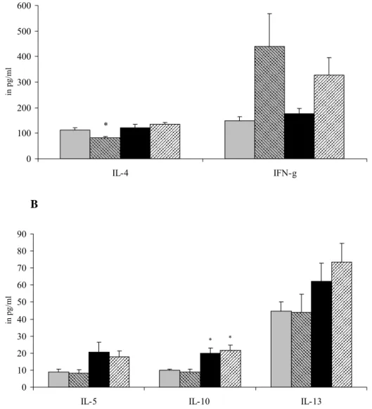
3.1.3 Cytokine production by spleen cells stimulated with ConA
After in vitro stimulation with ConA spleen cells from female mice that were sensitized with OVA produced more IL-5, IL-10 and IL-13 (Figure 4A). B. breve did not affect this cytokine production. IFN-γ production did not differ between control and sensitized mice. However,
B. breve increased IFN-γ in both control and sensitized females. Furthermore, in control mice
B. breve reduced IL-4 significantly this was not observed in sensitized females.
A 0 100 200 300 400 500 600 IL-4 IFN-g in p g/ml * B 0 10 20 30 40 50 60 70 80 90
IL-5 IL-10 IL-13
in
pg/
ml
* *
Figure 4: Female mice were sensitized and challenged with vehicle and received either saline (grey bars) or B. breve (striped bars) orally or were sensitized and challenged with
ovalbumin and received either saline (black bars) or B. breve (striped bars) .Cytokine production was assessed after ex vivo stimulation of spleen cells from female mice with 5 μg/ml ConA for 48 hours. Cytokines were measured with a bioplex Th1/Th2 kit on a Luminex® and expressed as mean ± SE in pg/ml supernatant.IL-4 and IFN-γ are shown in Figure A and IL-5, IL-10 and IL-13 in Figure B Statistical significant from the control group: *p<0.05.
A 0 50 100 150 200 250 300 350 IL-4 IFN-g in p g/ml ** B 0 10 20 30 40 50 60
IL-5 IL-10 IL-13
in pg/ ml ** # *** ** * *
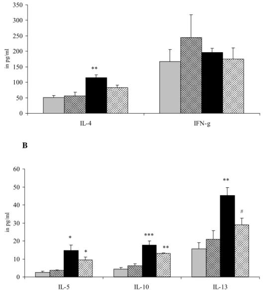
Figure 5: Male mice were sensitized and challenged with vehicle and received either saline (grey bars) or B. breve (striped bars) orally or were sensitized and challenged with
ovalbumin and received either saline (black bars) or B. breve (striped bars). Cytokine production was assessed after ex vivo stimulation of spleen cells from female mice with 5 μg/ml ConA for 48 hours. Cytokines were measured with a bioplex Th1/Th2 kit on a Luminex® and expressed as mean ± SE in pg/ml supernatant. IL-4 and IFN-γ are shown in Figure A and IL-5, IL-10 and IL-13 in Figure B. Significantly different from the control group: * p<0.05, ** p<0.01, *** p<0.001. Significantly different from the OVA group: #
p<0.05.
Figure 5 shows the ConA-induced cytokine production in males. In sensitized males,
splenocytes produced more IL-4, IL-5, IL-10 and IL-13. IFN-γ production was not affected by either sensitization with OVA or treatment with B. breve. A decrease in Th2 cytokines was observed after treatment with B. breve, which was significant for IL-13.
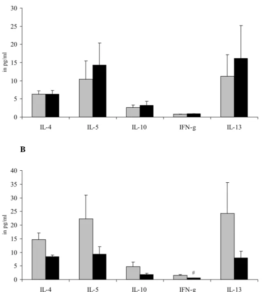
3.1.4 Cytokine production by spleen cells stimulated OVA
Spleen cells were also cultured with OVA to detect allergen-specific cytokine production. In females (Figure 6A), there was no effect of B. breve on cytokine production. However, in males (Figure 6B) B. breve reduced levels of all OVA-specific cytokines. This was significant for IFN-γ. A 0 5 10 15 20 25 30
IL-4 IL-5 IL-10 IFN-g IL-13
in pg/ml B 0 5 10 15 20 25 30 35 40
IL-4 IL-5 IL-10 IFN-g IL-13
in pg
/ml
#
Figure 6: Female (A) or male (B) mice were sensitized and challenged with ovalbumin and received either saline (grey bars) or B. breve (black bars). Cytokine production after ex vivo stimulation of spleen cells with 100 μg/ml OVA for 96 hours. Cytokines were assessed with a bioplex Th1/Th2 kit on a Luminex® and expressed as mean ± SE in pg/ml supernatant. Significantly different from the OVA group: # p<0.05.
3.2 Effects of B. breve on experimental autoimmune
encephalomyelitis
3.2.1 Body weight
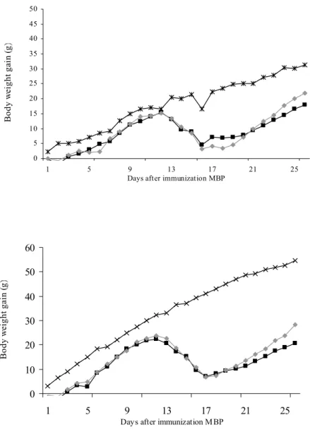
In Figure 7 the gain in body weight gain after immunization of female (Figure A) and male (Figure B) Lewis rats is shown. Growth is normal in both male and female control rats.
A 0 5 10 15 20 25 30 35 40 45 50 1 5 9 13 17 21 25 Days after immunization MBP
B ody w eight ga in ( g) B 0 10 20 30 40 50 60 1 5 9 13 17 21 25
Days after immunization MBP
B ody w ei ght ga in (g )
Figure 7: Female (A) or male (B) rats were immunized with MBP and received saline (-■-) or B. breve (-♦-). Non-immunized controls are also included in the figure (-x-). Body weight gain is expressed as the increase in body weight in grams from the day of immunization. Figure A shows mean body weight gain of females and figure B of males.
Weight loss was observed in all rats that were immunized with MBP. In females that did not receive probiotics this occurred from day 13 until 16 and in females that received B. breve from day 12 until 16. Then, rats started gaining weight again and the curve for females that received B. breve appears to be somewhat steeper, indicating a more rapid growth. Body weight on the final day of the experiment (day 26) was higher in females that received
B. breve (p=0.087).
In immunized males, weight loss was observed from day 12 until 17, in the probiotic and the control group. After day 17 males that were treated with B. breve gained weight faster compared to controls. Body weight was significantly higher at day 25 (p=0.045).
Table 4: Effects of B. breve on clinical parameters of EAE
Disease onset (days) Duration symptoms (days) Cumulative disease index females EAE 14.4 ± 0.6 6.8 ± 1.1 4.5 EAE + B. breve 13.6 ± 0.3 6.0 ± 0.7 4.9 males EAE 13.4 ± 0.4 7.4 ± 0.6 6.1 EAE + B. breve 13.9 ± 0.6 5.5 ± 0.3** 5.9
Disease onset is the first day that clinical signs were observed in a treatment group, the duration of symptoms is the total number of days clinical symptoms were visible in a treatment group, and cumulative disease index (CDI) is sum of the cumulative daily scores per group divided by the number of days that the clinical symptoms were observed in this group. All data are expressed as mean ± SE. Significantly different from the EAE group: ** p<0.01
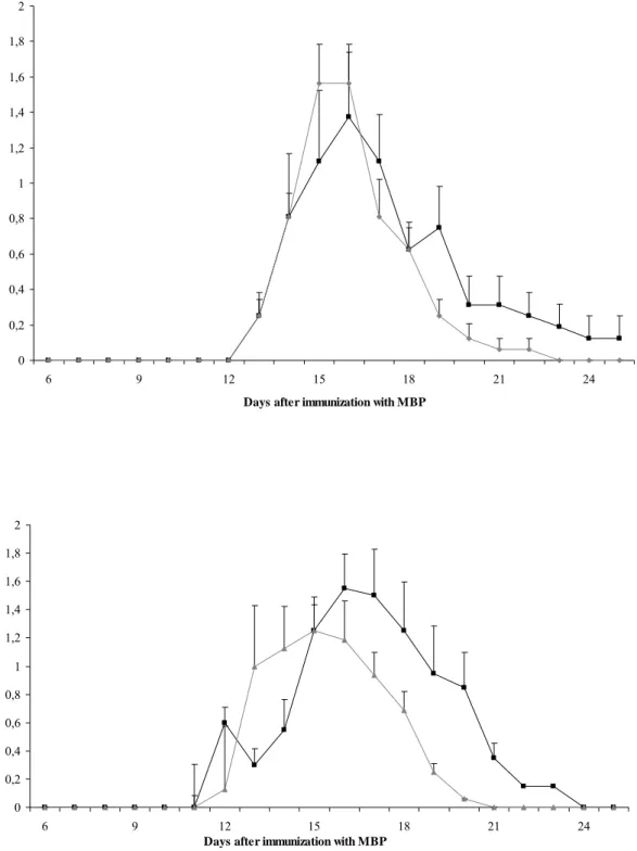
3.2.2 Clinical symptoms
All rats that were immunized with MBP developed EAE. The first clinical symptom that appeared was loss of tonicity of the tail. In Table 4 clinical parameters of EAE are
summarized. In females the day of onset was not influenced by B. breve. Furthermore, the duration of symptoms and the cumulative disease index was slightly increased in females that received the B. breve compared to those that received vehicle. In males, onset and cumulative disease index were not affected by B. breve. The duration of symptoms, however, was almost two days shorter in males that receive B. breve. The mean clinical score in time is shown in Figure 8 and the effects of B. breve on the duration of symptoms is illustrated by a shift of the
curve to the left (Figure 8b). A similar trend is observed for females, although this is less evident (Figure 8A).
A 0 0,2 0,4 0,6 0,8 1 1,2 1,4 1,6 1,8 2 6 9 12 15 18 21 24
Days after immunization with MBP
M ean c li n ic al s cor e B 0 0,2 0,4 0,6 0,8 1 1,2 1,4 1,6 1,8 2 6 9 12 15 18 21 24
Days after immunization with MBP
M ean cl in ic al s cor e
Figure 8: Female (A) or male (B) rats were immunized with MBP and received saline (-■-) or B. breve (-♦-). Time course of the mean clinical score per day (±SE) is shown.
In Figure 9 the cumulative clinical score is shown. This figure shows that in female rats that were immunized and received the vehicle orally, the cumulative score is still increasing at the end of the experiment. In females that received B. breve displayed the cumulative clinical score increased until day 19. In males the increase of cumulative score stops on day 21 and on day 18, for vehicle and B. breve treated rats, respectively.
A 0 1 2 3 4 5 6 7 8 9 10 12 15 18 21 24
Days after immunization
C um ula tiv e c lin ic al s co re B 0 2 4 6 8 10 12 12 13 14 15 16 17 18 19 20 21 22 23 24 25
Days after immunization
C um ula tiv e c lin ic al s co re
Figure 9: Female (A) or male (B) rats were immunized with MBP and received saline (-■-) or B. breve (-♦-). Cumulative score(±SE) per day is calculated by cumulating the clinical socre per day over time per animal.
3.2.3 MBP-specific IFN-γ production
Ex vivo stimulation with 10 and 25 μg/ml MBP resulted in a dose-dependent increase of IFN-γ production (Figure 10). In females treated with B. breve an increase of MBP-specific IFN-γ production was observed, which was significant for cells stimulated with 25 μg/ml MBP. In males a similar trend towards higher IFN-γ production after B. breve treatment was observed. These differences did not reach significance. MBP-specific IL-4 could not be detected in these samples.
A 0 200 400 600 800 1000 1200
EAE EAE+B. breve
IFN-g (p g/ m l) # B 0 100 200 300 400 500 600 700 800 EAE EAE+B.breve IFN-g (p g/ m l)
Figure 10: IFN-γ production by spleen cells from female (A) and male (B) Lewis rats stimulated with 10 (grey bars) or 25 (black bars) μg/ml MBP mg/ml for 96 hours. IFN-γ was detected with a sandwich ELISA. Significantly different from the control: # p<0.05
4. DISCUSSION
We have investigated the effects of B. breve on respiratory allergy in mice and on EAE in rats. In the respiratory allergy model, B. breve slightly reduced the inflammatory response in the lungs of females and males, by reducing the number of infiltrating eosinophils and lymphocytes. This reduction was not supported by a reduction of cytokines in the BAL fluid, such as IL-5. Cytokine profiles assessed in spleen cells stimulated with ConA showed that
B. breve increased IFN-γ production in control and sensitized females and reduced IL-4 in control females. This shift in cytokine profile did not result in reduced serum IgE levels, however. Remarkably, ovalbumin-specific IgG1 levels, which are also Th2 mediated, were increased in females. In previous allergy models, probiotics stimulated Th1 responses and as a consequence a reduction of allergen-specific serum IgE was observed (Matsuzaki et al., 1998; Matsuzaki and Chin, 2000; Sashihara et al., 2006). In this study, the shift in cytokine profile towards Th1 was probably not sufficient to reduce ovalbumin-specific IgE in our model. It is important to note that the shift towards Th1 was only observed after ex vivo stimulation with ConA and not with ovalbumin. In other studies the shift towards Th1 was observed after ex
vivo stimulation with ovalbumin (Matsuzaki et al., 1998; Matsuzaki and Chin, 2000). This
could mean that B. breve had an effect on spleen cells, but not on the ovalbumin-specific response, since stimulation with ConA induced Th1 responses in both control and sensitized females. In males, however, B. breve reduced ovalbumin-specific production of all assessed Th1 and Th2 cytokines. Apparently this was not sufficient to reduce IgE levels.
Previously we have investigated the effects of LcS in the same allergy model. LcS modestly enhanced ovalbumin-specific IgE levels both in adult mice and in mice that received LcS from lactation phase onward. This increase was accompanied by an increase of ovalbumin-specific cytokine production in adult mice (Ezendam et al., 2006). However, in mice that received LcS from lactation phase onward, no effects on cytokine profiles were found (Ezendam and Van Loveren, 2006). The inflammatory lung response was affected differently depending on the timing of administration. A reduction was observed in adult mice that received LcS (Ezendam et al., 2006), whereas a further increase was observed after early administration (Ezendam and Van Loveren, 2006). Thus, early administration of LcS aggravated lung inflammation, whereas adult exposure alleviated it. B. breve induced a different effect compared to LcS, since administration of B. breve from lactation phase onward slightly reduced lung inflammation.
Also in the EAE model in rats LcS and B. breve differentially affected the disease. B. breve significantly reduced the duration of the disease in males and induced a faster recovery after
weight loss. A similar trend was observed in the females. Remarkably, in both males and females MBP-specific IFN-γ production was increased. One would expect that stimulation of Th1 responses would lead to aggravation of EAE. Previously, alleviation of EAE symptoms was associated with a decrease of Th1 (12, IFN-γ) and an increase of Th2/Th3 (4, IL-10, TGF-β) cytokines (Xu et al., 2000; Monteiro de Castro et al., 2004;Yang et al., 2004). It is important to note that there was no correlation between MBP-specific IFN-γ levels and the assessed clinical parameters. Thus, it seems that IFN-γ is not involved in the alleviation of EAE. Effects of B. breve on MBP-specific Th2 and Th3 cytokines might provide more insight in the influence of these cytokines on EAE.
In contrast to B. breve, LcS aggravated EAE symptoms, both in adults and in rats that received LcS from lactation phase onward (Baken et al., 2006; Ezendam et al., 2006; Ezendam and Van Loveren, 2006). The effects of probiotics on EAE are clearly strain-dependent. This is less obvious in the respiratory allergy model, although effects on the inflammatory lung response are also different for the two probiotic strains. In general, LcS appears to stimulate immune responses in these two experimental models, whereas B. breve alleviates both allergy and autoimmunity.
In conclusion, we have demonstrated that immune effects of probiotics are clearly strain-dependent. Our data do suggest that proper strain selection is needed to reach the goal for which the specific application is meant. Thus, B. breve appears to be suitable for the treatment of allergic or autoimmune diseases, whereas LcS might be more appropriate for the
enhancement of resistance. Furthermore, in certain populations, LcS might induce unwanted effects. However, the consequences of these preclinical data cannot be directly extrapolated to the human situation. For extrapolation, well-designed human trials are needed. Information from such studies could provide insight in both the benefits and the risks of probiotic use.
REFERENCES
Baken, K. A., Ezendam, J., Gremmer, E. R., de Klerk, A., Pennings, J. L., Matthee, B., Peijnenburg, A. A., and van Loveren, H. (2006). Evaluation of immunomodulation by
Lactobacillus casei Shirota: Immune function, autoimmunity and gene expression. Int J Food Microbiol.
Brouwer, M. L., Wolt-Plompen, S. A., Dubois, A. E., van der Heide, S., Jansen, D. F., Hoijer, M. A., Kauffman, H. F., and Duiverman, E. J. (2006). No effects of probiotics on atopic dermatitis in infancy: a randomized placebo-controlled trial. Clin Exp Allergy 36, 899-906.
Chapat, L., Chemin, K., Dubois, B., Bourdet-Sicard, R., and Kaiserlian, D. (2004).
Lactobacillus casei reduces CD8+ T cell-mediated skin inflammation. Eur J Immunol 34, 2520-2528.
Christensen, H. R., Frokiaer, H., and Pestka, J. J. (2002). Lactobacilli differentially modulate expression of cytokines and maturation surface markers in murine dendritic cells. J
Immunol 168, 171-178.
Cross, M. L., Ganner, A., Teilab, D., and Fray, L. M. (2004). Patterns of cytokine induction by gram-positive and gram-negative probiotic bacteria. FEMS Immunol Med
Microbiol 42, 173-180.
Cross, M. L., Mortensen, R. R., Kudsk, J., and Gill, H. S. (2002). Dietary intake of
Lactobacillus rhamnosus HNOO1 enhances production of both Th1 and Th2
cytokines in antigen-primed mice. Med Microbiol Immunol (Berl) 191, 49-53. Drakes, M., Blanchard, T., and Czinn, S. (2004). Bacterial Probiotic Modulation of Dendritic
Cells. Infect. Immun. 72, 3299-3309.
Ezendam, J., Baken, K. A., and Van Loveren, H. (2006). Immune effects of Lactobacillus casei Shirota. RIVM report 340320004.
Ezendam, J., and Van Loveren, H. (2006). Long-term immune effects of Lactobacillus casei Shirota during lactation. RIVM report 340320005.
Ezendam, J., and Van Loveren, H. (2006). Probiotics: immunomodulation and evaluation of safety and efficacy. Nutr Rev 64, 1-14.
He, F., Morita, H., Ouwehand, A. C., Hosoda, M., Hiramatsu, M., Kurisaki, J., Isolauri, E., Benno, Y., and Salminen, S. (2002). Stimulation of the secretion of pro-inflammatory cytokines by Bifidobacterium strains. Microbiol Immunol 46, 781-785.
Helin, T., Haahtela, S., and Haahtela, T. (2002). No effect of oral treatment with an intestinal bacterial strain, Lactobacillus rhamnosus (ATCC 53103), on birch-pollen allergy: a placebo-controlled double-blind study. Allergy 57, 243-246.
Hendriks, J. J., Alblas, J., van der Pol, S. M., van Tol, E. A., Dijkstra, C. D., and de Vries, H. E. (2004). Flavonoids influence monocytic GTPase activity and are protective in experimental allergic encephalitis. J Exp Med 200, 1667-1672.
Isolauri, E., Arvola, T., Sutas, Y., Moilanen, E., and Salminen, S. (2000). Probiotics in the management of atopic eczema. Clin Exp Allergy 30, 1604-1610.
Kalliomaki, M., Salminen, S., Arvilommi, H., Kero, P., Koskinen, P., and Isolauri, E. (2001). Probiotics in primary prevention of atopic disease: a randomised placebo-controlled trial. Lancet 357, 1076-1079.
Kalliomaki, M., Salminen, S., Poussa, T., Arvilommi, H., and Isolauri, E. (2003). Probiotics and prevention of atopic disease: 4-year follow-up of a randomised
placebo-controlled trial. Lancet 361, 1869-1871.
Maassen, C. B., van Holten, J. C., Balk, F., Heijne den Bak-Glashouwer, M. J., Leer, R., Laman, J. D., Boersma, W. J., and Claassen, E. (1998). Orally administered
Lactobacillus strains differentially affect the direction and efficacy of the immune
response. Vet Q 20 Suppl 3, S81-83.
Madsen, K. L. (2001). The use of probiotics in gastrointestinal disease. Can J Gastroenterol 15, 817-822.
Matsuzaki, T., Chin, J. (2000). Modulating immune responses with probiotic bacteria.
Immunol Cell Biol 78, 67-73.
Matsuzaki, T., Yamazaki, R., Hashimoto, S., and Yokokura, T. (1998). The effect of oral feeding of Lactobacillus casei strain Shirota on immunoglobulin E production in mice. J Dairy Sci 81, 48-53.
Mohamadzadeh, M., Olson, S., Kalina, W. V., Ruthel, G., Demmin, G. L., Warfield, K. L., Bavari, S., and Klaenhammer, T. R. (2005). Lactobacilli activate human dendritic cells that skew T cells toward T helper 1 polarization. Proc Natl Acad Sci U S A 102, 2880-2885.
Monteiro de Castro, G., Eduarda Zanin, M., Ventura-Oliveira, D., Aparecida Vilella, C., Ashimine, R., and de Lima Zollner, R. (2004). Th1 and Th2 cytokine
immunomodulation by gangliosides in experimental autoimmune encephalomyelitis.
Cytokine 26, 155-163.
Peng, G.-C., and Hsu, C.-H. (2005). The efficacy and safety of heat-killed Lactobacillus paracasei for treatment of perennial allergic rhinitis induced by house-dust mite.
Penner, R., Fedorak, R. N., and Madsen, K. L. (2005). Probiotics and nutraceuticals: non-medicinal treatments of gastrointestinal diseases. Current Opinion in Pharmacology
Gastrointestinal/Endocrine and metabolic diseases 5, 596-603.
Petrof, E. O., Kojima, K., Ropeleski, M. J., Musch, M. W., Tao, Y., De Simone, C., and Chang, E. B. (2004). Probiotics inhibit nuclear factor-kappaB and induce heat shock proteins in colonic epithelial cells through proteasome inhibition. Gastroenterology 127, 1474-1487.
Rautava, S., Arvilommi, H., and Isolauri, E. (2006). Specific Probiotics in Enhancing Maturation of IgA Responses in Formula-Fed Infants. Pediatr Res.
Rosenfeldt, V., Benfeldt, E., Nielsen, S. D., Michaelsen, K. F., Jeppesen, D. L., Valerius, N. H., and Paerregaard, A. (2003). Effect of probiotic Lactobacillus strains in children with atopic dermatitis. J Allergy Clin Immunol 111, 389-395.
Rosenfeldt, V., Benfeldt, E., Valerius, N. H., Paerregaard, A., and Michaelsen, K. F. (2004). Effect of probiotics on gastrointestinal symptoms and small intestinal permeability in children with atopic dermatitis. J Pediatr 145, 612-616.
Sashihara, T., Sueki, N., and Ikegami, S. (2006). An analysis of the effectiveness of heat-killed lactic acid bacteria in alleviating allergic diseases. J Dairy Sci 89, 2846-2855. Smit, J. J., van Loveren, H., Hoekstra, M. O., Nijkamp, F. P., and Bloksma, N. (2003).
Influence of the macrophage bacterial resistance gene, Nramp1 (Slc11a1), on the induction of allergic asthma in the mouse. Faseb J 17, 958-960.
Smits, H. H., Engering, A., van der Kleij, D., de Jong, E. C., Schipper, K., van Capel, T. M. M., Zaat, B. A. J., Yazdanbakhsh, M., Wierenga, E. A., van Kooyk, Y., and
Kapsenberg, M. L. (2005). Selective probiotic bacteria induce IL-10-producing regulatory T cells in vitro by modulating dendritic cell function through dendritic cell-specific intercellular adhesion molecule 3-grabbing nonintegrin. Journal of
Allergy and Clinical Immunology 115, 1260-1267.
Viljanen, M., Savilahti, E., Haahtela, T., Juntunen-Backman, K., Korpela, R., Poussa, T., Tuure, T., and Kuitunen, M. (2005). Probiotics in the treatment of atopic
eczema/dermatitis syndrome in infants: a double-blind placebo-controlled trial.
Allergy 60, 494-500.
Wang, J. (2006). The Efficacy and Safety of Heat-Killed Lactobacillus paracasei for Treatment of Perennial Allergic Rhinitis Induced by House-Dust Mite. Pediatrics 118, S23-.
Weston, S., A., H., Richmond, P., and Prescott, S. L. (2005). Effects of probiotics on atopic dermatitis: a randomised controlled trial. ADC.
Wheeler, J. G., Shema, S. J., Bogle, M. L., Shirrell, M. A., Burks, A. W., Pittler, A., and Helm, R. M. (1997). Immune and clinical impact of Lactobacillus acidophilus on asthma. Ann Allergy Asthma Immunol 79, 229-233.
Xu, L. Y., Huang, Y. M., Yang, J. S., Van Der Meide, P. H., Link, H., and Xiao, B. G. (2000). Suppression of ongoing experimental allergic encephalomyelitis (EAE) in Lewis rats: synergistic effects of myelin basic protein (MBP) peptide 68-86 and IL-4. Clin Exp
Immunol 120, 526-531.
Yang, J. S., Xu, L. Y., Xiao, B. G., Hedlund, G., and Link, H. (2004). Laquinimod (ABR-215062) suppresses the development of experimental autoimmune encephalomyelitis, modulates the Th1/Th2 balance and induces the Th3 cytokine TGF-beta in Lewis rats.
Datum 25 juli 2007 Ons kenmerk Blad 1/1 Tel (030) 274 3447 Fax (030) 274 4446 janine.ezendam@rivm.nl Onderwerp
Erratum RIVM rapport 340320006
Begin dit jaar heeft u het RIVM rapport ‘Immune effects of the probiotic
Bifidobacterium breve’ ontvangen. Aan dit rapport zal onderstaand erratum worden
toegevoegd.
Erratum:
De naamgeving van de in de beschreven studies gebruikte probiotische stam is
Bifidobacterium animalis in plaats van Bifidobacterium breve.
Met vriendelijke groet,