RIVM report 230086001/2004
Is there evidence for a link between Crohn’s disease and
exposure to Mycobacterium avium ssp. paratuberculosis?
A review of current literature
A.A.P.M. Herrewegh1, P.J.M. Roholl2, P. Overduin3, J.W.B. van der Giessen4 and D. van Soolingen3
BioMedics Scientific Consultancy, Maarssen1; Laboratory for Toxicology, Pathology and Genetics (TOX)2, Diagnostic Laboratory for Infectious Diseases and Perinatal Screening (LIS)3 and Microbiological Laboratory for Health Protection (MGB)4, RIVM
This investigation has been performed under the authority of The Food and Consumer Product Safety Authority (VWA2004/28780)
Abstract
Crohn’s Disease is characterized by a severe, non-specific, chronic inflammation of the intestinal wall. The inflammation of CD most commonly affects the last part of the ileum, and often includes the colon and sigmoid. CD is a Th1 disease characterized by the
production of pro-inflammatory cytokines like TNF-α, which is responsible for development of the lesions, and by the production of IFN-γ and Il-2. CD is a multi-factorial disease; based on epidemiological and geographical observations, several genetic (familial, racial) and environmental (geographic, hygienic) factors (especially microbial) have been associated with the disease. Mutations in the human CARD15 gene and differential expression of Toll-like receptors (TLRs) 2, 3 and 4 have been associated with CD. CARD15 and TLRs are part of the body’s innate defence system against bacteria. They activate the immune system after recognizing specific bacterial components. Presence of intestinal bacteria seems to be a prerequisite for CD; the disease cannot develop or perpetuate without the presence of an intestinal flora. One of the bacteria that has been frequently associated with CD is
Mycobacterium avium subspecies paratuberculosis (Map). Map causes a severe chronic
intestinal disease in ruminants, Paratuberculosis. CD and Paratuberculosis share several clinical, immunological and histo-pathological characteristics. Map is present in many dairy herds and probably can be transmitted to humans via foodstuff. Many investigators have tried to prove, or controvert a common aetiology for CD and Paratuberculosis, and have applied several detection methods to accomplish this. Unfortunately, the quest for Map using PCR and culture methods, and the studies on the immune responses against Map in CD patients, have yet not resulted in conclusive data to support or discount the hypothesis that Map is the etiologic agent of CD. The fact that Map can be found in a high percentage of apparently healthy individuals, and that CD patients have significantly higher immune responses against several food antigens compared to healthy individuals, raises the question whether Map is a common passer-by of the human intestinal tract, or that a particular cofactor (a genetic aberration) is needed before Map can cause disease. The current hypothesis about the patho-physiology of CD is that in a genetic susceptible host, the intestinal flora triggers an aberrant immune response that results in a chronic intestinal inflammation and a damaged (leaky) intestinal mucosal barrier. Although a multi-factorial cause for CD is expected, the
possibility, however, that an infectious agent like Map can play a key role in the causation of even a sub-set of CD patients remains, and clearly needs to be taken seriously.
Preface
Crohn’s disease (CD) is a high burden of disease in humans, and has a dramatic negative impact on the patients’ quality of life. Since more than 30 years, numerous researchers have indicated Mycobacterium avium ssp. paratuberculosis (Map) as the possible causative agent of CD in humans. This assumption is predominantly based on the clinical and pathological similarities between CD and the disease ‘Paratuberculosis’ in ruminants, which is
unambiguously caused by Map, and the presence of Map in a subset of CD patients. In contrast, just as many investigators have discounted Map as the cause of CD based on the absence of Map in a subset of CD patients, and its presence in many healthy individuals. Nevertheless, since Map is an animal pathogen that may be transmitted from animals to humans via milk, meat, water and other foodstuff 37,38,84,145,146, concern has risen about the potential risk of Map-contaminated food for the development of CD. In this report we aim to answer the question: ‘is there scientific evidence for a causal link between CD in humans and exposure to Map?’ This report reviews the available information on the nature of CD and its likely causation, especially concerning a possible link with exposure to Map. Furthermore, the results of an extended cohort study in 59 CD patients and 79 control persons, conducted at the RIVM, are summarized. On the basis of what is known to date about CD and Map we will substantiate whether a link between Map and CD is conceivable.
Contents
Samenvatting 9
1. Crohn’s disease 17
2. Features of CD 19
2.1 Classification of CD 19
2.2 Immune responses in CD patients 19
2.3 Aetiology of CD 20
3. The role of genetic factors in CD 23
3.1 CARD15/NOD2 mutations and CD 23
3.2 Toll-like receptors and CD 24
4. Intestinal flora and CD 27
5. M. avium ssp. paratuberculosis and CD 29
6. Is CD caused by Map? 33
6.1 Detection of Map in CD tissue 33
6.2 Map does not always cause disease 36
6.3 Immune responses against Map in CD 37
6.4 Antibiotics against Map in CD 39
6.5 Prevalence of CD versus prevalence of Map 40
7. Interpretation of the data in literature 41
7.1 Summary 41
7.2 Loss of tolerance and disturbed mucosal barrier 43
8. Conclusion 45
Appendix 1 Tables 47
Samenvatting
Doel van dit rapport
Mycobacterium avium ssp. paratuberculosis (Map) wordt door veel onderzoekers beschouwd
als mogelijke verwekker van de ziekte van Crohn (morbus Crohn, MC) bij de mens. Dit is vooral gebaseerd op klinische en pathologische overeenkomsten tussen MC en de ziekte Paratuberculose bij runderen (herkauwers), die zonder twijfel veroorzaakt wordt door Map, en de aangetoonde aanwezigheid van Map bij een deel van de patiënten met MC. Echter, evenzoveel onderzoekers zijn van mening dat Map niet de verwekker is van MC omdat Paratuberculose en MC ook verschillen in een aantal kenmerken. Map kan bijvoorbeeld niet worden aangetoond bij alle MC patiënten. Verder kan Map vaak wel worden aangetoond bij een aanzienlijk deel van de onderzochte gezonde personen. Omdat Map niettemin een ziekteverwekker is die via melk, vlees, water en andere levensmiddelen de consument kan bereiken, bestaat er ernstige bezorgdheid over het mogelijke risico van dit Map-besmet voedsel voor het ontstaan van MC bij de consument. In dit rapport wordt een antwoord gegeven op de vraag ‘is het verband tussen de verwekker van Paratuberculose,
Mycobacterium avium ssp. paratuberculosis, en de ziekte van Crohn overtuigend bewezen?’
De huidige wetenschappelijke kennis over MC wordt beschreven, waarbij het accent wordt gelegd op de informatie die direct of indirect betrekking heeft op, of een indicatie kan zijn voor een mogelijke relatie tussen MC en Map.
De ziekte van Crohn
Nederland telde in 2000 tussen de 27.000 en 56.000 mensen met de chronische
darmontsteking ‘Inflammatory Bowel Disease’ (IBD) 97. Hiervan stierven in 2000 in totaal 60 personen aan de gevolgen van deze ziekte 97. De twee vormen van IBD zijn Colitis Ulcerosa en Morbus Crohn (MC, de ziekte van Crohn). Elk jaar worden in Nederland ongeveer 2.000 nieuwe patiënten gediagnosticeerd met IBD 97. Vooral de incidentie van de MC lijkt toe te
nemen. De meeste patiënten worden door MC getroffen in de leeftijdsfase tussen 15 en 35 jaar. MC is een niet-specifieke, chronische ontsteking van het maagdarmkanaal. De ziekte heeft een zeer dramatische invloed op het fysieke- en sociale leven van de patiënt, en is niet te genezen. Medicijnen die worden gebruikt voor de behandeling zijn er vooral op gericht om de ontstekingsreacties en de symptomen van de ziekte te onderdrukken.
Achtergrondinformatie over MC
MC kan op verschillende manieren tot uiting komen. Bij de meeste patiënten is het laatste deel van de dunne darm (terminale ileum) ontstoken, en vaak is ook de dikke darm (colon) aangetast. Minder vaak zijn andere delen van het maagdarmkanaal erbij betrokken, zoals mond, slokdarm (oesophagus) en twaalfvingerige darm (duodenum), en soms worden ook
ontstekingen buiten het darmgebied aangetroffen, zoals oogontstekingen en
gewrichtsontstekingen. Het is evident dat MC niet een eenduidig en gelokaliseerd ziektebeeld geeft en daarom soms moeilijk is te diagnosticeren. Afhankelijk van de plaats van de
ontstekingen en de aard van de afwijkingen wordt de ziekte in verschillende categorieën ingedeeld.
Bij ontstekingen speelt het afweersysteem een belangrijke rol. Het afweersysteem beschermt het lichaam tegen binnendringen van ziekteverwekkers en andere lichaamsvreemde stoffen. Er is een aangeboren en een verworven afweersysteem. Het aangeboren afweersysteem onderneemt actie tegen indringers voordat een specifieke afweerreactie (door het verworven afweersysteem) op gang komt, en maakt daarbij gebruik van speciale structuren (eiwit- en suikerstructuren) die aanwezig zijn bij micro-organismen (bacteriën, virussen) die proberen binnen te dringen. In tegenstelling tot het aangeboren deel is het verworven afweersysteem specifiek gericht op één bepaalde lichaamsvreemde stof, en is opgebouwd uit een cellulair en een humoraal deel. Om een goed geheugen tegen lichaamsvreemde stoffen te verwerven maken het cellulaire en humorale afweersysteem gebruik van T-helper (Th) cellen die deze afweerreactie besturen en coördineren. Er zijn twee belangrijke soorten T-helper (Th) cellen:
• Th1 cellen, die de afweerreactie sturen in de richting van het afdoden van door virussen of bacteriën geïnfecteerde cellen.
• Th2 cellen, die de afweerreactie sturen in de richting van antilichaamproductie. Th2 cellen spelen een belangrijke rol bij het afdoden van nog niet door macrofagen opgenomen virussen en bacteriën, onder andere bij acute infecties.
Bij gezonde personen is er een balans in de sturing door deze Th1/Th2 cellen. In de darm van gezonde personen ligt de balans naar de Th2 kant. Bij MC patiënten blijkt echter een
verstoring te zijn opgetreden, waardoor er een te hoge activiteit van de Th1 cellen is ontstaan. Deze Th1 cellen sporen macrofagen aan om stoffen (cytokines) te produceren die
ontstekingsreacties en weefselschade veroorzaken. Enkele van deze cytokines die een belangrijke rol spelen in MC zijn: Interferon gamma (IFN-γ), Tumor Necrosis Factor alpha (TNF-α) en Interleukine 6 (Il-6). Vooral TNF-α is verantwoordelijk voor de weefselschade bij MC.
Factoren betrokken bij MC
Sinds 1913, toen MC voor het eerst werd beschreven, zijn er allerlei hypotheses opgesteld over de mogelijke oorzaak van de ziekte. Bestudering van epidemiologische en geografische gegevens, die te maken hebben met het optreden en vóórkomen van MC, hebben geleid tot het identificeren van twee belangrijke elementen die betrokken zijn bij het ontstaan van MC: erfelijke factoren en omgevingsfactoren.
Dat erfelijke factoren betrokken zijn bij MC blijkt uit een relatie met:
• Bloedverwantschap: iemand die een eerstegraads bloedverwant heeft met MC heeft een grotere kans om de ziekte ook te krijgen dan iemand die geen bloedverwant met MC heeft. Dat blijkt vooral bij eeneiige tweelingen, waar de ziekte bij allebei vaker voorkomt dan bij twee-eiige tweelingen.
• Etnische achtergrond: MC komt vaker voor bij blanke mensen dan bij gekleurde mensen, en vaker bij joodse mensen dan bij niet-joodse mensen.
De betrokkenheid van omgevingsfactoren blijkt uit:
• De afgelopen 50 jaar is er een toename van MC te zien in het aantal nieuwe gevallen per jaar.
• Er is een geografische noord-zuid gradiënt te zien in het aantal MC gevallen per jaar. In noord-west Europa komt MC vaker voor dan in zuid-Europa, en vaker in Europa dan in Azië en Afrika.
• Mensen die vanuit een streek waar MC nauwelijks voorkomt emigreren naar een streek waar MC veel voorkomt, krijgen dezelfde kans om MC te ontwikkelen als de plaatselijke bevolking van die streek.
• Kinderen die opgroeien onder goede hygiënische omstandigheden lijken meer kans te lopen later MC te krijgen.
• Rokers hebben een verhoogde kans om MC te krijgen.
Door de grote verscheidenheid aan factoren die betrokken lijken te zijn bij MC en het feit dat tot nu toe geen individuele factor gevonden is die doorslaggevend is voor het ontstaan van de ziekte, wordt MC gezien als een multi-factoriële ziekte, waarbij het optreden van
verschillende factoren samen bepalen of een persoon de ziekte ontwikkelt of niet.
Erfelijke factoren
De aanwijzingen voor betrokkenheid van erfelijke factoren bij het ontstaan van MC hebben onderzoekers ertoe gebracht op zoek te gaan naar afwijkingen in het erfelijk materiaal (het genoom, de genen op de chromosomen) van MC patiënten. Inmiddels zijn minstens zeven plaatsen in het genoom geïdentificeerd waar mogelijk afwijkingen (mutaties) optreden die leiden tot een verhoogd risico op het ontwikkelen van MC. De belangrijkste mutaties die tot nu toe ontdekt zijn liggen in het CARD15 gen en deze mutaties komen bij 15-20% van de MC patiënten voor. Het CARD15 gen codeert voor het NOD2 eiwit. Dit eiwit is een onderdeel van het aangeboren afweersysteem van het lichaam, en speelt een rol bij de afdoding van door de cel opgenomen bacteriën. Een ander onderdeel van het aangeboren afweersysteem waarbij afwijkingen zijn gevonden in MC patiënten zijn de Toll-like receptoren. Evenals CARD15 herkennen Toll-like receptoren speciale structuren bij microorganismen en zetten het specifieke afweersysteem in gang. Het feit dat bij MC
patiënten afwijkingen zijn aangetroffen in twee verschillende onderdelen van het aangeboren afweersysteem tegen microorganismen (CARD15 en Toll-like receptoren) doet vermoeden dat erfelijke factoren die betrokken zijn bij de herkenning van microorganismen mede het ontstaan en/of het voortduren van MC bepalen.
Darmbacteriën
De theorie dat micro-organismen, en dan vooral de darmbacteriën (darmflora), een cruciale rol spelen bij MC wordt ondersteund door de waarneming dat proefdieren (muis en rat) geen
darmontsteking ontwikkelen als ze worden gekweekt in een steriele omgeving (waarin ze geen darmflora hebben). Verder bleek dat bij MC patiënten met een stoma waarbij een deel van de aangetaste darm afgesloten werd van voedseldoorstroming (en dus de darmflora ter plaatse verdween) na 6 maanden geen MC meer optrad, terwijl dat in controle MC patiënten, waar wel een normale darmflora aanwezig is, wel het geval was. Nadat het stoma was verwijderd en de darm weer een darmflora kreeg, ontwikkelde zich bij alle ex-stoma patiënten toch weer CD-achtige ontstekingen.
Mycobacterium avium ssp paratuberculosis (Map)
De bacterie die het meest met MC in verband wordt gebracht is Mycobacterium avium ssp.
paratuberculosis (Map). Map hoort tot de familie van de Mycobacteriacae, waartoe ook Mycobacterium tuberculosis (de veroorzaker van tuberculose), Mycobacterium leprae (de
verwekker van lepra) en Mycobacterium avium (deze bacterie veroorzaakt vaak infecties bij AIDS patiënten) behoren. Vooral de verwantschap tussen Map en M. avium is erg hoog. Map veroorzaakt in ieder geval Paratuberculose, een chronische darmontsteking bij herkauwers (rund, schaap, geit). De klinische, immunologische en pathologische verschijnselen van Paratuberculose lijken sterk op die van MC. Het is dan ook niet verwonderlijk dat onderzoekers op zoek zijn gegaan naar Map bij MC patiënten.
Detectie van Map
Verschillende methoden zijn toegepast om de betrokkenheid van Map bij MC aan te tonen: • Isoleren en kweken van Map bacteriën uit weefsels van MC patiënten.
• Aantonen van Map in weefsels met behulp van moleculaire detectiemethoden zoals PCR en in-situ hybridisatie.
• Aantonen van een afweerreactie tegen Map in MC patiënten.
• Gebruik van antibiotica die werkzaam zijn tegen mycobacteriële infecties.
In de afgelopen 20 jaar zijn meer dan 40 publicaties verschenen die betrekking hebben op de aanwezigheid van Map in MC patiënten. De resultaten van die verschillende onderzoeken zijn in drie categorieën in te delen:
1. Map werd significant vaker aangetroffen bij MC patiënten dan bij personen zonder MC (controle personen).
2. Map werd even vaak aangetroffen in MC patiënten als in controle personen. 3. Map werd niet aangetroffen in MC patiënten en niet in controle personen.
In de meeste onderzoeken werd Map zowel bij een deel (tot 90%) van de MC patiënten als bij een deel (tot 40%) van de controle personen aangetoond. Het blijft hierbij vreemd dat als
Map de enige oorzaak zou zijn van MC, deze bacteriën niet kunnen worden aangetoond bij
alle CD patiënten, en dat Map ook bij veel gezonde personen wordt aangetroffen. Mogelijke reden hiervoor zijn:
• Map is altijd betrokken bij MC, maar alleen bij het ontstaan van de ziekte. Na binnendringen van Map komt het afweersysteem op gang en wordt Map snel
opgeruimd. Echter door een (genetisch) defect in het afweersysteem is er als het ware een defect aan de rem die het systeem weer tot rust moet brengen; het systeem slaat op hol met MC als gevolg.
• Map is niet altijd betrokken bij MC. Ook andere factoren (microorganismen) spelen een belangrijke rol.
• Map is betrokken bij MC maar kan niet altijd worden aangetoond. Soms zijn de aantallen bacteriën in een geïnfecteerde patiënt zo laag dat er niet altijd voldoende van aanwezig zijn in het weefselmonster dat voor analyse wordt gebruikt. In dat geval geldt: hoe meer weefselmonsters er geanalyseerd worden, hoe groter de kans is dat de bacterie wordt aangetroffen.
• Map is helemaal niet betrokken bij MC. De reden dat Map aangetroffen wordt in het darmkanaal van MC patiënten en controle personen komt doordat Map een toevallige passant van de darm is. Mycobacteriën komen vrij algemeen in de natuur voor en kunnen dus gemakkelijk via het voedsel (al dan niet in levende vorm) worden aangevoerd. Dat Map in sommige onderzoeken vaker in MC patiënten wordt
aangetroffen dan in controle personen kan komen doordat de darm van MC patiënten zodanig is aangetast dat Map zich er gemakkelijker in kan nestelen. In dit geval is dus de aanwezigheid van Map niet de oorzaak van MC, maar het gevolg.
Afweerreacties tegen Map
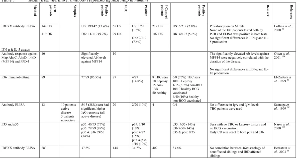
In alle onderzoeken die tot nu toe zijn uitgevoerd naar antilichamen tegen Map worden eiwitten gebruikt die niet specifiek zijn voor Map, maar ook voorkomen bij infecties met andere mycobacteriën. Als er dus in MC patiënten een afweerreactie gemeten wordt tegen ‘Map’, dan betekent dit dat de patiënt ook geïnfecteerd zou kunnen zijn (geweest) met andere, nauw verwante mycobacteriën zoals M. avium. In de meeste onderzoeken worden antilichamen aangetoond tegen mycobacteriën in zowel een deel van de MC patiënten als een deel van de controle personen. Antilichamen tegen Map worden dus niet in alle MC patiënten aangetroffen. Zoals eerder vermeld is in deze ziekte vooral het cellulaire afweersysteem van belang en minder het humorale (antilichamen). Het is daarom de vraag in hoeverre de
aanwezigheid van specifieke antilichamen tegen Map in het serum een belangrijke parameter is in de rol die Map wellicht speelt in deze ziekte. Bovendien werd in alle onderzoeken alleen gekeken naar afweerreacties in het bloed van patiënten. Bekend is dat er in de darm lokale afweerreacties optreden die niet terug te vinden zijn in het bloed. Het zou dus beter zijn om specifiek de ontstoken darm te onderzoeken op afweer tegen Map (of elke andere mogelijke veroorzaker van MC) en de aanwezigheid van specifieke IgA antilichamen te bepalen en niet het perifere bloed als bron te gebruiken.
Intolerantie en een lekkende darm
De meest recente hypothese over het ontstaan en voortduren van MC gaat uit van het idee dat er drie factoren bij MC betrokken zijn:
• Erfelijke aanleg.
• Prikkeling van de darm door de darmflora. • Afwijkende afweerreacties in de darm.
Dus in een persoon die daar genetisch gevoelige voor is wekken één of meerdere
componenten van de darmflora een abnormale afweerreactie op die leidt tot de chronische darmontsteking die karakteristiek is voor CD. Dit proces gaat gepaard met een lekkend darmslijmvlies (mucosa) waardoor allerlei voedselcomponenten en microorganismen gemakkelijker kunnen binnendringen, en op hun beurt ook weer voor een afweerreactie kunnen zorgen. Het afweersysteem in de darm wordt voortdurend geprikkeld door alle mogelijke antigenen (dit zijn stoffen die een afweerreactie opwekken, zoals
micro-organismen) die door het voedsel worden aangevoerd. Afweercellen staan permanent paraat om snel schadelijke antigenen te neutraliseren. Aan de andere kant moeten gunstige of niet schadelijk antigenen, zoals die van de darmflora wel worden getolereerd. In gezonde personen is daarom in de darm permanente paraatheid van het afweersysteem noodzakelijk, zonder dat het evenwicht omslaat naar de verkeerde kant. Als er een ontregeling plaatsvindt in het evenwicht tussen tolerantie van, en afweer tegen antigenen kan in genetisch gevoelige personen een ongecontroleerde reactie ontstaan die leidt tot een chronische ontsteking van de darm. Ontregeling van het evenwicht in de darm zou kunnen ontstaan als gevolg van een veranderde samenstelling van de darmflora, bijvoorbeeld door verhoogde koolhydraatinname, roken en andere milieufactoren. Opvallend is bijvoorbeeld dat zeer veel MC patiënten
antilichamen hebben tegen bakkersgist (Saccharomyces cerevisiae). Dit duidt erop dat er bij MC patiënten een verminderde tolerantie is tegen normale voedselantigenen zoals
bakkersgist, doordat deze als gevolg van een lekkend darmslijmvlies gemakkelijker tot de diepere lagen van de darm kunnen doordringen.
Conclusie
In dit review laten we zien dat er veel verschillende factoren betrokken zijn bij MC. Tot nu toe is echter geen enkele factor gevonden waarvan onomstotelijk bewezen is dat deze individueel verantwoordelijk is voor het ontstaan van MC, of die in samenwerking met andere factoren als een causatief agens kan worden aangemerkt; dit geldt ook voor Map. Wel bestaat de reële mogelijkheid dat Map, in samenwerking met andere factoren, als trigger betrokken is bij een deel van de MC patiënten. Bijvoorbeeld door:
• de darmwand te beschadigen en daarmee een afwijkende afweerreactie op te wekken. • van de beschadigde (lekke) darmmucosa gebruik te maken om binnen te dringen en in
• als normale, niet pathogene darmpassant gebruik te maken van een ontstoken darm om zich in te nestelen en te vermeerderen.
Hoewel MC gezien wordt als een ziekte waarbij meerdere factoren samen een rol spelen bij het ontstaan en voortduren van de ziekte, blijft de kans aanwezig dat in een deel van de MC patiënten een infectieus organisme zoals Map wel een hoofdrol speelt. Ondanks het feit dat een duidelijk verband tussen MC en Map (nog) niet is bewezen blijft het dus noodzaak om hun mogelijke relatie serieus te nemen.
1.
Crohn’s disease
Inflammatory bowel disease (IBD) is recognized as an important gastrointestinal disease in human adolescents and adults. IBD exist can manifest in two major forms, Crohn’s disease (CD) and ulcerative colitis (UC). Both diseases are characterized by a chronic inflammation of the intestinal wall. CD is a non-specific chronic inflammation, which extends through all the layers of the intestinal wall, and may involve the mesentery as well as the regional lymph nodes. The inflammation of CD most commonly affects the last part of the ileum, and often includes the colon and sigmoid. In more rare cases other segments of the alimentary tract (mouth, oesophagus, stomach and duodenum) are affected, but this usually occurs in
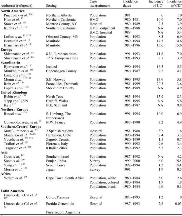
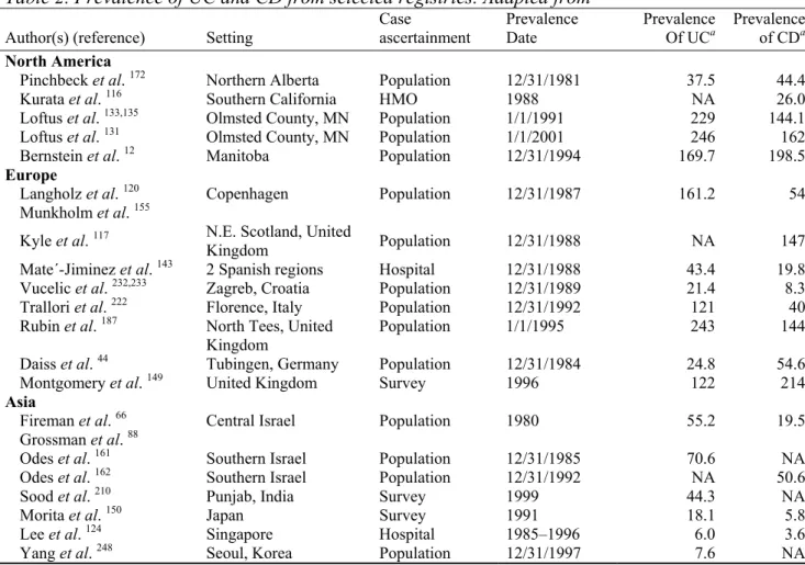
association with involvement of the distal ileum. CD may be diagnosed at any age, although most diagnoses are made between the age of 15 to 35. Men and women have an equal chance to develop the disease. Figures from the US indicate that 20% of cases are diagnosed before the age of 20, and 40% between 20-29 134. The prevalence (existing cases) of CD in Europe varies between 10 to 200 cases per 100,000 persons (Appendix 1, Table 2). The incidence (new cases) of CD in southern Europe ranges from 1-4 cases per 100,000 persons per year, and in northern Europe from 4-10 cases per 100.000 persons per year (Appendix 1, Table 1)
132. In the Netherlands, the prevalence of IBD in 2000 was estimated between 10.000 and
21.000 cases for CD and between 17.000 and 35.000 for UC; mortality in 2000 was determined at 34 cases for CD and 26 for UC 97. There are somewhat indications that the incidence of CD is increasing, especially in low incidence areas 11,97.
Early pathological features of CD include hyperaemia and oedema of the intestinal mucosa and of the mesentery, associated with enlarged inflammatory lymph nodes. Another typical, pathological lesion of CD is a mucosal ulceration, commonly located in areas with Peyer’s patches in the small intestine and over the lymphoid aggregates in the colon. Later during the course of the disease the ulcerations tend to enlarge and to coalesce. Histologically, CD is characterized by lymphoid infiltration of the submucosa and to a lesser degree of the muscularis mucosa. Moreover, non-caseating granulomas are often present in the intestinal mucosa and submucosa, as well as in the mesentery, peritoneum and lymph nodes. The predominant clinical symptoms observed in CD patients are diarrhoea, abdominal pain and weight loss. The clinical course of CD in most patients is characterized by periods of relapses and remissions. Although spontaneous improvement and disappearance of clinical symptoms may be seen in some patients without medical or surgical treatment, well over 60% of all patients with CD will require surgery at one or another phase during the course of their disease, and about half of these patients will require more than one operation over time. About 5-10% of all CD patients will die of their disease, primarily due to massive infection
63.
The pathology and epidemiology of CD share many features with those of UC, though they can be distinguished on histological and immunological characteristics:
• The inflammation of CD may be discontinuous, meaning that areas of involvement in the intestine may be separated by normal, unaffected segments of intestine.
• The inflammation of CD affects all layers of the intestinal wall, while ulcerative colitis affects only the inside layer of the intestine. Also, ulcerative colitis is usually limited to the rectum without extension to the other colon segments or the ileum. • Immunologically, CD is a Th1 disease, whereas UC is a Th2 disease (this will be
discussed later).
CD lesions occur predominantly where there is a high density of lymphoid follicles: in the small bowel where the follicles are grouped in Peyer’s patches, and in the colon where they are isolated. In the small intestine, lesions are more frequent in the distal ileum where Peyer’s patches are most present. In addition to this spatial relationship, a temporal relationship has also been suggested 228. Peyer’s patches are more numerous in younger individuals and become less prominent with age 4. Interestingly, the age-dependent incidence curve for CD is roughly parallel to the number of Peyer’s patches with a peak in the third decade of life 100. A delay of 5–10 years is seen between the development of Peyer’s patches and the occurrence of symptoms. Such a delay is compatible with chronic infections. Conversely, the ileal location of CD is less frequent in elderly people after the involution curve of Peyer’s patches
174. In the colon, in contrast with the small intestine, the number of lymphoid follicles is less
subject to variation. This observation can account for the fact that colonic CD can occur at any age 100. At present there is no cure for the disease, which tends to pursue a variable course, characterized by periods of activity interspersed with remissions, when the disease is either absent or relatively quiescent. A range of factors is thought to increase the risk of relapse of quiescent disease, including stress and dietary factors. Many therapies have been tried to induce or maintain remissions. Typically, treatment is by a combination of medical therapies (anti-inflammatory drugs to reduce the inflammation, drugs to treat the symptoms, and antibiotics to reduce infections associated with the disease), attention to diet to control weight loss, and surgery to deal with the structural effects of the inflamed intestines. Although only carried out as a last resort, a significant proportion of patients needs a
colostomy or an ileostomy. It is clear that CD has a dramatic negative impact on the patients’ quality of life and frequently also has serious consequences for the rest of their family.
• Summary: CD is an incurable chronic inflammation of the human intestinal wall. CD
has a dramatic negative impact on the patient’s quality of life. CD occurs particularly in the Western community.
2.
Features of CD
2.1
Classification of CD
It is becoming more and more recognized that CD is not a homogeneous pattern of disease but a variety of different patterns of inflammatory disease of the intestinal tract 87. Previous attempts of classification have been based primarily on anatomic location and behaviour of disease. During the World Congresses of Gastroenterology in Vienna in 1998, an
international Working Party has developed a simple classification of CD based on objective variables 71. Eight outcome-related variables relevant to CD were identified and evaluated in detail. Three variables were finally chosen: ‘Age at Diagnosis’ (below 40 years, A1 ; equal to or above 40 years, A2), ‘Location’ (terminal ileum, L1 ; colon, L2; ileocolon, L3; upper gastrointestinal, L4), and ‘Behaviour’ (non-stricturing non-penetrating, B1; structuring, B2; penetrating, B3). This Vienna classification of CD provides distinct definitions to categorize CD patients into 24 subgroups (2 x 4 x 3) or disease clusters. A cross-table analysis reveals associations between Age at Diagnosis and Location, and between Behaviour and Location (all p < 0.001) 71. However, the initial ‘behavioural’ classification of B1 (stricturing non-penetrating) at the onset of CD hardly ever remains stable over the lifetimes of the patient but almost invariably progresses in time to either B2 (stricturing) or B3 (penetrating) disease.
• Summary: in terms of pathological criteria, CD is not a homogeneous pattern of
disease and can be categorized into 24 subgroups.
2.2
Immune responses in CD patients
The present knowledge about the immunological mechanisms involved in perpetuation of the chronic inflammation in CD is that the mucosa of patients with established CD is dominated by CD4+ lymphocytes with a Th1 phenotype, characterized by the production of cytokines like Interferon gamma (IFN-γ) and Interleukin-2 (Il-2) 173. In contrast, the mucosa in
ulcerative colitis is dominated by CD4+ lymphocytes with a Th2 phenotype, characterized by production of Transforming Growth Factor beta (TGF-β) and Il-5 173. In Crohn disease the
Th1 cytokines activate macrophages, which in turn produce Il-12, Il-18, and macrophage migration inhibitor factor and thus further stimulate Th1 in a self-sustaining cycle 137,139,183. Macrophages produce a potent mix of broadly active inflammatory cytokines, including Tumor Necrosis Factor (TNF), Il-1 and Il-6; in CD, Il-6 levels correlate with the severity of the disease 136. Recruitment of additional leukocytes from the vascular space to sites of disease activity is especially important in maintaining inflammation. In CD, TNF-α is the key pro-inflammatory cytokine in the development of the lesions and in the persistence of IFN-γ production by Th1-cells 4. On the other hand, production of anti-inflammatory cytokines such
as Il-10, Il-4 and Il-13 seems deficient or at least insufficient to counteract the pro-inflammatory loop of the immune response 4,137,159.
• Summary: CD is a Th1 disease characterized by the production of pro-inflammatory
cytokines like TNF-α, which is responsible for development of the lesions, and by the production of IFN-γ and Il-2.
2.3
Aetiology of CD
Since Thomas Dalziel’s initial description of CD in 1913, many ideas have been postulated about the cause of the disease 45. Based on epidemiological and geographical observations, two major factors were identified that contribute to the development of IBD: genetic factors and environmental factors.
Genetic factors
At present, substantial evidence is available that suggest genetic factors are important
determinants of susceptibility and disease behaviour in IBD. Reports of familial clustering of IBD date back to the 1930’s. Concordance rates in siblings and monozygotic twins suggest that the genetic contribution to disease pathogenesis is at least equivalent to that in other common immune mediated diseases (Multiple Sclerosis, insulin-dependent Diabetes
Mellitus). First-degree relatives of an affected patient have a risk of IBD that is 4 to 20 times as high as that among the background population; the absolute risk of IBD is approximately 7% among first-degree family members of IBD patients 173. A substantial higher rate of disease concordance has been observed in monozygotic twins than in dizygotic twins,
especially in those with CD 173. A positive family history is more common with CD than UC, and relatives of patients with CD have a higher risk of developing IBD than do relatives of patients with UC 173. Furthermore, Caucasian people are more frequently affected than other racial groups, and the incidence of IBD among Jewish people compared with non-Jewish are 16.8% versus 7.0%, respectively, for CD and 4.6% versus 0.9%, respectively, for UC. Within Jewish populations, Ashkenazi (Eastern European) Jews have a higher incidence of IBD than Sephardic (Middle Eastern, African, or Spanish) Jews. These data strongly suggest a genetic background for IBD; so persons can have a predisposition to CD. However, the absence of a simple mendelian inherited pattern of disease suggest that IBD is a polygenic disease, and that multiple gene products, whether or not in combination with environmental factors, contribute to a person’s risk of IBD 173.
Environmental factors
The incidence of CD has increased in developed countries over the past 50 years 205. There is a slight north to south gradient of IBD incidences worldwide as well as in Europe; for
instance, the incidence rates in Scandinavia, UK and the Netherlands are 2-3 fold higher compared to southern Europe (Appendix 1, Table 1)132,206. In developing countries infectious
intestinal diseases are common, whereas IBD’s, especially CD, are rare. However, the incidence and prevalence of IBD are beginning to rise in low-incidence areas such as southern Europe, Asia, and much of the developing world 132. Since people who have immigrated from regions with a low IBD prevalence to regions with a high prevalence acquired the same disease prevalence as the local population 94,132 there is little doubt that environmental factors are involved: Western lifestyle and hygiene. This is supported by the finding that good domestic hygiene in infancy has been shown to be a risk factor for CD, but not for UC 63,72. The risk of developing CD was shown to be increased 3-fold if a separate toilet is available and 5-fold if there is hot tap water in the household 63,72. Furthermore, CD
and UC patients in childhood were less likely to have been breast-fed 112 and less likely to have ingested unpasteurised milk. CD seems to occur more frequently in members of small families. Since intra-familial transmission of common pathogens is frequent, the single child is particularly prone to be raised under more hygienic conditions with lower risk of acquiring gut infections 74,171. Most likely these various factors associated with the incidence of CD serve as indicators of a rather clean environment, leading to a diminished confrontation with (non)-pathogenic micro-organisms. As a result, the intestinal innate immune system is probably not trained to confront minor infections without recruiting the full array of specific immune functions that act only at the expense of a relevant inflammation 63. Another
interesting new aspect of the hygiene hypothesis is the decline in infections from helmintic parasites in the developed world, which is in line with more hygienic conditions. Helmintic infections are mechanistically associated with the rise in prevalence of CD 60,205. Helminths are associated with a Th2 response that would counterbalance the Th1 response of CD. There are however also publications that indicate that infections in childhood increase the chance of IBD. Babies with a recorded ‘prenatal health event’ (i.e., infection or serious illness in mother or child) had a four-fold increased risk for IBD, and infants from families of low socio-economic status (often associated with low hygiene) were three times more likely to develop IBD later in life. Furthermore, several studies have noted a higher frequency of gastroenteritis or diarrhoeal illness during infancy among future IBD patients. Unfortunately, in many retrospective population studies, recall bias is of great concern. Nevertheless, despite these somewhat controversial data, micro-organisms seem to play an important role in
development of IBD, and perhaps also in preventing from IBD. CD has also been associated with increased intake of carbohydrates (refined sugar) 179 and oral contraceptives. The strongest environmental factors identified so far are cigarette smoking and appendectomy
19,55,132. Whereas smokers have a significantly decreased risk of UC, they have an increased
risk of CD, and appendectomy appears to be protective for the development of UC, whereas it is associated with future risk of CD 132,189.
• Summary: CD is a multi-factorial disease. Several genetic (familial, racial) and
environmental (geographic, hygienic) factors -especially microbial- have been associated with the disease.
3.
The role of genetic factors in CD
3.1
CARD15/NOD2 mutations and CD
At present, at least seven genomic regions located on chromosomes 1, 5, 6, 12, 14, 16 and 19 are thought to be involved in the predisposition to IBD 18,34,54,90,91,101,180,181. Thus far, most emphasis is put on mutations found in the CARD15 gene, located on chromosome 16q12. CARD15 encodes NOD2, a cytoplasmic protein that is mainly expressed in
monocytes/macrophages and granulocytes, but has also been identified in epithelial cells
77,173. Three main mutations in the CARD15 gene (R702W, G908R and
1007fsinsC/3020insC) have been identified that are carried by 20-25% of Caucasian CD patients and only 7-15% of healthy controls 101,102,163. Interestingly, mutated homozygotes and compound heterozygotes (double dose mutation carriers) are significantly more frequent than expected by chance in CD patients (15-20%), which demonstrates a mutation dose effect
102. However, so far no association between CD and CARD15 has been found for Japanese
patients, suggesting that a subgroup of CD is not related to a dysfunction of CARD15 104,247, and little is known about presence of CARD15 mutations in other ethnic groups. The
frameshift mutation (1007fsinsC) of the CARD15 gene is associated with ileal involvement and the fistulizing or fibrostenotic phenotype, which are characterized by an increased expression of Th1 cytokines, such as TNFα. CARD15 is a component of the innate immune system. Innate immunity is the most ancient system of defence against pathogens in humans and animals. This system takes action against an invading organism before a specific,
acquired response has been established towards that particular organism. The innate immune system relies on the expression of a restricted set of receptors that enable the recognition of highly conserved microbial motifs, often also named ‘pathogen-associated molecular patterns’ (PAMPs); these PAMPs are not present on the host cell. PAMPs are generally structural bacterial components that display little or no variation because of selection pressure. In bacteria, examples of such PAMPs include lipopolysaccharides (LPS), lipoproteins, flagellin, CpG DNA and peptidoglycan 77,130. Peptidoglycan is a major
constituent of the cell wall of Gram-positive bacteria. In Gram-negative bacteria a thin layer of peptidoglycan is located in the periplasmic space 77. The 1007fsinsC frameshift mutation, the most frequent CARD15 variant associated with CD, fully abrogates NOD2-dependent detection of peptidoglycan 76.
NOD2 plays a role in the intracellular killing of bacteria with its function as an intracellular receptor for PAMPs and activates the ‘nuclear factor-kappa B’ (NF-κB) pathway following inflammatory stimuli, which results in increased transcription of proinflammatory cytokines
211. NF-κB plays a central role in the cellular immune system. The C-terminal domain of
CARD15 is required for bacterial PAMP dependent activation of NF-κB activity 16,163. The
three main CD associated mutations are located within this C-terminal domain, suggesting that an important causative factor in CD may be an initial defect in the response to bacterial
PAMPs. This loss of function model, also suggested by the dosage effect observed for CARD15 mutations, has been confirmed in experiments with cells transfected with the 1007fsinsC mutated CARD15 gene, where mutated NOD2 protein had a lower ability to respond to PAMPs and to induce NF-κB activation 16.
In CD lesions, an excessive stimulation of the NF-κB pathway is observed. This seems in contradiction to the functional model of reduced NF-κB activation due to a mutated CARD15 gene. However, this paradox between the in vivo situation in CD and reduced NF-κB activity
in vitro is counterbalanced by Toll-like receptors. This will be discussed in the next
paragraph.
• Summary: mutations in the human CARD15 gene have been convincingly associated
with CD. CARD15/NOD2 is part of the body’s innate defence system against bacteria. It activates the immune system after recognizing bacterial components.
3.2
Toll-like receptors and CD
Recently, an additional component of the innate immune system was discovered in mammals, the Toll-like receptors (TLRs). Toll-like receptors are related to the Toll protein, a molecule that is implicated in defence against fungal infection in the fly 126,158. Toll-like receptors are anchored in the cell membrane. Out of the ten TLRs present in the human genome, at least eight detect PAMPs at the cell surface. TLRs are thought to play a significant role in IBD. In patients with active IBD, TLR4, TLR3 and TLR2 are differentially modulated compared to control patients 25. In biopsies from patients with CD or UC, an abundant expression of TLR4
by epithelial cells and lamina propria cells has been reported. In contrast, TLR4 is only minimally detectable in intestinal epithelial cells (IECs) of non-IBD intestines 25. TLR4 recognizes LPS from Gram-negative bacteria 217. Luminal LPS is usually well tolerated in large quantities within the healthy intestine. It has been shown that mice with a single point mutation in their TLR4 gene are highly susceptible to developing a more severe form of induced colitis.
In active CD biopsy specimens, the epithelial expression of TLR3 was significantly
decreased in the IECs of the colon, compared with specimens from UC patients and normal controls 25. TLR3 is constitutively expressed in the IECs of UC patients and controls. Reduced expression of TLR3 on IECs seems to be consistent in active CD, independent of location or inflammatory activity. So reduced TLR3 does not simply reflect the local effect of some inflammatory mediator.
TLR2 is involved in responses to Gram-positive bacteria, yeast, and LPS from Gram-negative bacteria 109,217,224,237, and is suggested to be essential for the induction of a protective immune response to Mycobacteria 224. TLR2 is present in monocytes, polymorphonuclear phagocytes, dendritic cells and epithelial cells 158. Stimulation of cells expressing TLR2 with LPS
strongly activates the NF-κB pathway 20,109. Point mutations in TLR2 abrogate inflammatory
TLR4 mutations do abrogate LPS signalling 223. Except for changes in TLR3 and TLR4 expression in CD patients, there is a significant up-regulation of TLR2 protein expression observed in active CD and UC. This up-regulation is restricted to inflammatory cells of the lamina propria 25.
Because TLRs are located on the outside of the cell membrane, they are considered as membrane pattern recognition receptors of the innate immune system 102. TLRs would thus define a membrane-associated counterpart of the NOD2 protein, which is located
intracytoplasmatic. Interestingly, both TLRs and NOD2 are involved in bacteria pattern recognition and both receptors may be abnormal in CD. Very recently, Watanabe et al. used NOD2-deficient mice to show that NOD2 signalling normally inhibits the TLR2-driven Th1 response by regulating NF-κB signalling. In addition, in the absence of NOD2-signalling, such that occur in the presence of a CARD15 mutation, NOD2-mediated inhibition is abrogated, resulting in increased TLR2-mediated NF-κB activation and more Il-12
production. Thus the CARD15 mutations lead to increased Th1 cytokine production 238. This suggests that, at the molecular level, CD results from an inappropriate activation of the mucosal immune system driven by bacteria like the intestinal flora. The different disease patterns found in CD are likely to be based on the type of receptor and presence of specific commensal (not necessarily pathogenic) luminal bacteria. These data give additional
indications that the innate response to the bacterial flora in the intestinal lumen is ineffective. • Summary: differential expression of Toll-like receptors 2, 3 and 4 has been associated
with CD. TLRs are part of the body’s innate defence system against bacteria. They activate the immune system after recognizing specific bacterial components. Enhanced expression of TLR2 followed with increased production of Th1-related cytokines is related with CARD15 mutations.
4.
Intestinal flora and CD
For many years, micro-organisms are thought to play a pivotal role in the development of CD. Microbial agents such as Yersinia enterocolitica, pseudomonas-like organisms,
Escherichia coli, Streptococcus faecalis, Chlamidiae, Mycoplasma, Measles virus,
Saccharomyces cerevisiae, Helicobacter pylori, and Mycobacteria have been postulated to
play a role in the aetiology of the disease 46,50,79,98,100,118,129,182,196,234,243. Accumulating evidence suggest that especially the intestinal flora is a prerequisite, and perhaps a central factor in the development of IBD. The discovery of mutations in genes involved in the reaction to bacteria in the gut, CARD15 and Toll-like receptors, has strengthened this theory. This assumption is supported by studies in mouse and rat models of colitis established
through genetic manipulation. The development of colitis in rats and mice under
experimental conditions appears to require the presence of a luminal flora; colitis does not occur in animals when they are maintained in a germ-free environment, but it develops rapidly when they are colonized by commensal bacteria. Knockout mice lacking several immunological relevant genes, including Il-2 or IL-10 develop experimental colitis only when raised under contaminated but not under sterile conditions 201. Furthermore, in CD
patients after curative resection, a diverting terminal ileostomy was constructed thereby excluding the neoterminal ileum and the colon from intestinal transit. After six months of exclusion, transit was restored. None of the patients had endoscopic lesions in the
neoterminal ileum after that six months of exclusion and biopsies did not show inflammatory changes characteristic of CD. In contrast, 71% of patients with one-step surgery had
recurrence in the neoterminal ileum within six months of surgery. All five patients had an important recurrence of disease, both endoscopically and histologically, six months after transit was restored 43,92,190,194. These data show that the intestinal flora plays an important role in the development of CD.
• Summary: CD cannot develop or perpetuate without the presence of an intestinal
5.
M. avium ssp. paratuberculosis and CD
One of the bacteria that has been frequently associated with CD is Mycobacterium avium subspecies paratuberculosis (Map). In 1972, Patterson and Allen put emphasis on the strong similarities between CD in humans and Paratuberculosis (Johne’s disease) in cattle 166. The characteristics of CD and Paratuberculosis are compared in Appendix 1 Table 3 and 4. Paratuberculosis is a severe chronic intestinal disease caused by Map. Besides Cattle, Map can also cause Paratuberculosis in other ruminants like sheep and goats. Map is widespread in nature, and has been isolated from many wildlife species including rabbit, fox, stoat, weasel, crow, rook, jackdaw, rat, wood mouse, hare, badger and deer 7,8,167. Indications of a possible etiological link between CD and Paratuberculosis came when Chiodini et al. isolated a
Mycobacterium species from surgical gut specimens of four patients with CD 32,33. This
Mycobacterium was biochemically and genetically similar to Map. The investigators
hypothesized that this slow growing mycobacterium played an etiological role in at least some cases of CD. Map belongs to the genus Mycobacteriacea. Well know members of this family are M. tuberculosis, the cause of tuberculosis, and M. leprae, the cause of Leprosy.
Map is very closely related to M. avium ssp. avium (Maa) and together with M avium ssp. silvaticum and M. intracellulare belong to the Mycobacterium avium-Complex (MAC). On
the nucleotide sequence level Map and Maa are 98% identical, but can be differentiated by the presence of a chromosomal insertion sequence, IS900, which is (thus far) unique to Map, and is present in 17-18 copies in its genome 85. Phenotypically, Map can be distinguished by
its in vitro dependence on Mycobactin-J, an iron-chelating compound, and its slow growth. In contrast to Map, Maa is not a major pathogen of cattle, but it in HIV-infected patients it can cause severe complications 6,27,177. Map has a complex cell wall, relatively impermeable and rich in lipids, which confers acid-fast properties. Due to its firm cell wall Map can survive for a long time in the environment (for more than a year in a dry fully shaded environment) 244. Neonatal and juvenile animals are at the highest risk for acquiring an infection of Map 95. Young animals are most commonly infected through the fecal-oral route. This occurs either by ingesting the organism through contaminated milk or food products or by accidental ingestion of the micro-organism from contaminated surfaces 95.
Preferentially Map targets the mucosa-associated lymphoid tissues of the upper
gastrointestinal tract, where it is endocytosed by the M-cells of the ileal Peyer’s patches and subsequently phagocytosed by subepithelial and intraepithelial macrophages 4,70,95,138. Data suggest that Map can arrest the development of the phagosomal compartment into a mature phagosome 99,115,220. The Peyer’s patches reach their maximum development about the time of birth and progressively disappear afterwards, though patches in the jejunum and ileocaecal valve can persist in adults 148,176. This could explanation why the highest susceptibility for a
Map-infection occurs in young animals. Map probably remains in the phagosome, where it
multiplies intracellularly 115. A recent study showed that Map-infected macrophages were able to stimulate production of IL-2 and IFN-γ in CD4+ cells 250. Cytokine production and the
granuloma, and a cellular response is initiated in the nearby lymph nodes in an attempt to clear the infection 95,138. This inflammatory process leads to the clinical manifestations of a corrugated intestinal epithelium and the corresponding characteristic malnutrition syndrome associated with Paratuberculosis 95. Cattle become infected with Map as calves but often do not develop clinical signs until 3-5 years of age. After this subclinical stage, extensive granulomatous inflammation developes in the terminal ileum, which leads to chronic diarrhea, shedding high numbers of Map, diffuse edema, malabsorption of nutrients and decreased milk production. Though, not all infected cattle will develop clinical disease signs
110,122.
Paratuberculosis shows a wide immunological and histopathological spectrum: at one end the tuberculoid form, which is located where the host offers a strong cellular immune response with a very low humoral response; at the opposite end, the lepromatous form, associated with a weak cellular response but a strong humoral response. Between these forms is the
borderline form, which shows the most severe clinical signs 4. Animals from an infected flock can present varying immunological and pathological pictures and can be located in different positions in the spectrum.
Gross changes in Map-infected sheep and goat are often lacking or difficult to detect, and may not resemble those of Paratuberculosis in cattle 4. In cattle, sheep and goats, two distinct types of pathology are present, based on the abundance of Mycobacteria and cellular infiltrate
22,170. The more common form (predominantly found in cattle), the multibacillary (also named
‘pluribacillary or lepromatous’) form, is characterized by numerous acid-fast Map in the cytoplasm of the many large macrophages that infiltrate the mucosa. Lymphocytes and granulocytes are present in much lower numbers. These changes cause marked thickening of the intestine 4. The other form of Paratuberculosis, the lymphocytic (also ‘paucibacillary or tuberculoid’) form (which is predominantly found in sheep and goat), is characterized by a more marked lymphocytic infiltrate with scattered, small focal granulomata and giant cells. Lesions may exhibit caseation, calcification or fibrosis. Acid-fast Map are sparse or
undetectable in lymphocytic lesions and are usually absent from caseous or calcified foci 4. The two types of pathology in Paratuberculosis correlate with different host responses to the bacterium 4, and perhaps also to the type of Map strain 1,190. Sheep with multibacillary disease
have a strong antibody response but a weak or absent cell-mediated immunity, predominant Th2-like cytokines Il-4 and Il-10, and Map appears to multiply in epitheloid cells in these lesions 4,24,75. Animals with the lymphocytic form show a strong CMI response and poor or absent antibody response and predominant Th1-like cytokines Il-2 and IFNg. In these lesions the bacteria appear to degenerate in epitheloid macrophages 4. This form of Paratuberculosis appears to be able to reduce the bacterial load in the intestine 4. This lymphocytic form of Paratuberculosis accords best with the pathological and immunological features of CD. Although CD and Paratuberculosis share numerous clinical, histo-pathological and immunological features, there are however also significant differences between these two diseases, as shown in Appendix 1 Table 3 and 4 4,227. This is one of the reasons why several investigators believe that CD and Paratuberculosis do not share a common etiologic agent
• Summary: Map causes a chronic intestinal inflammatory disease in animals,
Paratuberculosis. Especially the paucibacillary variant of Paratuberculosis shares clinical, histo-pathological and immunological characteristics with CD. Map is present in many dairy herds and probably can be transmitted to humans via foodstuff like milk, meat, water, and via direct contact with contaminated soil.
6.
Is CD caused by Map?
For many investigators the resemblance between CD and Paratuberculosis was sufficient to assume Map to be the mutual causative agent. However, gross pathology and clinical data alone are not sufficient to confirm this hypothesis. Therefore the question remains whether
Map is indeed the etiologic agent of CD. An important dogma concerning the aetiology of
infectious diseases is Koch’s postulates (Robert Koch, 1843-1910). According to Koch, to prove that any organism is the cause of an infectious disease, four postulates should be fulfilled:
1. The specific organism should be present in all individuals suffering from a specific disease, but should not be found in healthy individuals.
2. The specific organism should be isolated from the diseased individual and propagated in the laboratory.
3. This freshly propagated organism, when inoculated into a healthy individual, should cause the same disease seen in the original individual.
4. The organism should be re-isolated from the experimental infection.
Although Koch’s postulates are slightly out of date, many investigators have tried to prove, or discount Koch’s first postulate concerning CD and Map. They have applied different methods to achieve this
2,5,21,23,26,30,33,35,47,48,56,58,65,68,83,103,107,114,144,146,151,156,160,164,165,178,185,191-193,197,198,200,213,215,218,229,236,239:
• Isolation and culturing of Map bacteria from affected CD tissue.
• Demonstration of the presence of a cellular and/or humoral immune response against
Map antigens in CD patients.
• Detection of Map DNA in CD tissue using PCR and/or in situ hybridisation (ISH). • Investigation of the efficacy of anti-mycobacterial drugs.
In the next section we will evaluate the different attempts made to detect Map in human tissue. Furthermore, we will put forward arguments to support, or to discount the hypothesis about an etiologic link between CD and Map.
6.1
Detection of Map in CD tissue
The two techniques that are mostly used for the detection of Map in tissue samples from CD patients are culturing and PCR. Since it is rather difficult to culture Map bacteria from affected tissue, most studies have been done using the PCR technique, which is able to detect as little as a few copies of the Map genome. PCR has resulted in a much higher detection rate for Map in tissues. However, great care needs to be taken to avoid contamination of samples, and negative control samples should be included in the assays, because the high sensitivity
means inadvertent contamination can easily give rise to false-positive results. Furthermore, the assay will not discriminate between viable and dead cells. The fact that PCR also detects dead Map bacteria may account for its higher detection rate compared to culturing
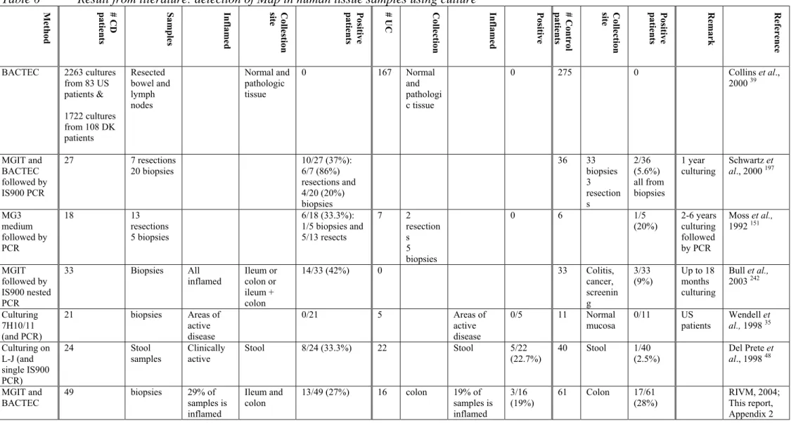
techniques. In Appendix 1 Table 5 and 6 the results of several recent studies on the presence of Map in tissue are depicted. An important issue to keep in mind to interpret these results is that there is neither consensus in patient selection nor in sample selection. Several
investigators took tissue samples from patients with only a history of CD, irrespective of the patients’ clinical state (whether or not an inflamed intestine) at the time of sampling. Other investigators only took samples from heavily inflamed intestines. In some studies either resections or biopsy samples were taken, while other studies used both. Resections cover the whole intestinal wall, but biopsies only the most outside layers (mucosa and lamina propria). Unfortunately, most if not all patients already had a history of IBD, and probably were (or had been) treated with medicines. Medicines change the course of the disease, and this of course could have influence on the endurance of a possible disease mediator, in particular micro-organisms.
The results of Map detection in CD tissue by PCR and culturing can be categorized into three groups:
1. Map was detected significantly more often in the CD group of patients than in the UC and control groups 47,48,65,103,156,192,193,197,198.
2. Map was detected in a number of CD, UC and control patients, but in neither the groups the detection rate was significantly higher 35,83,146(RIVM, Appendix 2). 3. Map could not be detected in CD, UC or control patients 2,5,26,30,56,68,107,178,185.
If CD was an infectious disease solely caused by Map, than according to Koch, Map should be present in all cases of individuals suffering from CD, but should not be found in healthy individuals. It is clear from the data in Appendix 1 Table 5 and 6 that Map cannot be detected in all individuals suffering from CD. But if Map were the etiologic agent of CD, why is it not possible to detect Map in all CD patients, and why does Map not cause disease in so many
Map-positive healthy individuals? Four hypotheses can be postulated to account for the
absence of Map (nucleic acid or antigens) in CD patients:
1. Map is always involved in CD, but solely at the onset of the disease. Map only
triggers the disease and is subsequently cleared from the gut. Th1 cell-mediated immune responses are considered as efficient against Mycobacteria. The continuous Th1 stimulation seen in CD may therefore be appropriate to eradicate offending Map bacteria. However, this relative success in killing the bacteria may be counteracted by an insufficiency feedback in the immune system associated with a genetically induced failure to produce those anti-inflammatory cytokines that can stop the immune response. This run-away immune response leads to disease perpetuation and mucosal damage. If this first hypothesis is right, Koch’s first postulate cannot be applied to CD.
2. Map is not always involved in CD. Apart from Map, other factors (micro-organisms,
chemical substances, genetic defects, impaired intestinal mucosa) are essential (etiological) agent(s) for development of CD. This will be discussed later.
3. Map is involved in CD but cannot be detected. The landmark of most mycobacterial
infections is the presence of fast bacilli: in Paratuberculosis one can see swarms of acid-fast bacilli, in CD there are none. Failure to see acid acid-fast bacilli in Map infections is not uncommon. In the lymphocytic or paucibacillary form of Paratuberculosis, often seen in sheep and goat, acid-fast Map cells are sparse or undetectable in lymphocytic lesions, and are usually absent from caseous or calcified foci 4,41. Furthermore, Map cannot be cultured from all animals known to be infected with Map 41. From studies on Leprosy it is evident that sensitisation with Mycobacteria can cause different host immune responses. Depending on the type of immune response, the clinical spectra in mycobacteriosis can vary from no symptoms; diffuse inflammatory mycobacteriosis with bacillary load such as lepromatous leprosy; self healing with no demonstrable bacilli (paucibacillary) such as the lymphocytic (tuberculoid) form of leprosy; intermediate type often with the presence of bacilli such as borderline leprosy. Hence, CD may resemble the tuberculoid form of leprosy in which hardly if any Map can be detected.
Some investigators believe that Map subsists in a ‘cell-wall deficient’ form in CD
31,59,83,142,151,198,236. This cell-wall deficient form is supposed to be responsible for triggering
the abnormal immune response that leads to CD 86. Cell-wall deficient forms are very
difficult to isolate. Due to the lack of a protective cell wall they do not (or hardly) survive the harsh decontamination procedure applied for culturing. Although cell-wall deficient Map cells have been produced artificially in vitro, there is however no conclusive evidence that they do exist in natural Map infections.
Another possible reason why Map cannot be detected in most CD patients is the sampling error. Even a very sensitive detection assay like PCR can only yield a positive result if the target (in this case Map DNA) is present in the reaction mixture. If Map is not uniformly distributed in CD tissue, and/or only present in minimal amounts, the place of sampling becomes very critical. It is a matter of chance to pick the right spot.
4. Map is not involved in CD. Map is just an opportunistic passenger that was
coincidentally passing through the intestinal tract of the individual from whom it was isolated. Leading to the chicken and egg scenario of which came first, CD or Map. This scenario may explain why in about half of the published studies Map is uniformly distributed between Crohn’s patients and controls. Other mycobacterial species are also known to be uniformly distributed between CD patients and controls 56,213. This is consistent with the known environmental distribution of Mycobacteria, which are present in up to 30-50% of all environmental samplings, including water, soil and even air. For instance, M avium ssp.
silvaticum is equally distributed among people with or without CD. In contrast, in the other
half of the reported studies Map is detected in a significant higher percentage of CD patients compared to control patients. This may suggest that Map has an affinity for inflamed tissue, which is in accordance with the observations that chronic inflamed tissues are readily colonized by other microbial agents. However, in that case Map would also be more
6.2
Map does not always cause disease
Recently, the RIVM has performed a large survey on the presence of Map in CD, UC and control patients. Detection of Map was performed using different techniques: PCR, two different culture methods and immunohistochemistry (immunoperoxidase staining, IP). The results, which are presented in the Appendix 2 section of this report, clearly show that Map is widely distributed between both IBD and control persons. When culturing and IP-staining techniques were applied, RIVM found even a higher prevalence of Map among control persons compared to CD patients; 29% versus 20% for IP-staining, 28% versus 27% for MGIT and 25% versus 22% for BACTEC culturing, respectively. Hence, Map is frequently present in healthy individuals. This is not unexpected, because Map (and most other
pathogenic micro-organisms) does not always cause disease after infection 95. The (in)ability of Map to cause disease may be attributed to the genetic background of the host, the virulence of Map in the host species and/or the nutritional state of the host 110,111. In experimentally infected animals, Map infections do not consistently reproduce disease symptoms. For example, oral inoculation of rabbits with approximately 108 CFU of Map gave clinical and histopathological lesions in only 62 to 75% of the animals 147. In contrast, 100% of calves orally inoculated with 106 CFU of Map developed disease 123. Certain inbred and outbred
strains of mice, such as the C57 black and Swiss white strains, are more susceptible to Map infections than the CBA strain 28,29,95. Resistance to mycobacterial infections in mice, including Map, is associated with the Bcg locus on mouse chromosome 1 69,95,208,209. Two allelic forms of the Bcg gene, Bcgs and Bcgr, confer susceptibility or resistance to infection, respectively. The nutritional and hormonal status of an animal may also influence its susceptibility to Map infections. Reduced dietary calcium protects beige mice from Map infections, but a corresponding increase in endogenous vitamin-D levels reverses the beneficial effects of low Calcium levels 95,211,212. Additionally, transient exposure of monocytes to growth hormone or prolactin enhances intracellular multiplication of Map in primary bovine monocytes 64. From these examples it is clear that individuals can be contaminated with Map without Map being infectious or causing disease symptoms. Therefore the second part of Koch’s first postulate ‘and should not be found in healthy individuals’ may not be applicable to Map infections (actually, this holds true for many other infectious agents). Nevertheless, if Map were the single etiologic agent of CD which can be found in up to 30% of healthy individuals (RIVM, Appendix 2)242, it is clear that the presence of Map in human intestines is very common but hardly leads to CD (as shown in Appendix 1 Table 2, the highest incidence of CD is only 15.6 cases per 100,000 persons per year). If it were possible to investigate a large number of intestinal samples per person, Map may be found in many more individuals with or without CD.
• Summary: so far, the quest for Map using PCR and culture methods, have not resulted
in conclusive data to support or discount the hypothesis that Map is the etiologic agent of CD. The fact that Map can be found in a high percentage of apparently healthy individuals raises the question whether Map is a common passer-by of the