RIVM report 230086002/2005
The possible involvement of Mycobacterium avium spp.
paratuberculosis in the aetiology of Crohn’s Disease
A case control study in the Netherlands
A.A.P.M. Herrewegh1,5, P. Overduin1, P.J.M. Roholl2, F.K. Gielis2, J.E. Robinson2, N. Mahmmod3, R.J. Lieverse3, R.J. Robijn3, A.G.M. van der Zanden4 and
D. van Soolingen1
Diagnostic Laboratory for Infectious Diseases and Perinatal Screening (LIS)1 and Laboratory for Toxicology, Pathology and Genetics (TOX, RIVM)2, RIVM, Bilthoven, the Netherlands. Department of Gastroenteroloy3 and Department of Medical
Microbiology and Infectious Diseases4, Gelre Hospitals, Apeldoorn, the Netherlands. BioMedics Scientific Consultancy, Maarssen, the Netherlands5
Contact: Dr. D. van Soolingen
Laboratory for Infectious Diseases and Perinatal Screening (LIS) email: d.van.soolingen@rivm.nl
Abstract
The possible involvement of Mycobacterium avium spp. paratuberculosis in the aetiology of Crohn’s Disease: a case control study in the Netherlands.
A case control study was performed to investigate the possible role of Mycobacterium avium ssp. paratuberculosis (Map) in the aetiology of Crohn’s disease (CD). Biopsy samples were collected from the ileum and colon of CD patients, Ulcerative Colitis (UC) patients and control persons. The biopsy samples were either cultured in MGIT and BACTEC medium, formaline-fixed, or immediately snap-frozen in liquid N2.
The presence of Map bacteria in the cultures was determined using a nested PCR assay targeted to the conserved IS900 DNA-sequence of Map. 27% of CD patients, 6% of UC patients, and 28% of control persons were positive for MGIT-PCR. The BACTEC cultures resulted in a slightly smaller percentage of PCR positive patients, 22% for CD patients, 7% for UC patients and 25% for control persons. The presence of Map was further studied applying the IS900-PCR directly on DNA from frozen biopsy samples. 7% of CD patients, 8% of UC patients and 5% of control persons were PCR positive for Map. The presence of Map was also investigated in formalin-fixed biopsies samples using a hyperimmune antiserum to the M. complex. With immunoperoxidase (IP) staining, M. avium-complex antigens were observed in biopsy samples from 20% of CD patients, 13% of UC patients and 29% of control persons. Surprisingly, these data show even a 50% higher
presence of Map-antigens in control persons (29%) compared to CD patients (20%). Our data clearly show that Map is present both in IBD patients (CD and UC) and in control persons. The results of culturing and IP staining show a good correlation; with both techniques, an average of 23% of CD patients and 27% of control persons is positive for Map. Remarkably, the results of culturing and IP staining show an even higher prevalence of Map in control persons compared to CD patients. Moreover, only in a few instances patients were positive in two different tests (PCR, IP, MGIT and BACTEC), and even with both culturing assays almost no overlap in positive results is observed. This result indicates that the number of Map cells in human tissue is very low, and that the prevalence of Map in the population of IBD patients and of control persons is difficult to estimate. Unexpectedly, we obtained evidence that members of the Chlamydiales family are present in high amounts in most of the CD and UC patients, whereas they were of low incidence in control persons.
In conclusion, our data do not support the hypothesis that Map is directly involved in Crohn’s disease, but does not exclude that Map infection may play a role in the aetiology of Crohn’s Disease for a susceptible subset of the population.
Keywords: Mycobacterium avium ssp. paratuberculosis, Crohn’s disease, Ulcerative colitis, biopsy, culturing, PCR, immunoperoxidase staining, Chlamydiae.
Rapport in het kort
Mycobacterium avium ssp. paratuberculosis als mogelijke oorzaak van de ziekte van
Crohn: resultaten van een patiëntenstudie in Nederland
De bacterie Mycobacterium avium ssp. paratuberculosis (Map) wordt beschouwd als een mogelijke oorzaak van de ziekte van Crohn (morbus Crohn, MC). In samenwerking met Gelre ziekenhuizen heeft het RIVM een onderzoek uitgevoerd naar het voorkomen van Map in darmbiopten van patiënten met MC, patienten met Ulcerative Colitis (UC) en
controlepersonen zonder darmontsteking. De aanwezigheid van Map in de darmbiopten werd onderzocht met behulp van kweekmethoden, een DNA amplificatiemethode (PCR) en een immunologische detectiemethode (immunoperoxidase kleuring, IP-kleuring). Met de PCR-methode werd Map aangetoond in 7% van de MC-patiënten, 8% van de UC-patiënten en 5% van de controlepersonen. Met de gecombineerde kweek- en PCR-methoden waren gemiddeld 25% van de MC-patiënten, 7% van de UC patiënten en 27% van de controlepersonen positief. Met behulp van de IP-kleuring werd Map aangetoond in 20% van de MC-patiënten, 13% van de UC-patiënten en 29% van de controlepersonen. De resultaten van de kweek- en PCR-methoden tonen geen significant verschil tussen de aanwezigheid van Map in MC-patiënten vergeleken met controlepersonen. De resultaten van de IP-kleuring tonen zelfs een hoger percentage Map-positieve controlepersonen vergeleken bij MC-patiënten. Onze resultaten tonen duidelijk aan dat Map zowel bij MC-patiënten als bij UC-patiënten en controlepersonen voorkomt. Naast de aanwezigheid van Map is ook gekeken naar de aanwezigheid van
Chlamydia in de biopten. Op grond van IP en in situ hybridisatie kleuringen bleken Chlamydiae in grote aantallen aanwezig te zijn in de biopten van MC- en UC-patiënten en nauwelijks aanwezig in de biopten van controle patiënten.
In conclusie: onze resultaten ondersteunen niet de hypothese dat Map direct betrokken is bij de ziekte van Crohn, maar sluiten ook niet uit dat Map een rol speelt bij het ontstaan van de ziekte van Crohn in een gevoelig deel van de populatie.
Trefwoorden: Mycobacterium avium ssp. paratuberculosis, ziekte van Crohn, Ulcerative Colitis, biopten, kweek, PCR, immunoperoxidase kleuring, Chlamydia.
Contents
Samenvatting 5
Introduction 7
1. Materials and Methods 8
1.1 Patients and control persons 8
1.2 Map culturing 8
1.3 DNA extraction 9
1.4 IS900 nested PCR 9
1.5 Immunoperoxidase staining (IP) 10
2 Results 11
2.1 Results nested PCR on biopsy samples 11
2.2 Results immunoperoxidase staining (IP) 11
2.3 Results Map culturing 12
3. Discussion and Conclusion 14
Samenvatting
In samenwerking met Gelre ziekenhuizen heeft het RIVM een onderzoek uitgevoerd naar het voorkomen van Mycobacterium avium ssp. paratuberculosis (Map) bij patiënten met de ziekte van Crohn (CD) en bij patiënten met Colitis Ulcerosa (UC). Als controle werden personen onderzocht die niet leden aan CD of UC. Tijdens routine coloscopie werden minimaal zes darmslijmvliesbiopten afgenomen uit het terminale ileum en het colon. Bij CD- en UC-patiënten werden deze biopten genomen uit ontstoken delen van de darm -indien aanwezig- en uit niet ontstoken delen.
Twee biopten werden gekweekt in MGIT en BACTEC medium, twee biopten werden gefixeerd in formaline, en twee of meer biopten werden direct ingevroren in vloeibare stikstof. Na een jaar incuberen bij 37 °C werd de aanwezigheid van Map in de kweken onderzocht met behulp van een nested-PCR gericht tegen het geconserveerde IS900 element dat aanwezig is in Map DNA. Dertien van de 49 CD-patiënten (27%), één van de 16 UC patiënten en 17 van de 61 controlepersonen waren positief in de MGIT-PCR. De BACTEC-PCR leverde iets lagere percentages positieve patiënten op, 22% voor CD, 7% voor UC en 25% voor controlepersonen. Daarnaast werd de aanwezigheid van Map bestudeerd in de vriesbiopten met behulp van de nested-PCR. Vier van de 59 CD-patiënten (7%), 2 van de 25 UC-patiënten (8%) en vier van de 79 controlepersonen (5%) waren PCR positief voor Map. Deze resultaten tonen geen significant verschil tussen de aanwezigheid van Map in
CD-patiënten vergeleken met controlepersonen. De aanwezigheid van Map werd ook bestudeerd in de formaline gefixeerde biopten met behulp van een immunoperoxidase (IP) kleuring en een serum dat immuno-reactief is tegen M. avium-complex antigenen. Biopten van 11 van de 55 CD-patiënten (20%), drie van de 23 UC-patiënten (13%) en 21 van de 73 controlepersonen (29%) waren positief in de IP kleuring. De resultaten van deze kleuring tonen zelfs een hoger percentage Map positieve controlepersonen (29%) vergeleken bij CD-patiënten (20%). Map bleek opmerkelijk vaker voor te komen in ontstoken biopten dan in niet ontstoken biopten; dit was onafhankelijk van de diagnose.
Onze resultaten tonen duidelijk aan dat Map zowel bij CD-patiënten als bij UC-patiënten en controlepersonen voorkomt. De resultaten van de kweek en IP kleuring tonen een goede overeenkomst. Met beide technieken is ongeveer 23% van de CD-patiënten en 27% van de controlepersonen positief voor Map. Opmerkelijk is dat de resultaten van kweek en
IP kleuring zelfs een nog hogere prevalentie van Map aantonen in controlepersonen dan in CD-patiënten. Ofschoon in totaal respectievelijk 10, 35, 31 en 25 personen PCR, IP, MGIT en BACTEC-positief zijn, werden slechts een paar personen positief bevonden in twee of meer tests. Zelfs met beide kweekmethoden is een zeer beperkte overlap in positieve resultaten te zien. Dit toont aan dat het aantal Map bacteriën in de onderzochte weefsels erg laag is, en daarmee is de prevalentie van Map in weefsel van de verschillende groepen
moeilijk in te schatten. Naast de aanwezigheid van Map is ook gekeken naar de aanwezigheid van Chlamydia in de biopten. Op grond van IP en in situ hybridisatie kleuringen bleken Chlamydiae in grote aantallen aanwezig te zijn in de biopten van CD- en UC-patiënten en nauwelijks aanwezig in de biopten van controle patiënten. In conclusie: onze resultaten
ondersteunen niet de hypothese dat Map direct betrokken is bij de ziekte van Crohn, maar sluiten ook niet uit dat Map een rol speelt bij het ontstaan van de ziekte van Crohn in een gevoelig deel van de populatie.
Introduction
A case control study was performed to investigate the possible role of Mycobacterium avium ssp. paratuberculosis (Map) in the aetiology of Crohn’s disease (CD). Intestinal biopsy samples were collected from CD patients, Ulcerative Colitis (UC) patients and control persons with intestinal disorder other than CD or UC. Patients attending for routine ileocolonoscopy as a normal part of their clinical care were enrolled prospectively in the study. Informed consent was obtained in each case. The case definition of CD was
established on the basis of standard clinical, radiological, endoscopic and histopathological criteria. The control group comprised patients attending for ileocolonoscopy who did not have a clinicopathological diagnosis of IBD. Inclusion criteria were compliance with case definitions and consent. Mucosal biopsies (2-8) were taken during colonoscopy from the terminal ileum and colon of all CD, UC and control patients. In CD and UC, biopsies were obtained from inflamed mucosa, if present, and from non-inflamed parts of the intestine. Two mucosal sample were fixed in phosphate buffered formaldehyde and embedded in paraffin for pathology and stained with immuno-peroxidase (IP) for the presence of Map-antigens using M. avium -complex specific antibodies. Two samples were stored in phosphate buffered saline (PBS) for culturing, and 2 to 4 samples were transferred to a cryo-vial and snap frozen in liquid N2 for DNA isolation to investigate the presence of Map DNA with a nested-PCR
using Map-specific primers targeted to the IS900 sequence. All patients and their physician in attendance were requested to fill in a detailed questionnaire, including questions about
general features, clinical picture and symptoms of their complaints, medication, (family) background, life-style, eating, drinking and smoking habits and physical condition.
1. Materials and Methods
1.1 Patients and control persons
All patients were obtained from the outpatient clinic of Gelre Teaching Hospital in Apeldoorn, the Netherlands. The mean age, height and weight of the persons in the three subgroups were comparable. The majority of the CD patients was in the age group of 20 to 30 years.
The percentage of female to male was relatively comparable in the CD and control group, while in the ulcerative colitis (UC) group (consistent with the epidemiological studies) were more males than females (14 and 12, respectively). The majority of the population in the study was born in the Netherlands (95%). The major indication for coloscopy in the control group was the screening for colon cancer. The second major indication was abdominal pain and polyps’ control. Coloscopy showed signs of inflammation in 71% of the CD patients (n = 59), 64% of UC patients (n = 25) and 6.1% of control persons (n = 82). The
inflammation was located in the ileum in 30.4 % of the CD patients. Two CD patients were also associated with colon inflammation. The ileum was not affected in any of the UC patients.
Histopathological examination of the persons revealed that the correlation between the macroscopic inflammation and the histopathological examination was very high. All persons showing signs of macroscopic inflammation by coloscopy also had signs of microscopic inflammation by histopathological examination. However, the appearance of a non-inflamed mucosa by coloscopy does not exclude a micoscopical inflammation. Histopathological examination of mucosa with a normal macroscopic appearance showed different ranges of microscopical inflammation in samples from 75% of CD patients, 44% of UC patients and 36% of control persons. The majority of these patients had a mild form of inflammation: 93% of the biopsies from CD patients, 80% of UC patients and 37% of control persons showed signs of inflammation of variable gradation.
1.2 Map culturing
The fresh biopsies were decontaminated and subsequently cultured in MGIT (BD Diagnostics) and BACTEC (BioMerieux) culture tubes at 37°C for up till now. Briefly, 0.5 ml NaOH-N-acetyl-L-cysteine (NALC: BBL Mycoprep, Becton Dickinson,) was added to each sample in 0.5 ml saline and incubated at room temperature for 20 min with occasional mixing by inversion. Samples were then centrifuged (10,000 x g for 10 min) and the
supernatant was removed by pipetting. The pellet was resuspended in 0.5 ml TEN buffer (2 mM NaEDTA; 400 mM NaCl; 10 mM TrisHCl pH 8.0: Sigma, UK) and transferred to the tubes containing MGIT and Bactec medium supplemented with 2 µg/ml Mycobactin J (Allied
Monitor, USA). After one year of incubation at 37°C, the culture media were harvested and the presence of Map bacteria was determined using the IS900 PCR as described
henceforward.
1.2 DNA extraction
DNA extraction was performed using the High Pure PCR Template Preparation Kit (Roche, Cat. No. 1.796.828) in combination with the protocol of Bull et al., 2003 1. Frozen biopsy sample were resuspended in 200 µl Tissue Lysis Buffer and 40 µg/ml Proteinase K (Roche kit) and incubated for 2 h at 55°C. Subsequently, 200 µl Binding buffer (Roche kit) was added and the mixture was transferred to a Lysing Matrix B ribolyser tube (Qbiogene, Cat. No. 6911-100). Tubes were chilled on ice for 5 min and mechanically disrupted in the FastPrep Ribolyser (Qbiogene) at a setting of 6.5 m/s2 for 45 s, centrifuged for 1 min at 8000 rpm and incubated for 10 min at 72°C. Subsequently, 100 µl isopropanol was added and the mixture was added to the upper reservoir of a High Pure filter tube, followed by 1 min centrifugation at 8000 rpm. Three consecutive washing steps with Inhibitor Removal buffer (Roche kit), Wash buffer and Wash buffer (Roche kit) were performed, respectively. Finally the DNA was eluted from the filter tube with 100 µl pre-warmed (70°C) Elution buffer (Roche kit).
1.4 IS900 nested PCR
A nested PCR test was developed targeting IS900. The primers AV1 and S535 were used for the first PCR and the primers S204 and AV2 for the nested PCR (Table 1). PCR comprised a first round PCR mix containing 5 µl of DNA extract in a final 50 µl volume with 2µM primers AV1 and S535, 1x HotStar reaction buffer, 200µM dATP, dGTP, dCTP, dTTP and 2.5U HotStar Taq polymerase (Qiagen, Cat. No. 203445). Cycling conditions were: 1 cycle of 95°C for 15 min then 40 cycles of 94°C for 1 min, 58°C for 1 min, 72°C for 3 min then 1 cycle of 72°C for 7 min. Nested PCR used 5 µl of the first round reaction added to a 50 µl reaction mix as in the primary mix but containing 2 µM primers S204 and AV2. Cycling conditions were: 1 cycle of 95°C for 15 min then 40 cycles of 94°C for 1 min, 58°C for 1 min, 72°C for 3 min then 1 cycle of 72°C for 7 min. Positive and negative controls were processed in parallel. Amplicons of expected size were visualised with ethidium bromide on 1.5% agarose gels.
Table 1 Primers used for nested PCR
Primer sequence (5' to 3') Orientation Reference AV1: ATG TGG TTG CTG TGT TGG ATG G sense Bull et al., 2003 1 S535: AGCGTCTTTGGCGTCGGTCTTG antisense Englund et al., 1999 3 S204: TGATCTGGACAATGACGGTTACGGA sense Englund et al., 1999 3
1.5 Immunoperoxidase staining (IP)
The presence of Map was studied on two formalin fixed biopsies from the ileum and/or the colon of patients with Crohn’s disease or ulcerative colitis and from control persons.
Formalin fixed biopsies were embedded in paraffin according to procedures routinely used in pathology laboratories. Presence of Map was determined on paraffin embedded material using a rabbit hyperimmune antiserum to M. avium-complex. This serum reacts with most members of the M. avium complex, including M. avium ssp. paratuberculosis, M. avium ssp. avium and M. avium ssp. intracellulare, but not with other members of Mycobaterium genus or with cells of the intestinal flora. Per biopsy, four paraffin sections were stained for Map. Sections were deparaffinized and endogenous peroxidase was blocked with methanol containing 0.3% H2O2. Next, sections were pre-treated with 0.5% blocking solution and
incubated with a rabbit Map in an optimal concentration. Peroxidase labelled swine anti-rabbit polyclonal antibody (DAKO) was used as secondary antibody. For detection, sections were incubated with diaminobenzidine/nickel substrate. The sections were counterstained with Nuclear Fast Red (Merck). Adjacent sections were incubated with an irrelevant rabbit antiserum (RIVM). Paraffin sections from ileum tissue from cow with Paratuberculosis disease were used as positive control. A cell was considered positive when cellular immunoreactivity was observed.
2 Results
2.1 Results nested PCR on biopsy samples
Fifty-nine CD patients, 25 UC patients and 79 control persons were examined by nested-PCR for the presence of Map DNA. From most patients, 2 biopsy samples were examined. The results are shown in Table 2. Four out of 59 (7%) CD patients, 2 of 25 (8%) UC patients and 4 of 79 (5%) control persons were PCR positive for Map. These data show no significant difference between the presences of Map-DNA in CD patients (7%) compared to UC patients (8%) and control persons (5%).
Table 2 Results nested PCR on biopsy samples
PCR positive PCR negative Patient
group
Patientsa Biopsiesb Positive
Patientsc Inflamedd Inflamede Not inflamedf Inflamedg Not inflamedh Crohn 59 118 4 (7%) 47 (40%) 4 (3%) 0 43 (36%) 71 (60%) UC 25 52 2 (8%) 7 (13%) 0 2 (4%)0 7 (13%) 43 (83%) Control 79 164 5 (6%) 0 0 5 (3%) 0 159 (97%)
aTotal number of patients examined; bTotal number of biopsies examined; cTotal number of PCR positive
patients; dTotal number of biopsies taken from involved (inflamed) areas of disease; eTotal number of PCR
positive biopsies from inflamed areas; fTotal number of PCR positive biopsies from non-inflamed areas; gTotal
number of PCR negative biopsies from inflamed areas; hTotal number of PCR negative biopsies from
non-inflamed areas.
2.2 Results immunoperoxidase staining (IP)
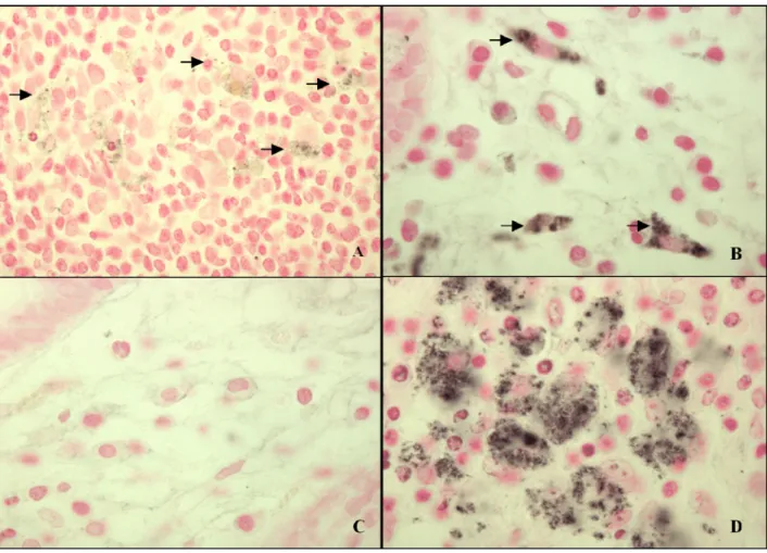
Immunoperoxidase staining (IP) of Map was observed in macrophages. Positive macrophages were low in number and very often located in the muscularis mucosa. A cytoplasmatic
granular staining pattern was frequently seen, but sometimes the cytoplasm was diffusely stained. Figure 1 shows that in infected areas variable amounts of Map-associated staining were observed within these cells. Macrophages with tiny speckles but also with moderate to large dots could be discriminated (Figure 1A – B). Macrophages in infected areas in the ileal of a cow with Paratuberculosis were often multi-nucleated and showed an intense immuno-reactivity for Map (Figure 1D). The results are presented in Table 3.
Table 3 Results immunoperoxidase staining of paraffin biopsy samples
Diagnosis Patientsa IP positive
patientsb
Inflamedc IP positive and
inflamedd % inflamed of IP positivee IP negative and Inflamedf CD 55 11 (20%) 43 (78%) 11 (20%) 100% (11/11) 32 (58%) UC 23 3 (13%) 12 (52%) 1 (4%) 33% (1/3) 11 (48%) Control 73 21 (29%) 20 (27%) 11 (15%) 55% (11/20) 9 (12%)
aTotal number of patients examined; bTotal number patients (biopsies) who are IP positive; cTotal number of
biopsies taken from involved (inflamed) areas of disease; dTotal number of IP positive biopsies from inflamed
areas; ePercentage of IP positive biopsies that are inflamed; fTotal number of IP negative biopsies from inflamed
areas.
Immuno-reactivity for M. avium-complex antigens was observed in biopsy samples from 11 out of 55 (20%) CD patients, 3 of 23 (13%) UC patients and 21 of 73 (29%) control persons. The data of IP staining show even a 50% higher presences of Map-antigens in control persons (29%) compared to CD patients (20%). The presence of Map-antigens is more often present in biopsies with tissue inflammation. For example all IP positive CD biopsies showed inflammation, whereas only one third and half of the IP positive biopsies of respectively UC and controle patients showed signs of inflammation. It should be noted that a correlation between tissue inflammation and the presence of Chlamydia-antigens and -DNA, which served as methodological controls for the detection of Map in this study, was also found.
2.3 Results
Map culturing
The results of Map culturing in MGIT and BACTEC media is presented in Table 4 and 5.
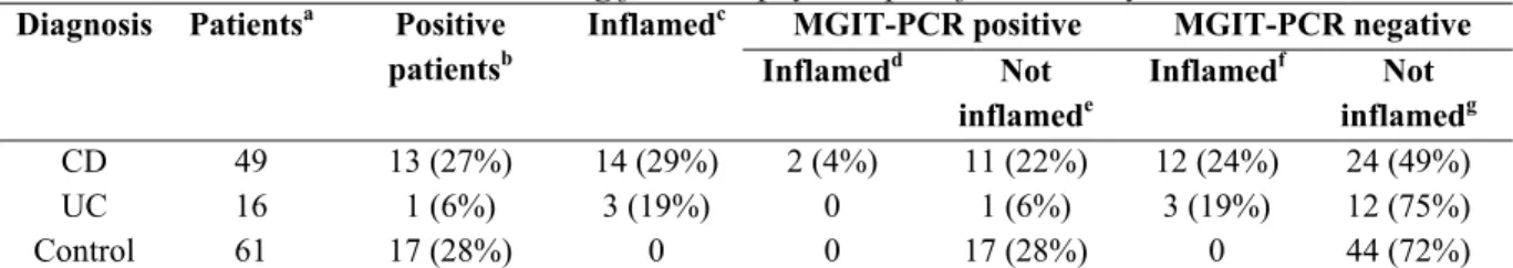
Table 4 Results MGIT culturing from biopsy samples followed by PCR
MGIT-PCR positive MGIT-PCR negative Diagnosis Patientsa Positive
patientsb Inflamedc Inflamedd Not inflamede Inflamedf Not inflamedg CD 49 13 (27%) 14 (29%) 2 (4%) 11 (22%) 12 (24%) 24 (49%) UC 16 1 (6%) 3 (19%) 0 1 (6%) 3 (19%) 12 (75%) Control 61 17 (28%) 0 0 17 (28%) 0 44 (72%)
aTotal number of patients examined; bTotal number of MGIT positive patients; cTotal number of biopsies taken
from involved (inflamed) areas of disease; dTotal number of MGIT positive biopsies from inflamed areas; eTotal
number of MGIT positive biopsies from non-inflamed areas; fTotal number of MGIT negative biopsies from
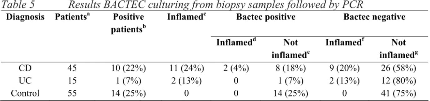
Table 5 Results BACTEC culturing from biopsy samples followed by PCR
Bactec positive Bactec negative Diagnosis Patientsa Positive
patientsb Inflamedc Inflamedd Not inflamede Inflamedf Not inflamedg CD 45 10 (22%) 11 (24%) 2 (4%) 8 (18%) 9 (20%) 26 (58%) UC 15 1 (7%) 2 (13%) 0 1 (7%) 2 (13%) 12 (80%) Control 55 14 (25%) 0 0 14 (25%) 0 41 (75%)
aTotal number of patients examined; bTotal number of BACTEC positive patients; cTotal number of biopsies
taken from involved (inflamed) areas of disease; dTotal number of BACTEC positive biopsies from inflamed
areas; eTotal number of BACTEC positive biopsies from non-inflamed areas; fTotal number of BACTEC
negative biopsies from inflamed areas; gTotal number of BACTEC negative biopsies from non-inflamed areas;
In Table 6, the combined data from PCR, culturing and IP staining are depicted. This table shows how many patients were positive in two different assays. Since none of the patients were positive in all three tests, combined data for three tests (PCR, IP and culturing) are not depicted.
Table 6 Combined data from PCR, Culturing and Immunoperoxidase staining
PCR positivea
PCR positive XXXX IP positive
IP positive 3 XXXX Culture positive
Culture positive 3 5 XXXX PCR negative
PCR negative XXXX 26 37 XXXX IP negative
IP negative 5 XXXX 33 77 XXXX
Culture negative 5 17 XXXX 63 48
3. Discussion and Conclusion
Our data clearly show that Map is present both in IBD patients and in control persons. Strikingly, the results of culturing and IP staining show an even higher prevalence of Map in control persons compared to CD patients. With IP staining and culturing Map can be found in at least 20% of the CD patients and control persons. This would suggest that this percentage would also be found in UC patients. However, data from IP staining and culturing show a remarkably lower prevalence of Map in UC patients. The reason for this is unclear. The results of culturing and immunoperoxidase staining show a good correlation; with both techniques, an average of 23% of CD patients and 27% of control persons is positive for Map and Map-antigen, respectively. In discrepancy with this result is the low percentage of PCR positive biopsies compared to culture positives. An explanation for this is that for culturing the complete biopsy sample is spent, while for PCR only one-fifth of the biopsy sample is used. Furthermore, during DNA extraction from biopsy samples, loss of some DNA template is inevitable, while on the contrary during culturing, life bacteria may replicate and thus providing even more template for the succeeding PCR amplification. It is concluded that the percentage of cells containing Map-proteins in a biopsy sample is very low. Besides, of both biopsy samples from PCR positive patient, a positive PCR result occurred only with one sample. Collins et al. tested over five biopsy samples on average per patient, and among PCR positive patients most often positive PCR results occurred with only a single biopsy per patient 2. Furthermore, although a total of 10, 35, 31 and 25 persons were PCR, IP, MGIT and
BACTEC positive, respectively, only in a few instances was a patient positive by two tests (Table 6). Even with both culturing assays, almost no overlap in positive results is observed: of the 115 persons who were tested in MGIT and in BACTEC, only 7 were positive in both culturing assays. These data show that the prevalence of Map in human tissue is very low. A very sensitive detection assay like PCR or culturing can only yield a positive result if the target (in this case Map-DNA or Map-bacteria) is present in the reaction mixture. Since Map is not uniformly distributed in the tissue, and apparently only present in minimal amounts, the place of sampling will be very critical. It is a matter of chance to pick the right spot. If it were possible to collect and analyse many biopsy samples per person, the percentage of Map-PCR, Map-IP and Map -culture positive persons would be higher.
Several investigators have detected Map in a higher percentage of inflamed tissues compared to non-inflamed tissues of CD patients 4,5,6,7. Our results from IP and PCR do confirm these observations: in these two assays the percentage of inflamed tissues that are Map-positive 26% and 9%, respectively) is higher than the percentage of non-inflamed tissues that are Map-positive (both 0%). Remarkably however, both culturing assays show quite the opposite result. In these assays the percentage of non-inflamed tissues that are Map positive (31% for MGIT and 24% for BACTEC) is higher than the percentage of Map-positive inflamed tissues (14% and 18%).
An interesting observation is the correlation between presence of Map antigens (MAC-antigens) and tissue inflammation in CD patients. All inflamed biopsy samples from CD
patients were IP positive (Table 3), while only 14% and 18% of inflamed tissues were MGIT and BACTEC positive, respectively. This might imply that this inflammation is not solely caused by Map antigens, but predominantly by other members of the MAC, or other pathogens. We obtained evidence that members of the Chlamydiales family are present in high amounts in most of the CD and UC patients, whereas they were of low incidence in control persons. Furthermore the presence of Chlamydiales was completely correlated with inflammation, not only in CD patients but also in UC patients and control persons (results not shown).
All patients have completed a detailed questionnaire, including questions about general features, clinical picture and symptoms of their complaints, medication, (family) background, life-style, eating, drinking and smoking habits and physical condition. These data will be studied in further detail, and will be combined to the data of PCR, IP and culturing to reveal correlations between epidemiological and molecular data related to CD and Map.
To conclude, our data do not support the hypothesis that Map is directly involved in Crohn’s disease, but at least suggest that infection plays an important role. Further research is needed to validate a role of Chlamydiales in the pathogenesis of IBD.
Figure 1. Immunoperoxidase staining for Map on paraffin sections of ileum from human (A-B) and cow with Johne’s disease (D). Figure C shows the use of an irrelevant rabbit
antiserum on ileum from human. Notice the granular staining pattern and the variation in the intensity of the staining per cell. Arrows indicate tiny (A) and moderate (B) spots
intracellularly in macrophages. This staining is absent in C. Large, often multinucleated macrophages are intensely stained for Map in case of cow with Johne’s disease. Pictures taken with a 40x (A) and 60x (B-D) objective.
References
1. Bull, T. J., E. J. McMinn, K. Sidi-Boumedine, A. Skull, D. Durkin, P. Neild, G. Rhodes, R. Pickup, and J. Hermon-Taylor. 2003. Detection and verification of Mycobacterium avium subsp. paratuberculosis in fresh ileocolonic mucosal biopsy specimens from individuals with and without Crohn's disease. J. Clin. Microbiol. 41:2915-2923.
2. Collins, M. T., G. Lisby, C. Moser, D. Chicks, S. Christensen, M. Reichelderfer, N. Hoiby, B. A. Harms, O. O. Thomsen, U. Skibsted, and V. Binder. 2000. Results of multiple diagnostic tests for Mycobacterium avium subsp. paratuberculosis in patients with inflammatory bowel disease and in controls. J. Clin. Microbiol. 38:4373-4381. 3. Englund, S., A. Ballagi Pordany, G. Bolske, and K. E. Johansson. 1999. Single PCR
and nested PCR with a mimic molecule for detection of Mycobacterium avium subsp. paratuberculosis. Diagnostic Microbiology and Infectious Disease. 33:163-171.
4. Fidler, H. M., W. Thurrell, N. M. Johnson, G. A. Rook, and J. J. McFadden. 1994. Specific detection of Mycobacterium paratuberculosis DNA associated with
granulomatous tissue in Crohn's disease. Gut 35:506-510.
5. Hulten, K., H. M. T. El Zimaity, T. J. Karttunen, A. Almashhrawi, M. R. Schwartz, D. Y. Graham, and F. A. K. El Zaatari. 2001. Detection of Mycobacterium avium subspecies paratuberculosis in Crohn's diseased tissues by in situ hybridization. American Journal of Gastroenterology 96:1529-1535.
6. Murray, A., J. Oliaro, M. M. Schlup, and V. S. Chadwick. 1995. Mycobacterium paratuberculosis and inflammatory bowel disease: frequency distribution in serial colonoscopic biopsies using the polymerase chain reaction. Microbios 83:217-228. 7. Ryan, P., M. W. Bennett, S. Aarons, G. Lee, J. K. Collins, G. C. O'Sullivan, J.
O'Connell, and F. Shanahan. 2002. PCR detection of Mycobacterium paratuberculosis in Crohn's disease granulomas isolated by laser capture microdissection. Gut 51:665-670.