ESC GUIDELINES
2014 ESC Guidelines on the diagnosis and
management of acute pulmonary embolism
The Task Force for the Diagnosis and Management of Acute
Pulmonary Embolism of the European Society of Cardiology (ESC)
Endorsed by the European Respiratory Society (ERS)
Authors/Task Force Members: Stavros Konstantinides
*
(Chairperson) (Germany/
Greece), Adam Torbicki
*
(Co-chairperson) (Poland), Giancarlo Agnelli (Italy),
Nicolas Danchin (France), David Fitzmaurice (UK), Nazzareno Galie` (Italy),
J. Simon R. Gibbs (UK), Menno Huisman (The Netherlands), Marc Humbert
†(France),
Nils Kucher (Switzerland), Irene Lang (Austria), Mareike Lankeit (Germany),
John Lekakis (Greece), Christoph Maack (Germany), Eckhard Mayer (Germany),
Nicolas Meneveau (France), Arnaud Perrier (Switzerland), Piotr Pruszczyk (Poland),
Lars H. Rasmussen (Denmark), Thomas H. Schindler (USA), Pavel Svitil (Czech
Republic), Anton Vonk Noordegraaf (The Netherlands), Jose Luis Zamorano (Spain),
Maurizio Zompatori (Italy)
ESC Committee for Practice Guidelines (CPG): Jose Luis Zamorano (Chairperson) (Spain), Stephan Achenbach (Germany), Helmut Baumgartner (Germany), Jeroen J. Bax (Netherlands), Hector Bueno (Spain), Veronica Dean (France), Christi Deaton (UK), Çetin Erol (Turkey), Robert Fagard (Belgium), Roberto Ferrari (Italy), David Hasdai (Israel), Arno Hoes (Netherlands), Paulus Kirchhof (Germany/UK), Juhani Knuuti (Finland), Philippe Kolh (Belgium), Patrizio Lancellotti (Belgium), Ales Linhart (Czech Republic), Petros Nihoyannopoulos (UK), Massimo F. Piepoli
*Corresponding authors. Stavros Konstantinides, Centre for Thrombosis and Hemostasis, Johannes Gutenberg University of Mainz, University Medical Centre Mainz, Langenbeckstrasse
1, 55131 Mainz, Germany. Tel:+49 613 1176255, Fax: +49 613 1173456. Email:stavros.konstantinides@unimedizin-mainz.de, and Department of Cardiology, Democritus University of
Thrace, Greece. Email:skonst@med.duth.gr.
Adam Torbicki, Department of Pulmonary Circulation and Thromboembolic Diseases, Medical Centre of Postgraduate Education, ECZ-Otwock, Ul. Borowa 14/18, 05-400 Otwock,
Poland. Tel:+48 22 7103052, Fax: +48 22 710315. Email:adam.torbicki@ecz-otwock.pl.
†Representing the European Respiratory Society
Other ESC entities having participated in the development of this document:
ESC Associations: Acute Cardiovascular Care Association (ACCA), European Association for Cardiovascular Prevention & Rehabilitation (EACPR), European Association of Cardio-vascular Imaging (EACVI), Heart Failure Association (HFA), ESC Councils: Council on CardioCardio-vascular Nursing and Allied Professions (CCNAP), Council for Cardiology Practice (CCP), Council on Cardiovascular Primary Care (CCPC)
ESC Working Groups: Cardiovascular Pharmacology and Drug Therapy, Nuclear Cardiology and Cardiac Computed Tomography, Peripheral Circulation, Pulmonary Circulation and Right Ventricular Function, Thrombosis.
Disclaimer: The ESC Guidelines represent the views of the ESC and were produced after careful consideration of the scientific and medical knowledge and the evidence available at the time of their publication.
The ESC is not responsible in the event of any contradiction, discrepancy and/or ambiguity between the ESC Guidelines and any other official recommendations or guidelines issued by the relevant public health authorities, in particular in relation to good use of healthcare or therapeutic strategies. Health professionals are encouraged to take the ESC Guidelines fully into account when exercising their clinical judgment, as well as in the determination and the implementation of preventive, diagnostic or therapeutic medical strategies; however, the ESC Guidelines do not override, in any way whatsoever, the individual responsibility of health professionals to make appropriate and accurate decisions in consideration of each patient’s health condition and in consultation with that patient and, where appropriate and/or necessary, the patient’s caregiver. Nor do the ESC Guidelines exempt health professionals from taking into full and careful consideration the relevant official updated recommendations or guidelines issued by the competent public health authorities, in order to manage each patient’s case in light of the scientifically accepted data pursuant to their respective ethical and professional obligations. It is also the health professional’s responsibility to verify the applicable rules and regulations relating to drugs and medical devices at the time of prescription.
National Cardiac Societies document reviewers: listed in the Appendix.
&The European Society of Cardiology 2014. All rights reserved. For permissions please email: journals.permissions@oup.com. European Heart Journal
doi:10.1093/eurheartj/ehu283
by guest on September 4, 2014
http://eurheartj.oxfordjournals.org/
(Italy), Piotr Ponikowski (Poland), Per Anton Sirnes (Norway), Juan Luis Tamargo (Spain), Michal Tendera (Poland), Adam Torbicki (Poland), William Wijns (Belgium), Stephan Windecker (Switzerland).
Document Reviewers: Çetin Erol (CPG Review Coordinator) (Turkey), David Jimenez (Review Coordinator) (Spain), Walter Ageno (Italy), Stefan Agewall (Norway), Riccardo Asteggiano (Italy), Rupert Bauersachs (Germany),
Cecilia Becattini (Italy), Henri Bounameaux (Switzerland), Harry R. Bu¨ller (Netherlands), Constantinos H. Davos
(Greece), Christi Deaton (UK), Geert-Jan Geersing (Netherlands), Miguel Angel Go´ mez Sanchez (Spain),
Jeroen Hendriks (Netherlands), Arno Hoes (Netherlands), Mustafa Kilickap (Turkey), Viacheslav Mareev (Russia), Manuel Monreal (Spain), Joao Morais (Portugal), Petros Nihoyannopoulos (UK), Bogdan A. Popescu (Romania),
Olivier Sanchez†(France), Alex C. Spyropoulos (USA).
The disclosure forms provided by the experts involved in the development of these guidelines are available on the ESC website www.escardio.org/guidelines.
-Keywords Guidelines † Pulmonary embolism † Venous thrombosis † Shock † Hypotension † Chest pain † Dyspnoea † Heart failure † Diagnosis † Treatment–Anticoagulation † Thrombolysis
Table of Contents
Abbreviations and acronyms . . . 3
1. Preamble . . . 3 2. Introduction . . . 4 2.1 Epidemiology . . . 5 2.2 Predisposing factors . . . 5 2.3 Natural history . . . 6 2.4 Pathophysiology . . . 6
2.5 Clinical classification of pulmonary embolism severity . . . 7
3. Diagnosis . . . 7
3.1 Clinical presentation . . . 7
3.2 Assessment of clinical probability . . . 8
3.3 D-dimer testing . . . 8
3.4 Computed tomographic pulmonary angiography . . . 10
3.5 Lung scintigraphy . . . 11
3.6 Pulmonary angiography . . . 11
3.7 Magnetic resonance angiography . . . 11
3.8 Echocardiography . . . 11
3.9 Compression venous ultrasonography . . . 12
3.10. Diagnostic strategies . . . 12
3.10.1 Suspected pulmonary embolism with shock or hypotension . . . 12
3.10.2 Suspected pulmonary embolism without shock or hypotension . . . 13
3.11. Areas of uncertainty . . . 14
4. Prognostic assessment . . . 15
4.1 Clinical parameters . . . 15
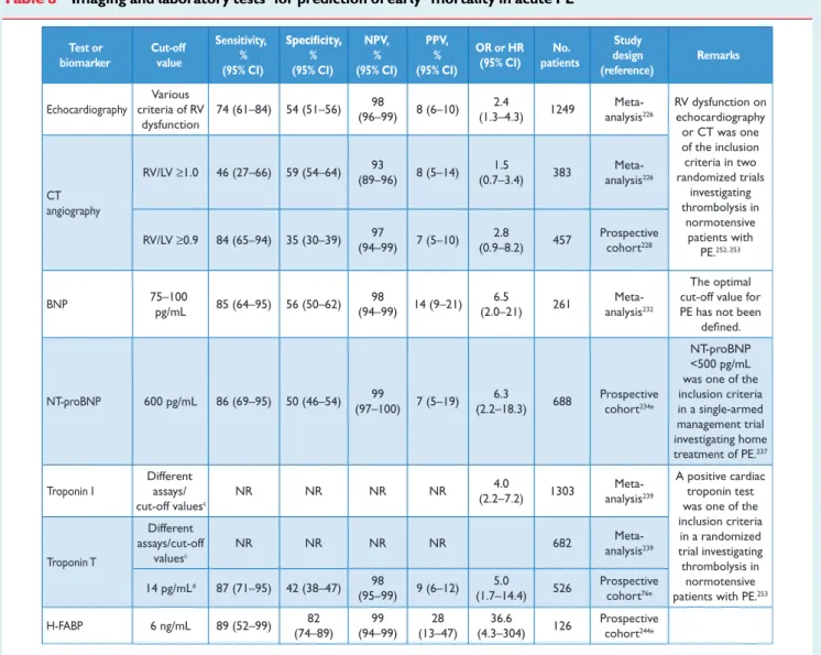
4.2 Imaging of the right ventricle by echocardiography or computed tomographic angiography . . . 16
4.3 Laboratory tests and biomarkers . . . 17
4.3.1 Markers of right ventricular dysfunction . . . 17
4.3.2 Markers of myocardial injury . . . 17
4.3.3 Other (non-cardiac) laboratory biomarkers . . . 18
4.4 Combined modalities and scores . . . 19
4.5 Prognostic assessment strategy . . . 19
5. Treatment in the acute phase . . . 20
5.1 Haemodynamic and respiratory support . . . 20
5.2 Anticoagulation . . . 20
5.2.1 Parenteral anticoagulation . . . 20
5.2.2 Vitamin K antagonists . . . 21
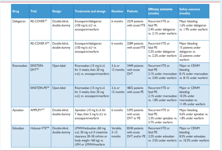
5.2.3 New oral anticoagulants . . . 22
5.3 Thrombolytic treatment . . . 23
5.4 Surgical embolectomy . . . 24
5.5 Percutaneous catheter-directed treatment . . . 24
5.6 Venous filters . . . 24
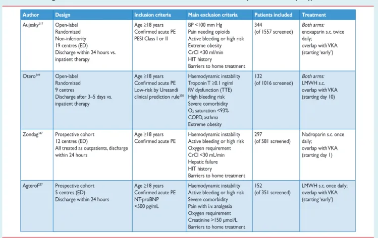
5.7 Early discharge and home treatment . . . 25
5.8 Therapeutic strategies . . . 26
5.8.1 Pulmonary embolism with shock or hypotension (high-risk pulmonary embolism) . . . 26
5.8.2 Pulmonary embolism without shock or hypotension (intermediate- or low-risk pulmonary embolism) . . . 26
5.9 Areas of uncertainty . . . 27
6. Duration of anticoagulation . . . 29
6.1 New oral anticoagulants for extended treatment . . . 30
7. Chronic thromboembolic pulmonary hypertension . . . 31
7.1 Epidemiology . . . 31
7.2 Pathophysiology . . . 31
7.3 Clinical presentation and diagnosis . . . 31
7.4 Treatment and prognosis . . . 32
8. Specific problems . . . 34
8.1 Pregnancy . . . 34
8.1.1 Diagnosis of pulmonary embolism in pregnancy . . . . 34
8.1.2 Treatment of pulmonary embolism in pregnancy . . . 34
8.2 Pulmonary embolism and cancer . . . 35
8.2.1 Diagnosis of pulmonary embolism in patients with cancer . . . 35
8.2.2 Prognosis for pulmonary embolism in patients with cancer . . . 35
8.2.3 Management of pulmonary embolism in patients with cancer . . . 35
8.2.4 Occult cancer presenting as unprovoked pulmonary embolism . . . 36
8.3 Non-thrombotic pulmonary embolism . . . 36
8.3.1 Septic embolism . . . 36
8.3.2 Foreign-material pulmonary embolism . . . 36
by guest on September 4, 2014
http://eurheartj.oxfordjournals.org/
8.3.3 Fat embolism . . . 36
8.3.4 Air embolism . . . 37
8.3.5 Amniotic fluid embolism . . . 37
8.3.6 Tumour embolism . . . 37
9. Appendix . . . 37
References . . . 37
Abbreviations and acronyms
ACS acute coronary syndrome
AMPLIFY Apixaban for the Initial Management of Pulmonary Embolism and Deep-Vein Thrombosis as First-line Therapy
aPTT activated partial thromboplastin time b.i.d. bis in diem (twice daily)
b.p.m. beats per minute
BNP brain natriuretic peptide
BP blood pressure
CI confidence interval
CO cardiac output
COPD chronic obstructive pulmonary disease
CPG Committee for Practice Guidelines
CRNM clinically relevant non-major
CT computed tomographic/tomogram
CTEPH chronic thromboembolic pulmonary hypertension
CUS compression venous ultrasonography
DSA digital subtraction angiography
DVT deep vein thrombosis
ELISA enzyme-linked immunosorbent assay
ESC European Society of Cardiology
H-FABP heart-type fatty acid-binding protein
HIT heparin-induced thrombocytopenia
HR hazard ratio
ICOPER International Cooperative Pulmonary Embolism
Registry
ICRP International Commission on Radiological Protection INR international normalized ratio
iPAH idiopathic pulmonary arterial hypertension
IVC inferior vena cava
LMWH low molecular weight heparin
LV left ventricle/left ventricular
MDCT multi-detector computed tomographic (angiography)
MRA magnetic resonance angiography
NGAL neutrophil gelatinase-associated lipocalin
NOAC(s) Non-vitamin K-dependent new oral anticoagulant(s) NT-proBNP N-terminal pro-brain natriuretic peptide
o.d. omni die (every day)
OR odds ratio
PAH pulmonary arterial hypertension
PE pulmonary embolism
PEA pulmonary endarterectomy
PEITHO Pulmonary EmbolIsm THrOmbolysis trial PESI pulmonary embolism severity index
PH pulmonary hypertension
PIOPED Prospective Investigation On Pulmonary Embolism Diagnosis
PVR pulmonary vascular resistance
RIETE Registro Informatizado de la Enfermedad Throm-boembolica venosa
RR relative risk
rtPA recombinant tissue plasminogen activator RV right ventricle/ventricular
SPECT single photon emission computed tomography sPESI simplified pulmonary embolism severity index TAPSE tricuspid annulus plane systolic excursion
Tc technetium
TOE transoesophageal echocardiography
TTR time in therapeutic range
TV tricuspid valve
UFH unfractionated heparin
V/Q scan ventilation – perfusion scintigraphy
VKA vitamin K antagonist(s)
VTE venous thromboembolism
1. Preamble
Guidelines summarize and evaluate all available evidence at the time of the writing process, on a particular issue with the aim of assisting health professionals in selecting the best management strategies for an individual patient, with a given condition, taking into account the impact on outcome, as well as the risk-benefit-ratio of particular diag-nostic or therapeutic means. Guidelines and recommendations should help the health professionals to make decisions in their daily practice. However, the final decisions concerning an individual patient must be made by the responsible health professional(s) in consultation with the patient and caregiver as appropriate.
A great number of Guidelines have been issued in recent years by the European Society of Cardiology (ESC) as well as by other soci-eties and organisations. Because of the impact on clinical practice, quality criteria for the development of guidelines have been estab-lished in order to make all decisions transparent to the user. The recommendations for formulating and issuing ESC Guidelines can be found on the ESC Web Site ( http://www.escardio.org/guidelines-surveys/esc-guidelines/about/Pages/rules-writing.aspx). ESC Guide-lines represent the official position of the ESC on a given topic and are regularly updated.
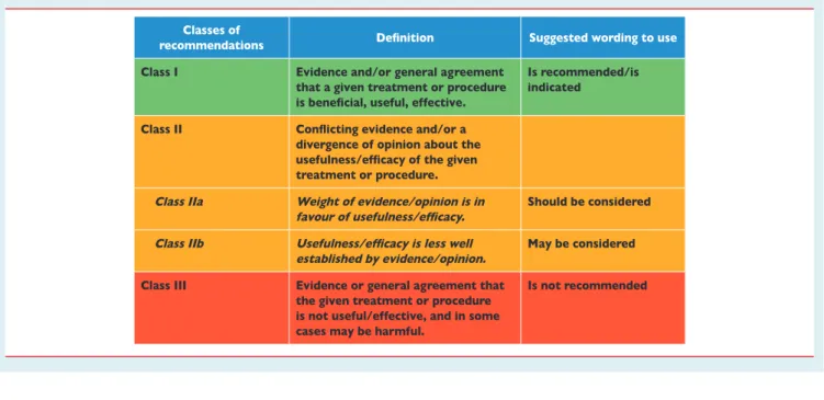
Members of this Task Force were selected by the ESC to represent professionals involved with the medical care of patients with this pathology. Selected experts in the field undertook a comprehensive review of the published evidence for management (including diagno-sis, treatment, prevention and rehabilitation) of a given condition according to ESC Committee for Practice Guidelines (CPG) policy. A critical evaluation of diagnostic and therapeutic procedures was performed including assessment of the risk-benefit-ratio. Estimates of expected health outcomes for larger populations were included, where data exist. The level of evidence and the strength of recom-mendation of particular management options were weighed and graded according to predefined scales, as outlined in Tables1and2. The experts of the writing and reviewing panels filled in declara-tions of interest forms which might be perceived as real or potential
by guest on September 4, 2014
http://eurheartj.oxfordjournals.org/
sources of conflicts of interest. These forms were compiled into one file and can be found on the ESC Web Site (http://www.escardio.org/ guidelines). Any changes in declarations of interest that arise during the writing period must be notified to the ESC and updated. The Task Force received its entire financial support from the ESC without any involvement from healthcare industry.
The ESC CPG supervises and coordinates the preparation of new Guidelines produced by Task Forces, expert groups or consensus panels. The Committee is also responsible for the endorsement process of these Guidelines. The ESC Guidelines undergo extensive review by the CPG and external experts. After appropriate revisions it is approved by all the experts involved in the Task Force. The fina-lized document is approved by the CPG for publication in the Euro-pean Heart Journal. It was developed after careful consideration of the scientific and medical knowledge and the evidence available at the time of their dating.
The task of developing ESC Guidelines covers not only the integra-tion of the most recent research, but also the creaintegra-tion of educaintegra-tional tools and implementation programmes for the recommendations. To implement the guidelines, condensed pocket guidelines versions, summary slides, booklets with essential messages, summary cards for non-specialists, electronic version for digital applications (smart-phones etc) are produced. These versions are abridged and, thus, if needed, one should always refer to the full text version which is freely available on the ESC Website. The National Societies of the ESC are encouraged to endorse, translate and implement the ESC Guidelines. Implementation programmes are needed because it has been shown that the outcome of disease may be favourably influenced by the thorough application of clinical recommendations. Surveys and registries are needed to verify that real-life daily prac-tice is in keeping with what is recommended in the guidelines, thus completing the loop between clinical research, writing of guidelines, disseminating them and implementing them into clinical practice.
Health professionals are encouraged to take the ESC Guidelines fully into account when exercising their clinical judgment as well as
in the determination and the implementation of preventive, diag-nostic or therapeutic medical strategies. However, the ESC Guide-lines do not override in any way whatsoever the individual responsibility of health professionals to make appropriate and ac-curate decisions in consideration of each patient s health condition and in consultation with that patient and the patient’s caregiver where appropriate and/or necessary. It is also the health professio-nal’s responsibility to verify the rules and regulations applicable to drugs and devices at the time of prescription.
2. Introduction
This document follows the two previous ESC Guidelines focussing on clinical management of pulmonary embolism, published in 2000 and 2008. Many recommendations have retained or reinforced their validity; however, new data has extended or modified our knowl-edge in respect of optimal diagnosis, assessment and treatment of patients with PE. The most clinically relevant new aspects of this 2014 version as compared with its previous version published in 2008 relate to:
Table 1 Classes of recommendations
Table 2 Levels of evidence
Level of evidence A
Data derived from multiple randomized clinical trials or meta-analyses. Level of
evidence B
Data derived from a single randomized clinical trial or large non-randomized studies.
Level of evidence C
Consensus of opinion of the experts and/ or small studies, retrospective studies, registries.
by guest on September 4, 2014
http://eurheartj.oxfordjournals.org/
(1) Recently identified predisposing factors for venous thrombo-embolism
(2) Simplification of clinical prediction rules (3) Age-adjusted D-dimer cut-offs (4) Sub-segmental pulmonary embolism
(5) Incidental, clinically unsuspected pulmonary embolism (6) Advanced risk stratification of intermediate-risk pulmonary
embolism
(7) Initiation of treatment with vitamin K antagonists
(8) Treatment and secondary prophylaxis of venous thrombo-embolism with the new direct oral anticoagulants
(9) Efficacy and safety of reperfusion treatment for patients at inter-mediate risk
(10) Early discharge and home (outpatient) treatment of pulmonary embolism
(11) Current diagnosis and treatment of chronic thromboembolic pulmonary hypertension
(12) Formal recommendations for the management of pulmonary embolism in pregnancy and of pulmonary embolism in patients with cancer.
These new aspects have been integrated into previous knowledge to suggest optimal and—whenever possible—objectively validated management strategies for patients with suspected or confirmed pul-monary embolism.
In order to limit the length of the printed text, additional informa-tion, tables, figures and references are available as web addenda at the ESC website (www.escardio.org).
2.1 Epidemiology
Venous thromboembolism (VTE) encompasses deep vein throm-bosis (DVT) and pulmonary embolism (PE). It is the third most fre-quent cardiovascular disease with an overall annual incidence of 100 – 200 per 100 000 inhabitants.1,2VTE may be lethal in the acute phase or lead to chronic disease and disability,3–6but it is also often preventable.
Acute PE is the most serious clinical presentation of VTE. Since PE is, in most cases, the consequence of DVT, most of the existing data on its epidemiology, risk factors, and natural history are derived from studies that have examined VTE as a whole.
The epidemiology of PE is difficult to determine because it may remain asymptomatic, or its diagnosis may be an incidental finding;2 in some cases, the first presentation of PE may be sudden death.7,8 Overall, PE is a major cause of mortality, morbidity, and hospitaliza-tion in Europe. As estimated on the basis of an epidemiological model, over 317 000 deaths were related to VTE in six countries of the European Union (with a total population of 454.4 million) in 2004.2Of these cases, 34% presented with sudden fatal PE and 59% were deaths resulting from PE that remained undiagnosed during life; only 7% of the patients who died early were correctly diag-nosed with PE before death. Since patients older than 40 years are at increased risk compared with younger patients and the risk approxi-mately doubles with each subsequent decade, an ever-larger number of patients are expected to be diagnosed with (and perhaps die of) PE in the future.9
In children, studies reported an annual incidence of VTE between 53 and 57 per 100 000 among hospitalized patients,10,11and between 1.4 and 4.9 per 100 000 in the community at large.12,13
2.2 Predisposing factors
A list of predisposing (risk) factors for VTE is shown in Web Addenda TableI. There is an extensive collection of predisposing environmen-tal and genetic factors. VTE is considered to be a consequence of the interaction between patient-related—usually permanent—risk factors and setting-related—usually temporary—risk factors. VTE is considered to be ‘provoked’ in the presence of a temporary or re-versible risk factor (such as surgery, trauma, immobilization, preg-nancy, oral contraceptive use or hormone replacement therapy) within the last 6 weeks to 3 months before diagnosis,14and ‘unpro-voked’ in the absence thereof. PE may also occur in the absence of any known risk factor. The presence of persistent—as opposed to major, temporary—risk factors may affect the decision on the dur-ation of anticoaguldur-ation therapy after a first episode of PE.
Major trauma, surgery, lower limb fractures and joint replace-ments, and spinal cord injury, are strong provoking factors for VTE.9,15Cancer is a well-recognized predisposing factor for VTE. The risk of VTE varies with different types of cancer;16,17 haemato-logical malignancies, lung cancer, gastrointestinal cancer, pancreatic cancer and brain cancer carry the highest risk.18,19 Moreover, cancer is a strong risk factor for all-cause mortality following an episode of VTE.20
In fertile women, oral contraception is the most frequent predis-posing factor for VTE.21,22When occurring during pregnancy, VTE is a major cause of maternal mortality.23The risk is highest in the third trimester of pregnancy and over the 6 weeks of the postpartum period, being up to 60 times higher 3 months after delivery, compared with the risk in non-pregnant women.23In vitro fertilization further increases the risk of pregnancy-associated VTE. In a cross-sectional study derived from a Swedish registry, the overall risk of PE (com-pared with the risk of age-matched women whose first child was born without in vitro fertilization) was particularly increased during the first trimester of pregnancy [hazard ratio (HR) 6.97; 95% confi-dence interval (CI) 2.21 – 21.96]. The absolute number of women who suffered PE was low in both groups (3 vs. 0.4 cases per 10 000 pregnancies during the first trimester, and 8.1 vs. 6.0 per 10 000 preg-nancies overall).24 In post-menopausal women who receive hormone replacement therapy, the risk of VTE varies widely depend-ing on the formulation used.25
Infection has been found to be a common trigger for hospitaliza-tion for VTE.15,26,27Blood transfusion and erythropoiesis-stimulating agents are also associated with an increased risk of VTE.15,28
In children, PE is usually associated with DVT and is rarely unpro-voked. Serious chronic medical conditions and central venous lines are considered to be likely triggers of PE.29
VTE may be viewed as part of the cardiovascular disease con-tinuum and common risk factors—such as cigarette smoking, obesity, hypercholesterolaemia, hypertension and diabetes melli-tus30–33—are shared with arterial disease, notably atheroscler-osis.34–37 However, at least in part, this may be an indirect association, mediated by the effects of coronary artery disease and,
by guest on September 4, 2014
http://eurheartj.oxfordjournals.org/
in the case of smoking, cancer.38,39Myocardial infarction and heart failure increase the risk of PE;40,41conversely, patients with VTE have an increased risk of subsequent myocardial infarction and stroke.42
2.3 Natural history
The first studies on the natural history of VTE were carried out in the setting of orthopaedic surgery during the 1960s.43Evidence collected since this initial report has shown that DVT develops less frequently in non-orthopaedic surgery. The risk of VTE is highest during the first two post-operative weeks but remains elevated for two to three months. Antithrombotic prophylaxis significantly reduces the risk of perioperative VTE. The incidence of VTE is reduced with increas-ing duration of thromboprophylaxis after major orthopaedic surgery and (to a lesser extent) cancer surgery: this association has not been shown for general surgery.44,45The majority of patients with symp-tomatic DVT have proximal clots, complicated by PE in 40 – 50% of cases, often without clinical manifestations.44,45
Registries and hospital discharge datasets of unselected patients with PE or VTE yielded 30-day all-cause mortality rates between 9% and 11%, and three-month mortality ranging between 8.6% and 17%.46–48Following the acute PE episode, resolution of pulmonary thrombi, as evidenced by lung perfusion defects, is frequently incom-plete. In one study, lung perfusion scintigraphy demonstrated abnor-malities in 35% of patients a year after acute PE, although the degree of pulmonary vascular obstruction was ,15% in 90% of the cases.49 Two relatively recent cohort studies covering 173 and 254 patients yielded incidences approaching 30%.50,51The incidence of confirmed chronic thromboembolic pulmonary hypertension (CTEPH) after unprovoked PE is currently estimated at approximately 1.5% (with a wide range reported by mostly small-cohort studies), with most cases appearing within 24 months of the index event.52,53
The risk of recurrence of VTE has been reviewed in detail.54–56 Based on historical data, the cumulative proportion of patients with early recurrence of VTE (on anticoagulant treatment) amounts to 2.0% at 2 weeks, 6.4% at 3 months and 8% at 6 months; more recent, randomized anticoagulation trials (discussed in the section on acute phase treatment) indicate that recurrence rates may have dropped considerably recently. The rate of recurrence is highest during the first two weeks and declines thereafter. During the early period, active cancer and failure to rapidly achieve therapeutic levels of anticoagulation appear to independently predict an increased risk of recurrence.56,57
The cumulative proportion of patients with late recurrence of VTE (after six months, and in most cases after discontinuation of anticoa-gulation) has been reported to reach 13% at 1 year, 23% at 5 years, and 30% at 10 years.56Overall, the frequency of recurrence does not appear to depend on the clinical presentation (DVT or PE) of the first event, but recurrent VTE is likely to occur in the same clinical form as the index episode (i.e. if VTE recurs after PE, it will most likely be PE again). Recurrence is more frequent after multiple VTE epi-sodes as opposed to a single event, and after unprovoked VTE as opposed to the presence of temporary risk factors, particularly surgery.58 It is also more frequent in women who continue hormone intake after a VTE episode, and in patients who have
suffered PE or proximal vein thrombosis compared to distal (calf) vein thrombosis. On the other hand, factors for which an independ-ent association with late recurrence have not been definitely estab-lished include age, male sex,59,60a family history of VTE, and an increased body mass index.54,56 Elevated D-dimer levels, either during or after discontinuation of anticoagulation, indicate an increased risk of recurrence;61–63on the other hand, single thrombo-philic defects have a low predictive value and anticoagulation manage-ment based on thrombophilia testing has not been found to reduce VTE recurrence.64,65
2.4 Pathophysiology
Acute PE interferes with both the circulation and gas exchange. Right ventricular (RV) failure due to pressure overload is considered the primary cause of death in severe PE.
Pulmonary artery pressure increases only if more than 30 – 50% of the total cross-sectional area of the pulmonary arterial bed is occluded by thromboemboli.66 PE-induced vasoconstriction, mediated by the release of thromboxane A2 and serotonin, contri-butes to the initial increase in pulmonary vascular resistance after PE,67an effect that can be reversed by vasodilators.68,69Anatomical obstruction and vasoconstriction lead to an increase in pulmonary vascular resistance and a proportional decrease in arterial compliance.70
The abrupt increase in pulmonary vascular resistance results in RV dilation, which alters the contractile properties of the RV myocar-dium via the Frank-Starling mechanism. The increase in RV pressure and volume leads to an increase in wall tension and myocyte stretch. RV contraction time is prolonged, while neurohumoral activation leads to inotropic and chronotropic stimulation. Together with sys-temic vasoconstriction, these compensatory mechanisms increase pulmonary artery pressure, improving flow through the obstructed pulmonary vascular bed, and thus temporarily stabilize systemic blood pressure (BP).71 The extent of immediate adaptation is limited, since a non-preconditioned, thin-walled right ventricle (RV) is unable to generate a mean pulmonary artery pressure above 40 mm Hg.
The prolongation of RV contraction time into early diastole in the left ventricle leads to leftward bowing of the interventricular septum.72The desynchronization of the ventricles may be exacer-bated by the development of right bundle-branch block. As a result, left ventricular (LV) filling is impeded in early diastole, and this may lead to a reduction of the cardiac output and contribute to systemic hypotension and haemodynamic instability.73
As described above, excessive neurohumoral activation in PE can be the result both of abnormal RV wall tension and of circulatory shock. The finding of massive infiltrates in the RV myocardium of patients who died within 48 hours of acute PE may be explained by high levels of epinephrine released as a result of the PE-induced ‘myo-carditis’.74This inflammatory response might explain the secondary haemodynamic destabilization which sometimes occurs 24 – 48 hours after acute PE, although early recurrence of PE may be an alter-native explanation in some of these cases.75
Finally, the association between elevated circulating levels of bio-markers of myocardial injury and an adverse early outcome indicates
by guest on September 4, 2014
http://eurheartj.oxfordjournals.org/
that RV ischaemia is of pathophysiological significance in the acute phase of PE.76–78Although RV infarction is uncommon after PE, it is likely that the imbalance between oxygen supply and demand can result in damage to cardiomyocytes and further reduce contractile forces.
The detrimental effects of acute PE on the RV myocardium and the circulation are summarized in Figure1.
Respiratory failure in PE is predominantly a consequence of haemodynamic disturbances.79Low cardiac output results in desat-uration of the mixed venous blood. In addition, zones of reduced flow in obstructed vessels, combined with zones of overflow in the capillary bed served by non-obstructed vessels, result in ventila-tion – perfusion mismatch, which contributes to hypoxaemia. In about one-third of patients, right-to-left shunting through a patent foramen ovale can be detected by echocardiography: this is caused by an inverted pressure gradient between the right atrium and left atrium and may lead to severe hypoxaemia and an increased risk of paradoxical embolization and stroke.80Finally, even if they do not affect haemodynamics, small distal emboli may create areas of alveo-lar haemorrhage resulting in haemoptysis, pleuritis, and pleural effu-sion, which is usually mild. This clinical presentation is known as ‘pulmonary infarction’. Its effect on gas exchange is normally mild, except in patients with pre-existing cardiorespiratory disease.
2.5 Clinical classification of pulmonary
embolism severity
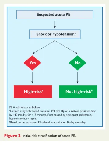
The clinical classification of the severity of an episode of acute PE is based on the estimated PE-related early mortality risk defined by in-hospital or 30-day mortality (Figure2). This stratification, which has important implications both for the diagnostic and therapeutic strategies proposed in these guidelines, is based on the patient’s clin-ical status at presentation, with high-risk PE being suspected or con-firmed in the presence of shock or persistent arterial hypotension and not high-risk PE in their absence.
3. Diagnosis
Throughout these Guidelines and for the purpose of clinical manage-ment, ‘confirmed PE’ is defined as a probability of PE high enough to indicate the need for PE-specific treatment, and ‘excluded PE’ as a probability of PE low enough to justify withholding PE-specific treat-ment with an acceptably low risk.
3.1 Clinical presentation
PE may escape prompt diagnosis since the clinical signs and symptoms are non-specific (Table3). When the clinical presentation raises the suspicion of PE in an individual patient, it should prompt further objective testing. In most patients, PE is suspected on the basis of dys-pnoea, chest pain, pre-syncope or syncope, and/or haemoptysis.81–83 Arterial hypotension and shock are rare but important clinical pre-sentations, since they indicate central PE and/or a severely reduced haemodynamic reserve. Syncope is infrequent, but may occur regard-less of the presence of haemodynamic instability.84Finally, PE may be completely asymptomatic and be discovered incidentally during diagnostic work-up for another disease or at autopsy.
Chest pain is a frequent symptom of PE and is usually caused by pleural irritation due to distal emboli causing pulmonary infarction.85 In central PE, chest pain may have a typical angina character, possibly reflecting RV ischaemia and requiring differential diagnosis with acute coronary syndrome (ACS) or aortic dissection. Dyspnoea may be acute and severe in central PE; in small peripheral PE, it is often mild and may be transient. In patients with pre-existing heart failure or pulmonary disease, worsening dyspnoea may be the only symptom indicative of PE.
Increased RV afterload RV O2 delivery TV insufficiency RV wall tension Neurohormonal activation Myocardial inflammation RV O2 demand RV ischaemia RV coronary perfusion RV output RV contractility Systemic BP Cardiogenic shock Death RV dilatation Low CO LV pre-load
BP = blood pressure; CO = cardiac output; LV = left ventricular; RV = right ventricular; TV = tricuspid valve.
Figure 1 Key factors contributing to haemodynamic collapse in acute pulmonary embolism
Suspected acute PE
Shock or hypotensiona?
Yes No
High–riskb Not high–riskb
PE = pulmonary embolism.
a
by ≥40 mm Hg, for >15 minutes, if not caused by new-onset arrhythmia, hypovolaemia, or sepsis.
bBased on the estimated PE-related in-hospital or 30-day mortality.
Figure 2 Initial risk stratification of acute PE.
by guest on September 4, 2014
http://eurheartj.oxfordjournals.org/
Knowledge of the predisposing factors for VTE is important in de-termining the likelihood of PE, which increases with the number of predisposing factors present; however, in as many as 30% of the patients with PE, no provoking factors can be detected.86In blood gas analysis, hypoxaemia is considered a typical finding in acute PE, but up to 40% of the patients have normal arterial oxygen saturation and 20% a normal alveolar-arterial oxygen gradient.87,88Hypocapnia is also often present. The chest X-ray is frequently abnormal and, al-though its findings are usually non-specific in PE, it is useful for exclud-ing other causes of dyspnoea or chest pain.89Electrocardiographic changes indicative of RV strain, such as inversion of T waves in leads V1 – V4, a QR pattern in V1, S1Q3T3 pattern, and incomplete or complete right bundle-branch block, may be helpful. These elec-trocardiographic changes are usually found in more severe cases of PE;90in milder cases, the only anomaly may be sinus tachycardia, present in 40% of patients. Finally, atrial arrhythmias, most frequently atrial fibrillation, may be associated with acute PE.
3.2 Assessment of clinical probability
Despite the limited sensitivity and specificity of individual symptoms, signs, and common tests, the combination of findings evaluated by clinical judgement or by the use of prediction rules allows to classify patients with suspected PE into distinct categories of clinical or pre-test probability that correspond to an increasing actual preva-lence of confirmed PE. As the post-test (e.g. after computed tomog-raphy) probability of PE depends not only on the characteristics of the diagnostic test itself but also on pre-test probability, this has become a key step in all diagnostic algorithms for PE.
The value of clinical judgement has been confirmed in several large series,91–93including the Prospective Investigation On Pulmonary Embolism Diagnosis (PIOPED).94 Note that clinical judgement usually includes commonplace tests such as chest X-ray and electro-cardiogram for differential diagnosis. However, clinical judgement lacks standardization; therefore, several explicit clinical prediction rules have been developed. Of these, the most frequently used
prediction rule is the one offered by Wells et al. (Table4).95This rule has been validated extensively using both a three-category scheme (low, moderate, or high clinical probability of PE) and a two-category scheme (PE likely or unlikely).96–100It is simple and based on information that is easy to obtain; on the other hand, the weight of one subjective item (‘alternative diagnosis less likely than PE’) may reduce the inter-observer reproducibility of the Wells rule.101–103 The revised Geneva rule is also simple and standardized (Table4).93Both have been adequately validated.104–106
More recently, both the Wells and the revised Geneva rule were simplified in an attempt to increase their adoption into clinical prac-tice (Table4),107,108and the simplified versions were externally vali-dated.105,109 Whichever is used, the proportion of patients with confirmed PE can be expected to be around 10% in the low-probability category, 30% in the moderate-low-probability category, and 65% in the high-clinical probability category when using the three-level classification.104 When the two-level classification is used, the proportion of patients with confirmed PE in the PE-unlikely category is around 12%.104
3.3 D-dimer testing
D-dimer levels are elevated in plasma in the presence of acute throm-bosis, because of simultaneous activation of coagulation and fibrin-olysis, The negative predictive value of D-dimer testing is high and a normal D-dimer level renders acute PE or DVT unlikely. On the other hand, fibrin is also produced in a wide variety of conditions such as cancer, inflammation, bleeding, trauma, surgery and necrosis. Accordingly, the positive predictive value of elevated D-dimer levels is low and D-dimer testing is not useful for confirmation of PE.
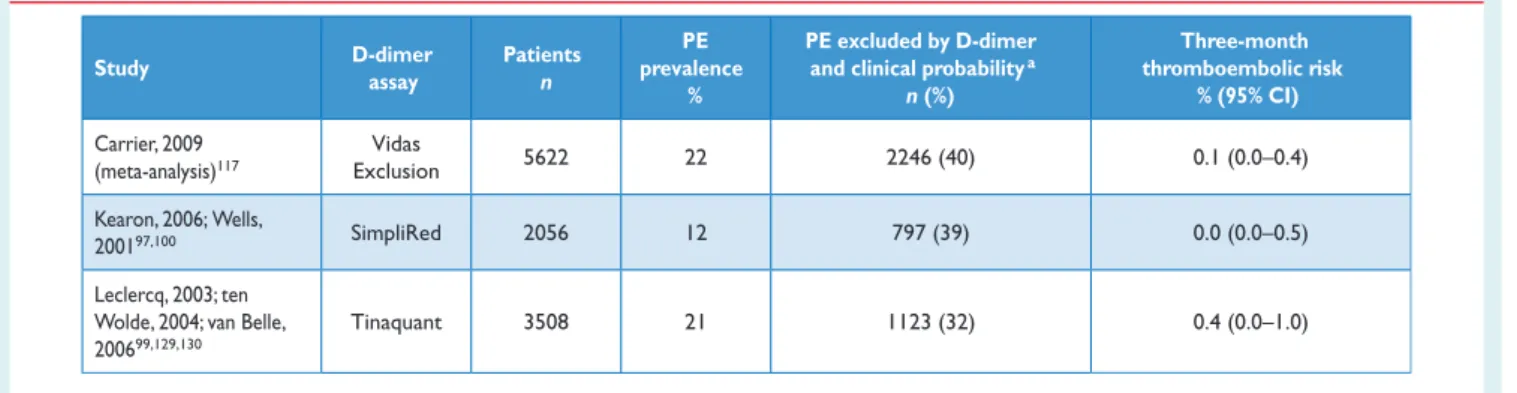
A number of D-dimer assays are available.110,111The quantitative enzyme-linked immunosorbent assay (ELISA) or ELISA-derived assays have a diagnostic sensitivity of 95% or better and can therefore be used to exclude PE in patients with either a low or a moderate pre-test probability. In the emergency department, a negative ELISA D-dimer, in combination with clinical probability, can exclude the disease without further testing in approximately 30% of patients with suspected PE.100,112,113Outcome studies have shown that the three-month thromboembolic risk was ,1% in patients left untreated on the basis of a negative test result (Table5);99,112–116these findings were confirmed by a meta-analysis.117
Quantitative latex-derived assays and a whole-blood agglutination assay have a diagnostic sensitivity ,95% and are thus often referred to as moderately sensitive. In outcome studies, those assays proved safe in ruling out PE in PE-unlikely patients as well as in patients with a low clinical probability.99,100,105Their safety in ruling out PE has not been established in the intermediate clinical probability cat-egory. Point-of-care tests have moderate sensitivity, and data from outcome studies in PE are lacking, with the exception of a recent primary care-based study using the Simplify D-dimer assay,118in which the three-month thromboembolic risk was 1.5% in PE-unlikely patients with a negative D-dimer.
The specificity of D-dimer in suspected PE decreases steadily with age, to almost 10% in patients .80 years.119Recent evidence sug-gests using age-adjusted cut-offs to improve the performance of D-dimer testing in the elderly.120,121 In a recent meta-analysis, age-adjusted cut-off values (age x 10 mg/L above 50 years) allowed increasing specificity from 34 – 46% while retaining a sensitivity Table 3 Clinical characteristics of patients with
suspected PE in the emergency department (adapted from Pollack et al. (2011)).82
Feature
(n = 1880) (n = 528)
Dyspnoea 50% 51%
Pleuritic chest pain 39% 28%
Cough 23% 23%
Substernal chest pain 15% 17%
Fever 10% 10%
Haemoptysis 8% 4%
Syncope 6% 6%
Unilateral leg pain 6% 5%
Signs of DVT (unilateral
extremity swelling) 24% 18%
DVT ¼ deep vein thrombosis.
by guest on September 4, 2014
http://eurheartj.oxfordjournals.org/
above 97%.122A multicentre, prospective management study evalu-ated this age-adjusted cut-off in a cohort of 3346 patients. Patients with a normal age-adjusted D-dimer value did not undergo computed tomographic pulmonary angiography and were left untreated and formally followed up for a three-month period. Among the 766 patients who were 75 years or older, 673 had a non-high clinical prob-ability. On the basis of D-dimer, using the age-adjusted cut-off
(instead of the ‘standard’ 500 mg/L cut-off) increased the number of patients in whom PE could be excluded from 43 (6.4%; 95% CI 4.8 – 8.5%) to 200 (29.7%; 95% CI 26.4 – 33.3%), without any addition-al faddition-alse-negative findings.123D-dimer is also more frequently elevated in patients with cancer,124,125 in hospitalized patients,105,126 and during pregnancy.127,128 Thus, the number of patients in whom D-dimer must be measured to exclude one PE (number needed to Table 4 Clinical prediction rules for PE
Items Clinical decision rule points
Wells rule Original version Simplified version
Simplified version
95 107
Previous PE or DVT 1.5 1
Heart rate ≥100 b.p.m. 1.5 1
Surgery or immobilization within the past four weeks 1.5 1
Haemoptysis 1 1
Active cancer 1 1
Clinical signs of DVT 3 1
Alternative diagnosis less likely than PE 3 1
Clinical probability Three-level score Low 0–1 N/A Intermediate 2–6 N/A High ≥7 N/A Two-level score PE unlikely 0–4 0–1 PE likely ≥5 ≥2
Revised Geneva score Original version93 108
Previous PE or DVT 3 1 Heart rate 75–94 b.p.m. ≥95 b.p.m. 3 5 1 2
Surgery or fracture within the past month 2 1
Haemoptysis 2 1
Active cancer 2 1
Unilateral lower limb pain 3 1
Pain on lower limb deep venous palpation and unilateral oedema 4 1
Age >65 years 1 1 Clinical probability Three-level score Low 0–3 0–1 Intermediate 4–10 2–4 High ≥11 ≥5 Two-level score PE unlikely 0–5 0–2 PE likely ≥6 ≥3
b.p.m.¼ beats per minute; DVT ¼ deep vein thrombosis; PE ¼ pulmonary embolism.
by guest on September 4, 2014
http://eurheartj.oxfordjournals.org/
test) varies between 3 in the emergency department and≥10 in the specific situations listed above. The negative predictive value of a (negative) D-dimer test remains high in these situations.
3.4 Computed tomographic pulmonary
angiography
Since the introduction of multi-detector computed tomographic (MDCT) angiography with high spatial and temporal resolution and quality of arterial opacification, computed tomographic (CT) angiog-raphy has become the method of choice for imaging the pulmonary vasculature in patients with suspected PE. It allows adequate visualiza-tion of the pulmonary arteries down to at least the segmental level.131–133The PIOPED II trial observed a sensitivity of 83% and a specificity of 96% for (mainly four-detector) MDCT.134PIOPED II also highlighted the influence of clinical probability on the predictive value of MDCT. In patients with a low or intermediate clinical prob-ability of PE as assessed by the Wells rule, a negative CT had a high negative predictive value for PE (96% and 89%, respectively), whereas this was only 60% in those with a high pre-test probability. Conversely, the positive predictive value of a positive CT was high (92 – 96%) in patients with an intermediate or high clinical probability but much lower (58%) in patients with a low pre-test likelihood of PE. Therefore, clinicians should be particularly cautious in case of discor-dancy between clinical judgement and the MDCT result.
Four studies provided evidence in favour of computed tomog-raphy as a stand-alone imaging test for excluding PE. In a prospective management study covering 756 consecutive patients referred to the emergency department with a clinical suspicion of PE, all patients with either a high clinical probability or a non-high clinical probability and a positive ELISA D-dimer test underwent both lower limb ultrasonog-raphy and MDCT.113The proportion of patients in whom—despite a negative MDCT—a proximal DVT was found on ultrasound was only 0.9% (95% CI 0.3 – 2.7).113In another study,99all patients classified as PE-likely by the dichotomized Wells rule, or those with a positive D-dimer test, underwent a chest MDCT. The three-month thrombo-embolic risk in the patients left untreated because of a negative CT was low (1.1%; 95% CI 0.6 – 1.9).99 Two randomized, controlled trials reached similar conclusions. In a Canadian trial comparing V/ Q scan and CT (mostly MDCT), only seven of the 531 patients
(1.3%) with a negative CT had a DVT, and one had a thromboembolic event during follow-up.135Hence, the three-month thromboembolic risk would have been 1.5% (95% CI 0.8 – 2.9) if only CT had been used.135 A European study compared two diagnostic strategies based on D-dimer and MDCT, one with- and the other without lower limb compression venous ultrasonography (CUS).116In the D-dimer – CT arm, the three-month thromboembolic risk was 0.3% (95% CI 0.1 – 1.2) among the 627 patients left untreated, based on a negative D-dimer or MDCT.
Taken together, these data suggest that a negative MDCT is an ad-equate criterion for excluding PE in patients with a non-high clinical probability of PE. Whether patients with a negative CT and a high clin-ical probability should be further investigated is controversial. MDCT showing PE at the segmental or more proximal level is adequate proof of PE in patients with a non-low clinical probability; however, the positive predictive value of MDCT is lower in patients with a low clin-ical probability of PE, and further testing may be considered, especial-ly if the clots are limited to segmental or sub-segmental arteries.
The clinical significance of isolated sub-segmental PE on CT angiog-raphy is questionable. This finding was present in 4.7% (2.5–7.6%) of patients with PE imaged by single-detector CT angiography and 9.4% (5.5– 14.2%) of those submitted to MDCT.136The positive predictive value is low and inter-observer agreement is poor at this distal level.137 There may be a role for CUS in this situation, to ensure that the patient does not have DVT that would require treatment. In a patient with iso-lated sub-segmental PE and no proximal DVT, the decision on whether to treat should be made on an individual basis, taking into account the clinical probability and the bleeding risk.
Computed tomographic venography has been advocated as a simple way to diagnose DVT in patients with suspected PE, as it can be combined with chest CT angiography as a single procedure, using only one intravenous injection of contrast dye. In PIOPED II, combining CT venography with CT angiography increased sensitivity for PE from 83% to 90% and had a similar specificity (around 95%);134,138however, the corresponding increase in negative predictive value was not clinically significant. CT venography adds a significant amount of irradi-ation, which may be a concern, especially in younger women.139As CT venography and CUS yielded similar results in patients with signs or symptoms of DVT in PIOPED II,138ultrasonography should be used instead of CT venography if indicated (see Section 3.10).
Table 5 Diagnostic yield of various D-dimer assays in excluding acute PE according to outcome studies
Study D-dimer assay Patients n PE prevalence % PE excluded by D-dimer
and clinical probabilitya
n (%) Three-month thromboembolic risk % (95% CI) Carrier, 2009 (meta-analysis)117 Vidas Exclusion 5622 22 2246 (40) 0.1 (0.0–0.4) Kearon, 2006; Wells, 200197,100 SimpliRed 2056 12 797 (39) 0.0 (0.0–0.5) Leclercq, 2003; ten Wolde, 2004; van Belle, 200699,129,130
Tinaquant 3508 21 1123 (32) 0.4 (0.0–1.0)
CI ¼ confidence interval; PE ¼ pulmonary embolism. a
Low or intermediate clinical probability, or PE unlikely, depending on the studies.
by guest on September 4, 2014
http://eurheartj.oxfordjournals.org/
The incidental discovery of clinically unsuspected PE on CT is an in-creasingly frequent problem, arising in 1– 2% of all thoracic CT exam-inations, most often in patients with cancer, but also among those with paroxysmal atrial fibrillation or heart failure and history of atrial fibril-lation.140–143There are no robust data to guide the decision on how to manage unsuspected PE with anticoagulants, but most experts agree that patients with cancer and those with clots at the lobar or more proximal level should be treated with anticoagulants.144
3.5 Lung scintigraphy
Ventilation – perfusion scintigraphy (V/Q scan) is an established diag-nostic test for suspected PE. It is safe and few allergic reactions have been described. The test is based on the intravenous injection of technetium (Tc)-99m-labelled macroaggregated albumin particles, which block a small fraction of the pulmonary capillaries and thereby enable scintigraphic assessment of lung perfusion. Perfusion scans are combined with ventilation studies, for which multiple tracers such as xenon-133 gas, Tc-99m-labelled aerosols, or Tc-99m-labelled carbon microparticles (Technegas) can be used. The purpose of the ventilation scan is to increase specificity: in acute PE, ventilation is expected to be normal in hypoperfused seg-ments (mismatch).145,146According to the International Commission on Radiological Protection (ICRP), the radiation exposure from a lung scan with 100 MBq of Tc-99m macroaggregated albumin parti-cles is 1.1 mSv for an average sized adult, and thus is significantly lower than that of CT angiography (2 – 6 mSv).147,148
Being a radiation- and contrast medium-sparing procedure, the V/Q scan may preferentially be applied in outpatients with low clinical probability and a normal chest X-ray, in young (particularly female) patients, in pregnancy, in patients with history of contrast medium-induced anaphylaxis and strong allergic history, in severe renal failure, and in patients with myeloma and paraproteinaemia.149 Lung scan results are frequently classified according to the criteria established in the PIOPED study: normal or near-normal, low, inter-mediate (non-diagnostic), and high probability of PE.94These criteria have been the subject of debate, following which they were revised.150,151To facilitate communication with clinicians, a three-tier classification is preferable: normal scan (excluding PE), high-probability scan (considered diagnostic of PE in most patients), and non-diagnostic scan.135,152,153Prospective clinical outcome studies suggested that it is safe to withhold anticoagulant therapy in patients with a normal perfusion scan. This was recently confirmed by a ran-domized trial comparing the V/Q scan with CT.135An analysis from the recent PIOPED II study confirmed the effectiveness of the high-probability V/Q scan for diagnosing PE and of the normal perfusion scan for ruling it out.154Performing only a perfusion scan is acceptable in patients with a normal chest X-ray; any perfusion defect in this situ-ation will be considered to be a mismatch.155The high frequency of non-diagnostic intermediate probability scans has been a cause for criticism, because they indicate the necessity for further diagnostic testing. Various strategies to overcome this problem have been pro-posed, notably the incorporation of clinical probability.91,156,157
Recent studies suggest that data acquisition in the tomographic mode in single photon emission computed tomography (SPECT) imaging, with or without low-dose CT may reduce the frequency of non-diagnostic scans.152,158–161SPECT imaging may even allow the use of automated detection algorithms for PE.162Large-scale pro-spective studies are needed to validate these new approaches.
3.6 Pulmonary angiography
Pulmonary angiography has for decades remained the ‘gold standard’ for the diagnosis or exclusion of PE, but is rarely performed now as less-invasive CT angiography offers similar diagnostic accuracy.163 Pul-monary angiography is more often used to guide percutaneous catheter-directed treatment of acute PE. Digital subtraction angiog-raphy (DSA) requires less contrast medium than conventional cinean-giography and has excellent imaging quality for peripheral pulmonary vessels in patients who can hold their breath; it is less useful for imaging of the main pulmonary arteries, due to cardiac motion artefacts. The diagnosis of acute PE is based on direct evidence of a thrombus in two projections, either as a filling defect or as amputation of a pul-monary arterial branch.94Thrombi as small as 1 – 2 mm within the sub-segmental arteries can be visualized by DSA, but there is substan-tial inter-observer variability at this level.164,165Indirect signs of PE, such as slow flow of contrast, regional hypoperfusion, and delayed or diminished pulmonary venous flow, are not validated and hence are not diagnostic. The Miller score may be used in quantifying the extent of luminal obstruction.166
Pulmonary angiography is not free of risk. In a study of 1111 patients, procedure-related mortality was 0.5%, major non-fatal complications occurred in 1%, and minor complications in 5%.167 The majority of deaths occurred in patients with haemodynamic compromise or respiratory failure. The risk of access-related bleed-ing complications is increased if thrombolysis is attempted in patients with PE diagnosed by pulmonary angiography.168
Haemodynamic measurements should always be recorded during pulmonary angiography for estimation of the severity of PE and because they may suggest alternative cardiopulmonary disorders. In patients with haemodynamic compromise, the amount of contrast agent should be reduced and non-selective injections avoided.169
3.7 Magnetic resonance angiography
Magnetic resonance angiography (MRA) has been evaluated for several years in suspected PE but large-scale studies were published only recently.170,171Their results show that this technique, although promising, is not yet ready for clinical practice due to its low sensitiv-ity, high proportion of inconclusive MRA scans, and low availability in most emergency settings. The hypothesis—that a negative MRA combined with the absence of proximal DVT on CUS may safely rule out clinically significant PE—is being tested in a multicentre outcome study (ClinicalTrials.gov NCT 02059551).
3.8 Echocardiography
Acute PE may lead to RV pressure overload and dysfunction, which can be detected by echocardiography. Given the peculiar geometry of the RV, there is no individual echocardiographic parameter that provides fast and reliable information on RV size or function. This is why echocar-diographic criteria for the diagnosis of PE have differed between studies. Because of the reported negative predictive value of 40–50%, a nega-tive result cannot exclude PE.157,172,173On the other hand, signs of RV overload or dysfunction may also be found in the absence of acute PE and be due to concomitant cardiac or respiratory disease.174 RV dilation is found in at least 25% of patients with PE, and its detec-tion, either by echocardiography or CT, is useful for risk stratification of the disease. Echocardiographic findings—based either on a disturbed RV ejection pattern (so-called ‘60–60 sign’) or on depressed
by guest on September 4, 2014
http://eurheartj.oxfordjournals.org/
contractility of the RV free wall compared with the RV apex (‘McCon-nell sign’)—were reported to retain a high positive predictive value for PE, even in the presence of pre-existing cardiorespiratory disease.175 Additional echocardiographic signs of pressure overload may be required to avoid a false diagnosis of acute PE in patients with RV free wall hypokinesia or akinesia due to RV infarction, which may mimic the McConnell sign.176Measurement of the tricuspid annulus plane sys-tolic excursion (TAPSE) may also be useful.177New echocardiographic parameters of RV function, derived from Doppler tissue imaging and wall strain assessment, were reported to be affected by the presence of acute PE, but they are non-specific and may be normal in haemo-dynamically stable patients, despite the presence of PE.178–181
Echocardiographic examination is not recommended as part of the diagnostic work-up in haemodynamically stable, normotensive patients with suspected (not high-risk) PE.157This is in contrast to sus-pected high-risk PE, in which the absence of echocardiographic signs of RV overload or dysfunction practically excludes PE as the cause of haemodynamic instability. In the latter case, echocardiography may be of further help in the differential diagnosis of the cause of shock, by detecting pericardial tamponade, acute valvular dysfunction, severe global or regional LV dysfunction, aortic dissection, or hypovol-aemia. Conversely, in a haemodynamically compromised patient with suspected PE, unequivocal signs of RV pressure overload and dysfunc-tion justify emergency reperfusion treatment for PE if immediate CT angiography is not feasible.182
Mobile right heart thrombi are detected by transthoracic or trans-oesophageal echocardiography (or by CT angiography) in less than 4% of unselected patients with PE,183–185but their prevalence may reach 18% in the intensive care setting.185 Mobile right heart thrombi essentially confirm the diagnosis of PE and their presence is associated with RV dysfunction and high early mortality.184,186,187 Consequently, transoesophageal echocardiography may be consid-ered when searching for emboli in the main pulmonary arteries in specific clinical situations,188,189and it can be of diagnostic value in haemodynamically unstable patients due to the high prevalence of bilateral central pulmonary emboli in most of these cases.190
In some patients with suspected acute PE, echocardiography may detect increased RV wall thickness and/or tricuspid insufficiency jet velocity beyond values compatible with acute RV pressure overload. In these cases, chronic pulmonary hypertension, and CTEPH in par-ticular, should be included in the differential diagnosis.
3.9 Compression venous ultrasonography
In the majority of cases, PE originates from DVT in a lower limb. In a study using venography, DVT was found in 70% of patients with proven PE.191Nowadays, lower limb CUS has largely replaced venog-raphy for diagnosing DVT. CUS has a sensitivity .90% and a specifi-city of approximately 95% for symptomatic DVT.192,193CUS shows a DVT in 30 – 50% of patients with PE,116,192,193and finding a proximal DVT in patients suspected of having PE is considered sufficient to warrant anticoagulant treatment without further testing.194
In the setting of suspected PE, CUS can be limited to a simple four-point examination (groin and popliteal fossa). The only validated diag-nostic criterion for DVT is incomplete compressibility of the vein, which indicates the presence of a clot, whereas flow measurements are unreliable. The diagnostic yield of CUS in suspected PE may be increased further by performing complete ultrasonography, which includes the distal veins. Two recent studies assessed the proportion
of patients with suspected PE and a positive D-dimer result, in whom a DVT could be detected by complete CUS.195,196The diagnostic yield of complete CUS was almost twice that of proximal CUS, but a high proportion (26–36%) of patients with distal DVT had no PE on thoracic MDCT. In contrast, a positive proximal CUS result has a high positive predictive value for PE, as confirmed by data from a large pro-spective outcome study, in which 524 patients underwent both MDCT and CUS. The sensitivity of CUS for the presence of PE on MDCT was 39% and its specificity was 99%.194The probability of a positive prox-imal CUS in suspected PE is higher in patients with signs and symptoms related to the leg veins than in asymptomatic patients.192,193
3.10 Diagnostic strategies
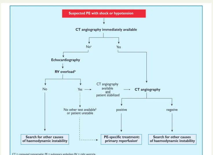
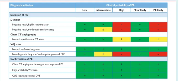
The prevalence of confirmed PE in patients undergoing diagnostic work-up because of suspicion of disease has been rather low (10 – 35%) in large series.99,100,113,116,197Hence, the use of diagnostic algo-rithms is warranted, and various combinations of clinical assessment, plasma D-dimer measurement, and imaging tests have been pro-posed and validated. These strategies were tested in patients present-ing with suspected PE in the emergency ward,99,113,114,116,197during the hospital stay and more recently in the primary care setting.118,126 Failure to comply with evidence-based diagnostic strategies when withholding anticoagulation was associated with a significant increase in the number of VTE episodes and sudden cardiac death at three-month follow-up.198The most straightforward diagnostic algorithms for suspected PE—with and without shock or hypotension—are pre-sented in Figures3and4, respectively; however, it is recognized that the diagnostic approach to suspected PE may vary, depending on the availability of—and expertise in—specific tests in various hospi-tals and clinical settings. Accordingly, Table6provides the necessary evidence for alternative evidence-based diagnostic algorithms.
The diagnostic strategy for suspected acute PE in pregnancy is dis-cussed in Section 8.1.
3.10.1 Suspected pulmonary embolism with shock or hypotension
The proposed strategy is shown in Figure3. Suspected high-risk PE is an immediately life-threatening situation, and patients presenting with shock or hypotension present a distinct clinical problem. The clinical probability is usually high, and the differential diagnosis includes acute valvular dysfunction, tamponade, acute coronary syndrome (ACS), and aortic dissection. The most useful initial test in this situation is bedside transthoracic echocardiography, which will yield evidence of acute pulmonary hypertension and RV dysfunction if acute PE is the cause of the patient’s haemodynamic decompensation. In a highly un-stable patient, echocardiographic evidence of RV dysfunction is suffi-cient to prompt immediate reperfusion without further testing. This decision may be strengthened by the (rare) visualization of right heart thrombi.184,199,200Ancillary bedside imaging tests include transoeso-phageal echocardiography which, if available, may allow direct visualiza-tion of thrombi in the pulmonary artery and its main branches,188,190,201 and bedside CUS, which can detect proximal DVT. As soon as the patient can be stabilized by supportive treatment, final confirmation of the diagnosis by CT angiography should be sought.
For unstable patients admitted directly to the catheterization la-boratory with suspected ACS, pulmonary angiography may be con-sidered as a diagnostic procedure after the ACS has been excluded, provided that PE is a probable diagnostic alternative and particularly if percutaneous catheter-directed treatment is a therapeutic option.
by guest on September 4, 2014
http://eurheartj.oxfordjournals.org/
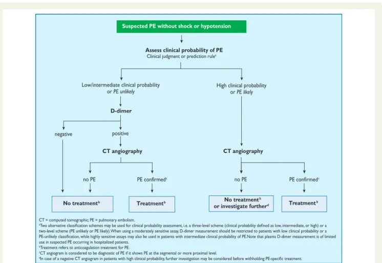
3.10.2 Suspected pulmonary embolism without shock or hypotension
Strategy based on computed tomographic angiography (Figure4) Computed tomographic angiography has become the main thor-acic imaging test for investigating suspected PE but, since most patients with suspected PE do not have the disease, CT should not be the first-line test.
In patients admitted to the emergency department, plasma D-dimer measurement, combined with clinical probability assess-ment, is the logical first step and allows PE to be ruled out in around 30% of patients, with a three-month thromboembolic risk in patients left untreated of ,1%. D-dimer should not be measured in patients with a high clinical probability, owing to a low negative pre-dictive value in this population.202It is also less useful in hospitalized patients because the number needed to test to obtain a clinically rele-vant negative result is high.
In most centres, MDCT angiography is the second-line test in patients with an elevated D-dimer level and the first-line test in patients with a high clinical probability. CT angiography is considered
to be diagnostic of PE when it shows a clot at least at the segmental level of the pulmonary arterial tree. False-negative results of MDCT have been reported in patients with a high clinical probability of PE;134however, this situation is infrequent, and the three-month thromboembolic risk was low in these cases.99Therefore, both the necessity of performing further tests and the nature of these tests in such patients remain controversial.
Value of lower limb compression ultrasonography
Under certain circumstances, CUS can still be useful in the diagnostic work-up of suspected PE. CUS shows a DVT in 30 – 50% of patients with PE,116,192,193and finding proximal DVT in a patient suspected of PE is sufficient to warrant anticoagulant treatment without further testing.194Hence, performing CUS before CT may be an option in patients with relative contraindications for CT such as in renal failure, allergy to contrast dye, or pregnancy.195,196
Value of ventilation – perfusion scintigraphy
In centres in which V/Q scintigraphy is readily available, it remains a valid option for patients with an elevated D-dimer and a Suspected PE with shock or hypotension
CT angiography immediately available
Echocardiography RV overloadb
Noa Yes
No
Search for other causes of haemodynamic instability
PE-specific treatment:
primary reperfusionc of haemodynamic instabilitySearch for other causes
Yes
No other test availableb
or patient unstable positive negative
CT angiography
CT angiography available
and patient stabilized
CT = computed tomographic; PE = pulmonary embolism; RV = right ventricle.
aIncludes the cases in which the patient’s condition is so critical that it only allows bedside diagnostic tests. b
chambers. Ancillary bedside imaging tests include transoesophageal echocardiography, which may detect emboli in the pulmonary artery and its main branches, and bilateral
cThrombolysis; alternatively, surgical embolectomy or catheter-directed treatment (Section 5).
Figure 3 Proposed diagnostic algorithm for patients with suspected high-risk PE, i.e. presenting with shock or hypotension.
by guest on September 4, 2014
http://eurheartj.oxfordjournals.org/
contraindication to CT. Also, V/Q scintigraphy may be preferred over CT to avoid unnecessary radiation, particularly in younger and female patients in whom thoracic CT may raise the lifetime risk of breast cancer.139V/Q lung scintigraphy is diagnostic (with either normal or high-probability findings) in approximately 30 – 50% of emergency ward patients with suspected PE.83,94,135,203The proportion of diag-nostic V/Q scans is higher in patients with a normal chest X-ray, and this supports the recommendation to use V/Q scan as the first-line imaging test for PE in younger patients.204
The number of patients with inconclusive findings may also be reduced by taking into account clinical probability.94Thus, patients with a non-diagnostic lung scan and low clinical probability of PE have a low prevalence of confirmed PE.94,157,203The negative predict-ive value of this combination is further increased by the absence of a DVT on lower-limb CUS. If a high-probability lung scan is obtained from a patient with low clinical probability of PE, confirmation by other tests may be considered on a case-by-case basis.
3.11 Areas of uncertainty
Despite considerable progress in the diagnosis of PE, several areas of uncertainty persist. The diagnostic value and clinical significance of sub-segmental defects on MDCT are still under debate.136,137 A recent retrospective analysis of two patient cohorts with suspected PE showed similar outcomes (in terms of three-month recurrence
and mortality rates) between patients with sub-segmental and more proximal PE; outcomes were largely determined by comorbid-ities.205The definition of sub-segmental PE has yet to be standardized and a single sub-segmental defect probably does not have the same clinical relevance as multiple, sub-segmental thrombi.
There is also growing evidence suggesting over-diagnosis of PE.206 A randomized comparison showed that, although CT detected PE more frequently than V/Q scanning, three-month out-comes were similar, regardless of the diagnostic method used.135 Data from the United States show an 80% rise in the apparent in-cidence of PE after the introduction of CT, without a significant impact on mortality.207,208
Some experts believe that patients with incidental (unsuspected) PE on CT should be treated,144especially if they have cancer and a proximal clot, but solid evidence in support of this recommendation is lacking. The value and cost-effectiveness of CUS in suspected PE should be further clarified.
Finally, ‘triple rule-out’ (for coronary artery disease, PE and aortic dissection) CT angiography for patients presenting with non-traumatic chest pain appears to be accurate for the detection of cor-onary artery disease.209However, the benefits vs. risks (including increased radiation and contrast exposure) of such a diagnostic ap-proach need thorough evaluation, given the low (,1%) prevalence of PE and aortic dissection in the studies published thus far.
Suspected PE without shock or hypotension
Assess clinical probability of PE Clinical judgment or prediction rulea
D-dimer
CT angiography
positive
CT angiography
negative
Low/intermediate clinical probability or PE unlikely
no PE PE confirmedc no PE PE confirmedc
High clinical probability or PE likely
No treatmentb Treatmentb No treatment
b
or investigate furtherd Treatmentb
CT = computed tomographic; PE = pulmonary embolism.
a
two-level scheme (PE unlikely or PE likely). When using a moderately sensitive assay, D-dimer measurement should be restricted to patients with low clinical probability or a
use in suspected PE occurring in hospitalized patients.
bTreatment refers to anticoagulation treatment for PE.
cCT angiogram is considered to be diagnostic of PE if it shows PE at the segmental or more proximal level. d
Figure 4 Proposed diagnostic algorithm for patients with suspected not high-risk pulmonary embolism.
by guest on September 4, 2014
http://eurheartj.oxfordjournals.org/