RIVM report 650040 002
Immunotoxicity and Developmental Toxicity in the Rat after Prenatal Exposure to
Diethylstilbestrol
A.H. Piersma, A. Verhoef, C.G.J. Sweep, W.H. de Jong, H. van Loveren
July 2001
This investigation has been performed by order and for the account of The Ministry of Public Health, Welfare and Sports, within the framework of project 650040, “Kennisbasis
Reproductietoxicologie”.
Abstract
In this study we explored the usefulness of including immune parameters in the prenatal developmental toxicity study in rats, as described in the OECD 414 guideline.
Diethylstilbestrol was used as the model compound, as it is known to be toxic both for the development and the immune system of the rat. The results show developmental toxicity in terms of decreased fetal survival and body weight in the presence of reduced maternal food consumption and body weight. Histopathologic effects in ovaries and uteri of pups were reminiscent of the estrogenicity of the test compound. In contrast, immune parameters were uncompromised. For some compounds, we speculate the early postnatal period to show more vulnerability to immunotoxic injury after xenobiotic exposure than the prenatal period, in which exposure occurred in this study.
Contents
SAMENVATTING...4 SUMMARY ...5 1. INTRODUCTION ...6 2. METHODS...8 3. RESULTS...10 4. DISCUSSION...12 REFERENCES...15APPENDIX I: FIGURES AND TABLES ...18
Samenvatting
Onderzoek aan dioxines en PCB’s heeft duidelijk gemaakt dat het immuunsysteeem in ontwikkeling een grote gevoeligheid voor xenobiotica vertoont in de perinatale periode. De huidige richtlijnen voor proefdiertesten voor reproductietoxiciteit bevatten echter geen immunologische parameters. In deze studie hebben we het mogelijke nut onderzocht van het inbouwen van immuunparameters in de “developmental toxicity study”, zoals beschreven in het OECD 414 protocol. Diethylstibestrol (DES) werd daarbij als modelstof gebruikt, vanwege de toxische eigenschappen van deze stof voor zowel de embryogenese als voor het immuunsysteeem. De resultaten lieten embryotoxiciteit zien in de vorm van embryonale sterfte en verlaagd foetaal gewicht, terwijl voedselconsumptie en groei van de moederdieren achterbleven. Daarnaast werden bij pups histopathologische effecten gezien in uterus en ovarium, die verklaard kunnen worden uit de oestrogene werking van de modelstof. Echter, immunologische parameters, waaronder histopathologie, hematologie, en SRBC-respons in pups op de leeftijd van vier weken, vertoonden geen afwijkingen. We speculeren dat voor sommige verbindingen in tegenstelling tot de prenatale periode, waarin in deze studie werd blootgesteld, de vroege postnatale periode meer gevoelig zou kunnen zijn voor
immunologische schade na blootstelling aan xenobiotica. In dat geval ligt het meer voor de hand om immuunparameters toe te voegen aan andere bestaande testen zoals de peri- en postnatale studie of een generatiestudie.
Summary
Studies with dioxins and PCB’s have shown that the developing immune system may be especially vulnerable to xenobiotics during the perinatal period. However, current guidelines for reproductive toxicity testing do not contain immune parameters. In the present study, we have explored the usefulness of including immune parameters within the prenatal
developmental toxicity study in rats, as described in the OECD 414 guideline.
Diethylstilbestrol was used as the model compound, as it is known to be toxic both for embryofetal development and for the immune system. The results show developmental toxicity in terms of decreased fetal survival and decreased pup body weight in the presence of reduced maternal food consumption and body weight. Female pups at four weeks of age showed histopathologic effects in ovaries and uteri which are characteristic for the
estrogenicity of the test compound. In contrast, immune parameters, including histopathology, hematology, and SRBC-response in pups at 4 weeks of age were uncompromised. It is
speculated that for certain compounds rather than the prenatal exposure protocol used here, the early postnatal period may be more vulnerable to immunotoxic injury after xenobiotic exposure. This would argue for the incorporation of immune parameters into other existing tests such as the peri- and postnatal study or a generation study.
1. Introduction
Within Europe, the reproductive toxicity of substances is evaluated using standardised OECD test guidelines, carried out according to a strategy based upon EU guidelines. The hazard and risk evaluations of new and prioritized existing substances are based upon the results of these tests. Their protocols are regularly reviewed to improve their usability for risk assessment. Experimental analysis and validation are essential for a meaningful evaluation of suggested protocol changes. In the present study the use of an increased number of dose groups as well as the addition of parameters for immune toxicity are validated, using diethylstilbestrol as the model compound.
The use of an increased number of dose groups fascilitates the description of the dose-response relation, probably without having to increase the total number of animals in the experiment. Present guidelines prescribe three dose groups and a control group. Interpretation of the results leads to a ‘No-Observed-Adverse-Effect-Level’ which is extrapolated to the sensitive human using interspecies and intraspecies uncertainty factors. An alternative for the NOAEL approach makes use of the complete dose-response curve. Starting from a predefined ‘Critical Effects Size’ (CES) the ‘Critical Effect Dose’ (CED) is derived. The present study employs ten dose groups with fewer animals per group resulting in the same total animal number as in the current OECD protocol.
Recently, parameters for immune toxicity have been incorporated in the OECD 407 28-day general toxicity study (van Loveren and Vos, 1989). These comprise predominantly histopathology of lymphoid tissues. Debate is ongoing whether immune function tests, i.e. the antibody response to SRBC, should be included. In view of the high sensitivity of the
developing immune system in the perinatal period the incorporation of immune parameters within the developmental toxicity study warrants further study. In the present study such parameters have been incorporated.
Diethylstilbestrol is used as a model compound, as both developmental toxicity and immunotoxicity of this compound have been described in animal studies as well as in man. Several studies from the group of Bradshaw (Rands et al., 1982; Wardell et al., 1982;
Cornwall et al., 1984) describe a.o. 18% resorptions and increased gestation length in Sprague Dawley rats at 45 µg/kg bw.day after oral exposure from day 6-18 of gestation. Male fetuses showed a low incidence of cryporchidism, increasing to 16% at 80 µg/kg bw.day. This dose induced 65% resorptions. Ashby et al. (1997) exposed AP rats to 8.6 µg/kg bw.dag via
drinking water and found decreased pup weights and reproductive organ anomalies in the pups. Maternal toxicity was not described at these dosages. On the basis of these findings we chose doses between 1 and 60 µg/kg bw.day in the present study. Immunotoxic effects of DES were reported by Luster et al. (1978, 1979), Ways et al. (1987), Turiel and Wingard (1988), Holladay et al. (1993).
2. Methods
Harlan-CPB-WU rats were kept under spf conditions and received water and feed (RMHGS Hope farms) ad libitum. At 10-12 weeks of age, females were randomly distributed over 10 groups and mated to male rats between 9 and 10 a.m. (=day 0 of pregnancy). Animals were exposed by gavage from day 6 to 20 of pregnancy to a daily dose of DES in corn oil at 1 ml per 200 g body weight based on the most recent body weight determination. Body weights and feed consumption were measured at days 0, 6 11,16, and 21 of pregnancy. Doses were 0, 1.0, 1.7, 2.8, 4.7, 7.8, 13, 22, 36 and 60 µg/kg body weight per day. Half of the pregnant animals in each dose group were sectioned at gestation day 21, the other half were allowed to deliver and nurse their pups. Maternal blood was collected before necropsy at gestation day 21 for serum estradiol determination (Dr.C.G.J.Sweep, Afd. Chemische Endocrinologie, Radboud Ziekenhuis, Nijmegen). The offspring of part of the animals was autopsied at day 28 after birth for immunotoxicity studies and histopathology. From each nest of pups one male animal was used for routine histopathology and determination of non specific immune parameters, two male animals were used for studying antibody formation after SRBC
immunisation, and one female animal was used for evaluation of female reproductive organs. Heart, lung, liver, spleen, thymus, kidneys, adrenals, testes, stomach, jejunum, colon, thyroid, oesophagus, brain, pituitary, mesenterial and poplital and mandibular lymph nodes, and bone marrow were studied histologically. Blood and bone marrow cells were differentially counted. Remaining offspring (one per nest) were challenged with SRBC (2.109) i.p., one week later blood was sampled for IgM determination, two weeks later the challenge was repeated, and three weeks later the animals were bled for IgG determination.
The data were analyzed by dose-response modeling (the “Benchmark approach”).
In the Benchmark approach a dose-response model is fitted to the data, a Critical Effect Size (CES) is defined, and the associated Critical Effect Dose (CED) is derived from the fitted model. The choice of the model for deriving the CED follows from a procedure of applying likelihood ratio tests on the members of the following nested family of models:
model 1: y = a
model 2: y = a exp( b x)
model 3: y = a exp( b xd)
model 4: y = a (c - (c - 1)) exp( b x)
model 5: y = a (c - (c - 1)) exp( b xd),
by choosing a more complicated model when the increase in number of parameters leads to a significantly better fit to the dose-response data. In these models the parameter a represents the background response; the parameter b reflects the ‘slope’ or the ‘strength’ of the response. These models are suitable for describing different (sub)populations by the same model. For example, when males and females are equally sensitive to the compound studied with respect to body weight, male and female body weights can be described by the same model, with only parameter a differing between males and females, to account for background body weights differing between sexes. When males and females are not equally sensitive, the parameter b should differ between sexes. The hypothesis that parameter a or parameter b (or both) differ between populations (e.g. sexes, or any experimental treatment) can be statistically tested by a likelihood ratio test (Slob and Pieters, 1999).
3. Results
Dams
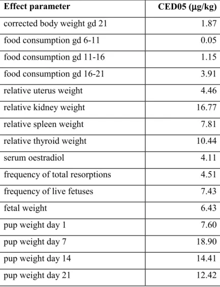
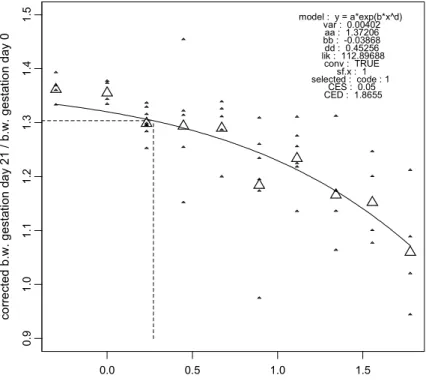
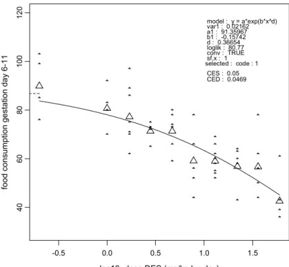
Dams did not show any clinical signs during the study. Body weight gain over three weeks of pregnancy, corrected for uterine weight, was dose-relatedly reduced (Figure 1). Food
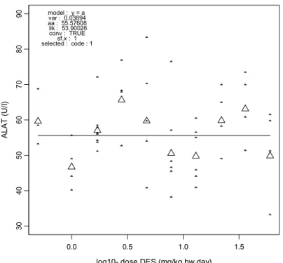
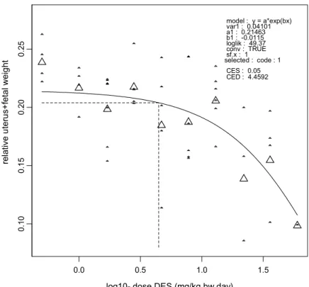
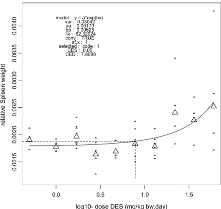
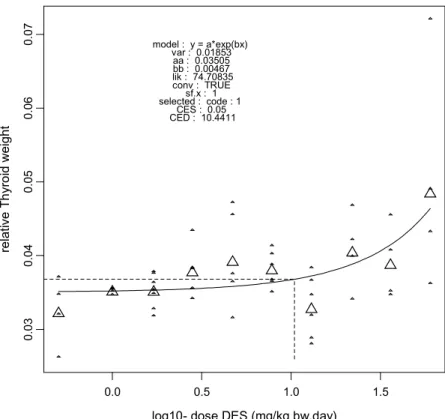
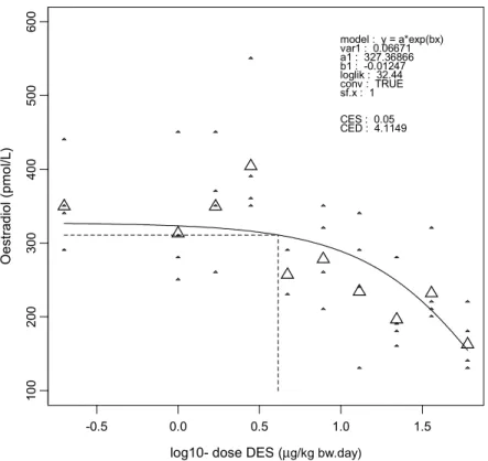
consumption was similar between dose groups before dosing (Figure 2), and showed a dose-related reduction during dosing (Figures 3, 4, and 5). Blood levels of liver enzymes ALAT and ASAT at gestation day 21 were not different between groups (Figures 6 and 7). Uterine weight (including contents) was dose-relatedly reduced with pronounced effects in the highest three dosages (Figure 8). Relative liver weight did not change (Figure 9), relative kidney weight (Figure 10) and relative spleen weight (Figure 11) increased with dose, relative thymus weight was unchanged after dosing (Figure 12), and relative thyroid weight increased with dose (Figure 13). Maternal blood estradiol levels at gestation day 21 decreased with dose (Figure 14).
Offspring
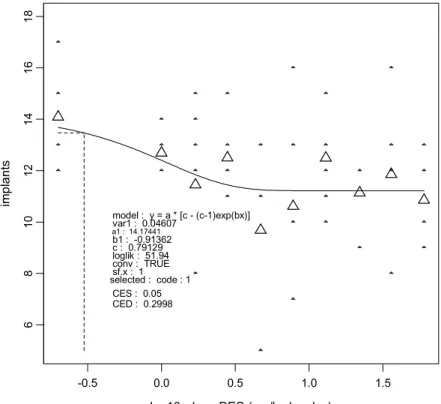
Corpora lutea numbers (Figure 15) did not show a dose-response relationship and also the number of implants per dam showed little variation between groups (Figure 16). Total resorptions per dam were increased in the highest three dosages (Figure 17), and
concommitantly the frequency of live fetuses, defined as the number of live fetuses divided by the number of implants, was reduced at these dosages (Figure 18). Fetal weights showed a dose-related reduction (Figure 19) with a 5% Critical Effect Dose of 6.4 µg/kg. Note that in this figure, the highest three dose groups contain considerably fewer data because of the high resorption rate. At these doses a selection has occurred of the less affected implants. Pup weights were dose-relatedly reduced at postnatal day 1 (Figure 20), but showed considerable catch-up growth at day 7 (Figure 21), day 14 (Figure 22), and day 21 (Figure 23). Offspring relative testis weight at 4 weeks of age did not show an effect of dosing (Figure 24).
Comparison of controls with the highest three doses showed that ovary stromal edema occurred significantly more often in the dosed groups (p=0.045) than in the control group. Ovary medullary hyperplasia and uterine stromal edema was found in several animals of the treated groups, but this phenomenon did not reach statistical significance (p=0.151). Apart from these findings histopathology and hematology were without specific effects. Blood concentrations of IgM (Figure 25) and IgG (Figure 26) after SRBC challenge were not
affected by treatment. Skeletal examination of fetuses did not reveal dose-related abnormalities (Tables 1 and 2).
4. Discussion
The idea of incorporation of immune parameters within regular developmental toxicity tests has been the subject of a series of studies in our laboratories over the past decade. The notion that the perinatal immune system may be specifically vulnerable to toxic insults stems e.g. from studies in harbour seals as well as in laboratory rats in which perinatal feeding with contaminated herring from the Baltic Sea resulted in a series of effects on the immune system (Ross, 1995). Since then, the OECD407 28-day subchronic toxicity study has been extended with socalled tier-1 immune parameters, comprising the histology immune tissues, to cover immune effects at the adult stage. Perinatal immune toxicity may differ significantly from effects in adulthood as the immune system passes critical stages of development and maturation during perinatal life. It was therefore considered worthwile to study whether immune parameters could be usefully incorporated into the existing OECD 414
developmental toxicity test.
We have shown earlier that a single exposure to the cytostatic agent cyclophosphamide on either day 11 or day 15 of gestation caused severe teratogenicity which was not accompanied by any observed immune toxicity (Hessel et al., 1994), as judged by histopathology as well as by flow cytometric analysis of subsets of immunecompetent cells in relevant organs such as thymus and spleen. In a subsequent study with diazepam, pharmacologically active doses given daily from day 6 to 21 of pregnancy, in accordance with the OECD414 protocol, did not induce either developmental toxicity or immune toxicity (de Waal et al., 1998). Diazepam had been shown in earlier studies to induce immune toxicity after prenatal exposure,
including an altered response to SRBC (Descotes et al., 1982; Covelli et al., 1991; Schreiber et al., 1993ab; Schlumpf et al., 1993, 1994; Ugaz et al., 1999). In the present study,
diethylstilbestrol (DES) was used as the model compound. This compound is highly
estrogenic and induces malformations in male and female reproductive organs of offspring of mothers treated in pregnancy (Giusti et al., 1995). In addition, DES has been reported to affect immune function (Ways et al., 1987, Turiel and Wingard, 1988).
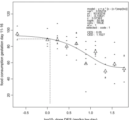
Diethylstilbestrol clearly showed developmental toxicity in this study (summarized in Table 3). Maternal food consumption and weight gain were dose-relatedly reduced. The effect on food consumption was most severe during the first 5 days of dosing (CED05 = 0.05 µg/kg),
and was reversible in the lower dose groups, with the CED05 reaching 3.91 µg/kg during the last 5 days of dosing. Maternal body weight gain was a sensitive parameter as well (CED05 = 1.87 µg/kg). The predominant developmental effect was an increase in resorptions (CED05 = 4.51 µg/kg), reflected in uterine weight effects with CED05 = 4.46 µg/kg. Fetal weight effects appeared as a somewhat less sensitive parameter (CED05 = 6.43 µg/kg). The finding that the predominant developmental effects occur with a CED05 of around 4 µg/kg is further
corroborated by the maternal oestradiol values, which result in CED05 = 4.11 µg/kg.
Table 3. Effect parameters and critical effect doses
at the 5% effect size
Effect parameter CED05 (µµµµg/kg)
corrected body weight gd 21 1.87
food consumption gd 6-11 0.05
food consumption gd 11-16 1.15
food consumption gd 16-21 3.91
relative uterus weight 4.46
relative kidney weight 16.77
relative spleen weight 7.81
relative thyroid weight 10.44
serum oestradiol 4.11
frequency of total resorptions 4.51
frequency of live fetuses 7.43
fetal weight 6.43
pup weight day 1 7.60
pup weight day 7 18.90
pup weight day 14 14.41
pup weight day 21 12.42
Furthermore, in 4 week old offspring, uterine and ovarian histopathology was studied and alterations were found in the three highest dose groups. These effects can be explained by the estrogenicity of the test compound, and are reminiscent of the effects found in human
offspring of pregnant women who used DES as a pharmaceutical compound. Immunopathological effects were not found in the present study. Both the tier-1
histopathology of immune organs and the SRBC challenge in young offspring did not reveal toxic effects on the immune system as a consequence of exposure to DES.
The present results reveal that the inclusion of immune parameters in the OECD414 protocol may not be sufficient to identify the immunotoxicity of all developmentally immunotoxic chemicals. Also in earlier studies in our laboratory, with cyclophosphamide and diazepam using different exposure regimens, no immune effects were found after prenatal exposure to pharmacologically and/or teratologically active dosages. The absence of immune toxicity in each of these studies may be related to the timing of exposure. In the OECD 414
developmental toxicity study exposure is done between days 6 and 21 of gestation. Development and maturation of the immune system takes place in part during this time period. However, also the postnatal period until weaning is critically important in this respect. Extending exposure until weaning may increase the sensitivity of the test system. This would however dramatically change the protocol, and in that respect it may be more useful to consider adding immune parameters to the generation study protocols (OECD 415-416). Another point to consider relates to animal use, expenses and duration of the study. For determination of tier-1 and functional immune parameters it is most meaningful to use offspring that has a mature immune system. For this reason we tested 4 week old offspring in the present study, requiring the number of pregnant rats per group to be doubled. As a result, expenses and duration of the study also increased considerably. This can only be justified if the information retrieved from such a protocol increases accordingly. Also in this respect, the generation study protocols may be favourable for including immune parameters, as they already include pup survival until weaning.
References
Ashby J, Tinwell H, Lefevre PA, Odum J, Paton D, Millward SW, Tittensor S, Brooks AN. Normal sexual development of rats exposed to butyl benzyl phthalate from conception to weaning. Regul. Toxicol. Pharmacol. 26:102, 1997.
Cornwall GA, Carter MW, Bradshaw WS. The relationship between prenatal lethality or fetal weight and intrauterine position in rats exposed to diethylstilbestrol, zeranol, 3,4,3’,4’,-tetrachlorobiphenyl, or cadmium. Teratology 30:341349, 1984.
Covelli V, Munno I, Decandia P, Altamura M, Nannuscio B, Maffione AB, Jirillo E. Effects of benzodiazepines on the immune system. Acta Neurol. 13(5): 418-423, 1991.
Descotes J, Tedone R, Evreux JC. Suppression of humoral and cellular immunity in normal mice by diazepam. Immunol. Lett. 5(1): 41-42, 1982.
De Waal EJ, Piersma AH, Van der Stappen AJ, Verhoef A, De Jong WH, Van Loveren H. Extension of the OECD414 rat teratogenicity protocol by increasing the duration of treatment and introduction of parameters of immunotoxicity: an exploratory study using diazepam as a model compound. RIVM Report no. 316601003, February 1998. RIVM Bilthoven.
Giusti RM, Iwamoto K, Hatch EE. Diethylstilbestrol revisited: a review of the longterm health effects. Ann. Intern. Med. 122: 778-788, 1995.
Hessel EM, Verhoef A, Van Loveren H, Piersma AH. Minimal immunologic changes in structurally malformed rats after prenatal exposure to cyclophosphamide. Toxicology 92: 287-299, 1994.
Holladay SD, Blaylock BL, Comment CE, Heindel JJ, Fox WM, Korach KS, Luster MI. Selective prothymocyte targeting by prenatal diethylstilbestrol exposure. Cell. Immunol. 152(1): 131-142, 1993.
Luster MI, Faith RE, McLachlan JA. Alterations of the antibody response following in utero exposure to diethylstilbestrol. Bull. Environ. Contam. Toxicol. 20(4): 433-437, 1978. Luster MI, Faith RE, McLachlan JA, Clark GC. Effect of in utero exposure to
diethylstilbestrol on the immune response in mice. Toxicol. Appl. Pharmacol. 47(2): 279-285, 1979.
Rands PL, White RD, Carter MW, Allen SD, Bradshaw WS. Indicators of developmental toxicity following prenatal administration of hormonally active compounds in the rat. I. Gestational length. Teratology 25:37-43, 1982
Ross PS. Seals, pollution and disease: environmental contaminant-induced immunosuppression. Thesis, University of Utrecht, 1995.
Schlumpf M, Lichtensteiger W, Ramseier H. Diazepam treatment of pregnant rats differentially affects interleukin-1 and interleukin-2 secretion in their offspring during different phases of postnatal develiopment. Pharmacol. Toxicol. 73(6): 335-340, 1993. Schlumpf M, Lichtensteiger W, van Loveren H. Impaired host resistance to Trichinella
spiralis as a consequence of prenatal treatment of rats with diazepam. Toxicology 94(1-3): 223-230, 1994.
Schreiber AA, Frei K, Lichtensteiger W, Schlumpf M. Alterations in interleukin-6 production by LPS- and Con A-stimulated mixed splenocytes, spleen macrophages and lymphocytes in prenatally diazepam-exposed rats. Agent Actions 38(3-4): 166-173, 1993a.
Schreiber AA, Frei K, Lichtensteiger W, Schlumpf M. The effect of prenatal diazepam exposure on TNF-alpha production by rat splenocytes. Agent Actions 38(3-4): 265-272, 1993b.
Slob W , Pieters MN. A probabilistic approach for deriving acceptable human intake limits and human health risks from toxicological studies: general framework. Risk Analysis 18:787-789, 1999.
Turiel J, Wingard DL. Immune response in DES-exposed women. Fertil. Steril. 49: 928, 1988.
Ugaz EM, Pinheiro SR, Guerra JL, Palermo-Neto J. Effects of prenatal diazepam treatment on Mycobacterium bovis-induced infection in hamsters. Immunopharmacology 41(3): 209-217, 1999.
Van Loveren H, Vos JG. Immunotoxicological considerations: A practical approach of immunotoxicity testing in the rat. In: Adv. Appl. Toxicol., Eds. A.D. Dayan and A.J.Paine, Taylor and Francis Ltd, pp 143-163, 1989.
Wardell RE, Seegmiller RE, Bradshaw WS. Induction of prenatal toxicity in the rat by diethylstilbestrol (DES), zeranol (ZN), 3,3’,4,4’-tetrachlorobiphenyl, cadmium, and lead. Teratology 26: 229-237, 1982.
Ways SC, Mortola JF, Zvaifler NJ, Weiss RJ, Yen SC. Alterations in immune responsiveness in women exposed to diethylstilbestrol in utero. Fertil. Steril. 48:193, 1987.
Appendix I: Figures and Tables
Legend to the FiguresSmall dots: individual data; large dots: geometric mean per dose group; dotted lines: CES and CED; figures 17 and 18: dots represent individual data per dam; figure 19: triangles represent male fetuses, circles represent female fetuses.
0.0 0.5 1.0 1.5
log10- dose DES (mg/kg bw.day)
0. 9 1 .0 1 .1 1.2 1.3 1.4 1.5 co rr e ct ed b. w. ge sta tio n d a y 21 / b. w. ges ta tion da y 0 model : y = a*exp(b*x^d) var : 0.00402 aa : 1.37206 bb : -0.03868 dd : 0.45256 lik : 112.89688 conv : TRUE sf.x : 1 selected : code : 1 CES : 0.05 CED : 1.8655
0.0 0.5 1.0 1.5
log10- dose DES (mg/kg bw.day)
2 0 4 0 60 80 100 120 14 0 fo od co ns um p tio n g es tat io n d ay 0 -6 model : y = a var : 0.01281 aa : 104.66487 lik : 95.6807 conv : TRUE sf.x : 1 selected : code : 1
Figure 2. Food consumption gestation day 0-6
-0.5 0.0 0.5 1.0 1.5
log10- dose DES (mg/kg bw.day)
40 60 80 10 0 120 food consumpt ion gest a tion day 6-11 version: 8.1 model : y = a*exp(b*x^d) var1 : 0.02162 a1 : 91.35967 b1 : -0.15742 d : 0.36654 loglik : 80.77 conv : TRUE sf.x : 1 selected : code : 1 CES : 0.05 CED : 0.0469
-0.5 0.0 0.5 1.0 1.5 log10- dose DES (mg/kg bw.day)
20 40 60 80 10 0 12 0 fo od consum pt ion gest at ion day 11-16 version: 8.1 model : y = a * [c - (c-1)exp(bx)] var1 : 0.05218 a1 : 93.93216 b1 : -0.10821 c : 0.57363 loglik : 55.66 conv : TRUE sf.x : 1 selected : code : 1 CES : 0.05 CED : 1.1527
Figure 4. Food consumption gestation day 11-16
-0.5 0.0 0.5 1.0 1.5
log10- dose DES (mg/kg bw.day)
20 40 60 80 100 120 food c ons um p tion ges ta tion day 16-21 version: 8.1 model : y = a*exp(bx) var1 : 0.07786 a1 : 97.532 b1 : -0.01311 loglik : 44.26 conv : TRUE sf.x : 1 selected : code : 1 CES : 0.05 CED : 3.9123
0.0 0.5 1.0 1.5
log10- dose DES (mg/kg bw.day)
30 40 50 60 70 80 90 ALAT (U /l) model : y = a var : 0.03894 aa : 55.57608 lik : 53.90026 conv : TRUE sf.x : 1 selected : code : 1
Figure 6. ALAT concentration in maternal serum on gestation day 21
0.0 0.5 1.0 1.5
log10- dose DES (mg/kg bw.day)
40 6 0 80 10 0 1 20 14 0 16 0 1 80 A SAT (U/l) model : y = a var : 0.06051 aa : 72.14045 lik : 43.31903 conv : TRUE sf.x : 1 selected : code : 1
0.0 0.5 1.0 1.5
log10- dose DES (mg/kg bw.day)
0. 10 0. 15 0. 20 0. 25 relative u ter u s+fetal weig ht version: 8.1 model : y = a*exp(bx) var1 : 0.04101 a1 : 0.21463 b1 : -0.0115 loglik : 49.37 conv : TRUE sf.x : 1 selected : code : 1 CES : 0.05 CED : 4.4592
Figure 8. Relative uterine + fetal weight at gestation day 21
0.0 0.5 1.0 1.5
log10- dose DES (mg/kg bw.day)
0.040 0.045 0.050 0.055 rel at ive L ive r w e ig ht model : y = a var : 0.00644 aa : 0.0452 lik : 101.13272 conv : TRUE sf.x : 1 selected : code : 1
0.0 0.5 1.0 1.5
log10- dose DES (mg/kg bw.day)
0.0050 0.0055 0.0060 0.0065 0.0070 re la tive Ki dne y w e ig ht model : y = a*exp(bx) var : 0.0051 aa : 0.00563 bb : 0.00291 lik : 106.95809 conv : TRUE sf.x : 1 selected : code : 1 CES : 0.05 CED : 16.7705
Figure 10. Relative maternal kidney weight at gestation day 21
0.0 0.5 1.0 1.5
log10- dose DES (mg/kg bw.day)
0.0015 0.0020 0.0025 0.0030 0.0035 0.0040 re la tiv e S p le e n w eig ht model : y = a*exp(bx) var : 0.03042 aa : 0.00179 bb : 0.00625 lik : 62.32024 conv : TRUE sf.x : 1 selected : code : 1 CES : 0.05 CED : 7.8088
0.0 0.5 1.0 1.5
log10- dose DES (mg/kg bw.day)
0.0004 0.0006 0.0008 0.0010 0.0012 0.0014 re la tive Thym us w e ig ht model : y = a var : 0.06766 aa : 0.00092 lik : 42.33024 conv : TRUE sf.x : 1 selected : code : 1
Figure 12. Relative maternal thymus weight at gestation day 21
0.0 0.5 1.0 1.5
log10- dose DES (mg/kg bw.day)
0.03 0.04 0.05 0.06 0.07 rel at ive T h yro id wei ght model : y = a*exp(bx) var : 0.01853 aa : 0.03505 bb : 0.00467 lik : 74.70835 conv : TRUE sf.x : 1 selected : code : 1 CES : 0.05 CED : 10.4411
-0.5 0.0 0.5 1.0 1.5
log10- dose DES (µg/kg bw.day)
10 0 2 00 30 0 40 0 5 00 60 0 Oes tr adi ol ( pmol /L) version: 8.1 model : y = a*exp(bx) var1 : 0.06671 a1 : 327.36866 b1 : -0.01247 loglik : 32.44 conv : TRUE sf.x : 1 CES : 0.05 CED : 4.1149
Figure 14. Maternal serum estradiol concentration at gestation day 21
0.0 0.5 1.0 1.5
log10- dose DES (mg/kg bw.day)
10 12 14 1 6 C o rpor a L u tea version: 8.1 model : y = a var1 : 0.01584 a1 : 13.76884 loglik : 77.06 conv : TRUE sf.x : 1 selected : code : 1
-0.5 0.0 0.5 1.0 1.5
log10- dose DES (mg/kg bw.day)
6 8 10 12 14 16 18 im pl ant s version: 8.1 model : y = a * [c - (c-1)exp(bx)] var1 : 0.04607 a1 : 14.17441 b1 : -0.91362 c : 0.79129 loglik : 51.94 conv : TRUE sf.x : 1 selected : code : 1 CES : 0.05 CED : 0.2998
Figure 16. Number of implantations per dam
0.0 0.5 1.0 1.5
log10- dose DES (mg/kg bw.day)
0.0 0.2 0.4 0.6 0.8 1.0 fr eq ue ncy of to ta l re sorp tion s model : B 3 bb : -0.2863 dd : 0.5205 th1 : -2.1279 sigma : 1 lik : -190.7524 conv : TRUE sf.x : 1 selected : code : 1 CED 1 : 4.5079 CES: 0.05
0.0 0.5 1.0 1.5 log10- dose DES (mg/kg bw.day)
0.0 0.2 0.4 0.6 0.8 1.0 fr equenc y of l iv e fetus model : B 2 bb : 0.0491 th1 : 1.6617 sigma : 1 lik : -194.7164 conv : TRUE sf.x : 1 selected : code : 1 CED 1 : 1.883 CES: 0.01
Figure 18. Frequency of live fetuses per dam
0.0 0.5 1.0 1.5
log10- dose DES (mg/kg bw.day)
2.5 3.0 3.5 4.0 4.5 5.0 fe ta l w eig ht (g ) model : y = a * [c - (c-1)exp(bx)] var : 0.00799 aa : 4.36244 aa : 4.15111 bb : -0.09631 cc : 0.89169 lik : 921.03803 conv : TRUE sf.x : 1 selected : fact1: sex CES : 0.05 CED : 6.4297 CED : 6.4297
0.0 0.5 1.0 1.5
log10- dose DES (mg/kg bw.day)
46 8 10 12 pu p w ei ght d a y 1 ( g) model : y = a * [c - (c-1)exp(bx)] var : 0.01336 aa : 7.50897 bb : -0.05547 cc : 0.85466 lik : 741.05035 conv : TRUE sf.x : 1 selected : CES : 0.05 CED : 7.601
Figure 20. Individual pup weights at postnatal day 1.
0.0 0.5 1.0 1.5
log10- dose DES (mg/kg bw.day)
5 10 1 52 02 5 pu p w e ight day 7 ( g) version: 8.1 model : y = a * [c - (c-1)exp(bx^d)] var1 : 0.02948 a1 : 15.01606 b1 : 0 c : 1.16241 d : 9.98731 loglik : 559.11 conv : TRUE sf.x : 1 CES : 0.05 CED : 18.9028
0.0 0.5 1.0 1.5
log10- dose DES (mg/kg bw.day)
20 25 3 0 3 5 40 45 pup w e ight d a y 14 ( g ) model : y = a * [c - (c-1)exp(bx^d)] var : 0.02042 aa : 27.95333 bb : -1e-005 cc : 1.24538 dd : 3.82573 lik : 630.24799 conv : TRUE sf.x : 1 selected : CES : 0.05 CED : 14.4062
Figure 22. Individual pup weights at postnatal day 14
0.0 0.5 1.0 1.5
log10- dose DES (mg/kg bw.day)
30 4 0 50 60 7 0 pup w e ight d a y 21 ( g ) model : y = a * [c - (c-1)exp(bx^d)] var : 0.0226 aa : 43.14642 bb : -2e-005 cc : 1.23913 dd : 3.69059 lik : 598.45474 conv : TRUE sf.x : 1 selected : CES : 0.05 CED : 12.4219
0.0 0.5 1.0 1.5
log10- dose DES (mg/kg bw.day)
0. 00 7 0 .0 08 0. 00 9 0. 0 10 0 .0 11 0. 01 2 pup r ela tive t e st is w e ight version: 8.1 model : y = a var1 : 0.00951 a1 : 0.00887 loglik : 239.45 conv : TRUE sf.x : 1 selected :
Figure 24. Pup relative testis weights at 4 weeks of age
-0.5 0.0 0.5 1.0 1.5
log10- dose DES (mg/kg bw.day)
0 246 8 1 0 Ig M version: 8.1 model : y = a var1 : 0.15056 a1 : 4.32146 loglik : 18.31 conv : TRUE sf.x : 1 selected :
-0.5 0.0 0.5 1.0 1.5 log10- dose DES (mg/kg bw.day)
0 2 4 6 8 10 12 14 Ig G version: 8.1 model : y = a var1 : 0.6457 a1 : 5.29861 loglik : -12.38 conv : TRUE sf.x : 1 selected :
Table 1. Skeletal abnormalities expressed as number of fetuses per group with the indicated anomaly
skeletal abnormalities total
dose (µg/kg bw.day) 0 7.8 13 22 36 60
number of fetuses examined 30 33 35 11 17 1 127
mascerated 2 0 3 0 0 0 5
zygoma dislocated 0 1 0 0 0 0 1
zygoma incompletely ossified 0 0 1 0 0 0 1
two or more ribs wavy 0 1 0 0 3 0 4
two or more ribs reduced in size 0 0 0 0 1 0 1
one rib reduced in size 0 0 1 0 0 0 1
accessory lumbar rib(s) 0 1 2 0 0 0 3
tarsals malformed 0 0 0 0 0 1 1
Table 2. Extent of ossification expressed as average number of bones ossified per fetus per dose group
extent of ossification total
dose (µg/kg bw.day) 0 7.8 13 22 36 60
number of fetuses examined 28 33 32 11 17 1 122
dental bones 0.4 0.5 0.3 0.6 0.4 1.0 0.44
vertebral caudal bodies 3.2 3.8 3.7 3.7 3.6 4.0 3.59
vertebral caudal arches 0.8 1.0 1.0 1.1 0.9 1.0 0.94
metacarpals 7.9 8.0 7.9 8.0 7.8 8.0 7.91
front proximal bones 2.2 2.5 2.4 1.7 2.5 0.0 2.33
front medial bones 0.0 0.0 0.0 0.0 0.0 0.0 0.00
front distal bones 8.3 8.5 8.1 8.3 7.6 8.0 8.20
metatarsals 8.0 8.1 8.0 8.1 7.9 8.0 8.04
hind proximal bones 0.0 0.0 0.0 0.0 0.0 0.0 0.00
hind medial bones 0.0 0.0 0.0 0.0 0.0 0.0 0.00
Appendix II: Mailing list
1. Dr.W.H.van Eck 2. Dr.H.Roelfzema 3. A.A.W.Kalis, arts
4. Voorzitter van de Gezondheidsraad
5. Depot Nederlandse Publikaties en Nederlandse Bibliografie 6. Directie RIVM
7. Dr.ir.G.de Mik, 8. Dr.A.Opperhuizen 9. Prof.dr.J.G.Vos
10. Dr.L.A.G.J.M. van Aerts 11. Dr.P.W.Wester 12. Dr.W.C.Mennes 13. Dr.M.Moerbeek 14. Prof.dr.W.Slob 15. Dr.C.G.J.Sweep, Radboudziekenhuis 16. Dr.A.H. Piersma 17. A.Verhoef 18. Dr.W.H.de Jong 19. Dr.H.van Loveren 20. SBD/VPR 21. Bureau rapportenregistratie 22. Bibliotheek RIVM 23. SBD/Voorlichting 24. – 33 Bureau Rapportenbeheer 25. – 38 Reserve-exemplaren