RIVM, P.O. Box 1, 3720 BA Bilthoven, telephone: 31 – 30 – 274 91 11; telefax: 31 – 30 – 274 29 71
Contact: J.W.A. Scheepmaker Expert Centre for Substances Jacqueline.Scheepmaker@rivm.nl RIVM report 601516013/2005
Factsheets for the (eco)toxicological risk
assessment strategy of the National Institute for Public Health and the Environment
Part V
J.W.A. Scheepmaker, C.E. Smit, M.T.M. van Raaij (eds.)
This investigation has been performed by order and for the account of the Board of Directors of the National Institute for Public Health and the Environment, within the framework of project 601516, Kennislacunes risicobeoordeling.
Authors
Chapter 1: G. Wolterink and G.H. Turkstra Chapter 2: P. van Hoeven-Arentzen
Chapter 3: A. Muller Chapter 4: C.E. Smit
Abstract
Factsheets for the (eco)toxicological risk assessment strategy of the National Institute for Public Health and the Environment - Part V
This report contains five factsheets describing risk assessment methods used at the Centre for Substances and Integral Risk Assessment (SIR) and the Expert Centre for Substances (SEC) of the National Institute for Public Health and the Environment (RIVM). The main aim is to enhance transparency and consistency in the risk assessment methods used at RIVM-SIR and RIVM-SEC. The factsheet on hepatic tumours in mice describes whether or not mouse liver tumours are considered relevant for human risk assessment. The factsheet on historical control data for tumour incidence provides the RIVM strategy on how to use historical control data in the evaluation of carcinogenicity data. The factsheet on Mononuclear Cell Leukaemia in the F344 rat strain discusses the relevance of an increased incidence of mononuclear cell leukaemia (MNCL) in F344 rats for humans and provides an approach to human hazard and risk
assessment. In the factsheet on energy and moisture content and assimilation efficiency of bird and mammal food, two large datasets on the energy and water content and assimilation
efficiency of various types of bird and mammal food, are merged and analysed. The last
factsheet on sorption of dissociating compounds describes the main principles and limitations of QSAR models and the HPLC method for determining the adsorption coefficients of organic ionisable substances. The five factsheets reflect a state-of-the-art approach and are meant to facilitate discussion with other national and international parties involved in risk assessment. Keywords: risk assessment; hepatic tumours; mononuclear cell leukaemia (MNCL); assimilation efficiency of food; QSAR models, HPLC method; organic ionisable substances
Rapport in het kort
Factsheets voor de (eco)toxicologische risicobeoordelingsstrategie van het Rijksinstituut voor Volksgezondheid en Milieu - Deel V
Dit rapport bundelt vijf factsheets waarin methodieken worden beschreven die worden gebruikt voor de risicobeoordeling van stoffen bij het Centrum voor Stoffen en Integrale
Risicobeoordeling (SIR) en het Stoffen Expertise Centrum (SEC) van het Rijksinstituut voor Volksgezondheid en Milieu (RIVM). Het voornaamste doel is om de inzichtelijkheid en eenduidigheid van de bij RIVM-SIR en -SEC gevolgde methodieken te vergroten.
De factsheet over levertumoren in de muis beschrijft onder welke voorwaarden levertumoren in de muis als relevant voor risicobeoordeling in de mens worden beschouwd. In de factsheet over historische controlegegevens van het voorkomen van tumor wordt aangegeven hoe het RIVM bij de evaluatie van carcinogeniteit omgaat met historische controlegegevens. De factsheet over Mononucleaire Cel Leukemie in de F344 rat bespreekt de relevantie voor de mens van een toename in het voorkomen van mononucleaire cel leukemie (MNCL) in F344 ratten en levert een strategie voor de gevaar- en risicobeoordeling. In de factsheet ‘Energie- en vochtgehalte en assimilatie-efficiëntie van voedsel voor vogel- en zoogdieren’ zijn twee grote datasets met gegevens over het energie- en watergehalte van verschillende voedselbronnen voor vogels en zoogdieren samengevoegd en geanalyseerd. De laatste factsheet over sorptie van dissociërende stoffen geeft een uiteenzetting van de belangrijkste principes en beperkingen van de QSAR- modellen en HPLC-methoden voor het bepalen van het adsorptiegedrag van een stof. De vijf factsheets vormen de weerslag van de huidige stand van wetenschap. Ze zijn bedoeld om de discussie met andere (inter)nationale partijen op het gebied van risicobeoordeling te bevorderen.
Trefwoorden: risicobeoordeling; levertumoren; Mononucleaire Cel Leukemie; assimilatie-efficiëntie van voedsel; QSAR-modellen; HPLC-methoden
Preface
This report was written within the framework of the project ‘Kennislacunes Risicobeoordeling’ (‘Knowledge gaps in risk assessment’). The factsheets presented in this report have been reviewed by members of the peer review groups of the Centre for Substances and Integral Risk Assessment (SIR) and the Expert Centre for Substances (SEC), and in some cases experts were consulted. The following persons are acknowledged for their contribution: M.E. van Apeldoorn, R. Beems, J. van Benthem, S. de Boer, A.G.A.C. Knaap, J.B.H.J. Linders, R. Luttik,
Contents
Samenvatting...13
Summary ...15
Introduction ...17
1. Hepatic tumours in mice...19
2. Historical control tumour incidence; its practical use ...31
3. Mononuclear Cell Leukaemia in the F344 rat strain...43
4. Energy and moisture content and assimilation efficiency of bird and mammal food ...57
5. Sorption of dissociating compounds ...73
Samenvatting
In dit rapport worden vijf factsheets gepresenteerd die worden gebruikt voor de beoordeling van stoffen bij het Centrum voor Stoffen en Integrale risicobeoordeling (SIR) en het Stoffen
Expertise Centrum (SEC) van het Rijksinstituut voor Volksgezondheid en Milieu (RIVM). Bij blootstelling van de muis aan chemische stoffen wordt vaak een verhoogde incidentie van levertumoren waargenomen. De relevantie van levertumoren in de muis voor de humane
risicobeoordeling is vaak een onderwerp van discussie geweest. In de factsheet ‘Levertumoren
in de muis’ wordt de cellulaire en moleculaire pathologie van de hepatocarcinogenese
beschreven en er wordt aangegeven wat overeenkomsten en verschillen in levertumorvorming tussen de muis en de mens zijn. In de RIVM-strategie wordt beschreven onder welke
voorwaarden levertumoren in de muis als relevant voor risicobeoordeling in de mens worden beschouwd.
In de factsheet ‘Historische controle gegevens van tumorincidentie; praktische
toepasbaarheid’ wordt aangegeven hoe het RIVM bij de evaluatie van carcinogeniteit omgaat
met historische controlegegevens. In het algemeen worden in een chronische studie met ratten de mogelijke effecten na langdurige blootstelling onderzocht, waaronder ook de mogelijk
carcinogene potentie van de stof. Vaak echter, wanneer een toegenomen tumorincidentie wordt waargenomen, ligt de verhoogde tumorincidentie op de grens van biologische en/of statistische significantie. In dat geval kunnen historische controlegegevens gebruikt worden om in te schatten of de verandering biologisch significant is.
In de factsheet ‘Mononucleaire Cel Leukemie in de F344 rat’ wordt de beoordeling van de carcinogeniteit van stoffen gebaseerd op epidemiologie en/of chronische dierstudies. Echter, toename van bepaalde type tumoren in dierstudies worden soms als niet relevant voor de mens beschouwd. De relevantie van andere type tumoren is twijfelachtig. Een van de tumor typen met een twijfelachtige relevantie is mononucleaire cel leukemie (MNCL) in de F344 rat. In deze factsheet wordt de relevantie voor de mens van een toename in de incidentie van MNCL in F344 ratten besproken en een strategie voor de gevaar- en risicobeoordeling geleverd.
In de factsheet ‘Energie- en vochtgehalte en assimilatie-efficiëntie van voedsel voor vogel- en
zoogdieren’ zijn twee grote datasets met gegevens over het energie- en watergehalte van
verschillende voedselbronnen voor vogels en zoogdieren samengevoegd en geanalyseerd. De gemiddelde calorische waarde en het vochtgehalte zijn berekend voor 16 verschillende
categorieën voedsel en de assimilatie-efficiëntie wordt gepresenteerd voor 22 vogeltaxa en zeven groepen zoogdieren. De gegevens vormen de basis van de risicobeoordeling voor vogels en zoogdieren in het kader van de toelating van bestrijdingsmiddelen binnen de Europese Unie. In de factsheet ‘Sorptie van dissociërende stoffen’ wordt een uiteenzetting van de belangrijkste principes van de QSAR-modellen en HPLC-methoden voor het bepalen van het adsorptiegedrag van dissociërende stoffen beschreven. Bovendien beschrijft de factsheet de beperkingen die deze modellen en methoden hebben met betrekking tot geïoniseerde stoffen. Als aanvulling worden voor deze stoffen pragmatische benaderingen gegeven voor de risicobeoordeling. In dit
Summary
This report presents five factsheets for the risk assessment methods used in the Centre for Substances and Integral Risk Assessment (SIR) and the Expert Centre for Substances (SEC) of the National Institute for Public Health and the Environment (RIVM).
Upon exposure to chemicals, the liver of mice is a common and susceptible site of an increased tumour incidence. The relevance for human risk assessment of liver tumours in mice has been the subject of extensive debate. In the factsheet ‘Hepatic tumours in mice’ the cellular and molecular pathology of hepatocarcinogenesis is described and similarities and differences in liver tumorigenesis between mice and humans are indicated. The question whether or not mouse liver tumours are considered relevant for human risk assessment is addressed in the RIVM strategy.
In the factsheet ‘Historical control tumour incidence; its practical use’ the RIVM strategy is given on how to use historical control data in the evaluation of carcinogenicity data.
The potential long-term effects of toxic substances are usually determined in chronic toxicity studies with rodents. The outcome of a chronic test should enable a conclusion about the
carcinogenicity of a given compound. However, the results often involve an increased incidence of tumours in treated animals which lies at the borderline of biological and/or statistical
significance. In this case historical control values can be helpful in the interpretation of the biological significance of the change observed.
In the factsheet ‘Mononuclear Cell Leukaemia in the F344 rat strain’ the assessment of the carcinogenicity of substances is based on the results of epidemiology and/or chronic animal studies. However, increases of some types of tumours in animal studies are sometimes claimed not to be relevant to humans. The relevancy of other tumour types is questionable. One of the questionable tumour types is mononuclear cell leukaemia (MNCL) in the F344 rat. In this factsheet, the relevance of an increased incidence of MNCL in F344 rats for humans is discussed, and an approach for human hazard and risk assessment is provided.
In the factsheet ‘Energy and moisture content and assimilation efficiency of bird and
mammal food’, two large datasets on the energy and water content and assimilation efficiency
of various types of bird and mammal food are merged and analysed. Average caloric and moisture values are determined for 16 different food types, and assimilation efficiencies are presented for 22 bird orders and seven groups of mammals. The data are used as basic input values for the risk assessment for birds and mammals within the framework of pesticide authorisation in the European Union.
In the factsheet ‘Sorption of dissociating compounds’ some of the main principles of the QSAR models and the HPLC method are presented. Furthermore, the limitations they have in the treatment of organic ionisable substances with respect to adsorption coefficients are described. In addition, pragmatic approaches will be given for the risk assessment of organic ionisable
substances. This factsheet specifically gives guidance to some issues that fall outside the current EU Technical Guidance Document.
Introduction
One of the main tasks of the Expert Centre for Substances (SEC) and the Centre of Substances and Risk Assessment (SIR) of the National Institute for Public Health and the Environment (RIVM) is to assess the risk of compounds for public health and the environment. The
availability of adequate and up-to-date risk assessment methods is of the highest importance to fulfil this task. Some of these methods follow international guidance, but many have been
developed within the RIVM during the process of evaluation. These risk assessment methods are not rigid procedures but can be adapted based on new/developing scientific information, possibly triggered by questions from policy makers or by developments in (inter)national organisations. For specific problems or gaps in the assessment of (eco)toxicological effects, 'factsheets' are written by employees of SEC and SIR in co-operation with experts. These factsheets describe the current assessment strategies of SEC and SIR, and their main aim is to provide a transparent and accessible guidance for issues that are not covered by regular guidance documents. After
adoption of the factsheet by the advisory board and the head of the laboratories SEC or SIR all employees of SEC and SIR have to follow the risk assessment method described in the factsheet. In 2001, the first eight factsheets were published in an RIVM report1, followed by similar reports in 2002, 2003 and 20042,3,4. The present report contains five factsheets that were produced in 2004 and 2005 by SIR and SEC:
1. Hepatic tumours in mice
2. Historical tumour incidence; its practical use 3. Mononuclear Cell Leukaemia in the F344 rat strain
4. Energy and moisture content and assimilation efficiency of bird and mammal food 5. Sorption of dissociating compounds
We hope that by publishing these factsheets, the risk assessment methods followed by
RIVM/SEC and RIVM/SIR will become more transparent. The authors of each factsheet have tried to describe the state of the art of their subject.
Remarks, omissions or supplementary information will be appreciated and can be send to Jacqueline.Scheepmaker@rivm.nl and will be passed on to the responsible authors.
1 Luttik R, Van Raaij MTM, editors. Factsheets for the (eco)toxicological risk assessment strategy of the National Institute for Public Health and the Environment (RIVM). Bilthoven: National Institute for Public Health and the Environment; 2001. Report no. 601516007.
2 Luttik R, Pelgrom SMJG, editors. Factsheets for the (eco)toxicological risk assessment strategy of the National Institute for Public Health and the Environment. Part II. Bilthoven: National Institute for Public Health and the Environment; 2002. Report no. 601516009.
3 Luttik R, Van Raaij MTM, editors. Factsheets for the (eco)toxicological risk assessment strategy of the National Institute for Public Health and the Environment. Part III. Bilthoven: National Institute for Public Health and the Environment; 2003. Report no. 601516010.
4 Smit CE, Van Raaij MTM, editors. Factsheets for the (eco)toxicological risk assessment strategy of the National Institute for Public Health and the Environment. Part IV. Bilthoven: National Institute for Public Health and the Environment; 2004. Report no. 601516012.
1.
Hepatic tumours in mice
Factsheet FSV-014/00, date 24-08-2005 Authors: G. Wolterink, G.H. Turkstra
Contents
1.1 Introduction and aim... 19
1.2 Mechanism for the development of the effect, and background ... 20
1.2.1 Pathogenesis of liver cancer ...20
1.2.2 Mechanisms for liver tumour induction ...21
1.3 Normal values and natural variation ... 23
1.4 Susceptible species / Subpopulations ... 24
1.5 Assessment and RIVM strategy... 25
1.6 References... 27
1.1 Introduction and aim
For the evaluation of toxic substances, effects of chronic exposure are usually determined in chronic toxicity/carcinogenicity studies with rodents. These effects include the potency of a substance to induce carcinogenic effects. The relevance for human risk assessment of some types of tumours in rodent bioassays has been the subject of extensive debate [e.g. 1, 2, 3]. In rodents, in particular in mice, the liver is a common site of an increased tumour incidence associated with chemical exposure; one-third to one-half of all chemicals that are carcinogenic in rodents are hepatocarcinogens [4, 5]. Worldwide, hepatocellular carcinoma (HCC) is also one of the most frequent visceral tumours in humans [6, 7, 8].
Many experts have addressed the susceptibility of the mouse liver to tumour development. It is now recognised that there is a variety of mechanisms by which chemicals can give rise to tumour development in the liver, and that their mechanisms may have different implications for human risk assessment.
In this factsheet the cellular and molecular pathology of hepatocarcinogenesis is described and similarities and differences in liver tumorigenesis between mice and humans are indicated. Whether or not mouse liver tumours are considered relevant for human risk assessment is addressed in the RIVM evaluation strategy in paragraph 5.
Apart from hepatocellular adenoma (HCA) and HCC, a chemical may induce other types of liver tumours in mice, (e.g. haemangiomas, haemangiosarcomas, isolated hepatoblastomas) which are considered relevant for humans. It must be noted that the RIVM evaluation strategy only applies to the HCA and HCC types of liver tumours in mice.
1.2 Mechanism for the development of the effect, and
background
1.2.1 Pathogenesis of liver cancer
The differences and similarities in the pathogenesis of liver cancer in different species have been reviewed by Grisham [9]. Hepatocarcinogenesis has been analysed both at the cellular and molecular level. For many hepatocarcinogens the mechanism of action has been clarified. On the basis of the mechanism of action carcinogens can be classified as DNA-reactive or epigenetic hepatocarcinogens, but for a number of chemicals which induce liver tumours, sufficient information is not yet available for mechanistic classification (see ref. 20).
Cellular pathogenesis
Mice and rats
In rodents, hepatocarcinogenesis is a sequential, multi-step process, with stages termed
initiation, promotion, and progression which can be delineated experimentally [9]. The cellular pathway is thought to lead from foci of cellular alteration (FCA), which are presumed to contain initiated cells, through hepatocellular adenoma (HCA) to hepatocellular carcinoma. Hepatocytes, liver stem cells, or both may be initially affected by genetic damage that initiates the carcinogenic process. Some proliferative lesions may be less well-defined in mice; e.g. hyperplastic nodules in combination with hepatocellular damage are generally associated with regeneration rather than (pre)neoplasia. It is often difficult to distinguish between HCA and HCC in mice. Mouse liver tumours generally do not invade surrounding extrahepatic tissues and they rarely metastasise [10].
Humans
A similar, rather simple pathway is not applicable to all HCC in humans. In humans three pathways are known. Pathway 1 is the most frequent, occurring in about 75% of all cases. In this pathway chronic hepatitis and cirrhosis precedes the development of HCC, which appears normally to develop in rapidly expanding cirrhotic nodules. In a second pathway, occurring in about 25% of all cases, chronic hepatitis and focal parenchymal hyperplasia are prominent features preceding the development of HCC. In the third pathway, which represents less than 1% of HCC, the lesion that precedes HCA is unknown. The risk of progression from HCA to
HCC is very small and HCA may regress by unknown mechanisms [9]. The prognosis of HCC in humans is poor. HCC often metastasises and the 5-year survival rate
is 6% [11].
Molecular alterations in pathogenesis of liver tumours
With respect to the molecular pathogenesis of HCC in rodents and in humans many alterations involving expression of growth factors/receptors, proto-oncogens, and tumour suppressor genes have been identified at various stages of hepatocarcinogenesis. The most important alterations are [9]:
Increased expression of the growth factors TGFα or IGF-II, in mice and rats as well as humans.
Increased expression of the H-ras and myc proto-oncogenes.
The level of methylation of a gene is one of the mechanisms involved in the control of gene expression. A hypomethylated gene can be considered to possess an increased potential for expression as compared to a hypermethylated gene [12]. Hypomethylation of
proto-oncogenes has been demonstrated in HCC of rats and mice [e.g. 13, 14]. Elevated c-myc expression is also a common feature of HCC and preneoplastic lesions in humans. Mutated ras genes.
Frequently ras genes are mutated in HCC of mice. The H-ras proto-oncogene is mutated in 30-60% of the spontaneous HCC in B6C3F1 or C3H mice. The mutation is less frequent in HCC of strains of mice that are less prone to development of HCC [15]. Even in HCC-susceptible mouse strains other molecular pathways than the ras mutations exist. In contrast to mice, mutations of ras genes are infrequent in HCC of rats and humans.
Increased expression or mutations of p53 tumour suppressor gene.
Changes in expression or mutations of the p53 tumour suppressor gene have been extensively studied in human cancer, including HCC [16]. It has been suggested that the mutation of the p53 gene may have a role in the development of HCC in humans. Mutations of the p53 gene have not been identified in HCC in mice [17, 18, 19].
Changes in additional tumour suppressor genes.
Other known and putative tumour suppressor genes are also assumed to be involved in the pathogenesis of HCC in humans [9].
1.2.2 Mechanisms for liver tumour induction
In the liver, several mechanisms of chemically induced carcinogenesis have been identified. These mechanisms can be categorised as follows (according to ref. 20).
A. DNA-reactive hepatocarcinogens
Many DNA-reactive agents, such as aromatic amines [16], can induce cancer in the liver of rodents in addition to producing cancer at other sites. For hepatocarcinogens, there is a
relationship between DNA binding and carcinogenic potency. Most DNA reactive carcinogens are more hepatocarcinogenic in male rats and mice than in females rats and mice [20].
B. Epigenetic hepatocarcinogens
Some agents produce increases in rodent liver tumours by epigenetic mechanisms that do not involve reactivity of the chemical with DNA. These agents can be assigned to several classes based upon the mechanism of action through which they produce an increase in the occurrence of neoplasms.
Promoters
Tumour promoters act by facilitating the clonal expansion of initiated cells. FCAs are presumed to contain initiated cells and FCAs are considered to be precursors of liver neoplasms.
Promoter-mediated development of FCA could be a consequence of enhanced cell proliferation under the influence of the promoter (e.g. phenobarbital) [21, 22].
Phenobarbital was found to increase the occurrence of liver tumours in rodents when
administered chronically at high levels [23]. Phenobarbital was the first compound of a group of liver enzyme inducers found to be promoters of liver tumours. Thus, the concept arose that phenobarbital-type promoters acted through enzyme induction. Indeed, there is a good
correlation between enzyme induction and promoting activities [24]. However, no clear
mechanistic explanation has been provided for the role of enzyme induction in development of liver tumours.
In addition to phenobarbital and other barbiturates, a number of organochlorine compounds have been found to increase the occurrence of neoplasms in the livers of mice and rats; these include some pesticides, PCBs, and 2,3,7,8-TCDD [25].
Peroxisome proliferating agents
After chronic exposure to peroxisome proliferators, increases in the incidence of HCC have been observed in several rodent studies in conjunction with significant peroxisome proliferation
(see for a review ref. 26), especially in mice and rats. In a number of these studies also tumours in other tissues have been found. A strategy regarding peroxisome proliferating agents is presented in a separate factsheet [26].
Cytotoxins
Cytotoxicity gives rise to regenerative cell proliferation and this may be involved in the pathogenesis of the neoplasms [27]. In addition, selective growth of pre-neoplastic lesions, which are resistant to the cytotoxic agents, may also be involved [28]. Carbon tetrachloride and chloroform are two hepatotoxic agents that also increase liver tumour incidence in mice. However, these agents are not known to increase cancer in humans, probably because
exposures are not sufficiently high enough for chronic hepatotoxicity. Ethanol is hepatotoxic in humans, and repeated episodes of hepatotoxicity are associated with increases in liver cancer. Interestingly, it has not been possible to replicate this mechanism in rodents [29].
Hormones
In humans, oral contraceptive use is associated with increased risk of liver tumours [30]. In rats, liver tumours have been induced by synthetic estrogenic agents such as ethinylestradiol [31, 32] and diethylstilbestrol [33]. Estrogens have been shown to have a promoting effect in rat liver hepatocarcinogenesis and enhancement of cell proliferation by estrogenic hormones has been documented in rat liver [31]. No effect of diethylstilbestrol [33, 34] or estrogens or progestagens on mouse liver tumour incidence has been reported [35].
Viral infections
In humans, viral hepatitis is a major factor for liver cancer, possibly through development of cirrhosis and regeneration [36]. In woodchucks, hepatitis virus also leads to development of liver cancer [37]. In mice, however, hepatitis does not appear to be associated with an increase in liver tumour incidence, but an increase is seen in mice with Helicobacter hepaticus infection [38] and in transgenic mice expressing the RNA for the hepatitis B virus [39]. Increased cell turnover is a common factor in all these conditions.
1.3 Normal values and natural variation
Experimental animals
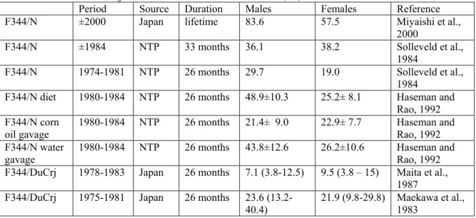
Table 1-1: Incidences of spontaneous hepatic tumours in rodent species in long-term studies
Species Strain Spontaneous incidence (%) References
Male Female HCA HCC HCA HCC Mice C3H 18-100* 0* [40] C3He 78* 0* [40] BalB/c 0* 0-1* [40] Charles River 2* 1.5* [40] CF-1 15-20* 0-13* [40] CBA 41* 27* [40] TF-1 13* 5* [40] B6C3F1 18-74 8-70 6-80 0-42 [41]
Rats Charles River CD 1.4* 2.7* [42]
Osborne-Mendel 0.5* 2.4* [42]
AES 2.6* 1.3* [42]
Sprague-Dawley 1.1* 0.7* [42]
Wistar 1.8* 1.1* [42]
Fischer 344 0-10 0-6 0-2 0-1 [41]
HCA= hepatocellular adenoma, HCC=hepatocellular carcinoma *: no distinction was made between HCA and HCC.
Table 1-1 indicates that the spontaneous occurrence of HCA/HCC is a very common finding in mice, and that there is a high variability in the incidences of HCA/HCC between mice of different strains. In rats the spontaneous incidence of liver tumours is lower than in mice. Three factors seem to influence the occurrence of spontaneous hepatocellular neoplasms in mice, namely strain, sex and food intake (ref. 43; see also paragraph 2).
Strain
As shown in Table 1-1 the incidence of spontaneous liver tumours in mice varies greatly among strains. Mice strains with a high incidence in spontaneous liver tumours also show an increased incidence in mutated H-ras genes (see paragraph 2, molecular pathogenesis). The relative susceptibilities of various strains to chemically induced hepatocarcinogenesis closely follows the incidence of spontaneous liver tumours in mice [20].
Sex
Male mice generally display higher incidences of spontaneous liver tumours and are more susceptible to hepatocarcinogenesis than females. The mode of action for this difference between males and females is unknown. However, since androgenic hormones have not been reported to induce rodent liver cancer, it is unlikely that it is a direct effect of male sex hormones. Castration or treatment of males with diethylstilbestrol reduced the occurrence of hepatocellular neoplasm in males [43, 44, 45]. In contrast, treatment of females with synthetic estrogenic agents increased liver tumour incidences [25].
Data from NCI/NTP studies indicate that there is a strong correlation between chemically induced male and female (liver) tumours in B6C3F1 mice, i.e. when liver tumours are observed in one sex they will also appear in the opposite sex [5, 46].
Food intake
Chronic dietary restriction induces an increase in apoptosis rate and a decrease in cell
proliferation rate in hepatocytes of 12-month-old B6C3F1 mice compared to ad libitum feeding [47]. This diet-induced shift in cell death/proliferation rates was associated with a marked reduction in spontaneous hepatoma and a marked increase in disease-free life span in dietary restricted mice compared to ad libitum fed mice. These results suggest that total caloric intake may modulate the rates of cell death and proliferation, showing a cancer-protective effect with dietary restriction and a cancer-promoting effect in ad libitum fed mice [47]. High-protein or high–fat diets also led to higher incidences of hepatic tumours in rodents [48]. It was shown that the inhibiting effect of dietary restriction on growth of diethylnitrosamine (DEN) induced glucose-6-phosphokinase-deficient (G6Pd) preneoplastic hepatic foci in Swiss OF1 mice was associated with reduced levels of insulin and IGF-I, two growth stimulating factors [49, 50]. Caloric restriction in SV129 mice was shown to modify nuclear receptor transcription (involved in regulation of growth factors and oxidizing enzymes) patterns which may play a role in liver tumour formation induced by hepatotoxic compounds [51].
Humans
In the age-adjusted standard population of humans, the incidence rate in hepatocellular carcinoma in Europe and North America is 0.003-0.005%. In Africa (0.030%) and in Asia (0.035%) incidence rates are relatively much higher [11]. The differences in incidence rates in humans from different parts of the world are mainly due to high incidences of chronic infection with the hepatitis B and C virus in Africa and Asia.
1.4 Susceptible species / Subpopulations
Interspecies differences
There are several differences between mice and humans with respect to the development of spontaneous or chemical-induced hepatic tumours.
Several strains of mice, e.g. B6C3F1, have very high and variable spontaneous tumour incidences, which indicates that their liver contains a significant population of ‘initiated’ or latent tumour cells [9]. These cells would be expected to be susceptible to the promoting effects of cellular proliferation. According to a report by the ‘international expert advisory committee to the Nutrition Foundation’, a similar susceptible population does not appear to exist in human livers [40].
Chemically induced liver tumours are often observed in mice. Analysis of the long-term carcinogenicity studies in rodents from the NCI/NTP database [46] revealed that from the 313 chemicals tested for liver tumours, 78 (25%) were positive in mice and 33 (11%) in rats. In comparison, as of July 1996 IARC had identified only a few chemicals or
therapeutic drugs that were considered to be associated with HCC in humans [30]. Of 183 pharmaceutical agents analysed by IARC, 3 substances (contraceptive steroids, azathiopine and anabolic steroids) were considered to be associated with risk of HCC in humans [30, 33]. Furthermore exposure to arsenic, aflatoxin B1, ethanol and vinyl chloride monomer are considered risk factors for development of HCC in humans. It must be noted that the exposure level to a chemical in experimental mice in long-term studies is much higher than in humans, which may be an important factor for the observed differences between mice and humans.
In contrast to human liver tumours, mouse liver neoplasms generally do not invade surrounding extrahepatic tissues and they rarely metastasise [10].
Hepatitis B and C viral infections are major factors for HCC in humans, whereas mice and rats are not known to be susceptible to a species-specific hepatitis virus.
There is a lack of concordance in the occurrence of chemically induced HCC in humans and mice. For instance, ethanol is hepatotoxic in humans and is associated with increased risk of liver tumours. These effects of ethanol are not observed in rodents. Similarly, aflatoxin B1 is a known hepatocarcinogen in rats and humans whereas mice appear to be resistant, probably due to a difference in metabolism of aflatoxine B1 [4, 5, 36].
In mice and rats the major hepatocarcinogens identified are chemicals. In human
epidemiological studies, the effect of a chemical on liver tumour induction is less clear and cannot be easily distinguished from other factors. An exception may be aflatoxin B1, for which most epidemiological studies show a correlation between exposure and increased incidence of liver cancer [52].
Human variability
The risk of developing HCC in humans is associated with several inheritable metabolic
abnormalities, e.g. primary hemachromatosis, tyrosinemia, glycogen storage disease, porphyria cutanea tarda and acute intermittent porphyria [9]. Most of these are characterised by inborn errors of metabolism that lead to accumulation of metabolic products in hepatocytes. In humans chronic infection with either hepatitis B or C greatly increases the risk of hepatic cell carcinoma (100-200-fold increased risks are found in epidemiological studies) [53].
Epidemiological evidence suggests that concurrent chronic infections with hepatitis B or C and exposure to aflatoxin [54], ethanol abuse [55], and possibly tobacco smoking [56], may amplify the risk of development of HCC.
1.5 Assessment and RIVM strategy
The biological relevance of chemically induced mouse liver tumours for human hazard identification has been the subject of considerable debate.
In contrast to some other rodent tumours where a specific mechanism has been identified (e.g. α2µ-globulin associated renal tumours in male rats [57]), the evidence regarding the relevance of hepatic tumours in mice for human risk assessment is less straightforward. The scientific evidence to date indicates that in many ways mouse liver tumours have different characteristics compared to HCC in humans [10].
The relevance of hepatic tumours in mice has been discussed by the ‘International Expert Advisory Committee to the Nutrition Foundation’ (1983 [40]), ‘ECETOC’ (1982 [58]), and in a ‘Society of Toxicology’-sponsored meeting (1997 [48]). In all discussions it was stated that for non-genotoxic hepatic tumour promoters, the weight of evidence indicates that a mouse liver tumour response is of questionable relevance to humans.
‘The International Expert Advisory Committee to the Nutrition Foundation’ stated that it is necessary that the total weight of evidence on carcinogenicity be considered in making regulatory decisions. More concern regarding the relevance of hepatic tumours in mice is warranted when there is:
1. unequivocal evidence of genotoxicity,
2. specific metabolic data demonstrating that the substance interacts with cellular genetic material,
3. evidence to demonstrate that the pharmacokinetic and metabolic data on the substance in the mouse more closely resembles man than other species tested,
4. evidence from epidemiological investigations indicating the substance has the potential to produce an increased risk to humans.
According to the committee a case-by-case approach should be applied to evaluate all available evidence.
With respect to risk assessment of pesticides, the use of hepatic tumour induction in mice has been addressed by JMPR on several occasions, as reviewed by the World Health Organization in the ‘Principles for the Toxicological Assessment of Pesticide Residues in Food’ [59]. In their review it is stated that ‘JMPR has generally considered it unwise to classify a compound as a carcinogen solely on the basis of an increased incidence of tumours of a kind that commonly occur spontaneously in the species and strain under study’. With respect to liver tumours it is stated that ‘the position of JMPR is that mouse liver tumours are of little relevance in predicting human cancer risk’ [60].
For the classification and labelling requirements for dangerous substances and preparations in the European Union (Annex VI of Directive 67/548/EEC, 2001[61]), compounds may not be classified as carcinogenic if the only available tumour data are liver tumours in certain sensitive strains of mice, without other supplementary evidence. No further reference was supplied in this guideline and the sensitive strains are not specified.
RIVM strategy
Apart from hepatocellular adenoma (HCA) and HCC, a chemical may induce other types of liver tumours in mice, (e.g. haemangiomas, haemangiosarcomas, isolated hepatoblastomas) which are considered relevant for humans. It must be noted that this RIVM evaluation strategy only applies to the HCA and HCC types of liver tumours in mice.
Overall, the data presented in paragraph 4 indicate that there are considerable differences in liver tumours between mice and humans. In order to establish the relevance of substance-related mouse liver tumours for human risk assessment the following points must be considered:
1.
The substance is genotoxic.
When the substance is genotoxic, it will be evaluated as if it presents a carcinogenic hazard to humans. Since genotoxic substances may induce irreversible damage to the DNA, it is generally assumed that there is no exposure level below which damage cannot occur. If a substance is genotoxic, all tumours, including mouse liver tumours, are considered relevant. In the risk assessment a non-threshold approach is applied to the mouse liver tumours, or if multiple sites or more than one species are affected, to the most critical tumour observed.
Note: The available database for a given substance may indicate that cytotoxicity rather than genotoxicity is the determining factor for mouse liver tumour induction. In that case, the available data should be subject to expert consultation, in order to decide on the primary mode of action and the way forward in risk assessment.
2.
The substance is not genotoxic
A. The substance is a peroxisome proliferator.
If liver tumours in the mouse are induced by a peroxisome proliferator, at doses at which peroxisome proliferation occurs, in most cases they will be considered not relevant for human risk assessment. Details of the RIVM strategy on interpretation of hepatocarcinogenesis induced by peroxisome proliferators are described in RIVM/SIR factsheet [26].
B. The substance induces only liver tumours in the mouse.
If the non-genotoxic substance induces only hepatic tumours in the mouse these tumours are considered not relevant for human risk assessment.
C. The substance induces other tumours.
Apart from liver tumours in the mouse, a chemical may induce other types of tumours in the mouse or tumours in another species. In this case, expert judgement is needed to establish the relevance of the liver tumours for humans.
The reasons for accepting or discarding mouse liver tumours as being relevant for human risk assessment should always be justified.
1.6 References
1. Alison RH, Capen CC, Prentice DE. 1994. Neoplastic lesions of questionable significance to humans. Toxicol. Pathol. 22: 179-186.
2. Maronpot RR, Flake G and Huff J. 2004. Relevance of animal carcinogenesis findings to human cancer predictions and prevention. Toxicol. Pathol. 32 (Suppl.1): 40-48 3. The Nutrition Foundation. 1983. The relevance of mouse liver hepatoma to human
carcinogenic risk. A report of the International Expert Advisory Committee to the Nutrition Foundation.
4. Gold LS, Slone TB, Manley NB and Bernstein L. 1991. Target organs in chronic bioassays of 533 chemical carcinogens. Env. Health Perspect. 93: 233-246. 5. Huff J, Cirvello J, Haseman J, and Bucher J. 1991. Chemicals associated with
site-specific neoplasia in 1394 long-term carcinogenesis experiments in laboratory rodents. Environ Health Perspect 93: 247-70.
6. Parkin DM, Pisani P, Ferlay J. 1993. Estimates of the worldwide incidence of eighteen major cancers in 1985. Int J Cancer. 54:594-606.
7. Parkin DM, Pisani P, Ferlay J. 1999. Estimates of the worldwide incidence of 25 major cancers in 1990. Int J Cancer. 80:827-41.
8. Pisani P, Bray, F, Parkin, MP. 2002. Estimates of the world-wide prevalence of cancer for 25 sites in the adult population. Int J Cancer. 97:72-81.
9. Grisham JW. 1996. Interspecies comparison of liver carcinogenesis: implications for cancer risk assessment. Carcinogenesis 18:59-81.
10. Newberne PM. 1983. How relevant are mice hepatomas to human risk? Toxicol. Pathol. 11: 113-4.
11. National Cancer Institute. 2001. SEER Cancer Statistics Review 1973-1998. Available at
http://seer.cancer.gov/publications/csr1973_1998/liver.pdf.
12. Strom SC, Faust JB. 1990. Oncongen activation and hepatocarcinogenesis. Pathobiology 58: 153-167.
13. Bhave MB, Wilson MJ, Poirier LA. 1988. c-H-ras and c-K-ras gene hypomethylation in the livers and hepatomas of rats fed methyl-deficient, amino acid-defined diets. Carcinogenesis 9: 343-348.
14. Ray JS, Harbison ML, McClain RM, Goodman, JI. 1994. Alterations in the methylation status and expression of the raf oncogene in phenobarbital-induced and spontaneous B6C3F1 mouse liver tumors. Mol. Carcinogenesis 9: 155-166.
15. Maronpot RR, Fox T, Malarkey DE, Goldsworthy TL. 1995. Mutations in the ras proto-oncogene, clues to etiology and molecular pathogenesis of mouse liver tumors. Toxicol. 101: 125–156.
16. Greenblatt MS, Bennett WP, Hollstein M, Harris CC. 1994. Mutations in the p53 tumor suppressor gene, clues to cancer etiology and molecular pathogenesis. Cancer Res. 54: 4855–4878.
17. Goodrow TL, Storer RD, Leander KR, Prahalada SR, Van Zwieten MJ, Bradley MO. 1992. Murine p53 intron sequences 5-8 and their use in polymerase chain
reaction/direct sequencing analysis of p53 mutations in CD-1 mouse liver and lung tumors. Mol. Carcinogenesis 5: 9-15.
18. Kress S, König J, Schweizer J, Löhrke H, Bauer-Hofmann R, Schwarz M. 1992. p53 Mutations are absent from carcinogen-induced mouse liver tumors but occur in cell lines established from these tumors. Mol. Carcinogenesis. 6: 148-158.
19. RumsbyPC, Davies MJ, Evans JG. 1994. Screening for p53 mutations in C3H/He mouse liver tumors derived spontaneously or induced with diethylnitrosamine or
phenobarbitone. Mol. Carcinogenesis 9: 71-75.
20. Williams GM. 1998. Carcinogenic responses to toxic liver injury in rodents. In: Comprehensive toxicology, Volume 9: hepatic and gastrointestinal toxicology (McCuskey RS, Earnest DL). Elsevier, Amsterdam.
21. Schulte-Hermann R, Ohde G, Schuppler J, Timmermann-Trosiener. 1981. Enhanced proliferation of putative preneoplastic cells in rat liver following treatment with the tumor promoters phenobarbital, hexachlorocyclohexane, steroid compounds, and nafenopin. Cancer Res. 41: 2556-2562.
22. Watanabe K, Williams GM. 1978. Enhancement of rat hepatocellular-altered foci by the liver tumor promoter phenobarbital: evidence that foci are precursors of neoplasms and that the promoter acts on carcinogen-induced lesions. J. Natl. Cancer Inst. 61: 1311-1314.
23. McClain RM, 1990. In 'Mouse Liver Carcinogenesis Mechanisms and Species
Comparisons,' eds. Stevenson DE, McClain RM, Popp JA, et al Wiley-Liss, New York, 345-365.
24. Rice JM, Diwan BA, Hu H , Ward JM, Nims RW, Lubet RA. 1994. Enhancement of hepatocarcinogenesis and induction specific cytochrome P450-dependent
monooxygenase activities by the barbiturates allobarbital, aprobarbital, pentobarbital, secobarbital and 5-phenyl- and 5-ethylbarbituric acids. Carcinogenesis 15: 395-402. 25. International Agency for Research on Cancer 'IARC Monographs on the Evaluation of
Carcinogenic Risks to Humans Overall Evaluations of Carcinogenicity. 1987. An Updating of IARC Monographs Volumes 1-42, IARC, Lyon, Suppl. 7.
26. Mennes W, Blaauboer BJ. 2003. The interpretation of hepatic peroxisome proliferation and associated hepatocellular carcinogenesis. RIVM/SIR factsheet FSV/010/00. 27. Larson JL, Wolf DC, Butterworth BE. 1994. Induced cytotoxicity and cell proliferation in
the hepatocarcinogenicity of chloroform in female B5C3F1 mice: comparison of administration by gavage in corn oil vs. ad libitum in drinking water. Fundam. Appl. Toxicol. 22: 96-102.
28. Farber E, Sarma DSR. 1987. Hepatocarcinogenesis: a dynamic cellular perspective. Lab. Invest. 56: 4-22.
29. International Agency for Research on Cancer. 1988. IARC Monographs on the
Evaluation of Carcinogenic Risks to Humans: Alcohol Drinking. IARC, Lyon, 44: 207-214.
30. International Agency for Research on Cancer. 1996. IARC Monographs on the
Evaluation of Carcinogenic Risks to Humans: Some medicinal drugs. IARC, Lyon, 66.
31. International Agency for Research on Cancer 'IARC Monographs on the Evaluation of Carcinogenic Risks to Humans Overall Evaluations of Carcinogenicity. 1987. An Updating of IARC Monographs Volumes 1-42, IARC, Lyon, Suppl. 7.
32. Yager JD, Zurlo J, Ni N. 1991. Sex hormones and tumor promotion in the liver. Proc. Soc. Exp. Biol. Med. 198: 667-674.
33. Marselos M, Vainio H. 1991. Carcinogenic properties of pharmaceutical agents evaluated in the IARC Monographs programme. Carcinogenesis 12: 1751-1766.
34. International Agency for Research on Cancer. 1979. IARC Monographs on the
Evaluation of Carcinogenic Risks to Humans: Sex hormones (II). IARC, Lyon, 21. 35. International Agency for Research on Cancer. 1999. IARC Monographs on the
Evaluation of Carcinogenic Risks to Humans: Hormonal contraception and post-menopausal hormone therapy. IARC, Lyon, 72.
36. International Agency for Research on Cancer. 1994. IARC Monographs on the
Evaluation of Carcinogenic Risks to Humans: Hepatitis Viruses. IARC, Lyon, 59. 37. Popper H, Roth L, Purcell RH, Tennant BC, Gerin JL. 1987. Hepatocarcinogenicity of the
woodchuck hepatitis virus. Proc. Natl. Acad. Sci. USA 84: 866-870.
38. Ward JM, Fox JG, Anver MR, Haines DC, George CV, Collins MJ Jr, Gorelick PL, Nagashima K, Gonda MA, Gilden RV, et al. 1994. Chronic active hepatitis and associated liver tumors in mice caused by a persistent bacterial infection with novel Helicobacter species. J. Natl. Cancer Inst. 86: 1222-1227.
39. Dunsford HA, Sell S, Chisari FV. 1990. Hepatocarcinogenesis due to chronic liver cell injury in hepatitis B virus transgenic mice. Cancer Res. 50: 3400-3407.
40. The Nutrition Foundation. 1983. The relevance of mouse liver hepatoma to human carcinogenic risk. A report of the International Expert Advisory Committee to the Nutrition Foundation.
41. National Toxicology Program. 2000. Historical control data. Available at
http://ehis.niehs.nih.gov/ntp/docs/ntp_hcrs.html.
42. Plaa GL, Hewitt WR. 1982. Toxicology of the liver. (Target organ toxicology series). Raven Press Books, New York, USA.
43. Frith CH, Ward JM, Turusov VS. Tumours in the liver. 1994. In: Pathology of tumours in laboratory animals. Volume 2 - tumours in the mouse. Second edition. (eds. Turusov VS, Mohr U) IARC Scientific Publications, Lyon, France. 111: 223-269.
44. Vesselinovitch SD and Mihailovitch N. 1967. The effect of gonadectomy on the development of hepatomas induced by urethane. Cancer Res 27: 1788-91.
45. Vesselinovitch SD, Itze L, Mihailovitch N and Rao KVN. 1980. Modifying role of partial hepatectomy and gonadectomy in ethylnitrosourea-induced hepatocarcinogenesis. Cancer Res 40: 1538-42.
46. Haseman JK, Lockhart A-M. 1993. Correlations between chemically related site-specific carcinogenic effects in long-term studies in rats and mice. Environ Health Perspect. 101: 50-4.
47. James SJ, Muskhelishvili L, Gaylor DW, Turturro A, Hart R. 1998. Upregulation of apoptosis with dietary restriction: implications for carcinogenesis and aging. Environ Health Perspect 106: Suppl. 307-312.
48. Dragan Y, Klaunig J, Maronpot R, Goldsworthy T. 1998. Forum, Meeting overview Mechanisms of susceptibility to mous liver carcinogenesis. Toxicological Sciences 41: 3-7.
49. Lagopoulos L, Sunahara GI, Wurzner H, Dombrowsky I, Stalder R. 1991. The effects of alternating dietary restriction and ad libitum feeding of mice on the development of diethylnitrosamine-induced liver tumours and its correlation to insulinaemia. Carcinogenesis; 12(2):311-315.
50. Lagopoulos L, Sunahara GI, Wurzner H, Fliesen T, Stalder R. 1991. The correlation of body growth with diethylnitrosamine-induced hepatocarcinogenesis in relation to serum insulin and somatomedin-C. Carcinogenesis; 12(2):211-215.
51. Corton, J.C. et al. 2004. Mimetics of caloric restriction include agonists of lipid-activated nuclear receptors. J.Biol.Chem. 279(44): 46204-46212.
52. WHO. 1998. Safety evaluation of certain food additives and contaminants. Prepared by the forty-ninth meeting of the Joint FAO/WHO Expert Committee on Food Additives (JECFA). WHO Food Additives Series No. 40. Geneva.
53. International Agency for Research on Cancer. 1994. Hepatitis viruses. IARC on the evaluation of Carcinogenic risks to humans, IARC Scientific Publications, Lyon, France, 59: 286.
54. International Agency for Research on Cancer. 1994. Some occurring substances: Food items and constituents, amines and mycotoxins. IARC Monographs on the evaluation of carcinogenic risk to humans, IARC Scientific Publications, Lyon, France, 56: 245–395.
55. International Agency for Research on Cancer. 1988. Alcohol drinking. IARC
Monographs on the evaluation of Carcinogenic risks to humans, IARC Scientific Publications, Lyon, France. 44: 207–215.
56. Yu MC, Chen C-J, Luo J-C, Brandt-Rauf PW, Carney WP, Santella RM. 1994 Correlations of chronic hepatitis B virus and cigarette smoking with elevated expression of neu oncoprotein in the development of hepatocellular carcinoma. Cancer Res. 54: 5106–5110.
57. Turkstra GH, van Raaij MTM. 2001. Alpha2u-globulin associated nephropathy and renal-cell neoplasms. RIVM/SIR Factsheet FSV/006/00.
58. European Chemical Industry Ecology &Toxicology Centre. 1982 Hepatocarcinogenesis in laboratory rodents: relevance for man. Brussels, Belgium. Monograph 4.
59. Environmental Health Criteria 104. Principles for the toxicological assessment of pesticide residues in food. International Programme on Chemical Safety 1990. 60. FAO/WHO. 1985 Pesticide residues in food – 1984. Report of the Joint Meeting on
Pesticide Residues, Rome, Food and Agricultural Organization of the United Nations (FAO plant Production and Protection Paper 62).
61. Annex VI of Directive 67/548/EEC. 2001. General classification and labeling requirements for dangerous substances and preparations.
2.
Historical control tumour incidence; its practical
use
Factsheet FSV-015/00, date 11-01-2005 Author: P.A. Van Hoeven-Arentzen
Contents
2.1 Introduction... 31 2.2 Background information... 31 2.3 Normal values and variation... 32 2.4 Aspects that influence spontaneous tumour incidences ... 35 2.5 The use of historical control values ... 36 2.6 Opinions of (inter)national bodies... 38 2.7 RIVM/SIR strategy... 38 2.8 References... 39 Annex 1. ‘Guidance notes for analysis and evaluation of chronic toxicity and
carcinogenicity studies’ ... 41
2.1 Introduction
For the evaluation of toxic substances, the potential long-term effects are usually determined in chronic toxicity studies with rodents. Potential effects after chronic exposure also include possible carcinogenic effects.
Under ideal conditions, the outcome of a chronic test should enable a conclusion about the carcinogenicity of a given compound. However, the actual test results do not always allow such a clear distinction. Experience has shown that the results often involve an increased incidence of tumours in treated animals which lies at the borderline of biological and/or statistical significance. Evaluations about the significance of such findings in animals (and man) are complex [1]. In this case ‘historical control values’, i.e. data on the normal variation of a parameter in the test species, can be helpful in the interpretation of the biological significance of the change observed. However, the incidence of spontaneous changes is often highly
variable among control groups of the same species and strain in different studies conducted [2]. This factsheet will describe when and how historical control data are helpful in the evaluation of carcinogenicity studies.
2.2 Background information
The primary goal of the chronic rodent carcinogenicity study is to assess the development of tumours in animals exposed to a chemical of concern as compared with controls. Four types of neoplastic responses are considered to be evidence of chemically induced carcinogenesis:
1. An increase in the frequency of one or several types of tumours that also occur in the controls; 2. The development of tumours not seen in controls;
3. The occurrence of tumours at an earlier time point than in controls;
4. An increase in the number of tumours per individual animal (multiplicity), compared to the controls.
Further a dose response relationship should be apparent. However, one should be aware that there may be a lower tumour incidence in the high(er) dose group(s) resulting from poor survival in these animals [3,4,5].
The tumours in the experimental animals may not be at the same stage of development. The stages include, for example, atypical hyperplasia (putative preneoplastic), benign tumours, carcinomas in situ, invasion of adjacent tissues and metastasis to other parts of the body. Although tumours of the same type but at different neoplastic stages could be separately tabulated, they should be combined for statistical purposes [3,5].
Factors to be weighed in the analysis of tumorigenesis include: (i) the occurrence of (cyto)toxic effects in the target organ(s), (ii) the occurrence of toxic effects in non-target organs (iii) the tumour incidence in concurrent controls, (iv) the presence and nature of preneoplastic lesions, (v) the species- and organ- specific sensitivity of tumourigenic response (e.g. liver tumours in B6C3F1 mouse), and (vi) the presence of a shift from ‘benign’ towards ‘malignant’ tumours with increasing dose.
In table 2-1 and 2-2, a top ten rank order of chemically induced site specific tumours in mice and rats based on the NTP database (B6C3F1 mice and F344 rats) is given [4].
Table 2-1: Top ten organs/systems developing tumours in
carcinogenesis studies – mice a
Table 2-2: Top ten organs/systems developing tumours in
carcinogenesis studies – rats a ranking males females ranking males females
1 Liver Liver 1 Liver Liver
2 Lung Lung 2 Kidney Mammary gland 3 Forestomach Forestomach 3 Zymbal gland Zymbal gland 4 Circulatory
system
Haematopoietic system
4 Forestomach Thyroid gland 5 Haematopoietic
system
Circulatory system
5 Thyroid gland Forestomach 6 Thyroid gland Mammary gland 6 Skin Urinary bladder
7 Harderian gland Ovary 7 Haematopoietic system
Clitoral gland 8 Adrenal gland Thyroid gland 8 Urinary bladder Haematopoietic
system 9 Kidney Uterus/cervix 9 Intestines Kidney 10 Five sites b Harderian gland 10 Nasal cavity Uterus/cervix
a. Based on 379 long-term chemical carcinogenesis studies from the National Toxicology program (NTP) data base
a. Based on 379 long-term chemical carcinogenesis studies from the National Toxicology program (NTP) data base b. Heart, nasal cavity, preputial gland, skin,
urinary bladder
2.3 Normal values and variation
Spontaneous carcinogenesis occurs in rodents as well as in humans. Thus, a degree of background tumour formation is always observable in control animals from chronic rodent carcinogenicity studies. Certain organs/tissues seem to be more susceptible to spontaneous tumour formation than others. Spontaneous tumour incidences vary by species, strain, stock or breeder and by gender [4,6,7,8]. Therefore, knowledge of the natural incidence of neoplastic lesions is essential for interpretation of experiments designed to reveal the effects of potential carcinogens [6,9].Organs most susceptible to spontaneous tumour formation are not always the
ones most susceptible to chemically induced carcinogenesis as occurs in chronic rodent bioassays [4,10].
Examples of differences in tumour incidences between strains are: C57BL/6 mice have a much lower liver tumour rate than do C3H mice, female Sprague Dawley rats have considerably more mammary tumours than do Fisher 344 rats, and Fischer 344 rats have a higher rate of leukaemias and tumours of the testis [10].
Examples of differences in tumour incidences between genders are: the most frequently occurring neoplasms in untreated male F344 rats are testicular adenoma (89.1%) and mononuclear cell leukaemia (50.5%) and for untreated female F344 rats pituitary gland neoplasms (54.2%) and mammary gland fibroadenoma (41.2%). For B6C3F1 mice the most frequently occurring neoplasms for untreated males are liver adenoma/carcinoma (42.2%) and lung adenoma/carcinoma (20.5%) and for untreated female mice liver adenoma/carcinoma (23.6%) and malignant lymphoma (haematopoietic system) (20.9%). These examples are based on the 1997 NTP data [11].
In table 2-3 and 2-4, a ranking on incidence of spontaneous tumour formation (benign and malignant) is given for the organs of mouse and rats, based on the mean of the highest incidence found for various strains (CD1 and B6C3F1 mice and F344, Sprague Dawley and Wistar rats) [4]. These tables are only meant as illustration.
Table 2-3: Ranking of mouse organs based on the incidence of spontaneous tumour formation (benign and malignant)
males females
organ incidence a (%) organ incidence a (%)
1 Liver 28.7 1 Blood/lymphoid tissue b 29.1
2 Lung/trachea 24.3 2 Lung/trachea 22.9 3 Adrenal 14.7 3 Pituitary 11.6 4 Blood/lymphoid tissue b 13.8 4 Liver 8.8
5 Stomach 3.0 5 Uterus/vagina 7.6 6 Circulatory system 2.9 6 Mammary gland 4.3 7 Skin/subcutaneous 2.4 7 Ovary 2.8 8 Pancreas 2.1 8 Circulatory system 2.4 9 Thyroid 1.6 9 Adrenal 2.2 10 Kidney 1.5 10 Stomach 2.1 11 Pancreas islets 1.2 11 Skin/subcutaneous 1.8 11 Testes 1.2 12 Thyroid 1.7 12 Urinary bladder 1.1 13 Kidney 1.4 13 Pituitary 0.5 14 Urinary bladder 1.2 14 Intestines 0.4 15 Brain 1.1 15 Brain 0.1 16 Pancreas <1.0 15 Heart 0.1 16 Pancreas (islets) 0.8 15 Body cavities 0.1 17 Body cavity 0.3 18 Intestines 0.2
19 Heart 0.1
a mean of highest reported percent incidence of spontaneous tumour formation for various mouse strains b Leukaemia/lymphoma
Table 2-4: Ranking of rat organs based on the incidence of spontaneous tumour formation (benign and malignant)
males females
organ incidence a (%) organ incidence a (%) 1 Pituitary 42.1 1 Pituitary 61.4 2 Testes 38.8 2 Mammary gland 43.7 3 Adrenal 31.4 3 Adrenal 24.5 4 Pancreas 28.9 4 Uterus/vagina 18.1
5 Blood/lymphoid tissue b 20.7 5 Blood/lymphoid tissue b 14.1
6 Thyroid 12.5 6 Thyroid 11.4 7 Skin/subcutaneous 12.1 7 Liver 6.0 8 Pancreas (islets) 10.9 8 Skin/subcutaneous 4.0 9 Brain 6.3 9 Brain 2.7 10 Body cavities 3.5 10 Lung/trachea 2.1 10 Mammary gland 3.5 11 Pancreas (islets) 1.9 11 Lung/trachea 3.4 11 Ovary 1.9 12 Liver 3.2 11 Intestines 1.9 13 Circulatory system 2.5 12 Preputial gland 1.8 14 Preputial gland 2.4 12 Circulatory system 1.8 15 Intestines 2.1 13 Pancreas 1.7 16 Kidney 1.7 14 Body cavity 1.4 17 Stomach 1.2 15 Urinary bladder 1.1 18 Urinary bladder 0.9 15 Stomach 1.1 19 Heart 0.2 16 Kidney 1.0 17 Heart <0.1 a mean of highest reported percent incidence of spontaneous tumour formation for various mouse strains b Leukaemia/lymphoma
For up to date information on the historical control values of individual tumour types in certain organs, one can look on the internet for actual databases on historical controls like the NTP database [12], the database of Charles River Laboratories [13] and Mouse Tumor Biology database [14]. Other databases that provide a standardised and reliable source of historical control data are the North American Control Animal Database (NACAD) [15] and the
European Registry of Industrial Toxicology Animal-data (RITA) system [8]. Both can be found on the world wide web, but need membership to use it.
For certain commonly-occurring rodent tumour types, which give rise to controversy in relation to their relevance for man, an overview of historical control values can be found in earlier factsheets dealing with this subject:
- Factsheet FSV 003/00 [16]: pheochromocytomas
- Factsheet FSV 006/00 [17]: kidney tumours related to alpha2u- globulin - Factsheet FSV 007/00 [18]: follicular thyroid tumours
- Factsheet FSV 010/00 [19]: liver tumours related to peroxisome proliferation - Factsheet FSV 011/00 [20]: forestomach tumours
2.4 Aspects that influence spontaneous tumour incidences
Many factors can be of influence on spontaneous tumour formation and generally fall into two categories: a) study design and b) histological procedures.
a) Study design
The factors related to study design are diet (restricted diet vs. ad libitum), body weight, housing conditions (individually vs. group), duration of the study, environment (e.g. the laboratory), genetic drift, survival differences (age), time (calendar year), presence of infectious organisms such as Helicobacter (increase of liver neoplasms in B6C3F1 mice), exposure scenario (gavage exposure vs. dietary exposure) and/or type of control treatment (e.g. untreated, corn oil gavage) [4,6,7,8,11,15,22].
For example, a comparison of the historic control database from the NTP in 1997 with that of a decade earlier showed higher body weights and lower survival for rats and a similar trend for mice. It showed that many spontaneous tumour rates had increased and in some cases the higher body weight could account for these increases (e.g. liver tumours in mice (m/f) and mammary tumours in female rats) [11].
Some of the key environmental influences include lighting and numbers of animals per cage. However, one of the most important experimental variables is age, as differences in tumour rates may simply reflect the greater survival of one group over another [15]. When tumour rates found in lifespan studies were compared with 2-year historic control tumour rates in F344 rats, it was found that the variety of neoplastic lesions in animals that were allowed to live their life-span was not greater than that in animals that were killed between 110 and 116 weeks of age, thus older age was not characterised by unique neoplasms. A second finding was that the incidence of certain neoplasms increased markedly after 110-116 weeks. No tumours were found in animals of either sex that died before 59 weeks of age and most of the common types of neoplasms were already present between 85 and 97 weeks of age [9]. A similar finding that increasing age is correlated with increasing incidence of spontaneous tumours was observed for Sprague Dawley rats. The spontaneous tumour frequencies of all rats (male and female
together) were only 6% at 15 months, 13% at 21 months, 66% at 26 months, and 86% at 32 months [23]. The data with F344 rats showed that hepatocellular neoplasms (neoplastic nodules) and pancreatic acinar cell adenomas occurred relatively late in lifespan. These were seen for the first time in animals that died between 98 and 100 weeks of age and between 111 and 128 weeks of age, respectively [9].
b) Histopathologic procedures
The second major category of factors include methods for tissue preparation and
histopathologic evaluation like gross necropsy, tissues selected, orientation in trimming, number of sections surveyed, slide preparation procedures, staining procedures and diagnostic criteria applied by the pathologist (histopathology diagnoses and nomenclature of
pre-neoplastic lesions and tumours) [6,8,15,22,24].
For example a standard dissection is necessary to ensure that all animals are examined in a similar manner for the detection of gross lesions and collection of all required organs. As number of tumours can increase with multiple sections or changes in orientation [25,26], all groups within a carcinogenicity study should have tissues prepared in the same way. Staining procedures should also be as consistent as possible with animals from each group processed concurrently such that any variations in staining will be spread across all groups [15].
2.5 The use of historical control values
In the OECD guideline 451 for carcinogenicity study [27] it is stated: ‘A good knowledge of the tumour profile of the animal strain throughout the life span is highly desirable in order to evaluate the results of experiments in a proper way’ and ‘The incidence of tumours and other suspect lesions normally occurring in the strain of animals used (under the same laboratory conditions – i.e. historical control) is desirable for assessing the significance of changes observed in exposed animals’.
The overall opinion of many authors [5,8,11,22,24,28,29,30] is that historical control information is useful in the interpretation of experimental results from similar studies, especially for rare neoplasms and for borderline effects, though all emphasize that the most appropriate and important comparison of an experimental group is with its concurrent control. However, there are specific instances in which historical control rates provide relevant data needed to interpret results of rodent carcinogenicity studies and as such are useful in deciding whether or not the apparent increase in neoplasm incidence (either statistically or
not-statistically significant) is a biological meaningful effect:
– the evaluation of rare neoplasms (i.e. with spontaneous tumour rates of 1% or less) may require less stringent statistical evidence in a given study if the low spontaneous rate can be demonstrated from an established historical control database.
– historical control data may be helpful in the evaluation of a neoplasm showing a marginal increase in incidence relative to concurrent controls.
– historical control data may also be useful from a quality control point of view, to help determine if concurrent control neoplasm rates are consistent with past experience. Like in the case of aberrant frequencies (i.e. control frequencies outside the expected control range) of neoplastic or pre-neoplastic lesions, there are certain limitations of the concurrent control. A lower than normal tumour frequency in the concurrent control animals may lead to a statistically significant increased incidence of lesions in the dose groups (false
positive). A higher than normal tumour frequency in the concurrent control, on the other hand, might mask a carcinogenic response in dosed animals (false negative). In such cases historical control data (which may provide a more appropriate demonstration of the true mean for the population and the variability of that mean) may support substantially the assessment of a potential carcinogenic risk.
One should be aware that false positive results may occur at sites with high and variable tumour rates and that these results are dependent on the rate of spontaneous tumour incidence.
When it is decided to use historic control data, it is essential that the study being evaluated is comparable to the studies in the historic control database with respect to those factors that are known to influence tumour occurrence (see paragraph 3 and 4). And, due to time-related variability of tumour incidences, historical control data should be limited to more recent studies. For that reason, windows of 3-5 years are proposed [8,11,22,24].
In the literature only few examples were found showing how historical data have been used in the evaluation process. Two examples (see below) are copied from Haseman (1992) [24] and are based on the NTP technical Reports. The other two examples are based on the RITA database and are copied from Deschl et al. (2002) [8]. One should be aware that these are only examples without giving definite rules of how to interpret the results of a study.
Example 1: rare tumour
In the kidney of female F344 rats a striking increase in transitional cell hyperplasia of the renal pelvis was observed (control 7/50; low dose 20/50; high dose 37/50). However, only two transitional cell tumours (carcinomas) were found in the high dose group, which was not