Contact: J.W.A. Scheepmaker Expert Centre for Substances Jacquelne.Scheepmaker@rivm.nl RIVM report 601516014/2006
Factsheets for the (eco)toxicological risk
assessment strategy of the National Institute for Public Health and the Environment
Part VI
J.W.A. Scheepmaker (ed.)
This investigation has been performed by order and for the account of the Board of Directors of the National Institute for Public Health and the Environment, within the framework of project 601516, Kennislacunes risicobeoordeling.
Authors
Chapter 1: P.J.C.M. Janssen Chapter 2: W. ter Burg Chapter 3: S.M.G.J. Pelgrom Chapter 4: R. Fleuren
Abstract
Factsheets for the (eco)toxicological risk assessment strategy of the National Institute for Public Health and the Environment (RIVM)- Part VI
This report contains four factsheets describing risk assessment methods used at the Centre for Substances and Integral Risk Assessment (SIR) and the Expert Centre for Substances (SEC) of the National Institute for Public Health and the Environment (RIVM). The first three factsheets concern human risk assessment, and the fourth, environmental risk assessment.
The first factsheet, Relevance of changes in selected blood biochemical parameters, deals with biochemical blood parameters, such as the bilrubin level, as related to liver damage in test animals. The toxicological significance of increases in these parameters is evaluated here. The second factsheet, Strategy for quantitative risk assessment pertaining to skin sensitisation, describes the strategy using the Local Lymph Node Assay (LLNA), a method to assess the potential of substances to bring on hypersensitive reactions. This hypersensitivity is called skin sensitisation. The LLNA test method provides insight into the relationship between the dose and its effect. A strategy for the new approach – quantitative risk assessment – is proposed in relation to skin sensitisation.
The third factsheet deals with the Leydig Cell tumour, one of the three types of tumours that can occur in testicles. The factsheet discusses the relevance of Leydig cell tumour in animals, caused by exposure to chemicals, to human risk assessment.
The fourth factsheet, Proposal for interpreting leaching study data for wood preservatives (biocides), is important for the risk assessment of chemical substances in the environment. The leaching rate of the substance from the wood is crucial in the risk assessment of wood
preservatives, since the substance ends up in the environment by means of this route. This factsheet evaluates a few different models and proposes an efficient and simple method for the otherwise complicated establishment of this leaching rate.
Key words: biochemical parameters, liver damage, local lymph node assay, LLNA, skin sensitisation, Leydig cell tumour, testicle tumour, wood preservatives, leaching.
Rapport in het kort
Factsheets voor de (eco)toxicologische risicobeoordelingsstrategie van het Rijksinstituut voor Volksgezondheid en Milieu (RIVM) - Deel VI
Dit rapport bundelt vier ‘factsheets’ over methodieken voor de risicobeoordeling van stoffen bij het Centrum voor Stoffen en Integrale Risicobeoordeling (SIR) en het Stoffen Expertise Centrum (SEC). De eerste drie factsheets behandelen onderwerpen die betrekking hebben op humane risicobeoordeling, de laatste gaat over risicobeoordeling in het mileu.
De eerste factsheet Relevance of changes in selected blood biochemical parameters, gaat in op biochemische bloedparameters die gerelateerd zijn aan leverschade (zoals bv. bilirubine gehalte) in proefdieren. De factsheet evalueert de toxicologische betekenis van toenames in deze
parameters.
De tweede factsheet Strategy for quantitative risk assessment for skin sensitisation using the Local Lymph Node Assay (LLNA), gaat over een methode die de potentie van stoffen kan bepalen om overgevoeligheid bij huidcontact te veroorzaken. Het gevoelig raken voor stoffen wordt huidsensibilisatie (skin sensitisation) genoemd. De gebruikte LLNA testmethode geeft inzicht in de relatie tussen de hoogte van de dosis en het uiteindelijke effect. In deze factsheet wordt een strategie voorgesteld voor een kwantitatieve risicobeoordeling van huidsensibilisatie, wat tot nu toe niet gebruikelijk is.
De derde factsheet gaat over Leydigcel tumoren. De Leydig cel tumor is één van de drie typen tumoren die in testikels kunnen voorkomen. De factsheet bediscussieert of de Leydig cel tumor als gevolg van blootstelling aan chemische stoffen bij dieren, relevant is voor de humane risicobeoordeling.
De vierde en laatste factsheet Proposal for the interpretation of leaching study data for wood preservatives (biocides), heeft waarde voor de risicobeoordeling van stoffen in het milieu. Bij de risicobeoordeling van houtverduurzamingsmiddelen speelt de snelheid waarmee het middel uit het hout verdwijnt (uitloging) een cruciale rol, aangezien het middel via deze route in het milieu belandt. Aan de bepaling van deze snelheid zitten momenteel diverse haken en ogen. Deze factsheet behandelt een aantal verschillende modellen en stelt een efficiënte en simpele aanpak voor.
Trefwoorden: biochemische parameters, leverschade, lokale lymfeknoop test, LLNA,
Preface
This report was written within the framework of the project ‘Kennislacunes risicobeoordeling’ (‘Knowledge gaps in risk assessment’). The factsheets presented in this report have been reviewed by members of the peer review groups of the Centre for Substances and Integral Risk Assessment (SIR) and the Expert Centre for Substances (SEC), and in some cases additional experts were consulted. The following persons are acknowledged for their contribution: M.E. van Apeldoorn, A.J. Baars, R.B. Beems, J. van Benthem, E. de Boer, J.G.M. van Engelen, B. Hakkert, A.G.A.C. Knaap, F.X.R. van Leeuwen, J.B.H.J. Linders, H. van Loveren, J.J.A. Muller, M.E.J. Pronk, M.T.M. van Raaij, A.P. Verschoor, P.W. Wester, G. Wolterink and M. van
Contents
Samenvatting...11 Summary ...13 Introduction ...15 1. Relevance of changes in selected blood biochemical parameters for the
assessment of liver damage...17 2. Strategy for quantitative risk assessment for skin sensitisation using the Local
Lymph Node Assays (LLNA) ...31 3. Leydig cell tumour ...49 4. Proposal for the interpretation of leaching study data for wood preservatives
(biocides) ...69
Samenvatting
In dit rapport worden vier factsheets gepresenteerd die worden gebruikt voor de beoordeling van stoffen bij het Centrum voor Stoffen en Integrale Risicobeoordeling (SIR) en het Stoffen
Expertise Centrum (SEC) van het Rijksinstituut voor Volksgezondheid en Milieu (RIVM).
Factsheet Relevance of changes in selected blood biochemical parameters
In proefdierstudies worden standaard monsters genomen waarin de hoogte van bepaalde
biochemische parameters worden bepaald. Deze factsheet evalueert de toxicologische betekenis van eventuele toenames in deze biochemische parameters. Deze factsheet beperkt zich tot de biochemische parameters die betrekking hebben op leverschade.
Factsheet Strategy for quantitative risk assessment for skin sensitisation using the Local Lymph Node Assay (LLNA)
Onze huid kan na contact met chemische stoffen overgevoelig raken. Het gevoelig raken voor stoffen wordt huidsensibilisatie (skin sensitisation) genoemd.
Tegenwoordig wordt de LLNA testmethode steeds vaker gebruikt. Deze methode geeft inzicht in de relatie tussen de hoogte van de dosis en het uiteindelijke effect. In deze factsheet wordt een strategie opgezet waarmee kwantitatieve gegevens kunnen worden beoordeeld en gebruikt voor de risicobeoordeling.
Factsheet Leydig Cell tumour
De Leydig cel tumor is één van de drie typen tumoren die in testikels voorkomen. De factsheet Leydig cel tumoren geeft een raamwerk voor het vaststellen van de relevantie van het
vóórkomen van tumoren bij dieren voor de humane risicobeoordeling. De factsheet beschrijft het mechanisme achter de vorming van dit type tumoren. Daarnaast wordt ingegaan op de
overeenkomsten en verschillen tussen dier en mens voor wat betreft de anatomie en regulering van het hypofyse-hypothalamus-testikel hormoonsysteem. De factsheet definieert de
omstandigheden waaronder Leydigcel tumoren kunnen worden beschouwd als niet-relevant voor de humane risicobeoordeling.
Factsheet Proposal for the interpretation of leaching study data for wood preservatives (biocides)
Bij de risicobeoordeling van houtverduurzamingsmiddelen speelt de snelheid waarmee het middel uit het hout verdwijnt een cruciale rol, aangezien het middel vanuit het hout in het milieu belandt. Aan de bepaling van deze snelheid zitten momenteel zowel experimentele als
modelmatige haken en ogen. Deze factsheet behandelt een aantal verschillende modellen en stelt een efficiënte en simpele aanpak voor die gebruikt kan worden in de risicobeoordeling als onderdeel van de toelatingsprocedure voor deze stoffen.
Summary
This report presents four factsheets that are used for the risk assessment of substances at the Centre for Substances and Integral Risk Assessment (SIR) and the Expertise Centre for Substances (SEC) of the National Institute for Public Health and the Environment (RIVM).
Factsheet Relevance of changes in selected blood biochemical parameters
In animal experiments clinical chemistry on blood is done in a standard way. In the samples the contents of certain biochemical parameters is determined. This factsheets evaluates the
toxicological meaning of increases in certain biochemical parameters. The factsheet restricts the biochemical parameters to those that are involved in liver damage.
Factsheet Strategy for quantitative risk assessment for skin sensitisation using the Local Lymph Node Assay (LLNA)
Dermal exposure to chemicals can result in skin senstisation leading to a hypersensitive state. The LLNA test method is increasingly being used. This method gains an insight in the relation between the dose and its effect. In this factsheet a stratety is being proposed for a quantitative risk assessment of skin sensitization.
Factsheet Leydig Cell tumour
The Leydig cell tumour is one of the three types of tumours that can occur in testicles. The factsheet gives an outline for the determination of the relevance of the occurrence of tumours in animals for the human risk assessment. The facsheet describes the mechanism behind the
induction of this type of tumour. Also, the factsheet addresses the similarities and the differences between animals and humans regarding the anatomy and regulation of the pituitary –
hypothalamus-testicle hormone system. The factsheet defines the cases in which Leydig cell tumours can be considered to be not relevant for the human risk assessment.
Factsheet Proposal for the interpretation of leaching study data for wood preservatives (biocides)
In the risk assessment of wood preservatives the leaching rate of the substance from the wood is crucial, since the substance end up in the environment through this route. The establishment of this leaching rate is complicated. This factsheet evaluates a few different models and proposes an efficient and simple method which can be used as part of the registration procedure of these substances.
Introduction
One of the main tasks of the Expert Centre for Substances (SEC) and the Centre of Substances and Risk Assessment (SIR) of the National Institute for Public Health and the Environment (RIVM) is to assess the risk of compounds for public health and the environment. The availability of adequate and up-to-date risk assessment methods is of the highest importance to fulfil this task. Some of these methods follow international guidance, but many have been developed within the RIVM during the process of evaluation. These risk assessment methods are not rigid procedures but can be adapted based on new/developing scientific information, possibly triggered by questions from policy makers or by developments in (inter)national organisations. For specific problems or gaps in the assessment of (eco)toxicological effects, ‘factsheets’ are written by employees of SEC and SIR in co-operation with experts. These factsheets describe the current assessment strategies of SEC and SIR, and their main aim is to provide a transparent and accessible guidance for issues that are not covered by regular guidance documents. After adoption of the factsheet by the advisory board and the head of the laboratories SEC or SIR all employees of SEC and SIR have to follow the risk assessment method described in the factsheet. In 2001, the first eight factsheets were published in an RIVM report1, followed by similar reports in 2002, 2003, 2004 and 20052,3,4,5. These reports can be downloaded via http://www.rivm.nl/bibliotheek/index-en.html. The present report contains four factsheets that were produced in 2005 and 2006 by SIR and SEC:
1. Relevance of changes in selected blood biochemical parameters for the assessment of liver damage
2. Strategy for quantitative risk assessment for skin sensitisation using the Local Lymph Node Assay (LLNA)
3. Leydig Cell tumour
4. Proposal for the interpretation of leaching study data for wood preservatives (biocides)
We hope that by publishing these factsheets, the risk assessment methods followed by RIVM/SEC and RIVM/SIR will become more transparent. The authors of each factsheet have tried to describe the state of the art of their subject.
Remarks, omissions or supplementary information will be appreciated and can be send to Jacqueline.Scheepmaker@rivm.nl and will be passed on to the responsible authors.
1 Luttik R, Van Raaij MTM, editors. Factsheets for the (eco)toxicological risk assessment strategy of the National
Institute for Public Health and the Environment (RIVM). Bilthoven: National Institute for Public Health and the Environment; 2001. Report no. 601516007.
2 Luttik R, Pelgrom SMJG, editors. Factsheets for the (eco)toxicological risk assessment strategy of the National
Institute for Public Health and the Environment. Part II. Bilthoven: National Institute for Public Health and the Environment; 2002. Report no. 601516009.
3 Luttik R, Van Raaij MTM, editors. Factsheets for the (eco)toxicological risk assessment strategy of the National
Institute for Public Health and the Environment. Part III. Bilthoven: National Institute for Public Health and the Environment; 2003. Report no. 601516010.
4 Smit CE, Van Raaij MTM, editors. Factsheets for the (eco)toxicological risk assessment strategy of the National
Institute for Public Health and the Environment. Part IV. Bilthoven: National Institute for Public Health and the Environment; 2004. Report no. 601516012.
5 Scheepmaker, JWA, Smit CE, Van Raaij MTM, editors. Factsheets for the (eco)toxicological risk assessment
strategy of the National Institute for Public Health and the Environment. Part V. Bilthoven: National Institute for Public Health and the Environment; 2004. Report no. 601516013.
1.
Relevance of changes in selected blood biochemical
parameters for the assessment of liver damage
Factsheet FSV/017, date 01/03/2006 Author: P.J.C.M. Janssen
Contents
1.1 Problem definition ... 19 1.2 Introduction... 19 1.3 The liver ... 20 1.3.1 Liver functions ...20 1.3.2 Liver disease...201.4 Major biomarkers of liver damage ... 21
1.4.1 Enzyme activities ...21
1.4.2 Bilirubin and prothrombin time...22
1.5 Normal variation in values and interpretation of study results ... 23
1.6 Normal values, reference values ... 23
1.7 Interpreting increases in ALAT, ASAT, AP, GGT, bilirubin and PT... 25
1.8 Conclusion ... 26
1.1 Problem definition
In animal toxicity studies changes in blood biochemistry parameters (e.g. ASAT, ALAT) may be observed. Sometimes these changes are not accompanied by clear histopathological changes or other effects. Whether or not such changes should be viewed as adverse, is to be evaluated in the present factsheet. Only limited literature review was possible in the available time and consequently not all blood biochemistry parameters could be dealt with. Those parameters (from the core battery recommended by the OECD) most intimately linked to liver function were selected for the present factsheet.
1.2 Introduction
In animal experiments clinical chemistry on blood (blood biochemistry) is done on one or several occasions throughout the test period. International regulatory guidelines by OECD, US-EPA, US-FDA etc. provide recommendations on the exact parameters to be determined. The core battery of parameters includes the following enzyme activities: alanine aminotransferase (ALAT or ALT), aspartate aminotransferase (ASAT or AST), alkaline phosphatase (AP or ALP) and gamma-glutamyl transferase (GGT). The primary aim of these determinations is detection of liver damage. However, the activity of these enzymes is widespread among tissues and some patterns of changes reflect damage to other organs rather than to the liver. Another point to be noted is that these enzyme activities are not the only parameters relevant for assessing hepatic function/damage; other blood-biochemical parameters such as bilirubin, bile acids, lipids and plasma proteins and even prothrombin time have to be considered as well. Further, specific tests for liver function exist and when applied in particular cases provide additional diagnostic information on liver function.
The present factsheet will deal with the above enzyme activities only in reference to possible liver damage. First regular liver functions will briefly be outlined. Then the biological variation in enzyme actvities will be adressed including the concept of reference values for these parameters. Finally conclusions will be drawn as to the risk assessment strategy at RIVM on the point of enzyme activities. In order to provide a somewhat more complete picture with regard to liver damage, bilirubin and prothrombin time are also considered in the present factsheet.
A large literature exists on animal and human blood biochemistry. A major source for the present fact sheet was the book Animal Clinical Chemistry, A Primer for Toxicologists by E.O. Evans (ed.) (1996). In addition important information has resulted from human clinical practice, such as embodied in the Guidelines for performace of laboratory tests of liver function and injury of the US National Academy of Clinical Biochemistry (Dufour et al., 2000a, 2000b). On the specific issue of the toxicological interpretation of blood biochemistry results from animals studies, which is the subject of the present fact sheet, only scant information was found. International bodies such as WHO or OECD or national bodies such as US-EPA, ATSDR apparently do not provide guidelines on this subject. The limited literature review that is possible within the present project should be seen as an initial exploration only and the resulting guidelines for dealing with blood biochemistry changes are inevitably of a general and pragmatic nature.
1.3 The liver
1.3.1 Liver functions
In the liver a wide variety of processes occurs. They include (Woodman, 1996): - synthesis of almost all plasma proteins;
- protein catabolism (formation of urea which is excreted via the kidneys); - synthesis of carbohydrates from fats and proteins;
- synthesis of ATP from glucose (Embden-Meyerhoff); - storage of carbohydrates as glycogen;
- contributes to regulation of blood glucose;
- synthesis of lipids such as cholesterol and trigylcerides; - haem synthesis;
- synthesis of cytochrome P450 detoxifying enzymes; - synthesis of insulin-like growth factors;
- detoxification of lipid-soluble molecules with subsequent excretion into the bile or into the bloodstream for transport to kidneys.
1.3.2 Liver disease
Liver injury and its diagnosis (for which enzyme activities are a tool) represents a complex field of study. An important distinction is that between predictable liver lesions that show good dose- and time-dependence and cross-species consistency (type I lesions) and unpredictable, idiosyncratic liver lesions (type II). In Health Canada (2004) various forms of liver disease based on experience with human drugs, are discussed.
Intrinsic hepatotoxicity is predictable, dose- and time-dependent and occurs in most, if not all, subjects exposed to appropriate doses of the causative substance. The lesions are usually readily reproducible in animals although different species may have significantly different susceptibility. The time to onset is generally very short. Some drugs, however, such as heparin, statins and nicotinic acid cause increases in transaminases after a few days of treatment but they do not cause significant liver damage. The mechanism here is unknown but the characteristics suggest a biochemical effect. The time to onset is generally short but longer than for intrinsic toxins.
- Cholestatic injury (blockage of bile excretion) is characterized by an increase in conjugated bilirubin and alkaline phosphatase in the absence of hepatic necrosis. It can have characteristics of either intrinsic toxicity or an idiosyncratic reaction.
- Mitochondria hepatotoxicity is the inhibition of mitochondrial DNA synthesis leading to liver failure. This type of toxicity can either be intrinsic or idiosyncratic. Elevated transaminases may occur.
- Most drugs that have had to be withdrawn because of liver toxicity have the characteristics of idiosyncratic liver necrosis. Better ways of detecting and
differentiating these reactions from the other types of liver toxicity are needed. Most of these reactions probably are immune-mediated.
- As will be evident from the above description, enzyme activities provide only a limited view on what may be a complex disease process and as a consequence residual
uncertainty will frequently remain as to their interpretation, especially so when there are no concomitant histopathological effects.
1.4 Major biomarkers of liver damage
1.4.1 Enzyme activities
The determination of activities of ASAT, ALAT, AP and GGT in animal experiments has been derived from human clinical medicine. As explained by Woodman (1996), plasma enzyme activities offer only an indirect reflection of tissue damage. The rate of synthesis and plasma clearance are important factors in addition to leakage due to toxic damage. The wide tissue distribution of enzymes makes organ specificity a difficult goal. The advantage of enzyme activities, however, is that they represent a sensitive measure: due to the great difference between normal plasma and normal intracellular levels, the blood levels can rise rapidly as a result of enzyme loss from a relatively small number of cells.
Other enzyme activities than those mentioned above, are also used for identifying liver toxicity. Glutamate dehydrogenase (GLDH) activity is a marker of hepatocellular damage, as is the well-known enzymes lactate dehydrogenase (LDH) and ornithine carbamoyl transferase (OCT). Sorbitol dehydrogenase (SDH) is another such marker. GLDH and OCT may also indicate mitochondrial damage. In the present factsheet only the enzyme activities ASAT, ALAT, AP and GGT will be dealt with.
ALAT and ASAT
As already stated, ASAT and ALAT are widely distributed in cells throughout the body. ALAT is present primarily in cytosol of cells and to a lesser extent in mitochondria. ASAT shows the reverse pattern, i.e. high activity in mitochondria and lower activity in cytosol. Both ALAT and ASAT are present in many tissues, including heart, kidneys, muscles and brain, but levels in liver are highest, hence their use as markers of liver damage. In humans ALAT and ASAT activities in liver are about 7000 and 3000 times that in blood serum (Dufour et al. 2000a). ALAT and ASAT are the prime diagnostic markers for hepatocellular damage. ALAT is the most universal marker of hepatocellular injury across species whereas ASAT is mostly used only in conjunction with ASAT. ASAT activity being highest in mitochondria, its elevation may indicate specific mitochondrial damage in hepatocytes. Due to their short half-lives, presence of ALAT and ASAT in plasma is thought to reflect recent cell damage.
In human medicine the term ‘transaminitis’ has been applied to mild elevations of ALAT and ASAT that occur without other clinical laboratory abnormalities in asymptomatic individuals. When observed after drug exposure these small elevations are not interpretable, Amacher (1998) points out. In human medicine the range of normal values usually is defined as the 97.5 (or 95) percentile cut-off in a population without known disease. In specific groups of patients not known to have liver disease percentages of abnormally testing individuals have been reported to be up to 8% or even 25-30% (Amacher, 1998).
AP and GGT
AP is usually referred to as a single enzyme but actually the common analytical assays cover a variety of alkaline phosphatases. AP is widely distributed in tissues, occurring in the border membranes of the bile canaliculi and on sinusoidal surfaces of the liver, the intestinal mucosa, the osteoblasts of bone, the renal proximal tubules, the placenta and mammary glands. In most species age-related changes of osseous AP are observed reflecting periods of bone growth. Thus, levels early in life are much higher than those at adult age. The distribution of AP in
specific organs shows variety across species. AP is the standard diagnostic marker for cholestasis (blockage of bile excretion). It however has shortcomings in performing this function. The low activity in rat and cat liver, the high intestinal component in rat plasma and the variability in primate plasma activities do not make it an ideal choice.
Alternative enzymes have been used as markers for hepatobiliary damage, most frequently gamma glutamyl transferase (GGT). This enzyme is found primarily in brush border cells of the renal convoluted tubules and on the canicular surfaces of the hepatic parenchymal cells. Despite its relatively high tissue concentrations in the kidney plasma GGT does not appear to alter following renal injury. In hepatic studies plasma GGT is a marker for cholestasis even in rats where plasma GGT levels are normally very low (<2 IU/litre). Even more specifically, GGT in this species has been reported as an indicator of bile duct lesions. In dogs and cats, similarly as in rats, GGT activity in blood is very low (Woodman 1996). In humans, GGT is a well-established marker for cholestasis, considered slightly more sensitive than AP. Lack of specificity, however, reduces its value in humans (in several diseases increased GGT-activities in serum occur without the cause being known).
1.4.2 Bilirubin and prothrombin time
Bilirubin is the breakdown product of haem, the porphyrin-part of hemoglobine. The liver is responsible for both bilirubin conjugation with glucuronic acid and its excretion. The most common measure is total bilirubin but conjugated and unconjugated bilirubin can also be measured seperately (and used for differential diagnosis of obstructive and haemolytic jaundice). In humans bilirubin levels above 70 μmol/litre lead to the clinical picture of jaundice. Cholestasis in humans may result in plasma levels above 340 μmol/litre. Yellow pigmentation is visible in separated plasma already at 30 μmol/litre. Dogs and rats – but not monkeys – have much lower levels of bilirubin in their blood and small increases are significant indicators of hepatic damage. Although increased total bilirubin indicates hepatobiliary damage, the increases should be evaluated critically because extrahepatic factors also influence bilirubin concentrations. Additional indicators of cholestasis may be needed at this point.
Prothrombin time (PT) measures the time for blood plasma to clot after addition of several clotting factors, one of which is prothrombin. Clotting factors are proteins synthesized in the liver and increased PT may point to liver injury. In human medicine PT is a well-established biomarker for hepatic failure. Specifically for acute hepatic injury PT has even been described as the best indicator (along with bilirubin) (Dufour et al., 2000b). Nevertheless changes in PT may also indicate non-hepatic injury, not only specific effects on blood clotting but also digestive disease.
PT as a measure for liver injury in animal toxicity experiments, however, has not been addressed extensively in literature. Woodman (1996) for instance not even lists PT as a measure for hepatotoxicity in animal studies.
1.5 Normal variation in values and interpretation of study
results
The variation in clinical biochemistry parameters as encountered in animal and human study results comprises (following Robinson and Evans, 1996):
- biological variation between species, sexes, ages, nutritional conditions, health conditions;
- analytical imprecision (connected with the technical conduct of studies); - methodological variation (due to differences in study design);
- interactions between the above three.
Enzyme activities are expressed mostly as International Units: one unit is the amount of enzyme that will catalyze the transformation of 1 μmol of substrate per minute. The activities are expressed as IU/L of mIU/ml. The actual value found depends on the biochemical conditions under which the assay is carried out:
- identity and pH of buffer;
- identity and concentration of substrate; - temperature;
- presence of activators; - specimen storage.
National and international recommendations on these point vary, which hampers comparisons between studies.
Another factor to influence the result of enzyme activity determinations is the conditions under which blood is collected, i.e. after feeding or fasting, whether or not anaesthetic is used, whether animals are bled randomly and the site of blood collection.
1.6 Normal values, reference values
Normal enzyme activities vary widely between species, sexes and individuals. Even within individuals wide variations occur. ALAT in humans, for instance, shows 45% variation during the day (highest in afternoon) and 10-30% from day to day. Normal bilirubin levels have a considerably smaller range than enzyme activities and the same goes for prothrombin time. In a rat study Carakostas and Banerjee (1990) studied biological and analytical variation in blood biochemical parameters in a total number of 120 rats (60m, 60f) with blood sampling after 3, 6, 9 and 12 weeks. Components of variation in results were expressed as percentage of mean for analytical variation and for intra- and inter-animal variation respectively.
Components of variation (as % of mean)
Mean Analytical intra-animal Inter-animal
r-Ratio1) ALAT 41.22 19.8 7.8 16.4 0.23 ASAT 71.92 24.1 7.9 16.9 0.22 AP 342.1 10.9 17.5 26.2 0.45 GGT-males 0.97 91.2 79.7 0.0 ND GGT-females 1.34 80.1 60.0 9.0 42.74 Bilirubin-males 0.27 25.7 34.2 3.7 64.66 Bilirubin-females 0.24 28.6 36.7 0.0 ND
As the authors point out, the large intra-animal variation in GGT and bilirubin probably was due to between-day analytical variation. The latter variation usually is high in rat measurements due to the low serum levels in this species (low analytical precision in rat reference range). As to the r-ratios, note that values smaller than unity indicate variation between animals is greater than within animals. For parameters with low r-ratios, population-based reference ranges (see below) may not be reliable indicators because individuals may have a much narrower range of normal results than the population reference range. Carakostas and Banerjee (1990) point out that particularly ALAT and ASAT have low r-ratios and that therefore for these parameters comparison with pre-test values may be preferable. More in general they stress that protocols as used in typical safety assessment studies have important practical limitations dictated by costs, desired levels of animal use, regulatory guidelines and requirements for other parts of safety assessment studies. Much more elaborate studies would be needed to account for the differences in biological and analytical variations in each blood biochemical test parameter (Carakostas and Banerjee, 1990).
Whithin medical practice judgement on blood biochemistry makes use of the concept of the reference interval, which indicates the range of values observed in healthy individuals. Typically this range is described by the upper and lower reference limits, which are commonly defined as the upper and lower and 95-percentiles respectively of results from healthy individuals tested under specific conditions (for example, fasting, drawnin the morning, with the patient seated for 10–15 min). Reference intervals may be established for different groups, such as for men and women or for children and adults. Partitioning is necessary when data showthat results are significantly different between particular subgroups.Although individual laboratories seldom perform extensivestudies to establish reference limits, the validity of the limitsused must be verified by testing a small number of healthy individualsto assure that the reference limits suggested in studies performedby the manufacturers of methods or reagents or published in the literature are acceptable for the population tested by the laboratory. Within medical practice upper reference limits have been established for cholesterol and glucose but for enzyme activity tests for liver injury similar values are yet to be developed. For developing more universally usable reference limits for enzyme activites harmonization in methods between laboratories is required.
The concept of reference intervals has not been developed as rigorously within experimental animal toxicology. Tables of normal values for experimental values for different animal laboratory species have been published but these are meant only as rough guidelines. These tables however do make clear that parameters of blood biochemistry tend to vary considerably within and between species. In addition there may be variation between strains, ages and, sometimes, sexes and most importantly also between laboraties. The following table, derived from a larger table compiled by the University Animal Care Center of the University of Arizona Center, illustrates inter- and intraspecies variation. As explicitly stated in the original presentation of these data, they represent only indications gathered from a variety of sources; the range limits should not be interpreted as firm boundaries.
Unit mouse rat Hamster rabbit cat dog Rhesus
ALAT IU/L 17-77 35-80 25-70 12-67 4-60 5-110 0-68
ASAT IU/L 54-298 57-1961) 28-140 14-113 0-48 10-47 19-197
AP IU/L 35-96 16-50 15-160 5-20 15-115 5-250 9-89
GGT2) IU/L n.a.7) 0.0 n.a. n.a. “negligi
ble” “negligible” 23-74 t-bilirubin Mg/d
L
0-0.9 0.2-0.5 0.2-0.8 0.2-0.7 0.0-0.6 0.0-0.7 0.1-0.2 PT Sec 7-193) 9-154) n.a. n.a. 8.65) 6-105) n.a. 6) 1) added based on Lillie et al. (1996) and Wolford et al. (1986)
2) added based on Lillie et al. (1996) (rat), Woodman (1996) (cat, dog), Wolford et al. (1986) (monkey) 3) added based on O’Brien & Holmes (1993)
4) for CD rats based on Lang (1993)
5) Normal values as given at: http://www.thepetcenter.com/exa/nv.html 6) Normal values in humans between 11 and 13.65 seconds:
http://www.nlm.nih.gov/medlineplus/ency/article/003652.htm)
7) n.a.= not available
Levine (2002) presents some detailed tables. Values for different age ranges for CD rats, F-344 rats, CD-1 and BLAB/c mice, Beagle dogs, New Zealand White rabbits and non-human primates. As explained by Levine the ranges of values presented are indications only.
1.7 Interpreting increases in ALAT, ASAT, AP, GGT,
bilirubin and PT
For human medicine specific criteria for liver injury have been defined by international consensus, involving a combination of ASAT, ALAT, AP and total bilirubin. Increases greater than two times the upper limit of normal values are used as cut-off points for diagnosis of liver injury. Increases below this criterium of two times the upper limit are to be designated as a “liver test abnormality” only (inconclusive evidence for liver injury). As already indicated, analogous decision schemes are not available for laboratory animals.
In diagnosing liver disease in humans generally accepted reference intervals are increasingly used. In part such intervals are still in the process of development. For the interpretation of blood biochemistry results from animal toxicity studies no generally accepted reference intervals are available. The tables of normal values for several animal species that have been published in a number of instances, provide only rough, informal guidance and do not represent reliable reference intervals against which blood biochemistry data from individual studies can be judged. Given the biological and analytical variation between species, ages and laboratories, the development of such reference intervals is not easily achieved and would require a large-scale concerted international effort. Probably reference ranges for different ages and different strains would be required. In the absence of acceptable reference ranges a more pragmatic approach is inevitable, one in essence describing the current practice of commense sense professional medical judgement focussed on detecting patterns of consistent abnormalities in the different toxicity studies available for a compound under evaluation. As Evans (1996) crucially points out in his introduction to the book Animal Clinical Chemistry, small group changes in blood biochemical parameters, even within accepted reference ranges, may indicate early toxicity. For ASAT, ALAT in particular it was already noted above that despite their variability they represent remarkably sensitive parameters for tissue damage.
For increases in blood biochemistry parameters in animal toxicity studies, an informal rule of thumb minimum increase of 50% relative to the concurrent control has been suggested as cut-off point for a (potentially) adverse change. For ASAT, ALAT and AP with their high variability this percentage seems appropriate. For GGT with ist low baseline value this percentage may be too high. The parameters bilirubin and PT have relatively low variability and for these a lower minimum percent of change is warranted. A cut-off point of 10% may be considered an appropriate choice here.
The Health Council of the Netherlands (GR 2003), in a report exploring the applicability of the Benchmark Dose-method, calculated observable effect sizes based on the relative standard deviations (RSDs) as derived from previously conducted rat experiments with 20 animals per sex per group. The following critical effect sizes were proposed in in this document GR (2003):
Parameter CES (%) PT 5 ALP 30 ASAT 20 ALAT 25
Bilirubin was not included in this evaluation. The above CES-values are based on statistical variances, i.e. they reflect what can be detected statistically. Their toxicological significance has not been established.
Apart from the size of the change in the relevant blood biochemical parameters and its statistical significance, presence or absence of a dose response in the observed increase is crucially important. Since the focus should be on detecting relevant patterns of findings, histopathological findings must always be evaluated carefully. In the liver for evidence, hepatocellular damage (confirming increased ALAT and ASAT as toxicologically relevant) and hepatobiliary changes (confirming increases in AP and GGT as toxicologically relevant) should be evaluated. Even in the absence of histopathological effects, however, increases in the blood biochemical parameters discussed in this factsheet that meet the criteria of statistical significance and/or the 50% rule of thumb, may be indicative of early toxic damage and accordingly be relevant. The presence or absence of dose dependency in the observed response and the presence or absence of similar or other liver effects in other toxicity studies should provide important additional information in the expert judgement that is needed to reach a conclusion.
1.8 Conclusion
Liver injury and its diagnosis (for which enzyme activities are a tool) represents a complex field of study. As will be evident from the foregoing discussion, enzyme activities provide only a limited view on what may be a complex disease process and as a consequence residual uncertainty will frequently remain as to their interpretation, especially so when there are no concomitant histopathological effects.
At the present stage no reliable reference ranges for ALAT, ASAT, AP, GGT, bilirubin and PT in different animal species are available. The following pragmatic criteria for expert judgement on the relevance of observed increases in these parameters are available:
- statistical significance against concurrent control combined with a rule-of-thumb of 50% increase for ASAT, ALAT and AP and 10% for bilirubin, PT and bilirubin as the minimum for biological meaningfulness;
- presence of histopathological effects in the same study;
- consistency of effects across data base for compound under scrutiny;
- general toxicological plausibility of liver effects for compound under scrutiny (comparison with known toxicity of chemical analogues).
Note that expert judgement in the application of these factors is indispensable. The focus is to be on detecting patterns of consistent changes.
In annex I a decision scheme shows how the above criteria can be applied.
References
Amacher DE (1998). Serum transaminase elevations as indicators of hepatic injury following the administration of drugs. Regulatory Toxicology and Pharmacology 27, 119-130. Amacher DE (2002). A toxicologist’s guide to biomarkers of hepatic response. Human and
Experimental Toxicology 21, 253-262.
Caisey JD and King DJ (1980). Clinical chemical values for some common laboratory animals. Clinical Chemistry 26 (13), 1877-1879.
Carakostas MC (1992). Interpreting clinical laboratory data in safety assessment studies. Toxicologic Pathology 20 (3), 480-483.
Carakostas MC and Banerjee AK (1990). Interpreting rodent clinical laboratory data in safety assessment studies: biological and analytical components of variation. Fundamental and Applied Toxicology 15, 744-753.
Davies DT (1996). Study design and regulatory requirements. In: EO Evans (ed.) Animal Clinical Chemistry – A primer for toxicologists. London, Taylor & Francis 1996.
Danan G. (2002). Presentation of the results of the International Consensus Meeting on Drug-Induced Liver Disorders, DIA Paris 10/2002.
Davies, DT (1992) Enzymology in preclinical safety evaluation. Toxicologic Pathology 20 (3), 501-505.
Dekker, S, de Heer, C, Rennen MAJ (2001). Critical effect sizes in toxicological risk assessment: a comprehensive and critical evaluation. Environmental Toxicology and Pahrmacology 10, 33-52.
Dufour DR, Lott JA, Nolte FS, Gretch DR, Koff RS and Seeff LB (2000a). Diagnosis and monitoring of hepatic injury. I. Performance characteristics of laboratory tests. Clinical Chemistry 46 (no.12), 2027-2049.
Dufour DR, Lott JA, Nolte FS, Gretch DR, Koff RS and Seeff LB (2000b). Diagnosis and monitoring of hepatic injury. II. Recommendations for use of laboratory tests in screening, diagnosis and monitoring. Clinical Chemistry 46 (12), 2050-2068.
Evans EO (1996). General introduction. In: EO Evans (ed.) Animal Clinical Chemistry – A primer for toxicologists. London, Taylor & Francis 1996.
Evans EO (1996). General enzymology. In: EO Evans (ed.) Animal Clinical Chemistry – A primer for toxicologists. London, Taylor & Francis 1996.
Fraser CG (2004). Inherent biological variation and reference values. Clinical Chemistry Laboratory Medicine 42 (7), 758-764.
Hall RL (1992). Clinical pathology for preclinical safety assessment: Current global guidelines. Toxicologic Pathology 20 (3), 472-476.
Health Canada (2004). Recommendations from the Scientific Advisory Panel Sub-groups on Hepatotoxicity: Hepatotoxicity of health products. Draft dated May 6, 2004.
Henny J and Petersen PH (2004). Reference values; from philosophy to a tool for laboratory medicine. Clinical Chemistry Laboratory Medicine 42 (7), 686-691.
Levine, BS (2002). Animal Clinical Pathology. In: MJ Derelanko, MA Hollinger (eds.) Handbook of Toxicology – Second edition. Boca Raton etc. CRC Press, 2002.
Lillie LE, Temple NJ and Florence LZ (1996). Reference values for young normal Sprague Dawley rats: weight gain, hematology and clinical chemistry. Human and Experimental Toxicology 15, 612-616.
Lindena J, Büttner D and Trautschold I (1984). Biological, analytical and experimental compontents of variance in a long-term stduy of plasma constituents in rats. Journal of Clinical Chemistry and Clinical biochemistry 22 (1), 97-104.
Matsuzawa T, Nomura M and Unno T (1993). Clinical pathology reference ranges of laboratory animals. Journal of Veterinary Medical Science 55 (3), 351-362.
NACB (2000). Laboratory Guidelines for Screening, Diagnosis and Monitoring of Hepatic Injury (Published Guidelines). At: http://www.nacb.org/lmpg/Hepatic_LMPG_PDF.stm
Robinson J and Evans EO (1996). Preanalytical and analytical variables. In: EO Evans (ed.) Animal Clinical Chemistry – A primer for toxicologists. London, Taylor & Francis 1996. Wolford ST, Schroer RA, Gohs, FX, Gallo PP, Brodeck M, Falk HB, Ruhren R (1986).
Reference range data base for serum chemistry and hematology values in laboratory animals. Journal of Toxicology and Environmental Health 18 161-188.
Woodman DD (1996). Assessment of hepatotoxicity. In: EO Evans (ed.) Animal Clinical Chemistry – A primer for toxicologists. London, Taylor & Francis 1996.
Internet:
http://www.charite.de/ch/neuro/expneuro/resources/pdf/chemical.pdf http://www.ahsc.arizona.edu/uac/invest/business/chemistry.pdf
2.
Strategy for quantitative risk assessment for skin
sensitisation using the Local Lymph Node Assays
(LLNA)
Factsheet FSV/019, date 02/02/2006 Author: W. ter Burg
Contents:
2.1 Introduction... 33 2.2 Background information... 33
2.2.1 General mechanism of skin sensitisation...33 2.2.2 Description of test methods...35 2.2.3 Factors of influence on LLNA ...37 2.2.4 Hazard identification...38
2.3 Approaches for quantitative risk assessment... 38
2.3.1 Determining point of departure ...38 2.3.2 Methods for extrapolation ...39
2.4 Sensitive groups... 42 2.5 RIVM risk assessment strategy ... 42
2.5.1 Point of Departure ...43
2.5.2 Conversion factor from weight percentage to area dose...43
2.5.3 Assessment factors...43 2.5.4 Examples...45
References ... 45
2.1 Introduction
Dermal exposure to chemicals can result in skin sensitisation leading to a hypersensitive state (also referred to as delayed type IV hypersensitivity). When a skin sensitized subject is re-exposed to the same chemical this may lead to allergic contact dermatitis (ACD). In 2000, the number of new contact eczema patients was estimated to be 420,000 in the Netherlands of which approximately 30,000 cases can be attributed to skin sensitisation due to chemical exposure (Coenraads et al. 2003). For this reason, it is important to identify the hazards and risks from chemicals to induce skin sensitisation (Kimber et al., 2002).
To this end, as early as 1935 guinea pig assays are used to assess the ACD potential of chemicals. The Guinea Pig Maximisation Test (GPMT) has been considered to be the preferred method for predicting skin sensitisation. Next to the GPMT the Buehler Assay (BA) is the other guinea pig assay. The general concept on skin sensitisation encompassed the assumption that for induction of skin sensitisation no threshold exists. A substance was considered either a skin sensitizer or a non-sensitizer. Current understandings on the mechanism of action of skin sensitisation have led to a change in perspective. Although not yet proven, there is reason to believe that a threshold exists for skin sensitisation as in other immunisation processes.
GPMT and BA predict the ability of a substance to sensitize the skin considerably well. The interpretation of results are rather qualitative than quantitative and disregards any threshold (Dean et al., 2001). Conversely, the murine local lymph node assay (LLNA) does provide quantitative data which are used to obtain a dose-response relationship. The decision criteria whether a chemical is considered a skin sensitizer in LLNA is based on a threshold level (a three-fold increase in lymph node cell proliferation). At present, the LLNA is solely used to assess the ability of a substance to sensitize the skin (EC, 2003). In addition to its use in classification and labelling, the quantitative data obtained in LLNA could also be used to perform a quantitative risk assessment (QRA). However, up until now QRA is not performed for skin sensitising substances. For this reason research has been conducted to propose quantitative risk assessment methodology based on data from LLNA.
In this fact sheet it will be discussed whether or not the LLNA is suitable as foundation for QRA. General mechanism of sensitisation and principles of the assays will be discussed. Furthermore, a strategy is set up for QRA for skin sensitisation.
2.2 Background information
2.2.1 General mechanism of skin sensitisation
Allergic contact dermatitis is an immunological mediated cutaneous reaction to a certain chemical. The process of skin sensitisation and resulting ACD proceed in two phases: the induction phase and the elicitation phase. During the induction phase a subject is exposed to an inducing contact allergen sufficient to provoke an immune response leading to hypersensitivity (EC, 1996, Kimber et al., 2002). Due to this hypersensitive state an accelerated and more extensive secondary immune response will be elicited when the subject is subsequently exposed to the same or cross reactive chemical contact allergens (Divkovic et al., 2005). At the site of contact a cutaneous inflammatory reaction will take place, which is recognized clinically as ACD (Kimber et al., 2002).
The cellular skin sensitisation process is not yet fully elucidated; on the other hand, current understanding on the skin sensitisation process provides a proper insight on the general
mechanism. In order for a chemical to act as a skin sensitizer the chemical must first be able to cross the skin barrier. A chemical should have certain characteristics to pass the stratum corneum and gain access to the viable epidermis (Kimber et al., 2001). Then, chemicals must be able to stimulate certain T-cells by acting as a hapten. As stated by Landsteiner and Jacobs (cited in Divkovic et al., 2005): ‘small organic molecules can only become a sensitizing entity when bound to a skin protein. Sensitizing chemicals are too small to be recognized by the immune system and need to be protein bound in order to elicit an immune response’. From this statement it was hypothesized that chemical allergens would bind to proteins to form hapten-conjugates (Divkovic et al., 2005).
Up until now it is known that skin sensitisation is dependent on the initiation of specific T- lymphocyte responses, which are provoked in lymph nodes. These local lymph nodes drain the site of exposure where a chemical sensitized the skin. In these local lymph nodes the antigen presenting cells (APC) accumulate. Briefly, Langerhans cells (LC) present in the epidermis can recognize and internalize hapten-protein conjugates. Regulated by local cytokines LC migrates as a dendritic cell (DC) towards the local lymph nodes, where they present the antigen on their surface with major histocompatibility complex (MHC) molecules of class II. From this point onwards the LC or DC is an antigen presenting cell (APC) which initiates activation of naïve T- cells (Grabbe and Schwarz, 1998; Kimber et al., 2002, 2002; Divkovic et al., 2005; Ryan et al., 2005) (see figure 2-1 for structural mechanism, adopted from US EPA 2004).
Clones of these naïve T-cells will circulate as memory cells through the bloodstream and/or reside in the skin. Upon re-exposure to the allergen, these memory cells infiltrate the site of contact and will release factors (such as cytokines) which attract inflammatory cells leading to a hypersensitive response (elicitation phase, see figure 2-1). The contacted skin will become erythematous and swollen which is recognized as ACD (Arts et al., 2005).
Figure 2-1: structural overview of skin sensitisation and elicitation.
The biological process of skin sensitisation (as described above) indicates that the response is related to the dose administered. The greater the level of exposure to contact allergen, the more vigorous will be the induced response and sensitisation acquired. Therefore, the potency is defined as a function of the amount of chemical required for the acquisition of skin sensitisation (Kimber et al., 2002). The table below displays a number of parameters which have influence on the potency of a certain chemical inducing skin sensitisation (Table 1
adopted from Griem et al., 2003). The potency is, however, partly determined by chemical-specific properties. Some of these chemical-chemical-specific properties are molecular weight (Arts et al., 2005), lipophilicity, ability to form hydrogen bounds, and ability to form haptens. These chemical characteristics could well influence the potency of a chemical to induce skin sensitisation. The other actors are the host-specific parameters and the availability of T- lymphocytes. However, the latter two will not play a role in differences in potency determined in guinea pig assays or LLNA, because experimental settings will be the same.
Table 2-1: Overview of parameters affecting potency of skin sensitization.
Parameter Explanation
Skin penetration Only after passing the skin barrier a chemical can interact with cells of the immune system and elicit a sensitisation; the penetration depends on the chemical itself (size, lipophilicity, and reactivity) and on circumstances (skin hydration, location of affected skin, presence of solvents that promote penetration).
Protein binding The chemical can only be recognized by T-lymphocytes after binding covalently to soluble proteins or membrane proteins. Inefficiently binding leads to lower local concentrations.
Metabolism Some sensitizing chemicals (prohapten) are not protein reactive as they come, but have to be metabolized. Efficiency of metabolism depends on enzyme expression and, eventually, genetic polymorphism.
Efficiency of uptake by
LC Only hapten bound to soluble proteins, membrane proteins or to damaged/dead skin cells can be taken up by LC and is thus available for presentation to T-cells. Induction and maturation
of LC
LC must be induced to leave the skin and to migrate to draining lymph node and to mature (e.g. upregulation of costimulatory membrane molecules) into DC; this activation can be caused by the sensitizing chemical itself (cytotoxic, irritative effect) or by circumstantial influences, such as physical injury, (chemical) irritation, or UV radiation.
Presentation of
haptenated peptide-MHC complexes
In order to activate T-lymphocytes, one or more kinds of haptenated peptides must be cut out of the haptenated protein in a partial degradation by proteases and the
haptenated peptide must fulfil the peptide binding motif allowing it to bind to a class II MHC molecule, so that it can be presented as a haptenated peptide-MHC complex at the DC surface.
Foreignness of
haptenated peptide-MHC complexes
T-lymphocytes must be available that carry a T-cell receptor specific for the presented complex of peptide-MHC molecule; suitable T-lymphocytes may be lacking due to genetic polymorphism in the T-cell receptor gene segments or due to immunological tolerance (inactivation or deletion of lymphocytes with certain T-cell receptors.
2.2.2 Description of test methods
The GPMT and the BA both have been described in EC (B.6) and OECD (406) guidelines. In the document was stated that the GPMT is favoured over BA. The LLNA is described in EC B.42 which is equivalent to the OECD TG 429 (2002). In addition, tests in humans are also addressed in risk assessment and are shortly described below. The US EPA also describes these tests in their health effect test guidelines: OPPTS 870.2600 Skin sensitisation. These test are in detail described in above mentioned documents, therefore the tests will only be discussed briefly.
2.2.2.1 Guinea Pig Maximisation Test (OECD 406)
Healthy young male or female albino guinea pigs (females should be nulliparous and non-pregnant) are initially exposed to the test substance by intradermal injections and/or epidermal application (induction phase). The application of the test substance can be co-administered with an adjuvant (commonly: Freunds Complete Adjuvant, FCA). The procedure includes
application of three injections with FCA alone, the test substance alone, and FCA and test substance simultaneously. At day 7 (6-8) after the intradermal application, skin treatment is repeated with a filter paper fully loaded with the test substance in a proper vehicle. The filter paper is occluded and held in contact with the clipped skin for 48 hours. Control groups are treated similarly, but are only exposed to the adjuvant and vehicle.
After a rest period of 10 to 14 days the challenge takes place (elicitation phase). A patch or chamber is loaded with either test substance or vehicle and placed on the flanks of the animals for 24 hours. Approximately 24 and 48 hours after removal of the patch the treated areas will be observed for effects. All skin reactions resulting from induction or elicitation should be observed and recorded according to the grading scale of Magnusson/Kligman. A substance is considered positive when 30% of the induced animals react to the challenge with substance.
2.2.2.2 Buehler Assay (OECD 406)
Healthy young male or female albino guinea pigs are exposed to test substances and/or vehicles. Exposure is performed with a test patch system topically applied to the test area on the skin (induction). After one and two weeks the same application is carried out as on day zero. After approximately four weeks the challenge takes place by an occlusive patch (elicitation phase) with either the test substance or vehicle to the posterior untreated flank of the animal. The treated and control animals will be observed for effects. All skin reactions resulting from induction or elicitation should be observed and recorded according to the grading scale of Magnusson/Kligman. A substance is considered positive when 15% of the induced animals react to the challenge with substance.
2.2.2.3 Human tests
Next to the animal studies, there are also human (volunteer) studies, such as the human repeat-insult patch test (HRIPT) and the Human Maximisation Test (HMT). In both HRIPT and HMT humans are exposed during induction and challenge to determine whether a substance can sensitize the skin. In patch tests subjects are exposed once to known allergens in order to test whether the subject is sensitized to a certain substance. In fact, elicitation of skin sensitisation is screened in human patch tests. Furthermore the exposures are relatively high to provoke a reaction in the human subject. Data obtained from human patch tests therefore cannot be directly used in QRA. On the other hand, results from human volunteer studies should be considered in risk assessment processes as background information (Kimber et al., 2001).
2.2.2.4 Local Lymph Node Assay (OECD 429)
For the LLNA, young adult female mice of strain CBA/Ca or CBA/J are used. The females should be nulliparous and non-pregnant. Other strains may be used when sufficient evidence and data are generated to demonstrate that specific strain differences do not exist. The test procedure is as follows: on the first day 25 µl of test substance, vehicle or positive control is applied to the dorsum of each ear. Doses are selected from the concentrations series 100%, 50%, 25%, 10%, 5%, 2.5%, 1%, 0.5% etc. Preferred positive substances are hexyl cinnamic aldehyde and mercaptobenzothiazole. On day two and three the application procedure carried out on day one is repeated (induction phase). On the sixth day the animals are injected with 250 µl phosphate-buffered saline containing 20 µCi of 3H-methyl thymidine or 2 µCi of 125 I-Iododeoxyuridine with 10-5 M-fluorodeoxyuridine into all mice via the tail vein. Five hours later the mice are sacrificed and auricular draining lymph nodes are excised. Single cell suspension of lymph node cells (LNC) is then prepared. With help of β-scintillation counting incorporation of 3H-methyl thymidine or 125I-Iododeoxyuridine is measured as disintegrations per minute (DPM). A mean DPM per group can be calculated (pooled data) or a DPM per animal can be calculated (non-pooled). The results of LLNA are presented as SI (Stimulation
Index). The SI is determined by the ratio of the proliferation in the treated groups to that in the vehicle control groups, where the average SI for vehicle group is set at one.
An indication of the quality of the LLNA should be presented alongside of the test results. This may be presented in the form of the test results from a positive control which should be chosen such that the induction is clear but not excessive and not exceed a half year before the test was performed. Furthermore the positive control should elicit a positive response with a stimulation index (SI) > 3 over the negative control group (vehicle). Supported by historical data the arbitrary choice was made to use an SI of 3 as indicator whether a test substance is a positive skin sensitizer or not (Dean et al., 2001).
2.2.3 Factors of influence on LLNA
2.2.3.1 Type of vehicle
The type of vehicle may influence the outcome in both guinea pig assays and LLNA. First of all the vehicle has to dilute the chemical properly and is not allowed to react with a chemical. Secondly, the vehicle should sufficiently adhere to the skin or should be properly occluded for an acceptable period of time during treatment. Hydrophillic chemicals should therefore be diluted into a vehicle system that wets the skin, but does not run off easily. Aqueous vehicles or runny liquids should be avoided (US EPA 2003). In order of preference, recommended vehicles are: acetone/olive oil (4:1), N,N-dimethylformamide, methyl ethyl ketone, propylene glycol, and dimethyl sulphoxide (Dean et al.,2001; US EPA, 2003). However olive oils may pose a problem in the LLNA. It was reported by Montelius et al. (1996) that SI levels of at least 16 were derived when tested at 100% and at least 2.9 when tested as acetone/olive oil (4:1). The ‘control’ group was in this case untreated animals. The author concluded that acetone/olive oil (4:1) should not be used as vehicle.
2.2.3.2 False positives and negatives
Although the LLNA was judged to be a suitable testing method for identifying potential hazardous skin sensitizers, it displayed during the evaluation a number of false positives. The chemicals which tested false positive were strong skin irritants. Strong irritants disrupt the structure of the epidermis. Irritants seem to have promoter effects on the sensitisation of chemicals (Felter et al 2002; 2003). Skin irritants induce an inflammatory reaction. Inflammatory reactions can lead to non-specific cell activation of dendritic cells, the migration of these dendritic cells, and finally causing a non-specific mediated stimulation of proliferation of LNC (Vohr and Ahr 2005). For skin sensitisation, only the cell-specific LNC proliferation due to chemical exposure is of interest. The fact that sodium lauryl sulphate (SLS; non-sensitizing irritant) was not able to influence the ability of DNCB (a potent skin sensitizer) to penetrate the skin, but did stimulate LNC proliferation in regard to DNCB tested alone, proved that irritants may promote sensitisation. Irritant pre-treatment may be used to increase the sensitivity of the LLNA for detection of weak sensitizers (De Jong et al., 2002).
When measuring the LNC proliferation without taking into account the irritant characteristics of a chemical thus leads to ‘false’ positive reactions. In addition, some chemicals that tested ‘false’ positive in LLNA were non-sensitizers in humans. No distinction, however, is made between an antigen-specific immune response and non-specific inflammatory reaction in LLNA (Dean et al., 2001; Vohr and Ahr, 2005).
In the GPMT the same problem occurs and will not indefinitely provide correct results. The LLNA was validated for testing of irritants (Dean et al., 2001). As there are no proper alternatives at present both the LLNA and/or GPMT can be considered for testing skin irritant for their ability to induce skin sensitisation. The LLNA is preferred over GPMT, because LLNA provides quantitative data.
There are other clinical outcomes that can be considered, besides the proliferation of LNC. These alternative endpoints include ear swellings, lesions, abrasive skin, caused by irritant. Another endpoint is the measurement of antigen responding cells by flow cytometry, thereby discriminating between contact sensitizers and non-specific proliferation (Dean et al., 2001; Kimber et al., 2002). However, these alternative endpoints are not validated yet and should be further investigated.
On the other hand the LLNA sometimes fails to identify skin sensitizers. In general, this is of greater concern for regulators. False negative results were found with some weak sensitizing agents and metals, e.g. nickel, undoubtedly a contact allergen (Dean et al., 2001; Kimber et al., 2002; Vohr and Ahr, 2005). Metals do not respond at all in LLNA and therefore ICCVAM stated that metals cannot be tested for skin sensitisation in the LLNA (Dean et al., 2001).
Guinea pig assays also displayed difficulties in identifying metals as potential skin sensitizers, for example nickel. One has to bear in mind that also the GPMT and BA show false positive and negative results.
2.2.4 Hazard identification
Both guinea pig assays and LLNA are used to identify skin sensitizers in the framework of EU classification and labeling. In guinea pig assays the animals are exposed and challenged to a substance, thereby actually measuring the ability to elicit an immune reaction in a sensitized animal. A substance is considered a positive skin sensitizer in the guinea pig assays when a minimum fraction of animals show skin lesions after treatment. Basically these tests will only provide a yes or no answer to whether a substance is able to sensitize the skin. Scaling the potency in these tests is limited when a single dose regime is followed, but possible when multiple doses are administered. The potency is then determined by the fraction of animals responding and the concentration of the substance administered. The potency categorization for GPMT and BA are displayed in an EU document (EC, 2003).
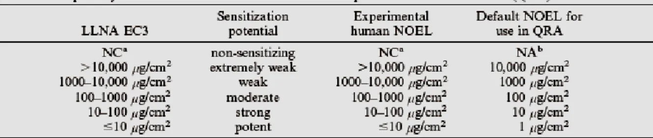
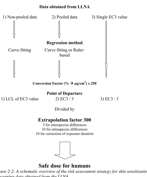
Data obtained from the LLNA are better suited to assign skin sensitizers into specific potency categories. Unlike the guinea pig assays the LLNA focuses on induction of sensitization only. Furthermore the LLNA provide objective and quantitative endpoints which can be used to obtain a dose-response relationship. As mentioned above a substance is a positive responder when a three-fold increase in LNC proliferation is observed compared to the control group. The concentration of the test substance, expressed as weight percentage in vehicle, at SI = 3 is the EC3 value. The lower the EC3 value, the more potent the substance as is displayed in an EU document (see also EC, 2003; Basketter et al., 2005).
As the guinea pig assays did not provide useful data for quantitative risk assessment (QRA); risk assessors were unable to conduct QRA for skin sensitising chemicals. The LLNA, however, does provide quantitative data which can be used in QRA. Up until now QRA is not performed for skin sensitizers. Performing a QRA for skin sensitising substances is desired, because it adds quantitative value to a characterisation of risk.
2.3 Approaches for quantitative risk assessment
2.3.1 Determining point of departure
As mentioned before, the GPMT and/or BA are not suited for QRA, because no quantitative data is provided. For this reason research was conducted to use the LLNA as foundation for QRA, because it can provide quantitative data. The endpoint measured in the LLNA is the LNC proliferation which directly links to the dose administered. Because multiple dose groups are