the infectivity of non-growing Campylobacter
jejuni.
L. Verhoeff-Bakkenes, F.M. van Leusden, R. de Jonge
This investigation has been performed by order and for the account of the Directory Board of RIVM, within the framework of project 251825, Quantitative safety aspects of exposure to
Campylobacter jejuni.
Abstract
Campylobacter (C.) jejuni is a causative agent of gastro-enteritis. Growth of this Gram
negative bacterium is restricted to a limited number of environmental conditions. Under conditions where growth of C. jejuni is not possible, e.g. at temperatures below 30oC,
C. jejuni can remain viable for a certain period of time, but its culturability decreases. In this
study we examined the effect of this loss of culturability on the infectivity in cell-lines. We hypothesised that if non-culturable cells affect the process of infection then the infectivity per culturable cell used in the infection assay is not constant, whereas if only culturable cell are infectious then the infectivity per culturable cell is. C. jejuni was stored for 40 days in nutrient poor and nutrient rich medium at three temperatures. During this period we
monitored its culturability, viability and infectivity. A decrease in culturability in time was observed. At low temperature (4oC) and in nutrient poor medium cells remained culturable for longer periods than at higher temperatures (12oC, 25oC) or in nutrient rich medium. Non-culturable cells remained viable, as determined by tetrazolium chloride staining. The absolute level of adhesion and invasion showed a decrease in time. At low temperatures and in
nutrient poor medium cells retained their capability to adhere and invade longer than at higher temperatures or in nutrient rich medium. While the absolute level of adhesion and invasion decreased, the infectivity per culturable cell added seemed to increase during the first 5 days of storage at 4oC. This result confirmed our hypothesis that non-culturable C. jejuni can affect the process of infection. However, the result can also be explained by the
presence of a limited number of bindingsites for C. jejuni in our testmodel. This suggested existence of bindingsites has great importance for the research into the influence of non-culturable cells on the infectivity. If non-non-culturable cells are not infectious but can block bindingsites, then they can play a protective role by competing with the culturable cells for the limited number of bindingsites.
Contents
SUMMARY... 7
SAMENVATTING ... 9
1. INTRODUCTION... 11
2. MATERIALS AND METHODS ... 15
2.1 EXPERIMENTAL SET UP... 15
2.1.1 Culturing C. jejuni and Salmonella (S.) enteritidis... 15
2.1.2 Preparing Erlenmeyer flasks for storage experiments... 15
2.2 MEASURING CULTURABILITY, MORPHOLOGY AND VIABILITY... 15
2.2.1 Culturability... 15
2.2.2 Morphology ... 16
2.2.3 Viability and total cell number ... 16
2.3 MEASURING INFECTIVITY... 16
2.3.1 Caco-2 cell line, growth media and conditions ... 16
2.3.2 Adhesion/Invasion assay... 17
2.3.3 Interleukin-8 assay... 17
3. RESULTS ... 19
3.1 CULTURABILITY, MORPHOLOGY AND VIABILITY... 19
3.2 INFECTIVITY... 19
4. DISCUSSION ... 21
REFERENCES ... 27
Summary
Under non-growing conditions the culturability of Campylobacter (C.) jejuni decreases, but cultures seem to retain their viability. The importance of viable non-culturable cells in the contamination cycle is questioned. This study was conducted to examine whether or not these non-culturable cells of C. jejuni are infective, and if the infectivity depends upon the
environmental conditions under which the non-culturable cells were formed.
During 40 days the culturability, viability and infectivity were determined of C. jejuni stored in two media at three temperatures. At low temperature (4oC) and in low nutrient medium (Phosphate Buffered Saline; PPBS) cells remained culturable for longer periods than at higher temperatures (12oC, 25oC) or in high nutrient medium (Brain Heart Infusion; BHI). Temperature did have a more profound effect than medium on survival. Non-culturable cells remained viable, as determined by tetrazolium chloride staining.
Infectivity was determined by the adhesion/invasion and Interleukine-8 (Il-8) assay. The Il-8 secretion was below the level of detection at all time points. Adhesion and invasion were only measurable when the number of cells in the suspension was over 105 per ml. The absolute level of adhesion and invasion showed a decrease in time. At low temperatures and in PPBS cells retained their capability to adhere to and to invade in Caco-2 cells for longer periods than at higher temperatures or in BHI.
The infectivity per culturable cell was calculated. Of an overnight culture 0.16 % adhered to and invaded in Caco-2 cells. This percentage increased during the first 5 days at 4oC and
decreased thereafter. This result confirms our hypothesis that non-culturable C. jejuni can be infectious. However, the result can also be explained by the presence of a limited number of binding sites for C. jejuni in our testmodel. As long as the initial number of culturable
C. jejuni exceeds the number of binding sites, an apparent increase in infectivity can be
observed during storage as the number of culturable C. jejuni decreases.
The suggested existence of bindingsites has great importance for the research into the influence of non-culturable cells on the infectivity. If non-culturable cells are not infective but can block the bindingsites, culturable cells have to compete with non-culturable cells for the bindingsites. Then a totally different effect of non-culturable cells would be shown, as non-culturable cells than would play a protecting rather then an infectious role.
In future research the infectivity assays have to be optimised. With the optimised assays the existence of bindingplaces will be studied. If the existence of bindingplaces can be proven, the influence of the presence of non-culturable cells on the infectivity will be studied further.
Samenvatting
Campylobacter jejuni is de meest frequente bacteriële veroorzaker van voedselinfecties.
Onder omstandigheden waarbij C. jejuni niet kan groeien, bijvoorbeeld bij temperaturen lager dan 30oC, verliest de bacterie zijn kweekbaarheid, maar lijkt de bacterie niet dood te gaan. In deze studie is onderzocht of een niet-kweekbare C. jejuni een infectie kan veroorzaken, en of de omstandigheden waaronder de kweekbaarheid verloren is gegaan van invloed zijn op een eventuele infectie. Gedurende 40 dagen zijn kweekbaarheid, levensvatbaarheid en vermogen om een infectie te veroorzaken bepaald van C. jejuni die was opgeslagen in twee verschillende media bij drie verschillende temperaturen. Wanneer opgeslagen in een nutriënt-arm medium bij lage temperatuur (4oC) bleek C. jejuni langer zijn kweekbaarheid te
behouden dan bij hogere temperaturen (12oC, 25oC) of in een rijk medium. Bacteriën bleven onder alle condities levensvatbaar. De mate van adhesie en invasie van de opgeslagen
cultures nam af in de tijd; bij lage temperatuur in buffer behielden de bacteriën het langst hun vermogen om aan darmepitheelcellen te hechten of om erin binnen te dringen.
Terwijl de infectiviteit van de totale cultuur afnam, nam de infectiviteit per kweekbare bacterie in die cultuur toe gedurende de eerste 5 dagen om daarna weer af te nemen. Dit resultaat suggereert dat een niet-kweekbare C. jejuni infectieus kan zijn. Echter, er kan ook een alternatieve verklaring zijn, namelijk dat er slechts een beperkt aantal specifieke bindingsplaatsen voor de hechting van C. jejuni aanwezig zijn. De eventuele aanwezigheid van specifieke bindingsplaatsen voor C. jejuni heeft mogelijk grote gevolgen voor onderzoek aan de invloed van niet-kweekbare cellen. Als niet-kweekbare cellen niet meer infectief zijn, maar wel kunnen hechten aan een bindingsplaats, dan kunnen zij met kweekbare cellen concurreren om een bindingsplaats. Dit zou een geheel nieuw rol betekenen voor niet-kweekbare cellen: niet-niet-kweekbare cellen zouden dan de kans op een infectie verlagen!
1. Introduction
Campylobacter jejuni has been identified as the major cause of bacterial gastro-enteritis in
the Netherlands 1. C. jejuni is a Gram-negative, motile microorganism, of which the
morphology varies from spiral-shaped to coccoid. C. jejuni is primarily micro-aerophilic and grows well in environments containing 3-5% CO2 and 3-15% O2. C. jejuni grows within a
short temperature range, being unable to multiply at temperatures either above 45oC or below 30oC. Under non-growing conditions the culturability of C. jejuni decreases, often
accompanied by a transformation from the spiral form into a coccoid form 2, 3, 4.
Temperatures below 15oC 2, 3 and nutrient limitation 2, 4 give lower transformation rates from culturable to non-culturable cells.
All required growth conditions are present in the gastrointestinal tract of warm-blooded animals. Poultry is identified as one of the major sources of C. jejuni. Pigs and cattle are a source of C. coli, but have not been identified as important reservoirs for C. jejuni 5. However, strains found in poultry have not been found in the human population, and only few of the human isolates have also been detected in poultry 6. For this apparent
contradiction, various reasons can be thought of:
- non-culturable cells might play a role as a source of contamination in the contamination cycle 7-11.
- other sources and/or reservoirs exist, but have not been identified yet.
- both chickens and humans harbour a large population of genotypically different
campylobacters, but the dominating, and thus most likely isolated genotype from poultry differs from the dominating genotype in human patients 12-15.
- the genotype of Campylobacter is not a constant, but changes 12, 16-18.
This study was conducted to examine the infectivity of non-culturable cells of C. jejuni. For risk assessment it is very important to know if non-culturable cells are infective. In practice the exposure is often measured as the number of culturable C. jejuni in a product, but when non-culturable cells are infective the risk is largely underestimated. In literature many contradictory articles on the infectivity of non-culturable cells of C. jejuni can be found. Some authors showed that non-culturable cells were not infective in chicks, mice and human volunteers 19-22. Other authors demonstrated that non-culturable cells were infectious in chicks and mice 7-9. The inconsistency in literature about the infectivity of non-culturable
cells might be the result of:
- different methods by which non-culturability was determined. In some articles selective enrichment instead of non-selective enrichment was used 8-10. The selective agents will negatively influence the culturability, as sub-lethally injured cells are sensitive to these agents 23, 24.
- different conditions under which the non-culturable cells were formed. In all studies where non-culturable cells were infective, the non-culturable cells were formed at 4ºC 7-9.
In experiments using higher temperatures no infectivity was found 19, 21. These results suggest that culturable cells formed at low temperatures are distinct from non-culturable cells formed at higher temperatures. Interestingly, Hazeleger et al. 2 showed
diversities between non-culturable cells formed at various temperatures. Non-culturable cells formed at 4oC showed characteristics, including intracellular / extracellular
ATP-ratio and membrane fatty acid composition, comparable to culturable cells, whereas those of non-culturable cells formed at 25oC were clearly different.
- different strains used. Some authors did not find any infectivity of non-culturable cells formed at 4oC 20-22. This might be explained by strain differences, as Jones et al. 7
reported that the capability of non-culturable cells to infect mice differed between strains. - presence of very few culturable cells in suspensions supposed to contain only
non-culturables.
Our reasoning is as follows:
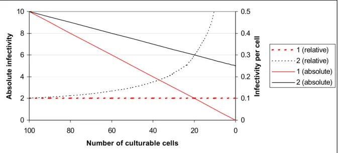
1. If non-culturable cells are not infective, a linear relationship exists between the number of culturable cells and the infectivity starting with a maximum infectivity at the maximum number of culturable cells down to no infectivity at zero culturable cells. The infectivity per culturable cell is a constant (Figure 1, case 1).
Figure 1. The relation between the infectivity and the number of culturable cells.
1. Non-culturable cells are not infective. 2. Non-culturable cells contribute to the infectivity. Relative means the infectivity per cell, absolute means the total number of infecting bacteria.
2. If non-culturable cells contribute to the infectivity, a linear relation can exist between the number of culturable cells and the infectivity, but at zero culturable cells
0 0.1 0.2 0.3 0.4 0.5 0 20 40 60 80 100
Number of culturable cells
Infectivity per cell
0 2 4 6 8 10 Absolute infectivity 1 (relative) 2 (relative) 1 (absolute) 2 (absolute)
infectivity still exists. The infectivity per number of culturable cells is not a constant, it increases, when the number of culturable cells decreases and the number of non-culturable cells increases (Figure 1, case 2).
We measured the decrease in culturability and infectivity of C. jejuni in time stored at various temperatures (4oC, 12oC and 25oC, respectively) and in two different media [brain heart infusion (BHI ) and phosphate buffered saline (PPBS), respectively]. These environmental conditions were based on the study of Hazeleger et al. 2 who reported dissimilarities between
non-culturable cells formed at various temperatures and in two different media. For our experiments C. jejuni C356 was chosen, because this strain showed the highest level of adhesion to and invasion in Caco-2 cells in a pilot study (unpublished result).
To be infectious cells at least have to be viable. To assess if there exist non-culturable cells that are still viable, the cellular respiration activity of the cells as measure for the viability was determined. The infectivity was determined by measuring the number of C. jejuni cells adhering to and/or invading in human epithelial Caco-2 cells. The culturability was measured by spread plating on Colombia agar plates with 5% lysed horse blood (CAB).
A drawback of the use of the adhesion and/or invasion assay is that infectivity is measured by determining the number of adhered and/or invaded culturable cells. If non-culturable cells can adhere or invade but remain non-culturable, no infectivity is measured by the
adhesion/invasion assay. Therefore, in addition the Il-8 assay was performed to measure the infectivity by determining the secretion of Il-8, a proinflammatory cytokine 29, by Caco-2 cells 30. The IL-8 assay is not dependent on whether or not cells are culturable.
2. Materials and Methods
2.1 Experimental set up
2.1.1 Culturing C. jejuni and Salmonella (S.) enteritidis
Bacterial strains were stored at –70ºC in Brain Heart Infusion broth (BHI, Beckton
Dickinson) plus 30% (v/v) glycerol in vials. For culturing C. jejuni C356, the content of one vial was thawed and put in an Erlenmeyer flask with 50 ml BHI. The Erlenmeyer flask was incubated while shaking in a custom made incubator (NuAire, Plymouth, Minnesota, USA) with a micro-aerobic atmosphere (10% O2, 5% CO2, 85% N2) at 37ºC for 24 hours. After
24 hours, the strain was subcultured in 100 ml BHI and incubated under the same conditions for 24 hours.
For culturing S. Enteritidis 97-198, the content of one vial was thawed and put in a tube with 10 ml BHI. The culture was used after overnight incubation at 37oC without shaking.
S. Enteritidis was chosen as positive control based on its good adherence and invasion
properties and IL-8 induction in Caco-2 cells (unpublished data).
2.1.2 Preparing Erlenmeyer flasks for storage experiments
The 24 hours old (stationary phase) cells were harvested by centrifugation at 3,000 x g and resuspended in either BHI (nutrient rich medium) or 50 mM potassium phosphate buffer containing 8.5 g/l NaCl (PPBS, nutrient poor medium). The cell suspensions were stored in Erlenmeyer flasks with a large opening in the dark, at various temperatures (4oC, 12oC and 25oC). Samples were taken after 5, 11, 17, 25, 32 and 40 days. For each sampling point a new set of Erlenmeyer flasks was used. All the experiments were done in duplicate.
2.2 Measuring culturability, morphology and viability
2.2.1 Culturability
Plate counts were performed by spread plating 0.1 ml of appropriate decimal dilutions of cell suspensions on Colombia Agar Base with 5% (v/v) lysed defibrinated horse blood (CAB). The plates were incubated micro-aerobically in a jar using BBL® Campypak (Becton Dickinson) at 37ºC for 72 hours.
2.2.2 Morphology
For morphological analysis, the cells were stained with acridine orange (Molecular Probes Europe BV, Leiden, the Netherlands) according to a modified technique of Hobbie et al. 25. Of a 1% acridine orange in distilled water, 100 µl was added to 1.0 ml of a bacterial
suspension. After incubation for 5 minutes in the dark, the morphology of the stained cells was monitored under a Zeiss epifluorescence microscope.
2.2.3 Viability and total cell number
The C. jejuni cells were stained with 5-cyano-2,3-ditolyl tetrazolium chloride (CTC,
Polysciences, Warrington, USA)) and 4’,6-diamino-2-phenylindole (DAPI, Sigma Chemical Co., St Louis, USA) as described by Cappelier et al. 26. CTC is an indicator of cellular respiratory activity. During respiration CTC is reduced into an insoluble red fluorescent CTC formazan salt, which accumulates intracellularly. DAPI stains DNA of intact cells and was used to determine total cell number.
To 0.5 ml of a bacterial suspension, 0.5 ml of BHI and 100 µl of a 0.04 g l-1 pyruvic acid solution (Sigma Chemical Co.) were added to stimulate cellular respiration. CTC was added to a final concentration of 5 mM. The mixture was incubated for 4 hours at 37ºC in a
microaerobic atmosphere. Cells were subsequently harvested by filtration through an isopore polycarbonate black membrane filter (0.2 µm pore size, 25 mm in diameter; Millipore, Ireland) and covered with a 5 µg ml-1 DAPI solution for 5 minutes. Finally, stains were removed by filtration. After washing the filter two times with 1 ml distilled water, the filter was air-dried and mounted in non-fluorescent immersion oil (Olympus, Japan) where after a coverslip was added.The number of respiring cells and the total number of cells were counted under a Zeiss epifluorescence microscope with a 405 nm excitation filter and a 455 nm dichromic mirror allowing simultaneous visualisation of both dyes.
2.3 Measuring infectivity
2.3.1 Caco-2 cell line, growth media and conditions
Caco-2 cells, human colon adenocarcinoma cells isolated from a primary colonic tumour in a 72-year-old Caucasian male were obtained from the American Type Culture Collection (Caco-2, ATCC HTB-37). In the experiments passage 30 to 45 were used.
We used Dulbecco’s Modified Eagle Medium 25 mM HEPES, containing 4500 mg l-1 D-glucose but no sodiumpyruvate (DMEM, GibcoBRL, Life Technologies Ltd, Paisley, Scotland) supplemented with 10% heat inactivated (30 minutes at 60ºC) fetal calf serum (FCS, Integro b.v., Zaandam, the Netherlands), 0.1% MEM non-essential amino-acids (Gibco), 6 mML-glutamine (Gibco) and 50 µg ml-1 gentamycin (Gibco) [DMEM10%].
Cells were stored at –135ºC in DMEM 10%, plus 10% (v/v) DMSO (Sigma Chemical Co., St Louis, USA). After quickly thawing in a waterbath at 37oC, the content of one vial (106 cells) was put in a culture-flask with 10 ml DMEM 10%. The cells were grown in a
CO2 5% (v/v) incubator at 37ºC. For experimental assays, confluent cultures were harvested
by mild trypsinisation and seeded into 12-wells tissue culture plates (Costar, Corning Costar Europe, Badhoevedorp, the Netherlands) at 160 000 cells/well and incubated in a CO2 5%
(v/v) incubator at 37ºC. Medium was changed 3 times a week and plates were used at 11-13 days post-confluence.
2.3.2 Adhesion/Invasion assay
The adhesion / invasion assay as developed in a former study was used 27. Briefly, 12 to 13 day old Caco-2 cells in the 12-well plates were inoculated with 40 µl bacterial suspension. The bacteria were allowed to adhere to and invade in the cells for 3 hours in a CO2 5% (v/v)
incubator at 37ºC.
To study adhesion and invasion, after incubation, the monolayers were rinsed 3 times with TCM and the cells were lysed with 1 ml 1% (v/v) Triton-x100 (Merck, Amsterdam, the Netherlands) in distilled water.
To study invasion, after incubation, the bacterial suspension was replaced by TCM containing 300 µg ml-1 gentamycin. After incubation for 1 hour, the monolayers were rinsed 3 times with TCM and the cells were lysed with 1 ml 1% (v/v) Triton-x100 in distilled water.
Adhesion/invasion and invasion alone were determined by plating serial dilutions of lysed Caco-2 cell suspensions on CAB and counting the resulting colony forming units (cfu), after 72 hours incubation at 37ºC under micro-aerobic conditions.
S. enteritidis 97-198 was used as positive control. The assays were performed in triplicate.
2.3.3 Interleukin-8 assay
The Il-8 assay as developed in a former study was used 27. Briefly, 12 to 13 day old Caco-2 cells in 12-well plates were inoculated with 40 µl bacterial suspension. The bacteria were allowed to adhere to and invade in the cells for 3 hours in a CO2 5% (v/v) incubator at 37ºC.
After incubation, the monolayers were rinsed 3 times with TCM. Finally, TCM with 50 µg/ml gentamycin was added to the cells, followed by incubation for 24 hours in a CO2 5% (v/v) incubator at 37ºC. After incubation, supernatants were collected and stored at
–70ºC to be analysed later. Il-8 concentrations were determined using IL-8 ELISA essentially according to Garssen et al. 28. Briefly, 96-well plates (NUNC-Immuno Plate, Roskilde, Denmark) were coated with 1 µg/ml antihuman IL-8 (Biosource, Nivelles, Belgium) in coating buffer (0.04 M carbonate buffer, pH 9.6). After 24 hours incubation at 4oC, the plates were incubated in blocking buffer [1% Bovine Serum Albumin (BSA, Sigma, Axel, the
Netherlands) plus 0.05% Tween-20 (Merck) in PBS] 24 hours at 4oC and washed in PBS plus 0.05% Tween-20. Recombinant human IL-8 (Medgenix diagnostics SA, Fleurus, Belgium) was used as a standard. Standard as well as serial dilutions of culture supernatant and
0.2 µg ml-1 biotinylated antihuman IL-8 (Medgenix diagnostics SA) was added to the plates. After incubation for 2 hours at 21oC, plates were washed and 0.1 µg ml-1 poly horseradish peroxidase-labeled streptavidin (Central Laboratory of the Blood Transfusion Service, Amsterdam, the Netherlands) was added. After incubation for 30 minutes at 21oC, plates were washed again and 50 µl 0.1 mg ml-1 tetra methyl benzidine (Sigma Chemical Co, St. Louis, MO) plus 0.006% H2O2 in 0.1M Na-acetate, pH 5.5 was added. The
colour-reaction was stopped after 5 minutes by adding 50 µl 2N H2SO4 (Merck). Plates were read at
450 nm using a Titertek Multiskan MCC/340 ELISA reader.
Caco-2 cells without bacteria and bacteria without Caco-2 cells were used as negative controls, Caco-2 cells with S. Enteritidis 97-198 were used as positive control. The assays were performed in triplicate.
3. Results
3.1 Culturability, morphology and viability
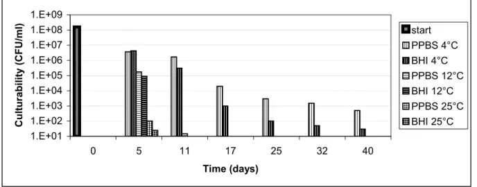
C. jejuni C356 resuspended in PPBS and BHI and stored at different temperatures showed a
decrease in culturability in time (Figure 2). The suspensions incubated at 4ºC remained culturable for over 40 days, while the suspensions incubated at 12ºC and 25ºC were below the detection limit of culturability in respectively 17 and 11 days. Besides the temperature effect a medium effect was seen. At all time points the suspensions incubated in PPBS showed a higher number of culturable cells than the cells stored in BHI.
The decrease in culturability did not have an effect on the morphology or viability. The shape of C. jejuni C356 cells was from the beginning rod-shaped and did not change in time. The viability, measured as number of CTC-stained cells, was at all time-points ± 95%. The number of cells stained by DAPI diminished slightly in time (not shown).
Figure 2. Culturability of C. jejuni C356 (CFU ml-1) kept in two media (PPBS or BHI) at various temperatures (4°C, 12°C or 25°C)
Both PPBS and BHI were inoculated from the same start culture.
3.2 Infectivity
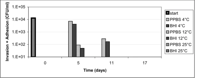
Invasion plus adhesion and invasion of Caco-2 cells by C. jejuni C356 stored in PPBS and BHI and incubated at different temperatures were measured (Figure 3 and 4). The results showed a decrease in adhesion/invasion at longer storage times. Cells incubated at 4ºC retained their ability to adhere and invade Caco-2 cells for 11 days, while cells incubated at 12ºC and 25ºC were below the detection limit of adhesion/invasion in 11 and 5 days, respectively. No clear medium effect was seen.
1.E+01 1.E+02 1.E+03 1.E+04 1.E+05 1.E+06 1.E+07 1.E+08 1.E+09 0 5 11 17 25 32 40 Time (days) Culturability (CFU/ml) start PPBS 4°C BHI 4°C PPBS 12°C BHI 12°C PPBS 25°C BHI 25°C
Figure 3. Invasion plus adhesion of Caco-2 cells by C. jejuni C356 kept in two media (PPBS or BHI) at various temperatures (4°C, 12°C or 25°C)
Figure 4. Invasion of Caco-2 cells by C. jejuni C356 kept in two media (PPBS or BHI) at various temperatures (4°C, 12°C or 25°C)
No Il-8 response was measured after exposure of the Caco-2 cells to C. jejuni C356, the secreted Il-8 was around the detection limit of 10 pg ml-1.In response to exposure to
S. enteritidis 97-198, the positive control, the Caco-2 cells secreted 70 pg Il-8 ml-1(not shown). 1.E+01 1.E+02 1.E+03 1.E+04 1.E+05 0 5 11 17 Time (days)
Invasion + Adhesion (CFU/ml)
start PPBS 4°C BHI 4°C PPBS 12°C BHI 12°C PPBS 25°C BHI 25°C 1.E+01 1.E+02 1.E+03 1.E+04 1.E+05 0 5 11 17 Time (days) Invasion (CFU/ml) start PPBS 4°C BHI 4°C PPBS 12°C BHI 12°C PPBS 25°C BHI 25°C
4. Discussion
This study was undertaken to find out whether or not non-culturable cells can be infective, and if so, does the infectivity depend on the environmental conditions under which the non-culturable cells were formed.
To determine the relation between the infectivity and the number of culturable cells, the culturability and infectivity, during storage under different environmental conditions, were measured. A decrease in culturability in time was detected, which mostly depended on the temperature and slightly on the medium used. At low temperatures cells remained culturable for longer periods than at higher temperatures. A transition from a spiral to a coccoid shape, often accompanying the decrease in culturability 2-4, 21, was not observed. In our experiments most non-culturable C. jejuni cells (± 95%) showed cellular respiration, as described
before 4,26, and thus were viable. Using CTC-staining as a measure for viability, this result confirmed the existence of non-culturable cells that were still viable and might be infectious. We hypothesized that if the number of culturable cells decreases during storage, the
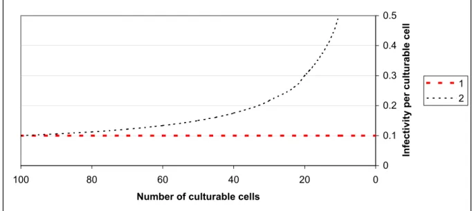
infectivity per culturable cell: 1.) will remain constant if the non-culturable cells are not infective; or 2.) will increase if the non-culturable cells contribute to the infectivity. This is shown in figure 5.
Figure 5. The hypothesized relations between the infectivity and the number of culturable cells.
1. Non-culturable cells are not infective. 2. Non-culturable cells contribute to the infectivity
During measuring the infectivity by the adhesion/invasion assay and the Il-8 assay, we encountered two problems:
- only at a limited number of time points the infectivity could be measured by the adhesion/invasion assay. When the culturability of the suspension was below
0 0.1 0.2 0.3 0.4 0.5 0 20 40 60 80 100
Number of culturable cells
Infectivity per culturable cell
1 2
105 CFU/ml, giving an infection dose below 10-2 culturable C. jejuni cells / Caco-2 cell, no adhesion/invasion could be detected.
- the adhesion/invasion assay only detects culturable cells. If non-culturable cells could adhere or invade but remain non-culturable, they will not be detected by this assay. The Il-8 assay, which does not depend on culturability, did not show sign of infectivity. No Il-8 was produced by Caco-2 cells after infection with C. jejuni C356.
The limited number of time points at which adhesion and invasion could be measured, showed a decrease in absolute adhesion and invasion in time. The decrease in infectivity depended mostly on the temperature and less on the medium used. At low temperatures cells retained their capability to adhere to and to invade in Caco-2 cells for longer periods than at higher temperatures.
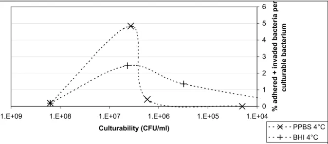
The infectivity per culturable cell was calculated for the few time points at which we measured infectivity by the adhesion/invasion assay (Figure 6 and 7).
Of an overnight culture 0.2 % adhered and invaded. This percentage increased to 4.8 % and 2.4 % after 5 days storage at 4°C in PPBS and BHI, respectively. Finally, the percentages decreased to 1.4 and 0.4% after 11 days and no adhesion and invasion was measurable after 17 days. The same trend was seen for invasion only (Figure 7).
Figure 6. The percentage of invading plus adhering bacteria relative to the number of culturable bacteria during storage at 4°C in PPBS or BHI
0 1 2 3 4 5 6 1.E+04 1.E+05 1.E+06 1.E+07 1.E+08 1.E+09 Culturability (CFU/ml)
% adhered + invaded bacteria pe
r
culturable bacterium
PPBS 4°C BHI 4°C
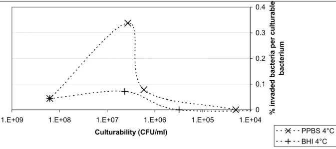
Figure 7. The percentage of invading bacteria relative to the number of culturable bacteria during storage at 4°C in PPBS or BHI
The decrease in culturability and concomitant rise in infectivity per bacterium indicated that non-culturable C. jejuni were infective. An increase in the efficiency at lower doses was shown by Hu and Kopecko31. They found the highest invasion efficiency of INT-cells by
C. jejuni 81-176 (3.5%) at 0.02 bacteria per epithelial cell, and a decrease in the efficiency at
higher doses. Their results suggest the existence of a maximum level of adhesion and invasion, or a limited number of bindingsites per Caco-2 cell for C. jejuni to adhere and invade.
At the start (t=0) of our experiments the number of culturable C. jejuni cells per Caco-2 cell was 40, while at t=5 the number of culturable C. jejuni cells per Caco-2 cell varied from 2.25x10⁻³ (BHI 12oC) to 1.1 (BHI 4oC). The maximum adhesion/invasion-efficiency (4.8%) was found in PPBS after 5 days at 4oC with an infection dose of ± 0.9 C. jejuni C356 per Caco-2 cell, which is higher than reported by Hu and Kopecko31. The use of different cell lines might explain this difference.
The existence of a limited number of bindingsites has great importance for the research into the influence of non-culturable cells on the infectivity. Until now, we only had two options:
1. the non-culturable cells are not infective.
2. the non-culturable cells can contribute to the infectivity. Now a third option arises:
3. the non-culturable cells are not infective, but can block a bindingsite. In that case culturable cells have to compete for a bindingsite with the non-culturable cells, and this would result in lower levels of infectivity. This third option is visualised in Figure 8 as line 3. 0 0.1 0.2 0.3 0.4 1.E+04 1.E+05 1.E+06 1.E+07 1.E+08 1.E+09 Culturability (CFU/ml)
% invaded bacteria per culturable
bacterium
PPBS 4°C BHI 4°C
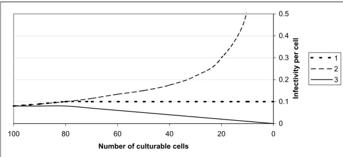
Figure 8. The relation between the infectivity and the number of culturable cells.
1. Non-culturable cells are not infective. 2. Non-culturable cells contribute to the infectivity. 3. Non-culturable cells are not infective, but can block a part of the limited number of bindingsites.
Figure 8, line 1 shows the relation between the culturability and the infectivity if non-culturable cells are not infective and do not block bindingsites. At first when the number of culturable cells is exceeding the number of bindingplaces the infectivity per cell will rise. Thereafter the infectivity per cell will be constant.
Figure 8, line 2 shows the relation between the culturability and the infectivity if non-culturable cells are infectious. At first when the number of infectious cells (non-culturable and non-culturable) is exceeding the number of bindingplaces the infectivity per cell will rise. Thereafter, the number of culturable cells diminishes, but as non-culturable cells contribute to an infection, the infectivity per culturable bacterium increases.
Line 3 in F igure 8 shows the relation between culturability and infectivity when non-culturable cells are not infectious, but block a part of the available bindingsites. At t=0 when only culturable cells are present, the infectivity is the highest. Thereafter the percentage of non-culturable cells increases. The culturable cells have to compete for binding sites to an increasing number of non-culturable cells, resulting in a decrease in infectivity per cell. The experimental data were too few to determine whether or not non-culturable cells did contribute to infectivity. The data did not allow determination of the maximum level of absolute adhesion and invasion and the subsequent trend after this maximum due to the high dose of C. jejuni required for a measurable response.
The results presented in this report however do not exclude a role for non-culturable C. jejuni in the infection process. In the non-culturable state cells might remain infectious for a certain period of time. Or, as non-culturable cell, it might lose its capacity to infect a host. If an infection with C. jejuni occurs after binding to a specific C. jejuni bindingsite and if non-culturable cells retain their capacity to bind to such sites, then the presence of non-non-culturable
C. jejuni reduces the probability of infection with C. jejuni.
0 0.1 0.2 0.3 0.4 0.5 0 20 40 60 80 100
Number of culturable cells
Infectivity per cell
1 2 3
Quantitative exposure data on C. jejuni obtained by direct culture techniques therefore will underestimate the real situation in case of non-culturables appear to be infectious. Using molecular techniques, the detection of C. jejuni DNA might overestimate the exposure to
C. jejuni if non-culturables are non-infective. Obviously, the role of non-culturable C. jejuni
References
1. Wit MAS de, Koopmans MPG, Kortbeek LM, Leeuwen WJ van, Vinjé J, Duynhoven YTPH van. Interim report of a study on gastroenteritis in sentinel practices in the Netherlands (NIVEL) 1996-1999. Results of the first two years. Bilthoven, 1999. (RIVM-report 216852003).
2. Hazeleger WC, Janse JD, Koenraad PM, Beumer RR, Rombouts FM, Abee T. Temperature-dependent membrane fatty acid and cell physiology changes in coccoid forms of Campylobacter jejuni. Appl. Environ. Microbiol. 1995; 61(7):2713-9.
3. Rollins DM, Colwell RR. Viable but nonculturable stage of Campylobacter jejuni and its role in survival in the natural aquatic environment. Appl.Environ. Microbiol. 1986; 52(3):531-8.
4. Boucher SN, Slater ER, Chamberlain AH, Adams MR. Production and viability of coccoid forms of Campylobacter jejuni. J. Appl. Bacteriol. 1994; 77(3):303-7. 5. Tauxe RV. Epidemiology of Campylobacter jejuni infections in the United States and other industrialized nations. In: Nachamkin I, Blaser MJ, Tompkins LS, editors. Washington, D.C.: ASM Press. 1992. P. 9-19.
6. Duim B, Wassenaar TM, Rigter A, Wagenaar J. High-resolution genotyping of
Campylobacter strains isolated from poultry and humans with amplified
fragment length polymorphism fingerprinting. Appl. Environ. Microbiol. 1999; 65(6):2369-75.
7. Jones DM, Sutcliffe EM, Curry A. Recovery of viable but non-culturable
Campylobacter jejuni. J. Gen. Microbiol. 1991; 137 ( Pt 10):2477-82.
8. Cappelier JM, Magras C, Jouve JL, Federighi M. Recovery of viable but non-culturable
Campylobacter jejuni cells in two animal models. Food Microbiol. 1999;
16(4):375-83.
9. Stern NJ, Jones DM, Wesley IV, Rollins DM. Colonization of chicks by non-culturable
Campylobacter spp. Lett. Appl. Microbiol. 1994; 18:333-6.
10. Pearson AD, Greenwood M, Healing TD et al. Colonization of broiler chickens by waterborne Campylobacter jejuni. Appl. Environ. Microbiol. 1993; 59(4):987-96.
11. Saha SK, Saha S, Sanyul SC. Recovery of injured Campylobacter jejuni cells after animal passage.Appl. Environ. Microbiol. 1991; 57(11):3388-9.
12. Thomas LM, Long KA, Good RT, Panaccio M, Widders PR.Genotypic diversity among
Campylobacter jejuni isolates in a commercial broiler flock. Appl. Environ.
Microbiol. 1997; 63(5):1874-7.
13. Petersen L, Nielsen EM, Engberg J, On SLW, Dietz HH. Comparison of genotypes and serotypes of Campylobacter jejuni isolated from danish wild mammals and birds and from broiler flocks and humans. Appl. Environ. Microbiol. 2001;
67(7):3115-21.
14. Steinbrueckner B, Ruberg F, Kist M. Bacterial genetic fingerprint: a reliable factor in the study of the epidemiology of human campylobacter enteritis? J. Clin. Microbiol. 2001; 39(11):4155-9.
15. Camarda A, Newell DG, Nasti R, Modugnoa G Di. Genotyping Campylobacter jejuni strains isolated from the gut and oviduct of laying hens. Avian Dis. 2000; 44(4):907-12.
16. Hanninen ML, Hakkinen M, Rautelin H. Stability of related human and chicken
Campylobacter jejuni genotypes after passage through chick intestine studied by
pulsed-field gel electrophoresis. Appl. Environ. Microbiol. 1999; 65(5):2272-5. 17. Wassenaar TM, Geilhausen B, Newell DG. Evidence of genomic instability in
Campylobacter jejuni isolated from poultry. Appl. Environ. Microbiol. 1998;
64(5):1816-21.
18. On SL, Nielsen EM, Engberg J, Madsen M. Validity of SmaI-defined genotypes of
Campylobacter jejuni examined by SalI, KpnI, and BamHI polymorphisms:
evidence of identical clones infecting humans, poultry, and cattle. Epidemiol. Infect. 1998; 120(3):231-7.
19. Beumer RR, Vries J de, Rombouts FM. Campylobacter jejuni non-culturable coccoid cells. Int. J. Food. Microbiol. 1992; 15(1-2):153-63.
20. Giessen AW van de, Heuvelman CJ, Abee T, Hazeleger WC. Experimental studies on the infectivity of non-culturable forms of Campylobacter spp. in chicks and mice. Epidemiol. Infect. 1996; 117(3):463-70.
21. Medema GJ, Schets FM, Giessen AW van de, Havelaar AH. Lack of colonization of 1 day old chicks by viable, non-culturable Campylobacter jejuni. J. Appl.
Bacteriol. 1992; 72(6):512-6.
22. Fearnley C, Ayling R, Cawthraw S, Newell DG. The formation of viable but nonculturable C. jejuni and their failure to colonise one-day-old chicks. In: Campylobacters, helicobacters, and other related organisms. New York: Plenum Press, 1996: 101-5.
23. Humphrey TJ. Techniques for the optimum recovery of cold injured Campylobacter
jejuni from milk or water. J. Appl. Bacteriol. 1986; 61(2):125-32.
24. Humphrey TJ. The influence of sub-lethal injury in Campylobacter jejuni on its subsequent resistence to antibiotics. J. Appl. Microbiol. 1984; 57:xvi. 25. Hobbie JE, Daley RJ, Jasper S. Use of nuclepore filters for counting bacteria by
fluorescence microscopy. Appl. Environ. Microbiol. 1977; 33(5):1225-8. 26. Cappelier JM, Lazaro B, Rossero A, Fernandez-Astorga A, Federighi M. Double
staining (CTC-DAPI) for detection and enumeration of viable but non-culturable
Campylobacter jejuni cells. Vet. Res. 1997; 28(6):547-55.
27. Verhoeff-Bakkenes L, Jonge R de, Leusden FM van. The use of Caco-2 cell lines for studying dose response relations of Campylobacter jejuni. Bilthoven, 2003. (RIVM report 251825001).
28. Garssen J, Briel RJ van de, Gruijl FR de, Wolvers DAW, Dijk M van, Fluitman A. UVB exposure-induced systemic modulation of Th1- and Th2-mediated immune responses. Immunol. 1999; 97(3):506-14. 228.
29. Jung HC, Eckmann L, Yang SK et al. A distinct array of proinflammatory cytokines is expressed in human colon epithelial cells in response to bacterial invasion. J. Clin. Invest. 1995; 95(1):55-65.
30. Eckmann L, Jung HC, Schurer Maly C, Panja A, Morzycka Wroblewska E, Kagnoff MF. Differential cytokine expression by human intestinal epithelial cell lines: regulated expression of interleukin-8. Gastroenterol. 1993; 105(6):1689-97. 31. Hu L, Kopecko DJ. Campylobacter jejuni 81-176 associates with microtubules and
dynein during invasion of human intestinal cells. Infect. Immun. 1999; 67(8):4171-82.
Appendix 1
Mailing list
1. Directeur-Generaal van het RIVM, Dr. M.J.W. Sprenger 2. Voorzitter van de Gezondheidsraad, Prof. Dr. A. Knottnerus 3. Dr. Ir. R.D.Woittiez, Directeur sector MEV,
4. Dr. Ir. R. Beumer, Wageningen Universiteit
5. Prof. Dr. Ir. M. Zwietering, Wageningen Universiteit 6. Ing. W.C. Hazeleger, Wageningen Universiteit 7. Dr. Ir. T. Abee, Wageningen Universiteit
8. Prof. Dr. F. Rombouts, Wageningen Universiteit 9. Dr. R. van Oostrom, Keuringsdienst van Waren 10. Drs. J. T. Jansen, Keuringsdienst van waren 11. Dr. Ir. P. in ’t Veld, Keuringsdienst van Waren 12. Dr. E. de Boer, Keuringsdienst van Waren 13. Dr. Ir. J.A. Wagenaar, ID Lelystad
14. Prof. Dr. J. van Putten, Universiteit van Utrecht 15. Dr. Ir. W. de Wit, Voedsel en Waren Autoriteit
16. Drs. A. G. Toorop-Bouma, Beleidsdirectie Voeding en Gezondheidsbescherming 17. Depot Nederlandse Publikaties en Nederlandse Bibliografie,
18. Prof. Dr. Ir. D. Kromhout, Directeur sector VCV, 19. Dr. Ir. A. H. Havelaar, MGB-RIVM,
20. Dr. Y. T. H. P. van Duijnhoven, CIE-RIVM 21. Dr. Ir. A. van der Giessen, MGB-RIVM 22. Auteurs
23. SBC/Communicatie
24. Bureau Rapportenregistratie (1 exemplaar) 25. Bibliotheek RIVM (1 exemplaar)
26-30. Bureau Rapportenbeheer 31-40. Reserve exemplaren