RIVM report 601516 007
Factsheets for the (eco)toxicological risk assessment strategy of the National Institute of Public Health and the Environment (RIVM)
Editors: R. Luttik and M.T.M. van Raaij
April 2001
This investigation has been performed by order and for the account of the Board of Directors of the National Institute of Public Health and the Environment, within the framework of project 601516, Risk Assessment of Substances: Science and Market.
RIVM, P.O. Box 1, 3720 BA Bilthoven, telephone: 31 - 30 - 274 91 11; telefax: 31 - 30 - 274 29 71 Authors
Chapter 1: W.M. Blom Chapter 2: M.T.M. van Raaij
Chapter 3: S.M.G.J. Pelgrom and M.T.M. van Raaij Chapter 4: T. Vermeire, M.. Pieters, M. Rennenand
P. Bos
Chapter 5: M.T.M. van Raaij Chapter 6: R. Luttik
Chapter 7: P.L.A. van Vlaardingen, J.A. de Knecht
and P.A.H. Janssen
Chapter 8: J.A. de Knecht, P.A.H. Janssen and
Abstract
Eight fact sheets describing risk assessment methods used at the Centre of Substances and Risk assessment (CSR) of the National Institute for Public Health and the Environment (RIVM) are presented here with the main aim of promoting greater transparency in the risk assessment methods used at the Institute in general and within the Centre in particular. The fact sheets, listed below, reflect a state-of-the-art approach; they are also meant to function as a platform for discussion.
1. Methemoglobine/Heinz bodies 2. Acetylcholinesterase inhibitors 3. Pheochromocytomas
4. Assessment factors for human health risk assessment 5. Delayed Neurotoxicity/NTE inhibition
6. Residues of plant protection products on food ingested by birds and mammals 7. Degradation of veterinary drugs in manure
8. Guideline for evaluating studies to determine excretion of veterinary drugs. The first five fact sheets are related to human risk assessment and the last three to
environmental issues. Remarks, omissions or additional information sent to the editors (first name) will be appreciated.
Preface
This report was written within the framework of the project ‘Risk Assessment of Substances: Science and Market’. The results as presented in this report have been discussed by members of the human and environmental peer review groups of the Centre of Substances and Risk assessment (CSR), and in some cases experts were consulted, all are acknowledged for their contribution. These members and experts are: A.J. Baars, P. van Beelen, R.B. Beems, B.J. Blaauboer, A.B.T.J. Boink, J. Janus, A.G.A.C. Knaap, A.M.A. van der Linden J.B.H.J. Linders, R. Luttik,W.C. Mennes, M.H.M.M. Montforts, M.T.M. van Raaij, A. Sips, W. Slob, G.J.A. Speijers, A. Verschoor, T.G. Vermeire, and P.W. Wester.
Contents
Samenvatting 9 Summary 11 Introduction 13 1 Methemoglobine/Heinz bodies 15 2 Acetylcholinesterase inhibitors 29 3 Pheochromocytomas 394 Assessment factors for human health risk assessment 49
5 Delayed Neurotoxicity/NTE-inhibition 71
6 Residues of plant protection products on food ingested by birds and mammals 83
7 Degradation of veterinary drugs in manure 95
8 Guideline for the evaluation of studies determining the excretion of veterinary drugs 103
Samenvatting
In dit rapport worden 8 factsheets gepresenteerd, die voor de risicoschatting van stoffen binnen het Centrum voor Stoffen en Risicobeoordeling (CSR) gehanteerd worden. De eerste 5 factsheets hebben betrekking op de humane risicoschatting en de overige 3 factsheets op het milieu.
In de factsheet ‘Methemoglobine/Heinz bodies’ wordt weergeven wat
methemoglobinevorming en Heinz bodies zijn, welke factoren deze effecten beïnvloeden, welke effecten als ‘adverse’ beschouwd dienen te worden en wat de beoordelingsstrategie voor deze effecten is.
In de factsheet ‘Acetylcholinesterase inhibitors’ wordt vastgelegd welke effecten met betrekking tot AChE inhibitie als toxicologisch relevant worden beschouwd en bij welke verandering in AChE activiteit er sprake is van een ‘adverse’ effect.
In de factsheet ‘Pheochromocytomas’ wordt vastgelegd wat de toxicologische relevantie is van een stof-geïnduceerde toename in pheochromocytomas in bijniermerg voor beoordeling en risicoschatting voor de mens.
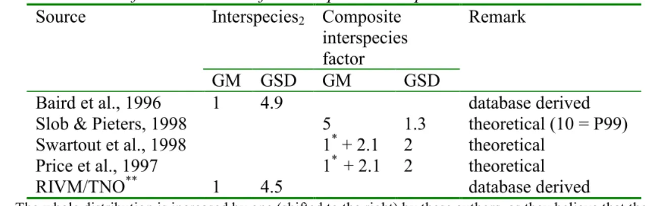
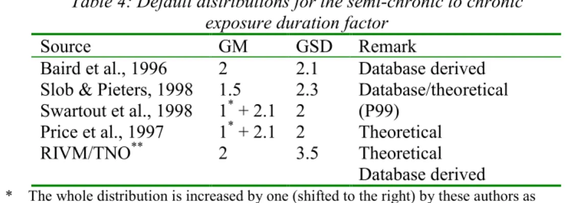
De factsheet ‘Assessment factors for human health risk assessment’ is gericht op de toepassing van probabilistische verdelingen van default assessment factoren in de risicobeoordeling van stoffen voor de mens (inter- en intraspeciesvariatie en de
correctiefactor voor de duur van de studies). De voorgestelde verdelingen zullen toegepast gaan worden in de risicobeoordeling van nieuwe en bestaande stoffen en bestrijdingsmid-delen voor de interpretatie van respectievelijk de ‘Margin of Safety’ (MOS) en de afleiding van de ADI.
De factsheet ‘Delayed Neurotoxicity/NTE-inhibition’ geeft een overzicht van wat op dit moment bekend is over OPIDPN, wat de significantie is van NTE-remming en welke rol NTE speelt bij het inschatten van mogelijk uitgestelde neurotoxische effecten van organo-fosfaatverbindingen.
De factsheet ‘Residues of plant protection products on food items for birds and
mammals’ geeft een samenvatting van recent gepubliceerd residu onderzoek, en geeft een
voorstel hoe deze nieuwe gegevens in de ecotoxicologische risicobeoordeling van bestrijdingsmiddelen te gebruiken.
In de factsheet ‘Degradation of veterinary drugs in manure’ wordt beschreven hoe de afbraakstudie van diergeneesmiddelen in mest beoordeeld moet worden binnen de toelatingsprocedure van diergeneesmiddelen.
In de factsheet ‘Guideline for the evaluation of studies determining the excretion of
veterinary drugs’ wordt beschreven hoe een excretiestudie van diergeneesmiddelen
Summary
This report presents 8 factsheets for the risk assessment methods used in the Centre for Substances and Risk assessment (CSR). The first 5 of these factsheets are dealing with issues related to human risk assessment and the other 3 with environmental issues.
The factsheet Methemoglobine/Heinz bodies describes the mechanism of methaemoglobin and Heinz body formation, the factors influencing these effects, those effects that must be labelled ‘adverse’, and the assessment strategy for these effects.
The factsheet Acetylcholinesterase inhibitors describes which effects associated with AChE inhibition are considered as toxicologically relevant and at what change in AChE activity an effect is labelled ‘adverse’.
The factsheet Pheochromocytomas aims to establish the toxicological significance of an agent-induced increase in adrenal medullar pheochromocytomas in experimental animals with respect to human risk assessment.
The factsheet Assessment factors for human health risk assessment is focussed on the Application of probabilistic distributions of default assessment factors in human health risk assessment (inter- and intraspecific variation and duration of the test). The proposed
distributions will be applied in risk assessment of new and existing substances and persticides for the interpretation of the Margin of Safety (MOS) and the derivation of the ADI,
respectively.
The factsheet Delayed Neurotoxicity/NTE-inhibition gives an overview of the current knowledge on OPIDPN, the role of NTE and the significance of NTE-inhibition data for the assessment of the delayed neurotoxic potential of organophosphorus compounds.
The factsheet Residues of plant protection products on food items for birds and
mammals a summary of recent published research on residue levels is presented and a
proposal is made how to use these new data in the ecotoxicological hazard/risk assessment for birds and mammals.
The factsheet Degradation of veterinary drugs in manure is a guideline for assessment of the reliability of the studies currently submitted for legislative purposes of veterinary drugs. The factsheet Guideline for the evaluation of studies determining the excretion of
veterinary drugs is meant as a preliminary guideline in which the requirements and criteria
for an excretion study are described and that can be used to decide whether or not the results are useful for the environmental risk assessment of veterinary drugs.
Introduction
One of the main tasks of the Centre for Substances and Risk assessment (CSR) of the National Institute of Public Health and the Environment (RIVM) is to assess the risk of compounds on public health and the environment. To carry out risk assessments it is of the highest importance that adequate and up-to-date risk assessment methods are available. Some of these methods are taken over (adopted) from other organisations, but many are, for a large part, developed within the RIVM. These risk assessment methods are not rigid procedures but can be adapted based on new/developing scientific information, possible triggered by
questions from policy makers or by developments in national or international organisations. For specific problems or gaps in the assessment of (eco)toxicological effects, ‘factsheets’ are written by employees of CSR in co-operation with experts. In these factsheets the assessment strategy of RIVM/CSR is described. After adoption of the factsheet by the advisory board and the head of the laboratory of CSR all employees of CSR have to follow the risk assessment method described in the factsheet.
In 1999 and 2000 eight factsheets were published ( 5 factsheets related to public health issues and 3 factheets related to environmental issues):
Factsheets concerning public health
Methemoglobine/Heinz bodies, Acetylcholinesterase inhibitors, Pheochromocytomas,
Safety factors,
Delayed Neurotoxicity/NTE-inhibition,
Factsheets concerning the environment
Residues of plant protection products on food items for birds and mammals, Degradation of veterinary drugs in manure,
Guidance document for summarising/interpreting of studies describing excretion of veterinary drugs by mammals.
We hope that by publishing these factsheets, the risk asessment methods followed by RIVM/CSR will become more transparent. The authors of each factsheet have tried to describe the state of the art of their subject. Remarks, omissions or supplementary
information will be appreciated and can be send to Robert.Luttik@RIVM.NL and will be passed on to the responsible authors.
1 Methemoglobine/Heinz bodies
Factsheet FSV-001/00 date 25-01-2000 Author:
W.M. Blom
1.1 Introduction and aim 16
1.2 Mechanism for the development of the effect, and background 16
1.3 Normal values and natural variation 17
1.4 Sensitivity species / groups 18
1.5 Factors that may influence the MetHb concentration 20
1.6 Assessment and strategy in the RIVM Centre for Substances and Risk Assessment (CSR) 20
1.7 Examples 23
1.8 References 23
Appendix 1 Compounds that can cause Methemoglobinemia 25
1.1 Introduction and aim
The standard toxicological evaluation of a chemical substance includes the measurement a number of standard haematological and biochemical parameters in animal experiments. Certain substances, however, such as nitrite, aromatic amines, and compounds with a strong oxidating capacity, may induce additional (non-standard) haematological effects, such as methaemoglobin and Heinz bodies (see appendix A for list with known substances). Certain characteristics, e.g. structural analogy, blue-coloring of extrimities (skin, nails), and associated signs at organ-level in animal studies, may be indicative of methaemoglobin formation and Heinz bodies. This factsheet describes the mechanism of methaemoglobin and Heinz body formation, the factors influencing these effects, the level of effect that must be labelled ‘adverse’, and the assessment strategy for these effects.
1.2 Mechanism for the development of the effect, and
background
Methaemoglobin
Haemoglobin (Hb) is an iron-containing, tetrameric protein, consisting of 4 protein chains forming the globin, and 4 identical haem groups. Each haem group is able to reversibly bind an O2-molecule to its ferrous group. However, a small proportion of the oxygen becomes
superoxide O2-, leaving one electron, so the iron is oxidized from the ferrous to ferric state, the
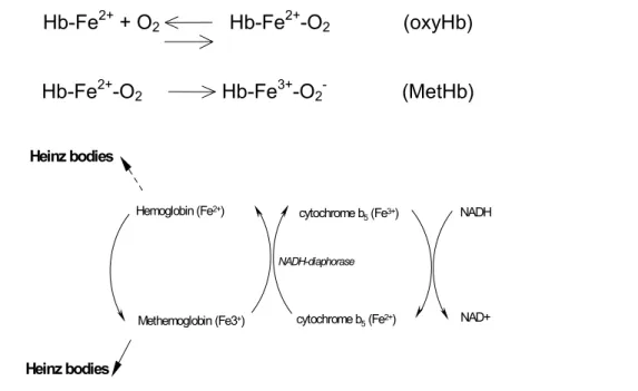
so-called methaemoglobin (MetHb, FeMethaemoglobin, HbFe3+; ferrihaemoglobin; see Fig. 1). Hb-Fe2+ + O2 Hb-Fe2+-O2 (oxyHb)
Hb-Fe2+-O2 Hb-Fe3+-O2- (MetHb)
Hemoglobin (Fe2+) Methemoglobin (Fe3+) NADH-diaphorase cytochrome b5 (Fe3+) cytochrome b5 (Fe2+) NADH NAD+ Heinz bodies Heinz bodies
Figure 1 Schematic representation of MetHb and Heinz bodies formation. Explanation in main body of the text.
Haemoglobin consequently loses its property of combining reversibly with oxygen, and furthermore the other O2-molecules are more strongly bound. This auto-oxidation occurs
cytochrome b5 reductase (also called NADH-diaphorase). This enzyme accounts for 60-95%
of the MetHb reduction.
Two other, quantitatively less important, reducing systems are cellular antioxidants, such as vitamin C, E and GSH (12-16%), and the NADPH-diaphorase (<5%) that is activated only by exogeneous electron carriers like methylene blue (Russell et al., 1982 ) (Calabrese, 1991 ). The ultimate MetHb concentration is the resultant of the equilibrium reaction between the (slow, spontaneous) formation of MetHb and the rate of the reducing systems (notably NADH-diaphorase). In humans this reducing capacity is 250 times larger than the rate at which MetHb is formed, so that under normal conditions MetHb in humans comprises less than 1% of the total Hb percentage (Jaffé, 1981 ).
MetHb levels will increase with increasing formation of MetHb, or through insufficiently protective reducing capacity (caused by impairment of the NADH diaphorase itself or through depletion of NADH-generating pathways in the erythrocytes). This may occur through oxidating compounds (toxic methaemoglobinaemia) or in rare genetic defects (Calabrese, 1991 ).
The following broad classification for clinical methaemoglobinaemia has been widely adopted, both in the literature and in handbooks: < 15% MetHb no effects; 15-40% fatigue, weakness, dizziness, headache, tachycardia; 40-70% hypoxia, unconsciousness, coma,
bradycardia, arrhythmias; > 70% fatal. This classification, however, is based on acute clinical problems (poisoning) and not on long-term exposure. Moreover, Coleman and Coleman (1996) point out that certain individuals will already show clinical signs at MetHb concentrations <10% (see section 6).
Heinz bodies
Heinz bodies are small or large precipitates in erythrocytes which develop after oxidation of Hb, or, to a lesser extent, from MetHb (through formation of haemichromes). Presumably, Heinz bodies do not necessarily develop under the influence of the same active metabolites that give rise to MetHb. But in both cases the protein in the erythrocyte cytoplasm undergoes a conformational change, denaturation, and next precipitation. This reduces the flexibility and functioning of the erythrocytes. The spleen usually removes small amounts of Heinz bodies from the blood. Heinz Bodies are demonstrable for a longer period in the blood than are increased MetHb concentrations. In general the presence of Heinz bodies nearly always points to MetHb formation, though indeed not in all cases (Smith, 1996; Russell et al., 1982).
1.3 Normal values and natural variation
MethaemoglobinThe MetHb concentration is determined by direct spectrophotometry (absorption peaks at 631 nm and 500 nm) and is expressed as percentage of the total Hb concentration. Another
(somewhat older) method makes use of cyanide: added to the sample it leads to the formation of cyanmethaemoglobin. Consequently, the 631 nm absorption peak will disappear and the 500 nm peak will shift to a broad peak at 542 nm (Evelyn and Malloy, 1938 ).
Normal values for MetHb levels have not been reported in handbooks or relevant journals. The MetHb blood levels in various control groups presented in Table 1 provide an indication of the values usually measured in experimental animals and humans.
In animal studies under evaluation, however, the MetHb concentrations in the control groups may differ from those mentioned in Table 1. For instance when animal measurements were done with equipment specifically developed to determine human MetHb: a difference in
spectrum will than result in higher values. For the assessment of effects in exposed groups the reference values in the study under consideration are ultimately more important than any historical reference values (Boink, personal commununication).
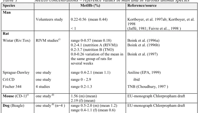
Table 1 MetHb concentrations – reference values in man and in various animal species a/b)
Species MetHb (%) Reference/source
Man
Volunteers study 0.22-0.56 (mean 0.44) < 1
Kortboyer, et al. 1997ab; Kortboyer, et al. 1998
(Jaffé, 1981; Faivre et al.., 1998 )
Rat
Wistar (Riv:Tox) RIVM studiesc) range 0-0.57 (mean 0.18)
0.2-4.1 (nutrition A (RIVM)) 0.2-3.7 (nutrition B (TNO) 0.0-0.26 variation of the mean in the same group of rats for several weeks
Boink et al. (1996a) Boink et al. (1996b) Boink et al. (1997)
Sprague-Dawley one study range 0.4-2.1 (mean 1.1) Aniline (EPA, 1999) Crl:CD one study range 0 - 2.9 ibid
Fischer 344 4 studies range 0.2-1.3 TNB (Choudhury, 1997 )
Mouse (CD-1)e) one study d) 1.56 (m) (mean)
2.19 (f) (mean)
EU-monograph Chlorpropham draft
Dog (Beagle) one study d) (n=4 ) range 0.5-2.0 (m) (mean 1.2)
range 0.4-1.1 (f) (mean 0.6)
EU-monograph Chlorpropham draft
a) Normal values derived from hematology handbooks available at RIVM/LPI, and various reference handbooks for laboratory animals (Loeb and Quimby, 1989; Lewi and Marsboom, 1981).
b) For other animal species, e.g. birds, see thesis Blaauboer (1978).
c) The values in controls did not exceed 0.5%, and values in the nitrite-exposed group ranged from 0.5-12% (Boink et al. 1996a, 1996b, 1997).
d) Heinz bodies were also determined in this study (0 in controls).
e) The mouse values in the Table are likely to be relatively high, but other reference levels are not available.
Heinz bodies
Heinz bodies are demonstrated using methyl-violet staining. In a leucocyte differential count, however, red blood cells will always be present, so that Heinz bodies may also be identified using routine Giemsa staining. Theoretically, the Heinz bodies level in blood of control animals is zero. (for more information see: (Russell et al., 1982), (Coleman and Coleman, 1996), (Chanarin, 1979), (Dabrow and Gabuzda, 1996).
1.4 Sensitivity species / groups
MethaemoglobinInterspecies differences have been investigated only in a limited number of studies, mainly in
in-vitro studies. Although these studies show that species differences are present, a distinct
ratio from animal species to humans cannot be established, since MetHb formaation in vivo is highly dependent on other factors, such as the metabolism of the substance, reducing
Various in vitro studies with erythrocytes, which particularly focussed on reducing capacity, yielded a rough dichotomy for the formation of MetHb: Rat/mous/rabbit/guinea pig/monkey are less sensitive to MetHb formation and generally show a more effective reduction of induced MetHb than do man/dog/cat. The cat is most sensitive to MetHb formation, primarily because of a different type of haemoglobin.
A number of old in vivo studies revealed a similar division as well. Administration of
acetanilid or acetophenetidin to various animal species resulted in slower MetHb formation in rat/rabbit/monkey (the latter two formed hardly any MetHb) than in man/dog/cat. However, it is not known whether for other substances a similar division exists. Following i.v.
administration of 4-dimethylaminophenol the reduction of induced MetHb in mice and rabbits was very rapid, whereas in dogs and cats it was much slower.
From this pattern it cannot be concluded that e.g. the dog is the most suitable experimental animal for MetHb effects, as several other factors are involved (e.g. the substance’s
metabolism) in addition to the reducing property of e.g. NADH diaphorase. For more information refer to (Calabrese, 1991), (Smith, 1996), and (Blaauboer, 1978).
As to intraspecies differences, the human population may be distinguished into these subpopulations:
- Faetuses and newborns: characterized by low NADH diaphorase (up to 50% less) and compared with adult Hb a more fluctuating faetal Hb (HbF). Both Hb and NADH diaphorase reach adult levels about 3-6 months after birth.
- Cytochrome b5 reductase deficiency: occurs very rarely. The individuals concerned
(hetero- and homozygotes) are extremely susceptible to toxic methaemoglobinaemia. - Elderly people: characterized by a more fluctuating Hb, which is more susceptible to
oxidation.
- Glucose-6 phosphate dehydrogenase deficiency: The individuals concerned show
defective NADH production, which may inhibit the reducing property of the NADH diaphorase (this deficiency was observed in American black males (11-13%),
Mediterranean Jews (11%), Greeks (1-2%), and Sardinians (1-8%); (Calabrese, 1991)).
- Deficiency of enzymes involved in the RBC energy balance.
- Mutations in Hb(THALASSAEMIA): owing to which it may become more susceptible to
oxidation to MetHb.
References: (Dabrow and Gabuzda, 1996; Griffin, 1997; Smith, 1996; Coleman and Coleman, 1996; Jaffé, 1981).
Proposing toxic threshold values for the whole population, the US-EPA uses an additional assessment factor to account for a difference in sensitivity between adults and newborns (see, for instance, Aniline: proposed acute exposure guideline levels (AEGLs)). RIVM holds the view that the commonly used factor 10 for intraspecies variation will suffice to account for the difference in sensitivity between adults and newborns. Hence, no additional assessment factor will be applied in determining a toxicological threshold value (e.g. an ADI) on the basis of MetHb effects.
It is not known whether similar intraspecies differences occur in laboratory animals, but from appendix B it appears that two Wistar rat strains (Riv:TOX and Wu:Harlan) for example, show a difference in MetHb formation during an 8 weeks’ exposure to nitrite.
Heinz bodies
Little is known about the formation of Heinz bodies, and Smith (1996) in his review points out that possible interspecies differences have not been systematically investigated. Yet he
provides a rough arrangement (omitting the rat!): the red blood cells of rabbit, monkey, chicken, and guinea pig are the least sensitive, followed by man, mouse, and dog, and finally the cat, which is most sensitive to the formation of Heinz bodies.
Strikingly, the mouse, in contrast to its sensitivity to MetHb formation, is here included in the group of sensitive species, together with man and dog. The review, however, does not provide a reason for this.
Little is known about sensitive groups within a species. But assuming that MetHb formation usually leads to formation of Heinz bodies, the above intraspecies differences are supposed to occur as well with respect to Heinz bodies formation in the human population (Smith, 1996 ).
1.5 Factors that may influence the MetHb concentration
A number of factors are known to influence the concentration of MetHb. These may be conditions during the experiment itself, but also after taking a blood sample.
• The timing of blood taking in a study is important, as the formation of MetHb is a relatively acute effect, and once formed MetHb is rapidly reduced. Therefore, when blood for a MetHb assay is collected at a time when the MetHb concentration in the blood is already falling or has reached baseline level, the effect is bound to be underestimated (see for instance study in EU-monograph Chlorpropham). See also the human volunteers experiment in appendix B, as well as the above studies in the literature in different animal species (see section 4). The maximum effect on MetHb strongly depends on the kinetics and metabolism of the substance.
• In addition, long-term exposure to a substance may cause adaptation of the reductase system. See the example in appendix B in which the maximum MetHb concentration in the blood of rats decreases during exposure to nitrite (total 8 weeks).
• Food intake may influence the MetHb concentration in the blood, for instance because the food itself will cause MetHb formation, or because it contains reducing components (GSH; oxidants) (Boink et al., 1996ab, and the list of substances that induce MetHb formation: Appendix A).
The conditions after blood sampling may also influence the percentage of measured MetHb (Sprokholt, 1987). For example, if red blood cells lyse, the released Hb is rapidly oxidized to MetHb. Energy depletion in the red blood cells may also result in higher MetHb level
because the NADH-diaphorase system is inhibited.
1.6 Assessment and strategy in the RIVM Centre for
Substances and Risk Assessment (CSR)
Until now, organisations like JMPR, WHO, or EPA have not published specific policies or guidelines for the assessment of substances that may cause methaemoglobin formation. There are two kinds of effects to be considered when assessing MetHb-inducing substances: clinical signs and haematological parameters.
Clinical signs
The first clinical signs indicating that a substance may induce MetHb-formation, are the brown/grey colour of the blood and the blue/grey appearance of the extremities (nails, nose, fingertips, skin). These may already occur at slightly raised MetHb concentrations (<6%) in experimental animals and humans. In a long-term study these effects might well be noticed in the initial phase only, and subsequently disappear due to adaptation.
These clinical signs could point at MetHb formation, but might also be the result of other mechanisms that lead to O2 deficiency (e.g. lung or cardiovascular effects). However, if these
signs are observed, the performance of MetHb measurements are certainly indicated.
MetHb concentration in the blood and related effects
General
Evaluation of MetHb measurements should always take notice of the experimental factors, such as the time points of measurement and treatment of the samples (see section 5). The accurate points of time are determined by the kinetics and metabolism data of the substance. Blood sampling at a relative late time point in a subacute (or semichronic) study may lead to underestimation of MetHb induction. However, at the same time this sampling time could have been too early to detect some related effects (see below) that will develop over time. In view of possible adaptation (particularly in the rat) the MetHb measurements should have been performed relatively early in the study. The optimum procedure consists of repeated measurements of the MetHb concentration during the first few days or weeks after administration of the substance.
The following general considerations play a pivotal role in the assessment strategy outlined below. If an effect on MetHb concentrations is observed, it is by definition considered an adverse effect because an increase in MetHb levels is possible only when the capacity of the reducing mechanisms is exceeded (notably NADH-diaphorase). The exposure in question has then already reached such a level that considerable energy will be spent on reduction of MetHb and production of reticulocytes. This is certainly considered an adverse effect on a long term basis, as it impedes the ‘biological fitness’ of the organism.
Increased MetHb concentration – when is it toxicologically relevant? The following rule of thumb applies for MetHb assessment:
Any significant increase in MetHb concentration compared to control level is in principle considered a toxicologically relevant (‘adverse’) effect if a dose-response relationship is present.
This could imply that also small, though significant, increases compared to control level are considered as adverse.
An increase in MetHb concentration which is not (yet) significant, is still considered a biologically relevant effect if a dose-response relationship is observed at the consecutive dosages. This dose at which the non-significant increase in MetHb is measured, is in principle considered as NOAEL in the study in question, unless other related effects have been
observed in the study in question or in other available studies at comparable dose(s) (see below). In the latter case a non-significant increase of MetHb concentrations may also be considered as toxicologically relevant.
Related effects
MetHb concentrations in exposed animals may occasionally show larger variation than those in control groups. Owing to a wide standard deviation, an increase of the MetHb
concentration at a certain dose might then turn out not to be statistically significant (see e.g. appendix B and Table 1, note c). A non-significant increase in MetHb may nevertheless be associated with other, related, effects. These MetHb-related effects in blood and organs are of importance as well in the absence of a dose-response relationship for effects on MetHb concentrations (see Table 2). This is why these related effects should always be taken into consideration.
In (sub-)chronic studies the whole blood differential cell count is generally more important than just the measured amount of MetHb, as the MetHb concentration may have dropped through adaptation. Prolonged exposure to MetHb-inducing compounds may bring about several adaptation-linked related effects, such as the presence of Heinz bodies in red blood cells and changes in the differential cell count indicative of anaemia: RBC↓, Hb↓, MCV↑, and reticulocytes↑. In addition, haematopoiesis in liver and spleen, hemosiderins (insoluble
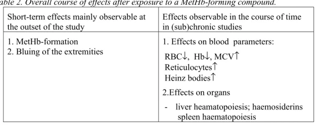
Table 2. Overall course of effects after exposure to a MetHb-forming compound.
Short-term effects mainly observable at the outset of the study
Effects observable in the course of time in (sub)chronic studies
1. MetHb-formation
2. Bluing of the extremities
1. Effects on blood parameters: RBC↓, Hb↓, MCV↑
Reticulocytes↑ Heinz bodies↑ 2.Effects on organs
- liver heamatopoiesis; haemosiderins spleen haematopoiesis
iron precipitates) in the liver, and possibly (in the beginning of the study) bluing of skin and/or nose are indicative of MetHb-formation as well.
The NOAEL in a study with a MetHb-inducing substance may, therefore, also be determined by effects on the above-mentioned parameters. The following ‘rule of thumb’ may be
applied:
The NOAEL in the study is based on a significant change in one of the
above-mentioned related parameters, provided that the next, higher doses result in a) a dose-effect relationship for the parameter in question, and b) the occurrence of minimally two other related effects.
Consequently, should the MetHb-formation have been determined incorrectly or not at all, and yet the effects listed in Table 2 are present, it is not always necessary to request for a new study in which MetHb-formation is measured. The overall picture of effects may then suffice to determine a NOAEL.
If there are indeed reasons to request for a new study, a short-term study lasting maximally 2-4 weeks, with repeated measurements of blood MetHb concentrations, will suffice in most cases.
1.7 Examples
Chlorpropham: Recommendation 06120B00, Draft dd. 28-12-1998. Authors: Apeldoorn ME van, Wouters MFA, Fouw JC de, et al. MetHb formation determined the NOAEL, and because MetHb was not established until the end of the 90-day study, a safety factor of 3X was applied to the ADI. See pp. 69-70.
1.8 References
Apeldoorn MEv, Wouters MFA, Fouw JCd, et al. Chloorpropham: CSR Advies rapport 06210B00, Bilthoven, The Netherlands: RIVM, 1998.
Blaauboer B. Formation and reduction of ferrihemoglobin in red cells. Effects of Phenylhydroxylamine and monoaminophenols in avian and mammalian erythrocytes. Utrecht: Thesis Rijksuniversiteit Utrecht, 1978.
Boink, A.B.T.J., Beekhof, P.K., Dormans, J.A.M.A., Speijers, G.J.A. (1996a) On the etiology of nitrite-induced hypertrophy of the zona glomerulosa of rats: I The possible role of
nitrate. Report 235802 003. RIVM, Bilthoven The Netherlands. 19p.
Boink, A.B.T.J., Beekhof, P.K., Dormans, J.A.M.A., Speijers, G.J.A. (1996b) On the etiology of nitrite-induced hypertrophy of the zona glomerulosa of rats: II The possible role of feed. Report 235802 004. RIVM, Bilthoven The Netherlands. 23p.
Boink, A.B.T.J., Beekhof, P.K., Dormans, J.A.M.A., Dortant, P.M. Speijers, G.J.A. (1997) A study on nitrite sensitivity in the rat : comparison between young adult rats and aged rats. Report 650050.001. RIVM, Bilthoven The Netherlands. 49p.
Calabrese EJ. Animal models for selected High-Risk groups - Heriditary blood disorders. Calabrese E, Ed. Principles of animal extrapolation. Chelsea, Michigan, USA: Lewis Publishers, Inc., 1991: 289-320.
Chanarin I. Haematological biochemistry. Brown SS, Mitchell FL and Young DS, Eds. Chemical diagnosis of disease. Amsterdam: Elsevier/North-Holland Biomedical Press, 1979: 853.
Choudhury H. Support Document for 1,3,5-trinitrobenzene (TNB) (Casno 99-35-4). Cincinnati, Ohio, USA.: United States (EPA), 1997.
Coleman MD and Coleman NA. Drug-induced Methaemoglobinaemia. Drug Safety 1996; 14(6):394-405.
Dabrow MB and Gabuzda TG. Nonimmune hemolysis and toxic methemoglobinemia. Bloom JC, Ed. Toxicology of the hematopoietic system, Volume 4 of Comprehensive Toxicology. Sipes IG,McQueen CA, Gandolfi AJ (Eds.). Vol. 4. Pergamon, 1996: 55-121.
EPA Environmental Protection Agency (1999) Aniline: Proposed acute exposure guideline levels (AEGLs). Public Draft. 36p
http://www.epa.gov/fedrgstr/EPA-TOX/1997/October/Day-30/aniline.pdf
Evelyn and Malloy. Microdetermination of oxy hemoglobin, methemoglobin and sulfhemoglobin in a single sample of blood. Journal of Biological Chemistry 1938; 126:655-62.
Faivre B, Menu P, Labrude P and Vigneron C. Hemoglobin autooxidation/oxidation mechanisms and methemoglobin prevention or reduction processes in the bloodstream. Literature review and outline of autooxidation reaction. Artificial Cells, Blood Substitutes, and Immobilization Biotechnology 1998; 26(1):17-26.
Griffin JP. Methaemoglobinaemia. Adverse Drug Reactions and Toxicological Reviews 1997; 16(1):45-63.
Jaffé ER. Methaemoglobinaemia. Clinics in Haematology 1981; 10(1):99-122.
Kortboyer, JM, Boink ABTJ, Zeilmaker, MJ, Slob, W, and Meulenbelt J. (1997a) The oral availability of sodium nitrite investigated in healthy adult volunteers. RIVM Report 235802 006, RIVM, Bilthovben, The Netherlands, 41p.
Kortboyer, JM, Olling, M, Zeilmaker, MJ, Slob, W, Boink ABTJ, Schothorst RC, Sips, AJAM and Meulenbelt J. (1997b) The oral availability of sodium nitrite investigated in healthy adult volunteers. RIVM Report 235802 007, RIVM, Bilthovben, The Netherlands, 94 p.
Kortboyer JM, Schothorst RC, Zeilmaker MJ and Meulenbelt J.(1998) Intravenous
administration of sodium nitrite to healthy volunteers: a single ascending dose study. RIVM Report 235802 011. RIVM, Bilthoven, The Netherlands, 65 p.
Lewi PJ and Marsboom, RP (1981) Toxicology reference data – Wistar rat. Body and organ weights , biochemical determinations, hematology and urinalysis, compiled at Janssen Pharmaceutica. Elsevier, North Holland Biomedical Press, Amsterdam, The Netherlands 358 p.
Loeb, WF and Quimby FW. (Ed) (1989) The clinical Chemistry of Laboratory animals. Pergamon Press, Oxford, Great Britain. 519p.
Russell NJ, Powell GM, Jones JG, Winterburn PJ and Basford JM. Red blood cells. Blood Biochemistry. 1985 reprint edition. Beckenham, Kent, UK: Croom Helm, 1982: 4-20. Smith RP. Toxic Responses of the Blood. Casarett and Doull’s Toxicology: The Basic
Science of Poisons. New York, USA: McGrawHill, 1996: 335-54.
Sprokholt, R. (1987) Quality control in bood gas chemistry.Thesis Universiteit Utrecht,The Netherlands.
Appendix 1
Compounds that can cause Methemoglobinemia
After Seger, 1992 Acetanilid Acetophenetidin Alloxans Alpha naphtylamine Aminophenols Ammonium nitrate Aniline and derivatesChloromethylaniline Paradichloraniline Paranitroaniline Aniline dyes Anilinoethanol Antipyrine Arsine
Benzene and derivates Dinitrobenzene Nitrobenzene Nitrosobenzene Benzocaine Chlorates Chloranilines Chlorobenzene Chloronitrobenzene Cobalt preparations Corning extract
Crayons, wax (red or orange) Dapsone
Diaminodiphenylsulfone Diesel fuel additives Dimethylamine Dimethylaniline Dinitrophenol Dinitrotoluene Hydrogen peroxide Hydroquinone Hydroxylacetanilid Hydroxylamine Inks Kiszka Lidocaine Menthol Meta-chloraniline Methylacetanilid Methylene blue Monochloroaniline Naphtylamines Nitrites and nitrates Amyl nitrite
Butyl/isobutyl nitrite Sodium nitrite
Nitrite and nitrate preservatives in meat Vegetables (carrots and spinach) in infants
Contaminated well water Nitrite salts used in industry Glyceryl trinitrate (nitroglycerin) Transdermal, sublingual, oral
nitrite/nitrate
Contaminants from aneasthesia cannisters with nitrous oxide Nitrogen oxide Nitrofurans Nitroglycerin Nitrophenol Ozone Parnaquine Para-aminopropiophenone Para-bromoaniline Para-chloraniline Para-toluidine Pentaerythritol tetranitrate Phenacetin Phenetidin Phenols Phenylazopyridine Phenylenediamine Phenylhydrazine Phenylhydroxylamine Phenytoin (Dilantin) Piperazine Plasmoquine Prilocaine Primaquine Propitocaine Pyridium Quinones Resorcinol
Shoe dye or polish Sodium nitroprusside Spinach
Sulfonamides Dapsone Prontosil Sulfanilamide Sulfapyridine Sulfathiazole Sulfones Tetranitromethane tetronal Tetralin Toluenediamine Toluidine Toluylhydroxylamine Trichlorocarbanilide (TCC) Trinitrotoluene Trional
Appendix 2
Factors that influence MetHb-levels
For the measurement of MetHb levels inblood, one should consider e.g. the time point of blood sampling. (see fig. 1), the possibility of adaptation (see fig. 2), and intraspecies differences. Such effects have been investigated at the RIVM within the framework of the nitrate/nitrite research. The acute methemoglobin formation was measured in a human volunteer study (RIVM report 235802 007, Kortboyer et al., 1998). The volunteers received nitrite intraveneously, after which MetHb-levels were measured in the blood for 10 hours. In figure 1, the MetHb-levels for 2 volunteers are depicted. It is shown that, after reaching a maximum level between 1-2 hours, MetHb levels decrease rapidly reaching control levels already after several hours. This indicates that measurements of MetHb levels several hours after a single treatment are useless. In another study, MetHb levels were determined after nitrite treatment in two different rat strains. Rats received drinking water containing nitrite for 8 weeks, and blood was sampled pre-exposure and than every two weeks. The study showed that nitrite treatment induced higher MetHb levels in Wu:Harlan Wistar rats compared to Riv:TOX Wistars.
In addition, repeated exposure of rats to nitrite resulted in a gradual adapatation in both rat strains.
Figure 1 MetHb-levels in intrvaveneous blood of human volunteers after exposure to nitrite.
Figure derived from Dr. ABTJ Boink; published in RIVM report 235802 007 (Kortboyer et al., 1998).
Figuur 2 Adaptation of MetHb-formation in 2 different Wistar rat strains.
Net effect (difference between test group and concurrent control (=KCL, 36 mmol.l-1) of exposition to nitrite (36 mmol.l-1) on the
methaemoglbin fraction of Riv:TOX (Ο) and Wu:Harlan Wistar ( ) rats during time (means ± s.d.). Uit: ‘Health aspects of nitrates and its metabolites (particularly nitrate)’ Proceedings International Workshop Bilthoven, The Netherlands 8-10 Nov 1994, p229-232.
2 Acetylcholinesterase inhibitors
Factsheet FSV-002/00 date 10-09-1999 Author:
M.T.M. van Raaij
2.1 Introduction and aim 30
2.2 Mechanism for the development of the effect, and background 30
2.3 Normal values and natural variation 31
2.4 Sensitive Species / Groups 32
2.5 Factors that may influence the measurements 32
2.6 Assessment and RIVM/CSR Strategy 33
2.7 Examples 36
2.1 Introduction and aim
Organophosporus esters (OPs) and carbamates are widely used insecticides. One of their major effects is inhibition of the enzyme acetylcholinesterase (AChE; EC 3.1.1.7). This enzyme is primarily located in the synapses of the somatic, autonomous, and central nervous systems, but also in erythrocytes and blood plasma. AChE is involved in the breakdown of the neurotransmitter acetylcholine, which diminishes or terminates the activation of
postsynaptic cholinergic receptors. Inhibition of AChE leads to acetylcholine-induced
overstimulation of the postsynaptic receptors, which in its turn results in so-called cholinergic toxicity or ‘cholinergic crisis’ [1,2,4]. Besides AChE inhibition in the central (CNS) or peripheral nervous system (PNS), OPs and carbamates can also inhibit AChE in the blood. This factsheet describes which effects associated with AChE inhibition are considered as toxicologically relevant and at what level of AChE inhibition is considered ‘adverse’.
2.2 Mechanism for the development of the effect, and
background
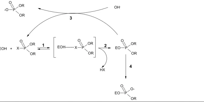
Although they both induce AChE inhibition, OPs and carbamates do not have quite the same mechanisms of action. OPs are analogues of the normal biological substrates of AChE. The various steps of the interaction between an OP and AChE are shown in Figure 1.
An OP reversibly binds to the hydroxyl group of a serine residue in the enzyme, resulting in a Michaelis-Menten complex (step 1). After separation of the residual group XH (step 2), the OP and AChE form a covalent bond (for acetylcholine the residual group is choline). The OP-enzyme complex may be reactivated (step 3). For the original substrate acetylcholine, step 3 will last a few microseconds only. However, for OPs the half-life times can be very long [2], resulting in long-term AChE inhibition. Some OPs, however, do not seem to
undergo any reactivation [2]. An additional complicating factor is ‘ageing’. This phenomenon refers to separation of one of the residual groups that are linked to the phosphorus through an oxygen atom (step 4). This irreversible reaction will inactivate the enzyme. The half-life times of ageing are 4 hours for dimethoxy-OPs, and about 10 hours for diethoxy-OPs. This implies that the rate of ageing (step 4) could far exceed that of reactivation (step 3), which makes the inhibition of AChE by OPs virtually irreversible [1,2,3]. Ageing is probably an essential step in the induction of OP-induced delayed neurotoxicity (OPIDN). This subject will be dealt with in a separate factsheet.
The behaviour of carbamates is different from that of OPs. They are hydrolized by AChE, but the enzyme-carbamate-complex has a long half-life time. This complex, unlike OPs, does not undergo ageing however, and AChE is thus reactivated according to step 3. Consequently, the AChE inhibition of carbamates is reversible (within a period lasting from minutes to hours). With regard to mechanisms of action refer to refs. for further information [1,2,3,4].
As mentioned, AChE is found particularly in the CNS and the PNS. The CNS is protected by the so-called ‘blood-brain-barrier’, preventing a number of AChE-inhibiting substances to reach their target site (the synapses) in the CNS. The PNS is surrounded by a perineurium, but this is less effective than the blood-brain-barrier, especially at the peripheral ganglia. Hence, exposure of peripheral nerves and ganglia may be occasionally higher than that of the CNS [3]. However, many organophosphorus esters pass the blood-brain-barrier relatively easily, with little or no difference in exposure between the CNS and the PNS [A. Moretto, personal communication].
P OR O OR X EOH + P OR O OR X EOH--- P OR O OR EO HX P O-O OR EO OH -P OR O OR -O 1 2 3 4
Fig. 1. Schematic representation of the interaction and enzymatic steps in the actions between an OP and AChE. (the various steps are explained in the main body of the text).
Both the erythrocytes and the blood plasma contain cholinesterases, which, in contrast to neural AChE, are not involved in cholinergic transmission. The enzyme in the erythrocytes is biochemically identical to that in the PNS, viz. acetylcholinesterase. The rate at which AChE is being resynthesized, is however much higher in the PNS than it is in erythrocytes [11,22]. Cholinesterase activity in plasma does not only comprise acetylcholinesterase but also butyrylcholinesterase (BuChE). In rat plasma the proportion of AChE and BuChE is 1:1, but in humans it is about 1:100 [2,5,11]. This means that plasma cholinesterase activity in humans almost exclusively relates to BuChE. This is a so-called ‘pseudo-cholinesterase’, which is also used as a biomarker. Most analytical methods determine total cholinesterase activity by measuring the conversion of acetylthiocholine in a colorimetric assay in which no actual differences between AChE and BuChE are established.
The physiological role of cholinesterase activity in erythrocytes and blood plasma is not known. From the available literature it appears that selective inhibition of erythrocyte or plasma ChE itself does not induce harmful effects [6]. Nevertheless, BuChE might be involved in the detoxification of other xenobiotics (e.g. cocaine), which suggests that inhibition of plasma ChE activity may indeed have physiological consequences [1].
2.3 Normal values and natural variation
In the toxicological literature effects on AChE activity are usually expressed as percentage inhibition relative to the concurrent control group, or to pre-exposure values in the exposed group. Absolute quantities of AChE activity are rarely reported in the public literature. Besides, as the reported data are largely influenced by the experimental methods applied (see also section 5), comparison of the various values obtained for AChE activity is less useful [see for absolute values e.g. refs. 4,5,6,7,8].
The intraspecies variation in AChE activity is dependent upon the target organ in which the activity is measured. In general, the brain shows less intraspecies variation of AChE activity than does the blood. The mean variation of AChE activity in the brain ranges from 10-15%, whereas in blood it ranges up to 20% [2,4,5,7,8,9].
The interspecies variation in total cholinesterase activity is characterized by both qualitative and quantitative differences between the various species. Blood ChE in particular shows great differences. Erythrocyte AChE activity in various species ascends in this order: cat << rat < rabbit, dog < horse < guinea pig < pig < cow < chimpanzee < human. The differences in erythrocyt AChE activity between rat and human could amount to a factor 5 to 10
[18,4,9,10]. Plasma ChE activity consists of AChE and BuChE (see section 2). These two enzymes show variable proportions in the various species [2,5,11]. As standard assays only measure total cholinesterase activity, they do not distinguish between AChE and BuChE. For a conclusion on interspecies variation in brain AChE activity, sufficient data were not available.
2.4 Sensitive Species / Groups
Although substantial species differences have been noted with respect to the sensitivity for OP-induced delayed neurotoxicity (OPIDN), we are currently not aware of specifically sensitive species and/or groups with regard to AChE inhibition that require a specific assessment procedure or should be considered a special risk group.
Nevertheless, there are some indications for differences in sensitivity between various species [21], between males and females (females more sensitive) [18], and for an increased
sensitivity in neonates [11,12,13,14,15,16,21].
However, the major differences in inter-individual sensitivity are probably caused by differences in metabolism and kinetics of the AChE inhibiting substances, notably in the detoxification routes [2].
2.5 Factors that may influence the measurements
There are several factors that influence the activity of AChE, such as age, sex, stress, endocrine status, and seasonal variation [4]. The ultimately measured AChE activity, however, is largely determined by experimental factors. The major factors influencing the actually measured/reported AChE levels are listed below.
• Timing of sampling. Especially when dealing with carbamates or after single doses of OPs, the time of sampling must be accurate in order to provide an optimal measurement of AChE inhibition (at the time of peak inhibition). Kinetic data or AChE activity measurements at different time points could be helpful in this respect [22].
• AChE inhibition might be underestimated if samples are not correctly treated and/or stored. Also when stored, samples may show, for instance, spontaneous reactivation of AChE. This is particularly a problem with carbamates. Samples should preferably be stored at a temperature of –60 or –80 °C [17,18,22].
• The assay conditions may exert a great influence on the results obtained through the so-called ‘Ellman-method’ (also available as standard kits): buffer composition, pH,
substrate concentration, temperature, sample dilution, and the measuring wavelength. For instance, the used wavelength can be increased from 412 to 480 nm in order to reduce the interference of haemoglobine (in the case of blood/RBC measurements) [17,18,22].
• According to Wilson et al. [17] it is essential to include tissue blanks in addition to substrate blanks, and to apply pre-incubation.
• Standard kits designed for measuring AChE activity in rat blood are not directly suitable for human samples [7].
The above indicates that the methods of measuring ChE should be carefully assessed,
especially in key studies. The latter implies that particularly in studies in which there is doubt about the analytical conduct, and in the study that yields the overall NOAEL (if based on cholinesterase inhibition), the analytical performance of the study should be carefully examined. References [4,7,8,17,18] provide detailed discussions and points for attention for such an analytical assessment.
2.6 Assessment and RIVM/CSR Strategy
In the past two decades the assessment of cholinesterase inhibition by OPs and carbamates has been subject to discussion, notably on the relevance of AChE inhibition in the blood. See for a historical review of the various discussions and propositions ref [19].
Three organisations in particular have published their policies on cholinesterase inhibition: WHO/UNEP [20], U.S. EPA [19,21], and JMPR [3,7,18,22,23] (The WHO’s opinion is based on the JMPR assessments). In spite of minor differences in the approach of
acetylcholinesterase inhibition assessment, the WHO, U.S. EPA, and JMP approaches show no fundamental differences. This is why the RIVM/CSR strategy outlined below basically links up with these international approaches.
AChE inhibition in toxicity studies should be assessed using a multi-stage approach (‘weight of evidence approach’ [19]) examing the following endpoints in the order of importance: 1) Clinical signs and/or other behavourial or neurophysiological effects in laboratory
animals or humans,
2) AChE inhibition measurements in the CNS and/or the PNS, 3) AChE inhibition measurements in erythrocytes or whole blood, 4) ChE inhibition measurements in plasma/serum.
NOTE: The analytical conduct of AChE measurements should be assessed as well (see section 5).
Clinical signs
The occurrence of (cholinergic) clinical signs, behavourial or neurophysiological effects provides direct indications for an adverse effect of ChE-inhibiting substances. Dependent on the distribution and kinetics of the substance (combination of PNS and/or CNS effects) a range of clinical patterns may be observed [2,19]. The most frequently observed cholinergic symptoms are: headache, dizziness, anxiety and restlessness, muscle-contractions, weakness, tremor, incoordination, vomiting, abdominal spasms, diarrhoea, miosis, sweating, salivation, and lacrimation. [2,19,24].
The incidence and/or severity of the clinical signs may decrease (‘adaptation’) upon repeated exposure to the substance. This suggests that in such a case the substance actually becomes less toxic. This ‘adaptation’, however, is merely the result of neurobiochemical adaptations at receptor level that impede the ‘biological fitness’ of the organism, or may be associated with other manifestations of neural dysfunction [25,26,27].
AChE inhibition in the CNS
AChE inhibition in the CNS is an indicator of an adverse effect as it directly interferes with the deactivation of acetylcholine, and thus influences the cholinergic activation of neurons. The inhibition of AChE in the brain may strongly differ between various regions of the brain (e.g. hypothalamus, striatum, hippocampus, cerebellum, cortex). Measurements at various specific brain areas could, therefore, be more conclusive than ‘whole brain’ measurements. For the present factsheet insufficient information was available to establish which regions of the brain are the most sensitive to AChE inhibition.
AChE inhibition in the PNS
AChE inhibition in the PNS is an indicator of an adverse effect. Although it is technically feasible to conduct PNS measurements, PNS AChE is rarely measured. The major PNS target tissues include: skeletal muscles, heart, diaphragm, salivary glands, and autonomous ganglia [21].
AChE inhibition in the erythrocytes
Inhibition of erythrocyte (and plasma) AChE itself does not lead (as far as known) to adverse effects. The enzyme in the erythrocytes is identical to that in the CNS and PNS [8,11]. Moreover, if measured correctly, a distinct correlation will be shown between
blood/erythrocyte AChE activity and CNS or PNS activity, although quantitatively this relation is different for various substances [2,5,14,15,28]. This is why erythrocyte AChE may serve as an indicator for AChE inhibition in the CNS and/or PNS. It is difficult (if not
impossible), however, to distinguish between the effects of AChE inhibition in the CNS and in the PNS.
During evaluation of a study three situations may arise:
1) Measurements of CNS, PNS, and erythrocyte AChE are all available. As in this situation data are available for the primary target organs (CNS and PNS), the erythrocyte
measurements are not (or less) relevant.
2) Measurements of CNS and erythrocyte AChE are available, but those in the PNS are lacking. This is the prevailing situation for most substances. The CNS AChE
measurements provide a direct indication for effects in the CNS, and erythrocyte measurements are, therefore, not relevant as indicator for the CNS. However, exposure of the PNS might have been higher than the CNS (see above); the erythrocyte AChE inhibition can then be used as indicator for PNS AChE inhibition. In this case erythrocyte AChE cannot be considered completely non-relevant.
A crucial element is the substance’s passage through the blood-brain-barrier1. If it effectively passes through the blood-brain-barrier, the use of erythrocyte AChE is relatively conservative; in that case the brain measurements are more relevant to the PNS. If the substance poorly penetrates into the blood-brain-barrier or not at all, the PNS may be higher exposed; in that case the erythrocyte measurements are indeed relevant to the PNS.
1 Whether a substance actually passes through the blood-brain-barrier can be deduced from kinetic data
(distribution of the substance among the organs/ brain) or from AChE inhibition data after acute exposure. Considerable inhibition of brain AChE (compared with blood/plasma) measured after acute exposure points at substantial passage of the blood-brain-barrier. If relatively little brain AChE inhibition (compared with blood/plasma) is observed, this could indicate poor passage of the blood-brain-barrier [A. Moretto, personal communication].
3) Measurements of CNS and PNS AChE inhibition are not available; only erythrocyte AChE measurements have been performed. In that case the erythrocyte measurements are used as indicator for AChE inhibition in both the CNS and the PNS.
Primarily in the case of short-term exposure (< 4 weeks) it is justified to use erythrocyte AChE inhibition as indicator for peripheral effects (PNS). Erythrocyte AChE measurements are probably very conservative in long-term exposure, as erythrocyte AChE is resynthesized more slowly than that in the PNS.
Exceptions to the above guidelines:
For several reasons it may be justified to use erythrocyte AChE inhibition as toxicological endpoint (also for establishing an ADI, RfD, or TDI) (see refs 19,21,23 for discussions on this issue). Examples are:
• Blood measurements are often the only available indicators for AChE inhibition. As CNS or PNS measurements are not available in humans, blood AChE actually forms the only or most critical toxicological endpoint for AChE inhibition in humans. Consequently, if human data are used to establish, for instance an ADI, erythrocyte AChE activity is used to this end.
• A substance with a steep dose-response curve, for which blood AChE is the most sensitive parameter.
• A substance for which the LOAELs and NOAELs for the various AChE inhibition endpoints prove to be more or less similar.
• A substance for which, on the basis of the available information, it may be assumed that it not or hardly passes through the blood-brain-barrier. Mainly peripheral cholinergic effects will arise in this situation, and erythrocyte AChE could then be the most relevant
parameter.
ChE inhibition in plasma or serum
As mentioned above, ChE activity in plasma (or serum) in rats consists partly of BuChE, and in humans almost entirely of BuChE. Moreover, the turnover of plasma ChE differs from that in erythrocytes and neural tissue. Inhibition of plasma ChE itself has (as far as it is known) no adverse effects. For these reasons inhibition of plasma ChE is considered only as indicator for exposure to the substance and is considered not toxicologically relevant.
Nevertheless, the correlation between cholinergic symptoms and plasma ChE activity may be the only one, or may be stronger than that of, e.g. erythrocyte AChE [2,14,15,19,23]. Plasma ChE inhibition, for instance, may be an important predictor for pesticide-related diseases, and may serve as biomarker for exposure [21]. Hence, plasma AChE inhibition as well should always be assessed accurately. Plasma AChE measurements may provide additional information for determining the substance’s toxicological profile (see section 2).
What is a toxicologically relevant (‘adverse’) effect
In line with the JMPR [22], RIVM/CSR regards a statistically significant inhibition of ≥ 20% as toxicologically relevant (‘adverse’). This applies both to the CNS and the PNS, as well as to erythrocyte AChE. The inhibition of 20% may be considered with respect to the concurrent control group or with respect to the ‘pre-exposure’ values in the treated group. In view of the fact that age is a modulating factor for AChE activity, (sub)chronic experiments certainly should include comparison with a concurrent control group.
A statistically non-significant inhibition of ≥ 20%, or a statistically significant inhibition of < 20% requires a more detailed evaluation of the effect. The toxicological relevance of such effects should be assed on a ‘case-by-case’ basis.
The JMPR does not provide specific argumentation for the 20% inhibition threshold value [22]. The normal inter-individual variation for brain and erythrocyte AChE activity is roughly ≤ 20% (see above). This implies that a < 20% change may fall within the range of normal variation. From studies investigating the correlation between brain/erythrocyte AChE and behaviour/symptoms it appears that clinical symptoms and/or behavioural changes will not become manifest until a level of ≥ 20% inhibition of brain/erythrocyte AChE activity has been reached [2,6,24,28].
The EPA [19,21] in its documents considers the value of 20% inhibition as a toxicological effect, but a 1997 policy document states with regard to brain AChE inhibition that
‘Statistically significant decreases in brain ChE are generally considered toxicologically significant…’ [19]. The recent ‘Guidelines for Neurotoxicity Risk Assessment’ of the EPA [26] do not include any statement on the toxicologically relevant level of AChE inhibition. The EPA opinion expressed in citation [19], however, does not collide with JMPR and RIVM basic assumptions.
2.7 Examples
- Ethoprophos (1999). P.H. van Hoeven, J.G.M. van Engelen, W. Mennes. Draft evaluation for JMPR. Toxis No. 6287.
2.8 References
1 Richardson, R.J. (1995) Assessment of the neurotoxic potential of chlorpyrifos relative to other organophosphorus compounds: a critical review of the literature. J. Toxicol.
Environm. Health 44: 135-165.
2 Ray, D. (1998) Organophosphorus esters: An evaluation of chronic neurotoxic effects. Report of the MRC Institute for Environment and Health, University of Leicester, U.K. 3 Marrs, T., Moretto, A. (undated) General issues relating to acetylcholinesterase inhibition.
Attachment 1 to Report of a consultation on interpretation of inhibition of acetylcholinesterase activity; Geneva 8-9 January 1998.
4 Silver, A. (1974) The Biology of Cholinesterases. Frontiers of Biology Vol. 36, Eds. A. Neuberger, E.L. Tatum, North-Holland Publishing Company, Amsterdam-Oxford. 5 Padilla, S., Wilson, V.Z., Bushell, P.J. (1994) Studies on the correlation between blood
cholinesterase inhibition and ‘target tissue’ inhibition in pesticide-treated rats. Toxicology 92, 11-25.
6 Padilla, S., Moser, V.C., Pop, C.N., Brimijoin, W.S. (1992) Paraoxon toxicity is not potentiated by prior reduction in blood acetylcholinesterase. Toxicol. Appl. Pharmacol. 117: 110-115.
7 Assay of Cholinesterase activity in toxicological studies submitted to the Office Of
Pesticide Programs. K. Hamernik, OPP, U.S. EPA. Presented at June 1997 FIFRA SAP on Cholinesterase Policy Issues.
8 Muthch, E., Blain, P.G., Williams, F.M. (1992) Interindividual variations in enzymes controlling organophosphate toxicity in man. Human Exp. Toxicol. 11, 109-116.
9 Callahan, J.F., Kruckenberg, S.M. (1967) Erythrocyte cholinesterase activity of domestic and laboratory animals: Normal levels for nine species. Am. J. Vet. Res. 28 (126): 1509-1512.
10 Loeb, W.F. and Quimby, F.W. (Eds). The clincal chemistry of laboratory animals. Pergamon Press, New York, USA, first edition 1989.
11 Chen, W.L., Sheets, J.J., Nolan, R.J., Mattsson, J.L. (1999) Human red blood cell
acetylcholinesterase inhibition as the appropriate and conservative surrogate endpoint for establishing chlorpyrifos reference dose. Regul. Toxicol. Pharmacol. 29: 15-22.
12 Moser, V.C. (1998) Age-related differences in aldicarb neurotoxicity in the rat. Neurotoxicology and Teratology 20(3); 364.
13 Moser, V.C., Padilla, S. (1998) Age- and gender-related differences in the time course of behavioral and biochemical effects produced by oral chlorpyrifos in rats. Toxicol. Appl. Pharmacol. 149(1); 107-119.
14 Pope, C.N. Chakraborti, T.K., Chapman, J.D., Farrar, J.D., Arthun, D. (1991) Comparison of in vivo cholinesterase inhibition in neonatal and adult rats by three
organophosphorothioate insecticides. Toxicology 68: 51-61.
15 Pope, C.N., Chakraborti, T.K. (1992) Dose-related inhibition of brain and plasma
cholinesterase in neonatal and adult rats following sublethal organophosphate exposures. Toxicology 73: 35-43.
16 Chakraborti, T.K., Farrar, J.D., Pope, C.N. (1993) Comparative neurochemical and neurobehavioral effects of repeated chlorpyrifos exposures in young and adult rats. Pharmacol. Biochem. Behavior 46: 219-224.
17 Wilson, B.W. et al. (1996) Factors in standardising automated cholinesterase assays. J. Toxicol. Environm. Health 48: 187-195.
18 Hamernik, K. (undated) Comments on the reliability of red blood cell cholinesterase activity measurements. OPP, U.S. EPA.
19 Sette, W.F. (1997) The use of data on cholinesterase inhibition for risk assessments of organophosphate and carbamate pesticides. OPP, U.S. EPA prepared draft document for the Scientific Advisory Panel (SAP) review. 30th April 1997.
20 WHO (1990) Environmental Health Criteria 104: Principles for the toxicological assessment of pesticide residues in foods. WHO-Geneva pp. 63-65.
21 Dorsey, L.C. Report of the FIFRA Scientific Advisory Panel (SAP) Meeting on cholinesterase Inhibition Policy. June 1997.
(http://www.epa.gov/opp00001/SAP/archive/june/inhibit.htm)
22 JMPR. Report of a consultation on interpretation of inhibition of acetylcholinesterase activity. Geneva 8-9 January 1998.
23 Hamernick, K. (undated) Case studies and some endpoints for consideration and
discussion in analyzing toxicity data for cholinesterase-inhibiting chemicals and applying the findings to hazard identification and risk assessment. Attachment 2 to Report of a consultation on interpretation of inhibition of acetylcholinesterase activity; Geneva 8-9 January 1998.
24 Sheets, L.P. Hamilton, B.F., Sangha, G.K., Thyssen, J.H. (1997) Subchronic neurotoxicity screening studies with six organophosphate insecticides: An assessment of behaviour and morphology relative to cholinesterase inhibition. Fund. Appl. Toxicol. 35: 101-119. 25 Costa, L.G. Interactions of neurotoxicants with neurotransmitter systems. Toxicology 49:
359-366.
26 U.S. EPA (1998) Guidelines for neurotoxicity risk assessment; Notice. Federal Register Vol. 63, no. 93, 14th May 1998, pp. 925-26937.
27 Pope, C.N., Chakraborti, T.K., Chapman, M.L., Farrar, J.D. (1992) Long-term
Neurochemical and behavioral effects induced by acute chlorpyrifos treatment. Pharmacol. Biochem. Behavior 42: 251-256.
28 Padilla, S. (1995) Regulatory and research issues related to cholinesterase inhibition. Toxicology 102: 215-220.
3 Pheochromocytomas
Factsheet FSV-003/00 date 22-05-2000 Authors:
S.M.G.J. Pelgrom and M.T.M. van Raaij
3.1 Introduction and aim 40
3.2 Mechanism for the development of the effect, and background 40
3.3 Normal values and natural variation 41
3.4 Sensitive Species / Groups 42
3.5 Polyol-induced pheochromocytomas 43
3.6 Assessment strategy in the RIVM centre for Substances and Risk Assessment (CSR) 44
3.7 Examples 45
3.1 Introduction and aim
The adrenal gland consists of the cortex and the central part, the medulla, which both have different endocrine functions. The cortex produces (adreno)corticosteroid hormones (e.g. glucocorticoids), the medulla produces catecholamines (CA, epinephrine, and
norepinephrine). Tumors (neoplasms) that are formed in the adrenal medulla are called pheochromocytomas.
The medulla of aging rats is characterized by spontaneously developing and commonly present hyperplasia en neoplasia [1,2]. Hyperplasia of the adrenal medulla has been observed in mice as well, though much less frequent than in rats [2,3]. Medullar hyperplasia and pheochromocytomas in humans are primarily (but not exclusively) associated with Sipple`s syndrome, type II Multiple Endocrine Neoplasia (MEN) [2,3,4]. This autosomal dominant inherited disease bears some resemblance to medullar lesions in de rat [2,3], but its incidence is very low.
Pheochromocytomas can be induced in experimental animals by exposing them to certain agents, for instance polyols (sugars or sugar alcohols). Polyols, except isomalt, induce
hyperplasia and/or (benign and malignant) pheochromocytomas in the adrenal medulla of rats after long-term dietary administration at 10%-20% concentrations [5,6,7,8,17].
This factsheet aims to establish the toxicological significance of an agent-induced increase in adrenal medullar pheochromocytomas in experimental animals with respect to human risk assessment.
3.2 Mechanism for the development of the effect, and
background
There is great terminological variety regarding cellular proliferation in the rat medulla [1,3,11]. Compression of the surrounding tissue has generally been accepted to discriminate between hyperplasia en pheochromocytoma. The universally accepted criterion for
malignancy of human pheochromocytomas is the appearance of metastases [1,9,10]. Characteristics hyperplasia/pheochromocytoma (according to Strandberg, 1983 [11]): 1. Diffuse hyperplasia: a diffuse increase in number and occasionally also in size
(hypertrophy) of the medullar cells, but no tumor formation. The cortex is not compressed or affected.
2. Nodular (focal) hyperplasia: one or more concentrated accumulations of medullar
cells. These cells neither affect surrounding medulla or cortex, nor compress them. 3. Benign, differentiated pheochromocytoma: a nodular process involving proliferation
of neoplastic cells, with compression of the adjoining medulla and cortex. 4. Malignant or non-differentiated pheochromocytoma: tumor invasion into the
adjoining medulla, cortex, capsule, and blood and lymphatics. Some will spread into the adipose tissue around the adrenal glands, or invade lymph nodes, lungs or other organs (metastasis).
These criteria have not been uniformly agreed on. For instance, the ‘European Registry of Industrial Toxicology Animal-data Group’ proposes to apply the term pheochromocytoma for hyperplasia affecting more than 50% of the medulla.