The Heineken Lectures were presented during a special session of the Royal Netherlands Academy of Arts and Sciences on 2 October 2000 in Amsterdam.
Amsterdam, 2002
Royal Netherlands Academy of Arts and Sciences
KNAW / Heineken Lectures
© 2002 Royal Netherlands Academy of Arts and Sciences
No part of this publication may be reproduced, stored in a retrieval system or transmitted in any form or by any means, electronic, mechanical, photo-copying, recording or otherwise, without the prior written permission of the publisher.
P.O. Box 19121, 1000 GC Amsterdam, the Netherlands Telephone: 020-5510700
Fax: 020-6204941
E-mail: knaw@bureau.knaw.nl WWW-address: http://www.knaw.nl isbn 90-6984-351-x
The paper in this publication meets the requirements of ∞ iso-norm 9706 (1994) for permanence
contents
Robert S. Reneman Preface 7
James E. Rothman
snare proteins – The basis of cellular membrane fusion and its specificity 9 Eric. R. Kandel
Genes, synapses and memory storage 23 Poul Harremoës
Scientific incertitude in environmental analysis and decision making 59 Jan de Vries
Towards a history that counts 71 Guido Geelen
A good work of art lifts you out of your earthly existence 81 About the winners of the Heineken Prizes 2000 87 List of prizewinners 93
preface
It is quite surprising that the value of science for our knowledge-driven society is hardly recognized, neither by the public, using applications of science and new technologies daily, nor by our politicians.
Apparently, we as scientists are not able to convince our politicians of the im-portance to invest in new developments in science and technology now! Is our political system wrong or are we not using the right arguments? The latter may quite well be. As scientists we are very capable of demonstrating our tour de forces, but only a very few of us are taking a more structural approach in explaining the importance of science for our society and actually for our daily life.
Science is not attractive nowadays. It is very hard to enrol youngsters into science programs. We are not alone in this respect; many Western countries are facing this situation. In high schools the children are inadequately confronted with the attractiveness of science and the perspectives for a basic science career are bad, in both universities and industry. To improve the situation within univer-sities a tenure track program has started in 2001, financially supported by the uni-versities, the Netherlands Organization for Scientific Research (nwo), the Royal Netherlands Academy of Arts and Sciences and the Dutch Government.
A tremendous challenge for our universities, enforcing them to focus on their core business, that is the training of undergraduates and graduates and the creation of a stimulating environment for scientists. Universities should stay away from commercial activities and the instalment of non-scientific training programs for the only reason of attracting students.
With the Heineken Prizes we are honouring great scientists and a creative artist, who were able to do the things they wanted in relatively great freedom. The lectures in this volume show that those conditions create a situation in which science and culture flourish.
Robert S. Reneman
Dr H.P. Heineken Prize for Biochemistry and Biophysics
James E. Rothman
snare proteins – The basis of cellular
membrane fusion and its specificity
The Dr H.P. Heineken Prize for Biochemistry and Biophysics 2000 was awarded to Dr James E. Rothman for clarifying the mechanism of
Ladies and Gentlemen, if you were wise and ate a good lunch before this afternoon’s lectures, right now your food is being digested. Glucose – blood sugar – is pouring into your bloodstream, and as a result insulin is being secreted by your pancreas into the bloodstream. This insulin is being detected by cells all over your body, signaling them to store the sugar away as carbohydrates.
Remarkably, these and many other physiological control processes utilize a common basic mechanism – membrane fusion – that is the subject of today’s lecture.
How does this work? Insulin is stored in ‘packets’ inside the cells of your pan-creas, and it is stored in little membrane envelope packets called ‘secretory’ or ‘transport’ vesicles. At the correct moment these vesicles merge with the outer surface of the cell – as their membranes fuse together – to release insulin into the blood. A few moments later, the insulin has circulated around the body, and signals cells throughout the body, via an insulin receptor to take up the sugar. This, too relies on membrane fusion. Most cells have a specialized protein in their outer (plasma) membrane that transports sugar into the cells. However, most of the glucose transporters are not on the outer surface of the cell, but rather they are inside the cell, once again stored in vesicular packets ready for delivery to the cell surface. So, when insulin signals a cell to take up more sugar, the cell responds by increasing the number of sugar transporters on the surface by fusing the trans-porter-containing vesicles with the surface membrane of the cell now in the walls. So, here are two cases where membrane fusion is critical for physiology – and there is a very long list.
Consider the synapse between nerve cells, which is the point at which information is passed, where memories are stored, and important computation is done. Synaptic communication involves the release of the substances called neurotransmitters – which are stored inside tiny sacs, little vesicles that merge with the surface membrane by membrane fusion at just the right moment, releasing neurotransmitters to traverse the synapse to signal the new neuron.
Membrane fusion, the merger of two lipid bilayers into one membrane, is analogous to the coalescence of two soap bubbles. Vesicles also fuse with intra-cellular membranes. A variety of different kinds of vesicles travel through the cytoplasm executing a complex pattern of protein traffic by delivering distinct groups of proteins and lipids to the different organelles they target for fusion. Intracellular transport is a universal and obligatory process for all eukaryotes. It is needed for cell division, since the surface and intracellular membrane systems must double in size to yield two daughter cells. It is also essential for homeostasis of the organism, producing both exocytic vesicles and their content. Vesicle
transport originating at the plasma membrane, termed endocytosis, is responsible for internalizing and distributing macromolecules and key nutrients such as vitamins, iron, and cholesterol. Endocytosis also allows the sensitivity of cells to external signals to be dynamically regulated by providing means to control the turnover of signaling receptors.
Remarkably, the underlying principle mediating membrane fusion events is the same for insulin release, neurotransmission, intracellular membrane assembly, and many other critical cellular and physiological processes. Of course, there are very significant differences in the regulation of these diverse processes – when to fuse, where to fuse – but the same core machinery is used, as I will describe to you today, an insight that has come from two decades of our work carried out successively at Stanford University, Princeton University and now at Sloan-Kettering.
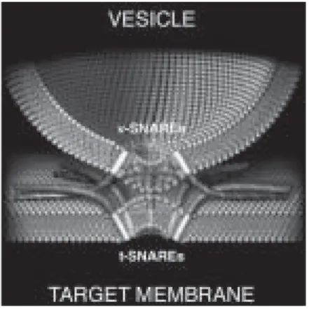
The answer that has emerged could not be more intuitive (Figure 1). ‘snare-pins’ assemble to link a vesicle to its target membrane. This pin, not unlike a ladies’ hairpin, has two ends. One is inserted into the vesicle, the other inserted into the target membrane. snare pins are assembled from two component parts. One part, the ‘v-snare’, is planted into the vesicle membrane. The other part, the ‘t-snare’, is planted into the target membrane with which the vesicle is destined to fuse. The cytoplasmically-facing portions of the v-snare and the t-snare proteins link up and fold into a stable rod-like structure – the snarepin – which is now anchored in both membranes. Pinning the vesicle to the target membrane triggers the process of membrane fusion when the energy released as the snarepin assembles is used to ‘shake-up’ the nearby membrane lipids.
By the early 1960’s, George Palade had captured and brilliantly interpreted images of ‘zymogen granules’ – vesicles storing digestive enzymes in the pancreas – with their membranes merging with the cell surface in the process of dischar-ging their content. In the decade that followed, Palade discovered the secretory pathway and it became clear that membrane fusion must be highly specific to ensure accurate delivery within the cell (Palade, 1975). The fundamental mecha-nism of membrane fusion and its exquisite specificity remained a central mystery of cell biology whose solution would await reconstitution of transport and fusion and the discovery of responsible proteins, especially the snare complex.
Early experiments – reconstitution and NSF
The line of experiments revealing the fusion machinery stemmed from the cell-free reconstitution of transport involving vesicle budding and fusion in the Golgi (Fries and Rothman, 1980; 1981; Balch et al., 1984a,b). Figure 2 shows the assay for transport of vsv G protein within the Golgi. 15B mutant cho cells lack GlcNAc transferase I; thus, oligosaccharides linked to proteins in these cells never acquire GlcNAc, although they are transported normally through the secretory pathway. A Golgi-containing membrane fraction from vsv-infected 15B cells (donor) is incubated with the Golgi-containing fraction (acceptor) from uninfected wild-type cells (plus atp and cytosol). Acquisition of [3H]GlcNAc by G protein
measures transfer between donor and acceptor Golgi stacks. This transport is
me-Figure 2. Assay for cell-free transport.
Transport-coupled glycosylation assay
detects transport between successive Golgi compartments
medial Golgi
cis Golgi
“Donor” Golgi-containing fraction from VSV-infected 15B mutant
“Acceptor” Golgi-containing fraction from uninfected wild-type cells
UDP UDP GIcNac Tr’ase
mix donor + acceptor fractions, ATP, cytosol ATP, cytosol
NH2 COOH
diated by vesicles budding and fusing between Golgi stacks. This was originally suggested by the electron micrographs in Figure 3 of Golgi fractions incubated with cytosol and atp for 15 minutes at 0oC (‘not primed’) or at 37oC (‘primed’),
respectively. Transport and vesicle budding are observed in the latter condition. Transport was inhibited by the sulfhydryl alkylating reagent N-ethylmaleimide (nem), allowing the N-ethylmaleimide Sensitive Factor (nsf) to be purified from cytosol fractions based on its ability to restore transport (Block et al., 1988). Elec-tron microscopy and other tests revealed that nsf is required for fusion since vesi-cles accumulate after nem inhibition (Malhotra et al., 1988).
We soon appreciated that nsf is an atpase required for vesicle fusion at many compartments in the cell, and that this mechanism is extremely well conserved in
Figure 3. Transport is mediated by vesicles budding from Golgi stacks.
Not primed Primed
evolution. nsf (Sec18p) from yeast will replace nsf in fusion with the Golgi of higher animals (Wilson et al., 1989), foreshadowing the universality of the fusion mechanism that became evident with the discovery of the snare complex. SNAPS and SNAP receptors
The Soluble Nsf Attachment Protein (snap) was purified according to its ability to bind nsf to Golgi membranes (Clary et al., 1990). snap binds to one or more saturable, high affinity ‘SNAP REceptors’ (which we termed ‘snares’) and then binds nsf in its atp-bound form. This complex sediments as a 20S particle after extraction from membranes with mild detergents. When nsf hydrolyzes the atp (requiring magnesium ion) it releases itself from the complex and from the membrane (Wilson et al., 1992).
It seemed likely that snares were integral membrane proteins because membra-nes retained the ability to bind snap after alkali extraction (Weidman et al., 1989). That put purification of this membrane protein(s) at the very top of our agenda because of the expectation that lipid bilayer fusion would require membrane-anchored proteins, and the snare proteins were the prime candidates.
The meaning of the seemingly futile cycle of atpase-driven membrane binding and release of nsf was then unclear, we imagined that energy from hydrolysis of atp somehow activated the membrane-anchored snares to power fusion. How-ever, the existence of the cycle had a huge impact on our strategy for identifying the snares, because the assembly and disassembly of 20S particles could be
exploited as sequential affinity purification steps to isolate snares by binding and release from nsf and snap. Previous experiments had shown that standard chromatographic methods and a single affinity step were inadequate; the 20S cycle would add a second level of biological specificity.
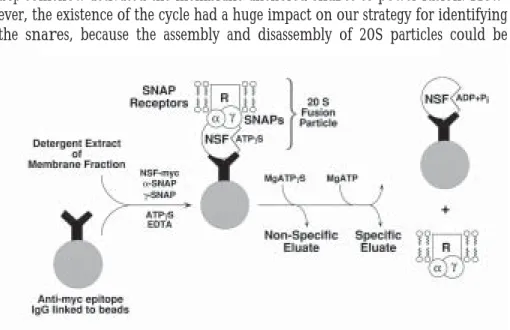
The to-be-identified snares were sequestered from the bulk of membrane protein by incorporation into 20S complexes formed with exogenously added re-combinant (bacterially-expressed) nsf and snap proteins. This incubation was done in the presence of atpgs (a non-hydrolyzable analogue of atp) and in the absence of free magnesium ion (Mg++ is required for hydrolysis of atp by nsf) to
promote 20S particle assembly. The recombinant nsf was expressed with an epitope tag to allow the 20S particles to be isolated with a monoclonal antibody (immobilized on beads) directed against the short peptide epitope (Figure 4).
The second biologically-specific step recapitulated the disassembly of 20S par-ticles. The to-be-identified snares are released when the beads are incubated with magnesium ion and atp to allow nsf to hydrolyze atp. Recombinant nsf remains bound to the beads by the antibody, but the recombinant snap proteins are released along with the to-be-identified snares from the brain membranes (Figure 4).
Because vesicle fusion occurs at many membrane compartments, we had sus-pected that cells would have a large family of snare proteins, related in sequence and differing in location. We were therefore surprised when the snare proteins derived from whole brain yielded a remarkably simple protein pattern (Figure 5) consisting of only four snare proteins, each present in the specific (MgATP) eluate and absent from the non-specific (MgATPgS) eluate.
Discovery of the SNARE complex
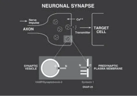
All four snares turned out to be proteins found in synapses (Figure 6). While they had all previously been cloned and sequenced, their function was still unknown. Two were isoforms of syntaxin, a plasma membrane protein identified by Richard Scheller (Bennett et al., 1992) based on its ability to bind synapto-tagmin, a synaptic vesicle calcium sensor (Geppert et al., 1994). The third snare protein was snap-25, short for synaptosome-associated protein of 25 kDa, cloned by Michael Wilson (Oyler et al., 1989). snap-25 mainly resides in the plasma membrane and was originally identified because of its abundance in synapses. Its connection to syntaxin and to membrane fusion was a surprise, as was the coin-cidental relationship of its acronym to that of the soluble nsf attachment protein, snap.
lynchpin because in contrast to snap-25 and syntaxin, vamp resides mainly in synaptic vesicles, immediately suggesting how a complex of the snare proteins (perhaps with nsf and snap) could link the vesicle to the membrane and initiate fusion (Figure 6). vamp/Synaptobrevin had been cloned by DeCamilli and Jahn, and independently by Scheller (Baumert et al., 1989; Elferink et al., 1989).
Supporting our simple model, vamp and syntaxin are membrane-anchored proteins with cytoplasmic domains, and snap-25 is anchored to the cytoplasmic side of the plasma membrane via covalently-attached fatty acids. Close to equi-molar amounts of vamp, syntaxin (its two isoforms considered together) and
snap-25 were recovered in the isolated complexes. Further, the snare proteins were isolated because they function together with nsf and snap, known to func-tion in fusion.
We interpreted the snare complex (Söllner et al., 1993) from the broad per-spective of vesicle traffic as distinct from the specialist’s point-of-view of synaptic exocytosis. First principles require that vesicles and targets are somehow marked to indicate which vesicles will fuse where. This, in turn, indicates that vesicle and target markers must be matched pairwise. We suggested that the simplest mecha-nism for matching is self-assembly, in which only matching pairs of ‘cognate’ vesicle (‘v’) and target (‘t’) markers bind each other between membranes, thereby forming a ‘v-t’ complex prerequisite for membrane fusion.
Based on our cognate vesicle and target marker concept, we proposed the ‘snare hypothesis’ in which the snares are the vesicle and target markers, which we termed v–snares and t-snares. vamp is the v-snare of the synaptic vesicle; syntaxin and snap-25 are the subunits of the cognate t-snare in the plasma membrane. The snare hypothesis provides the framework to generalize our results. We suggested that each vesicle would have its own characteristic v-snare, a homologue of vamp, and that each target membrane would be marked by a characteristic t-snare, homologues of syntaxin and snap-25.
SNARE proteins – minimal machinery for membrane fusion
We soon found that nsf and snap function to disrupt the snare complex using energy derived by atp hydrolysis. This critical step separates the v-snare from the t-snare only when they reside in the same bilayer (i.e., after fusion), but not when they are paired between bilayers (i.e., during fusion) (Weber et al., 2000). This allows nsf to recycle snare complexes after fusion while sparing fusion in progress. When it was shown that snap and nsf are not directly involved in bilayer fusion (Mayer et al., 1996), attention focused on the simplest remaining possibility, that the snare complex is all that is needed to mediate fusion. The extraordinary thermal stability of the complex (resisting) heat denaturation up to 90°c) was consistent with this source of energy for fusion (Hayashi et al., 1994) and its rod-like structure with all membrane anchors at one end, indicated that now the snare complex could bring the two membranes into close contact (Hanson et al., 1997).
The proof that the snare complex is the active principle of fusion could only come from demonstrating this function in the absence of any other proteins. Reconstituting recombinant exocytic/neuronal snares into liposomes established that the pairing of cognate snares between lipid bilayers – snarepins – indeed drives membrane fusion (Weber et al., 1998). Thus, when complementary v-snare and t-v-snare pairs engage, a productive fusion event is not only initiated – as we first imagined – but it is also completed (Figure 1).
The SNARE hypothesis – the specificity of membrane fusion
The snare hypothesis triggered extensive research that led to the identification of snares, which as predicted localize and function at compartments engaging in fusion (Bock et al., 2001). In the most direct test of the snare hypothesis to date, we established that the specificity for membrane fusion is encoded in the physical chemistry of the isolated snare proteins (McNew et al., 2000). Almost without exception, fusion only takes place with the rare combinations of v- and t-snares that correspond to flow patterns occurring in the living cell.
Matching snares establish which fusion events can potentially occur in a cell. Because t-snares are intrinsically auto-inhibited (Parlati et al., 1999) they can be locally activated, thereby allowing vesicles to be targeted to a distinct region with-in a membrane (such as the leadwith-ing edge of plasma membrane of a movwith-ing cell) and also adding a layer of specificity. Vesicles are initially captured by flexible protein tethers and they are ‘tethered’ before they can be ‘snared’ (Mellman and Warren, 2000). Current research focuses on how the snare complex is regulated
by additional proteins to permit membrane fusion to be spatially and temporally controlled.
Acknowledgements
The text of this Heineken Lecture is adapted from a discovery of the snare com-plex published on the website ergito.com. Figures 3 and 4 are from Nature 362, 318-324 (1993). Figure 6 is adapted from a cover of Cell 92 (1998). Figures 1 and 2 were adapted from Cell 39, 525-536 (1984) by ergito.com.
This occasion provides the chance to thank the many co-workers who have contributed to the understanding of vesicle transport and membrane fusion over the past 23 years. Especially, I would like to express my gratitude to two long-term collaborators who made special contributions, Dr. Lelio Orci and Dr. Thomas Söllner, and to the generosity of and superb environments of Stanford University, Princeton University and Sloan-Kettering Institute. Finally, I want to acknow-ledge the important influence of and inspiration by the two men who most im-pacted me as a scientist – Eugene P. Kennedy of Harvard (my Ph.D. thesis advi-sor) and Arthur Korn of Stanford (in whose department this body of work began when I joined as an Assistant Professor in 1978).
References
Balch, W., Dunphy, W., Braell, W., and Rothman, J.E. (1984a). Reconstitution of the transport of protein between successive compartments of the Golgi mea-sured by the coupled incorporation of N-acetylglucosamine. Cell 39, 405-16. Balch, W., Glick, B., and Rothman, J.E. (1984b). Sequential intermediates in the
pathway of intercompartmental transport in a cell-free system. Cell 39, 525-36. Baumert, M., Maycox, P.R., Navone, F., De Camilli, P., and Jahn, R. (1989). Synaptobrevin: an integral membrane protein of 18,000 daltons present in small synaptic vesicles of rat brain. EMBO J. 8, 379-84.
Bennett, M.K., Calakos, N., and Scheller, R.H. (1992). Syntaxin: a synaptic protein implicated in docking of synaptic vesicles at presynaptic active zones. Science 257, 255-9.
Block, M.R., Glick, B.S., Wilcox, C.A., Wieland, F.T., and Rothman, J.E. (1988). Purification of an N-ethylmaleimide-sensitive protein catalyzing vesicu-lar transport. Proc. Natl. Acad. Sci. USA 85, 7852-6.
Bock, J.B., Matern, H.T., Peden, A.A., and Scheller, R.H. (2001). A genomic perspective on membrane compartment organization. Nature 409, 839-41.
Clary, D.O., Griff, I.C., and Rothman, J.E. (1990). snaps, a family of nsf attachment proteins involved in intracellular membrane fusion in animals and yeast. Cell 61, 709-21.
Elferink, L.A., Trimble, W.S., and Scheller, R.H. (1989). Two vesicle-associated membrane protein genes are differentially expressed in the rat central nervous system. J. Biol. Chem. 264, 11061-4.
Fries, E., and Rothman, J.E. (1980). Transport of vesicular stomatitis virus glyco-protein in a cell-free extract. Proc. Natl. Acad. Sci. USA 77, 3870-4.
Fries, E. and Rothman, J.E. (1981). Transient activity of Golgi-like membranes as donors of vesicular stomatitis viral glycoprotein in vitro. J. Cell Biol. 90, 697-704.
Geppert, M., Goda, Y., Hammer, R.E., Li, C., Rosahl, T. W., Stevens, C.F., and Sudhof, T.C. (1994). Synaptotagmin i: a major Ca2+ sensor for transmitter re-lease at a central synapse. Cell 79, 717-27.
Hanson, P.I., Roth, R., Morisaki, H., Jahn, R., and Heuser, J.E. (1997). Structure and conformational changes in nsf and its membrane receptor complexes visualized by quick-freeze/deep-etch electron microscopy. Cell 90, 523-35. Hayashi, T., McMahon, H., Yamasaki, S., Binz, T., Hata, Y., Sudhof, T.C., and
Niemann, H. (1994). Synaptic vesicle membrane fusion complex: action of clo-stridial neurotoxins on assembly. EMBO J. 13, 5051-61.
Malhotra, V., Orci, L., Glick, B.S., Block, M.R., and Rothman, J.E. (1988). Role of an N-ethylmaleimide-sensitive transport component in promoting fusion of transport vesicles with cisternae of the Golgi stack. Cell 54, 221-7.
Mayer, A., Wickner, W., and Haas, A. (1996). Sec18p (nsf)-driven release of Sec17p (a-snap) can precede docking and fusion of yeast vacuoles. Cell 85, 83-94.
McNew, J.A., Parlati, F., Fukuda, R., Johnston, R.J., Paz, K., Paumet, F., Söllner, T.H., and Rothman, J.E. (2000). Compartmental specificity of cellu-lar membrane fusion encoded in snare proteins. Nature 407, 153-9.
Mellman, I., and Warren, G. (2000). The road taken: past and future founda-tions of membrane traffic. Cell 100, 99-112.
Oyler, G. A., Higgins, G. A., Hart, R. A., Battenberg, E., Billingsley, M., Bloom, F.E., and Wilson, M.C. (1989). The identification of a novel synaptosomal-associated protein, snap-25, differentially expressed by neuronal subpopula-tions. J. Cell. Biol. 109, 3039-52.
Palade, G. (1975). Intracellular aspects of the process of protein synthesis. Science 189, 347-58.
Parlati, F., Weber, T., McNew, J.A., Westermann, B., Söllner, T.H., and Rothman, J.E. (1999). Rapid and efficient fusion of phospholipid vesicles by the a-helical core of a snare complex in the absence of an N-terminal regula-tory domain. Proc. Natl. Acad. Sci. USA 96, 12565-70.
Söllner, T., Whiteheart, S.W., Brunner, M., Erdjument-Bromage, H., Geromanos, S., Tempst, P., and Rothman, J.E. (1993). snap receptors implica-ted in vesicle targeting and fusion. Nature 362, 318-24.
Weber, T., Parlati, F., McNew, J.A., Johnston, R.J., Westermann, B., Söllner, T. H., and Rothman, J.E. (2000). snarepins are functionally resistant to disrup-tion by nsf and asnap. J. Cell Biol. 149, 1063-72.
Weber, T., Zemelman, B.V., McNew, J.A., Westermann, B., Gmachl, M., Parlati, F., Söllner, T.H., and Rothman, J.E. (1998). snarepins: minimal ma-chinery for membrane fusion. Cell 92, 759-72.
Weidman, P.J., Melancon, P., Block, M.R., and Rothman, J.E. (1989). Binding of an N-ethylmaleimide-sensitive fusion protein to Golgi membranes requires both a soluble protein(s) and an integral membrane receptor. J. Cell Biol. 108, 1589-96.
Wilson, D.W., Whiteheart, S.W., Wiedmann, M., Brunner, M., and Rothman, J.E. (1992). A multisubunit particle implicated in membrane fusion. J. Cell Biol. 117, 531-8.
Wilson, D.W., Wilcox, C.A., Flynn, G.C., Chen, E., Kuang, W.J., Henzel, W.J., Block, M.R., Ullrich, A., and Rothman, J.E. (1989). A fusion protein required for vesicle-mediated transport in both mammalian cells and yeast. Nature 339, 355-9.
Dr A.H. Heineken Prize for Medicine
Eric R. Kandel
Genes, synapses and memory storage
Professor Eric R. Kandel received the Dr A.H. Heineken Prize for Medicine 2000 for his pioneering research on the molecular mechanisms underlying
In the last several decades of this century we have witnessed a remarkable increase in the explanatory power of biology, including the biology of the brain. The ability of biology to address central issues of brain function will only increase in the 21st century and will likely have a broad impact on many aspects of our lives.
As a result when the intellectual historians look back on these decades, they are likely to acknowledge that the deepest insights into the nature of mental processes will not come from the disciplines traditionally concerned with mind. They will have come not from philosophy, from the arts, or even from psychology of psy-choanalysis, but from biology. This is because in the last two decades biology has participated not simply in one but in two major unifications of thought which bear on our understanding of mind.
First, there has been a remarkable scientific synthesis, achieved through mole-cular biology, that has brought together the disciplines of cell biology, bioche-mistry, developmental biology, and oncogenesis. This unification derives from major advances in our understanding of the gene which have revealed how its structure determines heredity and how its regulation determines development and function. These remarkable insights have given us a marvelous sense of the conservation between different cells in any one organism and different organisms across evolution.
Second, there has been a parallel unification between neurobiology, the science of the brain, and cognitive psychology, the science of the mind. This second uni-fication is far less mature than that brought about by molecular biology, but it is potentially equally profound, for it promises to provide us with a new framework for the analysis of a variety of mental functions, such as perception, action, language and memory.
These two independent unifications stand at the extremes of the biological sciences: the one at the interface between biology and chemistry; the other at the interface between biology and psychology. This raises a question: to what degree can these two disparate strands be brought together? Can molecular biology enlighten the study of mental processes as it has enlightened the study of develop-ment and oncogenesis? Can we anticipate an even broader synthesis in the 21st
century, a synthesis ranging from molecules to mind? In this essay I outline the possibility of a molecular biology of cognition, and suggest that it will occupy cen-ter stage in the early part of the 21st century. I outline this development by using
as an example the study of memory.
We begin with a historical perspective because many of the themes that domi-nate current research on memory – including the distinction between systems and molecular approaches to memory – emery best in a historical overview. We then
will describe more recent molecular biological investigations of memory. Here we will focus in particular on one component of the memory process: the switch from short- to long-term memory. This component can in fact now be analyzed by combining cognitive psychology with modern molecular biology. Finally, we will suggest some possible future directions in memory research, focusing on the drawing together of strands of research that have historically been separated. The problem of memory has a systems component and a molecular component The work of Ramon y Cajal at the beginning of the century (Cajal, 1893) and of Donald Hebb in 1949 (Hebb, 1949) established a useful conceptual framework for the study of memory, based on the idea that memory is stored as changes in the strength of specific synaptic connections. This framework divides the study of memory into two components: the systems problem and the molecular problem. The systems problem of memory is concerned with where in the brain memory is stored and how neural circuits work together to create, process, and recall memo-ries. The molecular problem of memory is concerned with the mechanisms where-by synapses change and information is stored. Most early word on memory focused on the systems problem, focusing on the question, ‘Where is memory stored?’ We therefore begin our historical perspective with this question. Broca, Wernicke and the localization of language
In 1861, Pierre-Paul Broca, a French neurologist, described the first of nine pa-tients who suffered form a language impairment (now known as Broca aphasia) in which they could understand language but could not speak fluently. These defects were specific to the expression of language rather than to motor control of the vocal tract, as the patients could hum or whistle a tune but could not write fluently. Post-mortem examination of the patient’s brains revealed in each case a lesion in the posterior region of the frontal lobe (a region of cortex now called Broca’s area. Thus, for the fist time, Broca was able to assign a well-defined higher function to a specific region of cortex (Broca, 1865) Since all of these lesions were in the left hemisphere, Broca was also able to establish that the two hemispheres, although apparently symmetrical, have slightly different functions.
A decade later, in 1876, Carl Wernicke, a German neurologist, described a se-cond type of aphasia (Wernicke’s aphasia) that is in a way the opposite of Broca’s: an impairment not of the production of speech but of comprehension. Wernicke found that this syndrome was caused by a lesion in the posterior superior portion of the temporal lobe of the left hemisphere, a lesion distinct from that described
by Broca in an area we now call Wernicke’s area (Wernicke, 1908).
Taking his findings together with those of Broca, Wernicke put forward a theo-ry of how cortex is organized for language that, although simpler than our current understanding, is still central to how we now view the brain. Wernicke proposed that any complex behavior requires the activity not of one but of a number of different brain areas, and that these areas are interconnected in various ways. Mental activity is not unitary or seamless as might intuitively appear to be the case, bus can be broken down into multiple components, and each component can be assigned to a more or less specific brain region, much as the organologists had insisted. However, these different specialized areas do not function by themselves bus as part of large, interconnected networks. By application of this model Wernicke predicted, correctly, the possibility of a third kind of aphasia, conduction aphasia, resulting from lesion not of Broca’s area or Wernicke’s area but of the fibers (the arcuate fasciculus) passing between the two. Thus, while specific functions are localized as Gall had insisted, the function of the brain as a whole requires distributed processing somewhat reminiscent of that propounded by Flourens. Wernicke’s model of a distributed network of specialized areas has emerged as a dominant theme in the study of the brain.
Can memory storage be localized to specific regions of the brain?
The finding that the language can be localized within the brain led to the hunt for other areas concerned with specific higher functions. Areas concerned with motor control and with each of the senses were soon identified. It was only a matter of time before efforts to localize cognitive function would turn to memory (Ferrier, 1890; Jackson, 1884). However, attempts in the first half of the twentieth century to localize memory failed. The dominant figure in the period was Karl Lashley, Professor of Psychology at Harvard. Lashley began the experimental search for the locus of memory storage by training rats on specific memory tasks, systema-tically removing portions of cortex, and then testing them for recall. He repeated-ly failed to find any particular brain region that was special to or necessary for the storage of memory. On the basis of these findings, Lashley formulated the law of mass action, according to which the extent of a memory deficit is correlated with the size of a cortical lesion but not with the specific site of that lesion (Lashley, 1929). This law was reminiscent of the views of Flourens a century earlier.
The first clear evidence for the localization of memory came not from experi-mental animals but from clinical studies. In 1938, the neurosurgeon Wilder Penfield, working at the Montreal Neurological Institute, developed methods for
the surgical treatment of focal epilepsy, a form of epilepsy in which seizure is restricted to a relatively small region of the cortex. To functionally map the areas surroundings the epileptic center so as to avoid later damage to critical areas, such as Broca’s and Wernicke’s, Penfield electrically stimulated the cortical surface. Because the brain contains no pain receptors, the patients could remain unanaes-thetized and could report what they experienced during stimulation. In this way Penfield studied over one thousand patients and mapped out in each of them most of the exposed cortical surface. On rare occasions during such mappings, Penfield found a region of temporal cortex where stimulation gave rise to specific experiential responses, memory-like perceptions that the patients could describe. Penfield concluded that portions of the temporal lobe were specifically involved in memory (Penfield and Perot, 1963).
More conclusive evidence for the involvement of temporal lobe structures in memory came in 1957, when William Scoville, a neurosurgeon influenced by Penfield, and Brenda Milner, a psychologist and long-term collaborator of Pen-field’s, reported the now famous case of H.M. (Scoville and Milner, 1957). At age 9, H.M. sustained a head injury after being hit by a bicycle; over the next 18 years he suffered progressive seizures until he was completely incapacitated. As a last resort, H.M. underwent complete bilateral removal of the medial temporal lobe (where his seizures initiated). The surgery relieved his epilepsy, but he was left with a profound memory deficit: from the time of his surgery until this day he has been unable to form any new memories of people, facts, or events.
Brenda Milner studied H.M. and demonstrated that structures in the medial temporal lobe that Scoville had removed are specialized for memory. Her further studies with H.M. not only controverted Lashley’s influential views but also cast new light on the systems problem of memory: there are, within the brain, multi-ple, functionally specialized memory systems.
Memory is not unitary faculty of mind: there are multiple memory systems
The idea that there may be multiple memory systems is old, but it did not enter mainstream psychological thinking until Milner’s work in the 1960s. In the early parts of the nineteen century, the French philosopher Maine de Biran argued that memory can be subdivided into different systems for ideas, feelings and habits (Copplestone, 1977). In the 20th century, William James emphasized the idea that
memory has distinct temporal phases. Henri Bergson developed the distinction between conscious memory and habit (Bergson, 1913) In 1949 the British philo-sopher Gilbert Ryle proposed a similar distinction between ‘knowing that’
(con-scious recall of knowledge for facts and events) and ‘knowing how’ (knowledge of performance or skills without recourse to conscious awareness) (Ryle, 1949). A similar distinction was made by the psychologist Jerome Bruner, who termed ‘knowing that’ a memory with record and ‘knowing how’ a memory without record (Milner et al., 1998). The defining characteristic of memories with record is the ability to summon up a more or less detailed conscious recollection of facts and events about persons, places, and objects. The defining characteristic of a memory without record, by contrast, is a change in the way an organism responds to a situation or a stimulus, without access to the specific circumstance under which the memory was formed. The idea of distinct memory systems is in a sense implicit in Freud’s psychoanalytic writing. Central to Freud’s view of the brain is the distinction between conscious and unconscious memories.
Thus, even prior tot 1960 a fractionation of memory had already been proposed on the basis of content, function, and temporal profile. Nevertheless, the concept of multiple memory systems only drew the attention of the scientific community with the studies of H.M. After the profound nature of his memory deficit was recognized, Milner made the surprising further discovery that that despite his impairment he could learn a surprising amount of new information. First, H.M. was found to have perfectly good short-term memory: he could accurately repeat back a telephone number, and he can carry on a normal conversation. It is only when he is distracted from the topic or tasks at hand that his memory deficit reveals itself. Thus, the temporal lobe structure, which H.M. lacks, is not requi-red for short-term (or ‘working’) memory. This finding validated the early dis-tinction between short and long-term memory.
Second, Milner found that H.M. has reasonably good long-term memory for events prior to his operation. He maintains his overall intelligence and has good command of English. He remembers events from his childhood and adult life be-fore his surgery. There is a period of retrograde amnesia for events shortly bebe-fore the surgery, but for the most part H.M.s symptoms revealed that the medial temporal lobe is not the ultimate storage site for previously acquired knowledge. This finding supports the idea that knowledge is ultimately stored in whatever area of the cortex processes the relevant sort of information (see for example Zeki, 1993).
Third, and most surprising, H.M. is able to form certain types of long-term memory. In 1962 Milner and the psychologist Suzanne Corkin found that H.M. was able to acquire new motor skills (specifically, the ability to trace a complex figure in a mirror) (Corkin, 1965). When asked, he would deny that he had en-countered or practiced the task before; but his performance showed unequivocal
improvement over time. This finding showed that learning of this skill is preserved after severe temporal lobe damage and in the presence of profound amnesia for facts and events, and thus it demonstrated for the first time a fractio-nation of memory on the basis of content rather than just duration. Milner and Corkin thus validated the distinction between conscious memory and habit propounded by Bergson 50 years earlier and by Ryle in 1949.
The learning tasks that amnesia patients like H.M. are capable of mastering have several things in common. They have an automatic quality, and the forma-tion or expression of the memories is not dependent on awareness or cognitive processes such as comparison and evaluation. This type of memory typically builds up slowly over many trails and is expressed primarily by improved perfor-mance on certain tasks. The psychologist Lawrence Weiskrantz has noted that the spared learning skills are reflexive rather than reflective – typically the patient need only produce a physical response tot a stimulus or cue.
This distinction was soon validated on normal subjects. Larry Squire has framed the distinction particularly well by emphasizing the ability of humans to report verbally the contents of explicit memory but not of implicit memory: explicit memory is thus declarative whereas implicit memory is non-declarative (Squire and Zola-Morgan, 1991). Daniel Schachter framed Bruner’s distinction using the terms implicit for ‘knowing how’ and explicit for ‘knowing that’ (Schachter, 1996). These are the most widely used terms, and while specific defi-nitions of these various terms can differ in different contexts, all these authors are describing the basis distinction that was revealed in the studies of H.M.
The molecular component of memory storage
An examination of memory storage at the systems level revealed that memory is not a unitary faculty but has at least two forms: implicit (non-declarative) and explicit (declarative). This distinction raises the question: What are the cell and molecular mechanisms by which implicit and explicit memories are stored in the brain? Are the molecular storage mechanisms as different as is the logic of the explicit and implicit memory systems?
These two questions (1) the nature of storage mechanisms and (2) the generality and specificity of these mechanisms that have fascinated me most of my scientific career.
I began my studies of memory storage in 1957 by focusing a cell biological ap-proach on the hippocampus, a structure critical to explicit forms of memory sto-rage. But although we were fortunate in making an exciting start on the
hippo-campus, I soon realized that explicit memory storage was exceedingly complex. Without good plastic and pharmacological tools no progress could be made in relating cellular changes to organismic behavior. To achieve an intellectually satisfying understanding of memory storage one needed to take a completely dif-ferent approach to a memory – one needed to take a more radical reductionist approach. One needed to take an approach to mental processes such as memory similar to that used in other core areas of biology – an approach similar to that used, for example, by Thomas Hunt Morgan, Max Delbruck, and Salvador Luria in the early studies of that nature of the gene in Drosophila and bacterial phages. One needed to study not the most complex case but the simplest possible case of memory storage and to study it in the simplest and technically most advanta-geous animal available so that one could drive it into the ground.
After an extensive search, I focused for reasons I will tell you in a minute on the marine snail Aplysia where I have studied a very simple procedural memory. Within a few years after I began to study simple forms of procedural memory, we made the surprising discovery that this simple case of procedural memory shared certain key mechanisms with the most complex forms of explicit declarative memory.
The first clue to shared mechanisms came form the behavioral study of stages in memory storage. We found that this simple instance of procedural memory storage had stages that were surprisingly similar to those described earlier for declarative memory including human declarative memory. There was in each case a short-term memory that was labile and lasted only minutes and a long-term memory that was stable and self-maintained and can lest for days, weeks, or even years. We next found that in implicit, as well as in explicit memory repetition is responsible for converting short- to long-term memory. It is practice that makes perfect. Finally, we found that for implicit memory, as had several years earlier been found for explicit memory that long-term memory, but not short-term, requires new protein synthesis.
This suggested to me the possibility that the requirement for protein synthesis during the switch form short tot long-term memory is very general. It was evolutionarily conserved and was evident not only in vertebrates but ales in the invertebrate. Moreover, it held for both implicit and explicit forms of learning.
This generality suggest the possibility that some of the key proteins that make up the switch might also be conserved and used for both explicit and implicit forms of memory storage. If that were so the identifying the relevant proteins in any one system might provide molecular insight that are generally important for understanding other systems. Moreover, the information emerging in different
systems might be sufficiently complementary so as to provide a coherent outline, in molecular detail, of one important issue in the biology of mental process – how a transient short-term memory is converted into stable long-term memory. So with this logic as a background we focused on a molecular approach in Aplysia and then in mice. Before I tell you something about the molecular nature of this switch and complex forms of memory, let me introduce you to Aplysia.
Short-term storage for implicit memory involves functional changes in the strength of pre-existing synaptic connections
Implicit memory refers to memory about perceptual and motor skills. The sim-plest instances of such storage are the elementary forms of non associative and associative memory. These first emerged with particular clarity as distinctive forms of implicit memory from studies of the family of learning processes related to classical conditioning.
Learning refers to the acquisition of new information about the world and memory refers to the retention of that information over time. Classical condi-tioning is a form of learning in which an animal learns to associate two sensory stimuli, a neutral initiating sensory stimulus (called the to be conditioned stimulus or cs by behaviorist psychologists) that initially produces a weak reflex response, and a highly effective sensory stimulus (called the unconditioned stimulus or us by behaviorist psychologists) that produces an inborn, unlearned, reflex response (called the unconditioned response or ur). As a result of pairing these two sensory stimuli, (the cs and us) the animal learns to strengthen its pre-existing response to the neutral sensory stimulus (the cs) or to develop a completely new response to the cs.
In the course of studying classical conditioning, Ivan Pavlov and others disco-vered that when each of these two stimuli were repeatedly presented alone, they each gave rise to distinctive forms of learning and memory storage. Thus, re-peatedly presenting a neutral stimulus (cs) by itself gives rise to a form of learning called habituation, whereby the animal learned to recognize a stimulus as innocuous and comes to ignore it. By contrast, when the animal is presented with an aversive (us) stimulus, it gives rise to sensitization, a form of learning whereby the animal recognizes the stimulus as being highly noxious and learns to enhance its defensive and escape responses. Thus, simple forms of learning take one of two forms involve (1) in non-associative learning such as habituation and sensitization the animal learns about the properties of a single stimulus, (2) in associative learning the animal learns about the relation between two stimuli (Squire and Kandel, 1999).
What are the cellular changes that, result in the brain when animals learn these simple tasks? Initial insights into the cell biological nature of each of these three forms of memory storage first came from studies of the marine snail Aplysia. Aplysia has lent itself to the study of implicit memory storage because it offers a number of technical advantages for this study. First, the animal is quite smart; it can learn a number of different tasks and store them in both short- and long-term memory. Second, the animal has a relatively simple central nervous system, con-sisting of only about 20,000 neurons. Third, the neurons of Aplysia are particu-larly large, which allows them to be uniquely identified, so that one can return to the same cell in every animal of the species. Fourth, it is possible to map in detail the synaptic connections between individual cells and between a given cell and the sensory and motor periphery. As a result of these advantages, it is possible to work out significant parts of the neural circuitry of a given behavior – such as the gill and siphon withdrawal reflexes – in terms of uniquely identifiable cells and their pattern of interconnections (Kandel, 1976). Finally, one can culture Aplysia neurons and construct with them in vitro microcircuits of components of beha-vior such as the gill withdrawal reflex in ways that are not yet possible in other systems.
Studies of memory in Aplysia also first illustrated the advantages that accrue from using elementary forms of nonassociative and associative learning for study-ing memory storage. These simple, implicit forms of memory offer the advantage that memory can be tested (retrieved) at any time after learning by simply examining the time locked reflex response to a sensory test stimulus (the cs) (Kandel et al., 1995). This feature specifies that the key requirement for a neurobiological analysis of memory is to work out, in cellular detail, the neural circuitry of the behavior being modified by learning. Since each of these simple forms of learning leads to a change in the response of the reflex to the initiating sensory stimulus, one needs in particular to work out the pathway whereby the sensory stimulus of the reflex leads to a behavioral response. As a corollary, in learning tasks in which there is a clearly defined alteration in the behavioral res-ponse to the cs, all the important learning-related changes are contained within the circuit of the conditioned stimulus itself.
In Aplysia most work on implicit memory has been carried out on the with-drawal reflex of the gill and the siphon to a weak tactile stimulus applied to the siphon. This withdrawal reflex is mediated by both monosynaptic and polysy-naptic connections. The sensory neuron that innervates the siphon make direct monosynaptic connections to the motor neuron that withdraws the gill. In addi-tion, the sensory neurons also make polysynaptic connections to motor neurons
via interneurons (Figure 1.). While the polysynaptic connections contribute im-portantly to both the basal reflex and to learning, most studies have concentrated on the monosynaptic connections. These connections (1) form a significant com-ponent of the behavior, (2) their electrophysiological properties recapitulate the basic properties of both short- and long-term memory for various forms of lear-ning, (3) this component can be cultured and therefore studied in great detail morphologically and biochemically.
Although the gill and siphon withdrawal reflex is quite simple, it exhibits all three simple forms of learning. With each of these forms the animal learns to alter its behavioral response to the tactile stimulus to the siphon (which is the cs for these forms of learning), and in each case repetition converts a short-term form to a long-term form of implicit memory.
With habituation of the gill and siphon withdrawal reflex, the animal learns about the properties of a single, novel stimulus, a weak tactile stimulus to the si-phon. When this stimulus is first presented, the animal perceives it as novel and responds to it with a brisk reflex response. But when the same weak stimulus is repeated, the animal comes to recognize the stimulus as trivial and gradually
Figure 1. The gill withdrawal reflex in Aplysia is mediated by a simple circuit. Approxi-mately 50% of the learning observed in sensitization of this reflex by tail shock results from potentiation by serotonin of the direct synapse between the sensory neuron inner-vating the mantle shelf and the motor neuron innerinner-vating the gill. Individual identified neurons are indicated.
learns to ignore the stimulus and to stop responding to it. As a result the same weak siphon stimulus that once produced a brisk response now produces little or no response at all. This progressive decrease in the response to the weak siphon stimulus is reflected in the neural circuit of the gill-withdrawal reflex, as a weake-ning of the synaptic connections of the sensory input pathway, the synaptic con-nections between the siphon sensory neurons and their central target cells: the interneurons and motor neurons of the reflex (Figure 1). This weakening in syn-aptic strength of the sensory input pathway of the cs results from a decrease in the amount of the transmitter glutamate released from the presynaptic terminals of the sensory neurons.
With sensitization, Aplysia learns about the properties of an important aversive stimulus, a noxious shock to the tail (an unconditioned stimulus). The animal recognizes the stimulus as aversive and learns to enhance its gill and siphon with-drawal responses to the cs, to the weak touch to the siphon. Sensitization is re-flected in the neural circuit as an increase in synaptic strength in the input con-nections of the reflex, the pathway between the siphon sensory neurons end their targeted cells. This strengthening is due to an increase in the release of glutamate from, he terminals of the sensory neurons (the cs pathway) (Kandel, 1976; Byrne and Kandel, 1996; Carew and Sahley, 1986; Hawkins et al., 1993).
Aplysia also can learn to associate these two stimuli; it can learn classical condi-tioning. When a weak (cs) stimulus to siphon is repeatedly paired with a shock to the tail (the us), the reflex response to the siphon stimulus will be enhanced, and this enhancement of the response to the cs with classical conditioning is substan-tially greater than with sensitization when the weak siphon stimulus (cs) and the tail shock (us) are not paired. This classical conditioning is reflected in the neural circuitry as a greatly enhanced strengthening in the input connections of the sory neurons to their target cells, an enhancement that is greater than that of sen-sitization. In addition to a presynaptic mechanism, there is with classical condi-tioning a postsynaptic mechanism that comes into play (Squire and Kandel, 1999).
There is now a reasonably good understanding of how this association is achie-ved on the -cellular level (Glanzman, 1995; Squire and Kandel, 1999). As we have seen the sensory neurons release glutamate from their presynaptic terminals. Glutamate acts on two types of postsynaptic receptors: an ampa receptor and an nmda receptor. Under normal circumstances and with nonassociative learning such as habituation and sensitization, only the ampa receptors are utilized be-cause the mouth of the nmda receptor channel is blocked by Mg2+. To remove the
glutamate needs to bind to the postsynaptic nmda receptor, and the postsynaptic membrane needs to be depolarized substantially so as to extrude Mg2+ out of the
nmda receptor channel mouth. This coincident activation of the nmda receptor and postsynaptic depolarization only occur when the weak siphon stimulus (cs) and the strong tail shock (us), are paired together. Only then is each of the postsynaptic motor cells sufficiently depolarized to activate the nnwa receptors (Lin and Glanzman, 1994).
Detailed analyses of the distinctive role of the nnwa receptor in the postsynap-tic cell were first carried out in the hippocampus where, as we shall learn below, it was found that the extrusion of Mg2+ allows Ca2+ to flow into the postsynaptic
cell. The Ca2+ influx in turn activates a signaling cascade in the postsynaptic cell.
One component of long-term potentiation in Aplysia is a retro-grade signal that is generated in the postsynaptic cell and sent back from it to the presynaptic neuron where the signal acts to enhance presynaptic transmitter release to that it is even greater than occurs with sensitization alone. Thus, in the case of Aplysia, the facilitation of the connections between the sensory and motor neurons that occurs with classical conditioning has an additional, associative component superimposed on the facilitation produced by sensitization (Squire and Kandel, 1999).
These several studies in Aplysia revealed three insights into short-term memory storage that have proven quite general and apply as well to explicit as to implicit memory storage. First, these studies showed that learning can lead to alterations in synaptic strength and that the persistence of these synaptic changes in both the mono- and polysynaptic component of the neural circuit of the reflex represent the cellular storage mechanisms for memory. Second, a single synaptic connec-tion can participate in, and be modified in different ways by, different forms of learning and participate in different types of short-term memory storage. Finally, each of these three simple forms of learning – habituation, sensitization, and clas-sical conditioning - gives rise to both a short- and a long-term memory depending upon number of repetitions. Each of the long-term forms of memory is associated with a long-term change in synaptic strength in the monosynaptic connection between the sensory and motor neuron of the reflex. Thus, a single synaptic con-nection can not only participate in different types of short-term memory storage but the same connection can also be the site of both short- and long-term memo-ry storage.
Long-term storage for implicit memory involves the synthesis of new protein and the growth of new connections
Given that a single connection can participate in both short and long-term me-mory, what are the molecular mechanisms for these different phases of memory storage? In particular, how is short-term memory converted to long-term memo-ry? Can molecular biology with its ability to reveal homology relationship de-lineate commonalties in the mechanism of storage that might again encompass explicit as well as implicit storage?
The first insight into the molecular mechanisms of memory storage come from the discovery that there were phases of memory storage. The study of memory phases dates to 1885 and the work of Herman Ebbinghaus. By forcing himself to memorize lists of nonsense syllabus and then testing his own recall, Ebbinghaus determined that there are at least two phases to memory: a short term phase which contains much information but is transient, lasting minutes, and a long-term phase which is far more stable (Ebbinghaus, 1913). This is consistent with our everyday experience: we have access to far more of the information of the recent past (say, the last few minutes) than we will be able to remember a few hours hence. Ebbinghaus’ distinction is also consistent with observations of vic-tims of injury: a person who is struck on the head or shocked will typically lose memory for events that occurred shortly before the insult but not for more remote events (e.g., Zubin and Barrera, 1941). Modern work showed that the distinction between short- and long-term memory applies to both implicit and explicit memory.
In the 1960s Louis Flexner and his colleagues first found that short- and long-term memory are not only distinguished by their time course but also by their biochemical mechanism. Long-term memory differs from short-term memory in requiring the synthesis of new protein (Flexner et al., 1965; Agranoff, 1976). This requirement of protein synthesis for long-term memory has a specific time window, during and shortly after training, which is called the consolidation phase. Blockade of protein synthesis during the consolidation phase will disrupt long-term memory, but blockade before or after will have no effect (Bourtchouladze et al., 1998; Freeman et al., 1995).
The requirement for protein synthesis for long-term but not short-term me-mory, and the existence of a consolidation window during which memories are sensitive to disruption, has proven to be very general. lt has been demonstrated in explicit as well as implicit storage and in different vertebrates as well as in inverte-brates. This conservation in turn suggests that the proteins involved in the switch to long-term memory may also be conserved. If that were true, then a detailed
study of the molecules involved in the switch in any given memory storage process in any animal is likely to yield proteins that are of general importance. Moreover, a molecular study of several different instances of memory storage are likely to reveal the general nature of a cognitive process: the switch whereby a transient short-term memory is converted to a persistent, self-main-tained long-ten-n memory (Pittenger and Kandel, 1998). During the last decade, studies in Aplysia, Drosophila, and mice have begun to reveal some of the proteins essential for this switch.
Aplysia and Drosophila share some of the same genes and proteins for converting short- to long-term memory
The initial molecular insights into long-term storage of implicit memory came from studies of sensitization in Aplysia. As in other forms of learning, repetition of the sensitizing training in Aplysia increases the duration of the memory for sensitization. Thus, one tail shock produces an enhancement of the withdrawal response that lasts a few minutes. Five shocks produce an enhancement that lasts several days, and further training gives rise to memory that lasts weeks. This long-lasting sensitization requires protein synthesis during a critical time window, whereas short-term sensitization does not (Montarolo et al., 1986). Here, then, is a very simple model system of long-ten-n memory that shares some of the key mechanistic properties of more complex vertebrate systems.
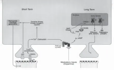
Sensitizing tail stimuli activate three different classes of modulatory inter-neurons that synapse on the axon terminals of the siphon sensory inter-neurons; all three have similar actions (Figure 1). Of the three, the interneurons that release serotonin (or 5-hydroxytryptamine, 5-ht) are thought to be particularly impor-tant. In the intact animal, in reduced preparations, and even in a dissociated cell culture consisting of a single sensory neuron, a single motor neuron and a single serotonergic facilitatory neuron (Figure 2, see next page), serotonin acts on the sensory neurons by increasing the intracellular concentration of the second mess-enger camp and by activating the camp dependent protein kinase (pka) and protein kinase C (Braha et al., 1990; Brunelli et al., 1976). Transient activation of these intracellu-lar signaling cascades by one tail shock or one pulse of 5-ht leads to a transient strengthening, or facilitation, of the synapse between the sensory and motor neurons by increasing the amount of neurotransmitter (glutamate) released by the sensory cell onto the motor cell when the siphon is touched. Repeated training or of five pulses of 5-ht produces long-lasting facilitation that can persist for 72 hr or more and is accompanied by the growth of new connections (Castellucci et al., 1986; Montarolo et al., 1986; Bailey and Kandel, 1993).
Whereas a single pulse of 5-ht activates the kinases pka and pkc transiently, five repeated pulses lead to a persistent activation of pk and to the recruitment of the map kinase signal transduction pathway (Figure 3). Both pka (Bacskai et al., 1993) and map kinase (Martin et al., 1997b) then translocate to the nucleus, where they activate the transcriptional activator crebla (Bartsch et al., 1998) and inactivate the transcriptional repressor creb2 (Bartsch et al., 1995). The creb fa-mily of transcriptional activators have been implicated in plasticity in many systems, as we will discuss below, and may be one of the most conserved mole-cular components of the mechanisms for switching on long-term plasticity. Once crebla is activated and the repressive action of creb2 is removed, a set of immediate-early genes is activated, of which two – the N-terminal ubiquitin hy-drolase and the transcriptional factor ApC/EBP (Alberini et al., 1994) – have been well characterized. ApC/EBP, forms both homodimers and heterodimers with another factor (activating factor l). The homodimers and heterodimers act diffe-rent on downstream genes that lead to the growth of new synaptic connections. This structural change, which represents the long-term, self-maintained, stable form of memory, is associated with a rearrangement of structural proteins such as the cell adhesion molecule ApCAM (Bailey and Kandel, 1993). This internali-zation of ApCAM, for example, is thought to be a necessary prerequisite for the growth of neuronal processes (Figure 3).
The requirement for transcription in long-term facilitation in Aplysia explains why long-term memory requires the synthesis of new protein. However, this
Figure 2. The circuit controlling this reflex can now be studied in reconstituted cell cul-ture. (S.N. – sensory neuron. 5-ht F.N. – facilitatory interneuron. M.N. – motorneu-ron.)
requirement poses a cell biological puzzle: since long-term plasticity relies on the activation of genes in the nucleus, one might expect that long-lasting changes in the connectivity of the neuron would be cell-wide. Recent experiments have re-vealed that each synapse or group of synapses can be modified independently. This spatially restricted plasticity requires the activity of crebi in the nucleus as well as local protein synthesis in the processes that were modulated by 5-ht. The mechanism of this specificity is revealed by a second phenomenon: synaptic cap-ture. When synapse- specific long-term facilitation is initiated at one of two branches, a single pulse of 5-ht – which alone is able to induce only a transient facilitation lasting minutes – is thereafter able to induce long-term plastic changes including the growth of new synaptic connections when applied to a second
Figure 3. Multiple molecular pathways are involved both in short-term facilitation and in long-term facilitation of the sensory neuron-motor neuron synapse in Aplysia.
branch (Figure 4) (Martin et al., 1997a). This phenomenon suggests that the new genes that are being activated in the nucleus have their products distributed widely, but that the products only persistently strengthen those synapses which have been somehow marked by short-term facilitation. This phenomenon is also evident in the vertebrate brain (Frey and Morris, 1997).
A similar set of genes important in the switch from short- to long-term memory has also emerged from studies of Drosophila. As an experimental system, Drosophila is in many ways the complement of Aplysia. The great advantage of Aplysia is that it is tractable in a top down approach for cell biological studies of learning, and allows in the limit the reconstitution of synaptic circuits in cell culture. Mutational (forward) genetics, however, is almost impossible in Aplysia. In Drosophila, on the other hand, cell biological and electrophysiological studies are difficult due to the tiny size of the neurons, but the genetics are extremely tractable and mature – more so than in any other multicellular organism. The great strength of the Drosophila work in learning, predictably, is therefore in its genetic analysis. Both genetic screens and reverse genetic analysis, in which specific genes have been disrupted to investigate their function, have been fruitful.
Figure 4. The ability to study two synapses from the same sensory neuron onto widely separated motor neurons in a modified Aplysia cell culture system allows synaptic tagging and synaptic capture to be explored for the first time.
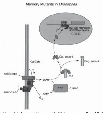
The pioneering work of Seymour Benzer, and the subsequent studies of Quinn, Tully, and Yin, has led to the identification of a number of genes required for memory storage (Weiner, 1999). Many of the genes identified in this way are the same as those implicated in plasticity in Aplysia, hinting at a dramatic conserva-tion of the mechanisms of synaptic plasticity over evoluconserva-tion. For example, the Drosophila genes dunce, rutabaga, and amnesiac all encode components of the camp-pka cascade (Figure 5). Other genes identified encode participants in other signal transduction cascades (for example, the gene leonardo; Skoulakis and Davis, 1998) or cell-cell adhesion molecules similar to ApCAM (Grotewiel et al., 1998). Based on the genes that have been identified, there seems to be a clear
Figure 5. Many of the learning-related genes identified by screens in Drosophila, inclu-ding dunce, rutabaga, and amnesiac, participate in the cyclic amp-pka pathway that is also implicated in Aplysia.